JOURNAL OFVIROLOGY, Oct. 2010, p. 9995–10003 Vol. 84, No. 19 0022-538X/10/$12.00 doi:10.1128/JVI.00505-10
Copyright © 2010, American Society for Microbiology. All Rights Reserved.
The Effect of Clade-Specific Sequence Polymorphisms on HIV-1
Protease Activity and Inhibitor Resistance Pathways
䌤
Rajintha M. Bandaranayake,
1Madhavi Kolli,
1Nancy M. King,
1Ellen A. Nalivaika,
1Annie Heroux,
2Junko Kakizawa,
3Wataru Sugiura,
3,4and Celia A. Schiffer
1*
Department of Biochemistry and Molecular Pharmacology, University of Massachusetts Medical School, 364 Plantation Street,
Worcester, Massachusetts 016051; Biology Department, Brookhaven National Laboratory, Upton, New York 11973-50002;
Laboratory of Therapeutic Research and Clinical Science, AIDS Research Center, National Institute of Infectious Diseases,
4-7-1 Gakuenn, Musashimurayama, Tokyo 208-0011, Japan3; and Department of Infection and Immunology,
Clinical Research Center, Nagoya Medical Center, Nagoya, Japan4
Received 6 March 2010/Accepted 14 July 2010
The majority of HIV-1 infections around the world result from non-B clade HIV-1 strains. The CRF01_AE (AE) strain is seen principally in Southeast Asia. AE protease differs by⬃10% in amino acid sequence from clade B protease and carries several naturally occurring polymorphisms that are associated with drug resis-tance in clade B. AE protease has been observed to develop resisresis-tance through a nonactive-site N88S mutation in response to nelfinavir (NFV) therapy, whereas clade B protease develops both the active-site mutation D30N and the nonactive-site mutation N88D. Structural and biochemical studies were carried out with wild-type and NFV-resistant clade B and AE protease variants. The relationship between clade-specific sequence variations and pathways to inhibitor resistance was also assessed. AE protease has a lower catalytic turnover rate than clade B protease, and it also has weaker affinity for both NFV and darunavir (DRV). This weaker affinity may lead to the nonactive-site N88S variant in AE, which exhibits significantly decreased affinity for both NFV and DRV. The D30N/N88D mutations in clade B resulted in a significant loss of affinity for NFV and, to a lesser extent, for DRV. A comparison of crystal structures of AE protease shows significant structural rearrangement in the flap hinge region compared with those of clade B protease and suggests insights into the alternative pathways to NFV resistance. In combination, our studies show that sequence polymorphisms within clades can alter protease activity and inhibitor binding and are capable of altering the pathway to inhibitor resistance.
Human immunodeficiency virus type 1 (HIV-1) is classified into three groups (M, N, and O), of which group M is further classified into nine major clades (A, B, C, D, F, G, H, J, and K) and 43 circulating recombinant forms (CRFs) based on viral genomic diversity (32, 37). The majority of HIV-1 infections across the globe result from non-B clade HIV-1 variants; clade B accounts for only ⬃12% of infections (15). However, the development of currently available anti-HIV therapies has been based on the virology of clade B variants. In recent years, several studies have shown that there are clear differences between clades when it comes to viral transmission and the progression to AIDS, an observation which raises questions about the effectiveness of the currently available anti-HIV therapies against the other clades and CRFs (16–18, 39).
HIV-1 protease has been an important drug target in the global effort to curb the progression from HIV infection to AIDS. However, the accumulation of drug-resistant mutations in the protease gene has been a major drawback in using HIV-1 protease inhibitors. The effects of mutations associated with drug resistance in HIV-1 clade B protease have been studied extensively over the years. For the most part, resistance mutation patterns are very similar in HIV-1 clade B and non-B clade proteases (19). However, several alternative resistance
pathways have been observed for non-B clade proteases com-pared with those of clade B protease (1, 12, 13, 26). Limited data are available on how sequence polymorphisms, some of which are associated with drug resistance in clade B protease, might influence the pathway to drug resistance in non-B clade proteases. Furthermore, very little is understood about how sequence polymorphisms in non-B clade proteases affect pro-tease function and inhibitor binding.
HIV-1 CRF01_AE (AE) was the first CRF to be observed in patient populations and is seen principally in Southeast Asia (2, 10, 25). AE protease differs by⬃10% in amino acid se-quence from that of clade B protease (Fig. 1A). Interestingly, AE protease develops a different resistance pathway from that of clade B protease to confer resistance to the protease inhib-itor nelfinavir (NFV) (1). In patients infected with AE, the protease acquires predominantly the N88S mutation in re-sponse to NFV therapy, whereas in patients with clade B in-fection, the protease acquires the D30N/N88D mutations. The fitness of AE viral strains is thought to be similar to that of HIV-1 group M viral strains (11, 41). However, the effect of AE-specific sequence variations as well as drug resistance sub-stitutions on viral fitness has not been studied extensively.
In the present study, biochemical and biophysical methods were used to determine the effect of sequence polymorphisms in AE protease on enzyme activity and inhibitor binding. Through determination of crystal structures and analysis of changes in hydrogen bonding patterns, a structural rationaliza-tion is described for the two different pathways observed for clade B and AE proteases to attain resistance to NFV.
* Corresponding author. Mailing address: Department of Biochem-istry and Molecular Pharmacology, University of Massachusetts Med-ical School, 364 Plantation Street, Worcester, MA 01605. Phone: (508) 856-8008. Fax: (508) 856-6464. E-mail: [email protected].
䌤Published ahead of print on 21 July 2010.
9995
on November 8, 2019 by guest
http://jvi.asm.org/
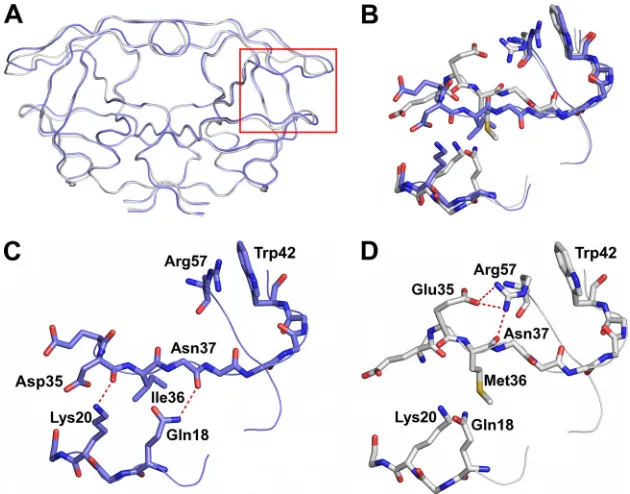
FIG. 1. (A) Amino acid sequence alignments of B-WT and AE-WT and NFV-resistant mutants. Residue positions that differ between clade B and AE are indicated in red. NFV resistance mutations are indicated in blue. (B) Ribbon diagram superposition of DRVAE-WT(blue) and
DRVAE-N88S(gray). (C) Double-difference plot comparing DRVAE-WTand DRVAE-N88S. (D) Ribbon diagram superposition of clade DRVAE-WT
(magenta) and clade DRVB-WT(gray). (E) Double-difference plot comparing DRVAE-WTand DRVB-WT. The color contours in the
double-difference plots indicate distance double-differences of⬍1.0 Å (black), 1.0 to 0.5 Å (green), 0.5 to 1.0 Å (blue), and⬎1.0 Å (red).
9996
on November 8, 2019 by guest
MATERIALS AND METHODS
Protease gene construction.The clade B wild-type (B-WT) protease gene was generated as previously described (34). The AE wild-type (AE-WT) protease gene was synthesized in fragments (Integrated DNA Technologies, Coralville, IA), with codons optimized for expression inEscherichia coli. The fragments were ligated to form the complete gene, which was then inserted into the pET11a expression vector (Novagen/EMD Chemicals, Gibbstown, NJ). The protease sequence was confirmed by DNA sequencing. The NFV resistance mutations, N88S in AE (AE-N88S) and D30N/N88D in clade B (B-D30N/N88D), were generated by directed mutagenesis using a Stratagene QuikChange site-directed mutagenesis kit (Agilent Technologies, La Jolla, CA). Mutagenesis was confirmed by sequencing. The Q7K substitution was introduced to all protease variants to prevent autoproteolysis (38).
Protein expression and purification.The clade B and AE variants were sub-cloned into the heat-inducible pXC35 expression vector (American Type Culture Collection [ATCC], Manassas, VA) and transformed intoE. coliTAP-106 cells. Protein overexpression, purification, and refolding were carried out as previously described (20). Protein used for crystallographic studies was further purified with a Pharmacia Superdex 75 fast-performance liquid chromatography column (GE Healthcare, Chalfont St. Giles, United Kingdom) equilibrated with refolding buffer (50 mM sodium acetate [pH 5.5], 10% glycerol, 5% ethylene glycol, and 5 mM dithiothreitol).
Crystallization and structure determination.Protease solutions between 1.0 and 2.0 mg ml⫺1
were equilibrated with a 5-fold molar excess of NFV, darunavir (DRV), and amprenavir (APV) for 1 h on ice. Crystals were grown over a reservoir solution consisting of 126 mM phosphate buffer (pH 6.2), 63 mM sodium citrate, and 18% to 23% ammonium sulfate by the hanging-drop vapor diffusion method. X-ray diffraction data for AE-WT were collected at a Bio-CARS beamline 14-BM-C at the Advanced Photon Source (Argonne National Laboratory, Argonne, IL) at a wavelength tuned to 0.9 Å with a Quantum 315 CCD X-ray detector (Area Detector Systems Corporation, Poway, CA). Diffrac-tion data for AE-N88S were collected by using beamline X29A at the NaDiffrac-tional Synchrotron Light Source (Brookhaven National Laboratory, Upton, NY) at a wavelength tuned to 1.08 Å with a Quantum 315 charge-coupled-device (CCD) X-ray detector (Area Detector Systems Corporation). Data for the B-D30N/ N88D variant was collected in-house with an R-Axis IV imaging plate system (Rigaku Corporation, Tokyo, Japan) mounted on a rotating-anode X-ray source (Rigaku Corporation). All data were collected under cryocooled conditions.
The data were indexed, integrated, and scaled using HKL/HKL-2000 software (HKL Research, Charlottesville, VA) (29). Structure determination and refine-ment were carried out using the CCP4 program suite (4) as previously described (35). The tensor (T), libration (L), and screw (S) parameter files used in TLS refinement were generated using the TLS motion determination server (30). Model building and real-space refinement were carried out with Coot molecular graphics software (8). Structure comparisons were made by superposing the structures using the C␣atoms of the terminal regions (residues 1 to 9 and 86 to 99) from the two monomers. In the case of the AE complexes, which have multiple orientations for the inhibitor, only the orientation common with the clade B structures was used for analysis. Structures were visualized using PyMol molecular graphics software (6).
Double-difference plots were generated for AE and clade B protease struc-tures to graphically visualize structural differences between the clades, as previ-ously described (35). Briefly, distances between all C␣atoms within the dimer were calculated for each complex. A distance difference matrix was then com-puted for each atom for a given pair of complexes. The distance difference matrix was then plotted as a contour plot using the gnuplot plotting software (44).
Nomenclature.The following nomenclature format will be used to refer to each crystal structure: inhibitorprotease variant. Thus, DRV in complex with AE-WT, clade B-AE-WT, AE-N88S, and AE-D30N/N88D protein are designated DRVAE-WT, DRVB-WT, DRVAE-N88S, and DRVB-D30N/N88D, respectively. Prime notation is used to distinguish the two monomers in the protease dimer. For example, residue 30 from the first monomer would be referred to as Asp30, and the same residue from the second monomer would be referred to as Asp30⬘.
ITC.Binding affinities and thermodynamic parameters of inhibitor binding to clade B and AE variants were determined by isothermal titration calorimetry (ITC) with a VP isothermal titration calorimeter (MicroCal, LLC, Northampton, MA). The buffer used for all protease and inhibitor solutions consisted of 10 mM sodium acetate (pH 5.0), 2% dimethyl sulfoxide, and 2 mM tris[2-carboxyethyl] phosphine. Binding affinities for all protease variants were obtained by compet-itive displacement titration using acetyl-pepstatin as the weaker binder. A solu-tion of 30 to 45M protease was titrated with 10-l injections of 200M acetyl-pepstatin to saturation. The pepstatin was then displaced by titrating 36
8-l injections of 200M APV or NFV or 41 7-l injections of 40M DRV. Heats of dilution were subtracted from the corresponding heats of reaction to obtain the heat resulting solely from the binding of the ligand to the enzyme. Data were processed and analyzed with the ITC data analysis module (Microcal) for Origin 7 data analysis and graphing software (OriginLab, Northampton, MA). Final results represent the average of at least two measurements.
Measurement of protease activity.Protease activity was assayed by following each variant’s ability to hydrolyze the fluorogenic substrate HiLyte Fluor 488-Lys-Ala-Arg-Val-Leu-Ala-Glu-Ala-Met-Ser-Lys (QXL-520) (AnaSpec, Inc., Fremont, CA) that corresponds to the HIV-1 CA-p2 substrate. The CA-p2 cleavage site was used since it is conserved between HIV-1 clades (7). The assay was carried out in a 96-well plate, and the enzymatic reaction was initiated by adding 20l of a solution of 100 to 250 nM protease to 80l of substrate solution. The buffer used in all reactions consisted of 10 mM sodium acetate (pH 5.0), 2% dimethyl sulfoxide, and 2 mM tris[2-carboxyethyl]phosphine. Final concentrations in each experiment were 0 to 40M substrate and 20 to 50 nM protease. Accurate concentrations of properly folded active protease were de-termined by carrying out ITC experiments for each variant with acetyl-pepstatin as described in the previous section. Fluorogenic response to protease cleavage was monitored at 23°C using a Victor3microplate reader (PerkinElmer, Wal-tham, MA) by exciting the donor molecule at 485 nM and recording emitted light at 535 nM. Data points were acquired every 30 s. The data points in relative fluorescence units (RFU) were converted into concentrations using standard calibration curves generated for HiLyte Fluor 488 at each substrate concentra-tion. In addition to the conversion of RFUs to concentrations, the generation of calibration curves at each substrate concentration allowed us to correct for the inner filter effect (5). Rates of each enzymatic reaction were determined from the linear portion of the data and were fitted against substrate concentrations to determineKmand catalytic turnover rate (kcat) values using VisualEnyzmics
enzyme-kinetics software (SoftZymics, Princeton, NJ). Final results for each variant represent the average from at least two experiments.
In order to determine the biochemical fitness of a particular variant in the presence of a given inhibitor, vitality values were calculated using the following equation, based on the vitality function described previously, whereKdis the
dissociation constant andkcat/Kmis the catalytic efficiency (14, 43).
Vitality⫽ 关Kd ⫻共kcat/Km兲兴mutant 关Kd ⫻共kcat/Km兲兴clade B-WT
The calculated vitality value for B-WT for a particular inhibitor would be 1.0, and vitality values greater than 1.0 would indicate that a given variant had a selective advantage over the same inhibitor, while values lower than 1.0 would indicate that the variant did not have a selective advantage.
RESULTS
Crystal structures.The AE-WT and NFV-resistant clade B and AE variants were cocrystallized with NFV, DRV, and APV to reveal the structural basis for the altered NFV resis-tance pathways. In addition, the effects of background poly-morphisms in AE-WT on inhibitor binding compared with that of clade B-WT were discerned. Crystals of AE protease in complex with NFV and APV did not diffract to a high resolution; therefore, structural comparisons were carried out for AE and clade B protease in complex with DRV. The structure of DRVB-WT was solved previously in the laboratory and was used for structural comparisons (Protein Data Bank [PDB] code 1T3R). Both DRVAE-WTand DRVAE-N88S crys-tallized with DRV bound in two orientations in the active site. Crystallographic data and refinement statistics for
DRVAE-WT, DRVB-WT, DRVAE-N88S, and DRVB-D30N/N88D
are given in Table 1.
Structural comparisons were carried out for AE and clade B DRV complexes by pairwise structural superposition and double-difference plots (Fig. 1B to E). The DRVAE-WTand
DRVAE-N88Scomplexes were structurally similar (Fig. 1B and C).
Although the DRVAE-WT and DRVB-WT could be superim-posed on each other very well (root mean square deviation
VOL. 84, 2010 HIV-1 PROTEASE, POLYMORPHISMS, AND RESISTANCE PATHWAYS 9997
on November 8, 2019 by guest
http://jvi.asm.org/
[RMSD] of 0.21 Å), there were clear and significant differences between the variants in the main chain at the flap hinge region (residues 33 to 39) and the protease core region (residues 16 to 22) (Fig. 1D and Fig. 2A to D). These differences were further evident by the presence of significant peaks in the double-difference plot (Fig. 1E). The Ile36 side chain in DRVAE-WT
packs well against the core region through favorable van der Waals interactions and is shorter than the Met36 in DRVB-WT (Fig. 2C). In addition, the shorter Asp35 in DRVAE-WTfurther enhances the packing by being flipped inward against the core, while in DRVB-WT, the longer Glu35 is flipped outward into the solvent and forms a salt bridge with Arg57 (Fig. 2D). The packing of the flap hinge and core regions in DRVAE-WT is further stabilized by a hydrogen bond between the carbonyl oxygen of Asp35 and Lys20 NZ atom and is not present in DRVB-WT.
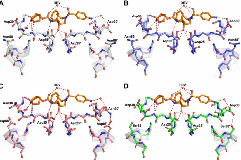
The Asp30⬘side chain of DRVB-WTdoes not directly form a hydrogen bond with DRV but indirectly interacts with the N1 atom of DRV through a water molecule-mediated hydrogen bond network (Fig. 3A). In contrast, the Asp30⬘side chain of
DRVAE-WTforms a direct hydrogen bond with the N1 atom of
DRV (Fig. 3B). Residue 30 of both NFV-resistant variants also interacts with the N1 atom of DRV through water molecule-mediated hydrogen bonding (Fig. 3C and D). However, in addition to this interaction, Asn30 of DRVB-D30N/N88D and Asp30 of DRVAE-N88Sare oriented away from the active site, enabling them to form hydrogen bonds with Asp88 and Ser88, respectively. In both cases, the NFV resistance mu-tations stabilize residue 30 away from the active site via hydrogen bonding.
[image:4.585.42.283.79.295.2]Binding thermodynamics.To determine the effects of back-ground sequence polymorphisms and NFV resistance muta-tions on inhibitor binding, the binding thermodynamic param-eters of NFV, DRV, and APV binding to WT and resistant AE and clade B variants were determined by isothermal titration calorimetry (Table 2). The AE-WT protease had a 6.9-fold-weaker affinity for NFV and a 2.7-fold-6.9-fold-weaker affinity for DRV
TABLE 1. Crystallographic statistics
Parametera Result for indicated variant
DRVB-WTb DRVB-D30N/N88D DRVAE-WT DRVAE-N88S
Inhibitor DRV DRV DRV DRV
Resolution (Å) 1.2 2.15 1.96 1.76
Space group P 212121 P 212121 P 61 P 61
Z 4 4 6 6
Cell dimensions (Å)
a 54.9 50.9 62.2 61.9
b 57.8 57.7
c 62.0 61.6 82.7 82.1
Total no. of reflections
302,022 108,838 89,284 79,445
No. of unique reflections
55,056 10,326 12,493 17,277
Rsymm(%) 3.8 6.7 5.5 4.6
Completeness (%) 95.5 99.6 93.9 97.4
I/I
25.0 9.6 11.2 19.6
Rwork(%) 14.1 18.1 20.0 19.6
Rfree(%) 17.9 23.6 25.9 23.9
RMSD
Bond length (Å) 0.004 0.009 0.009 0.007
Bond angle 1.5 1.9 1.5 1.7
PDB code 1T3R 3LZV 3LZS 3LZU
aZ, number of molecules in the unit cell; RMSD, root mean square deviation; Rsymm⫽ ⌺hkl兩Ihkl⫺ ⬍Ihkl⬎兩/⌺hklIhkl;I/I, signal-to-noise ratio;Rwork⫽ ⌺兩Fobs⫺
Fcal兩/⌺Fobs;Rfree⫽ ⌺test(兩Fobs兩⫺Fcal兩)2/⌺test兩Fobs兩2.
bSee King et al.(21) and Surleraux et al. (40).
FIG. 2. (A) Ribbon diagram superposition of DRVAE-WT(blue) and DRVB-WT(gray). The red box indicates the region of the protease
molecule highlighted in panels B to D. (B) Structural rearrangement of the flap hinge and core regions between DRVAE-WT(blue) and DRVB-WT
(gray). (C) Flap hinge and core regions of DRVAE-WT. (D) Flap hinge and core regions of DRVB-WTprotease. Hydrogen bond interactions are
indicated by red dashed lines.
on November 8, 2019 by guest
http://jvi.asm.org/
[image:4.585.134.449.439.686.2]than the affinities of B-WT protease for NFV and DRV, re-spectively (Table 2). This result indicates that the AE-WT protease has an inherently weaker affinity for NFV and DRV. No significant differences in the enthalpy of NFV binding
were observed among any of the variants. Although the bind-ing of DRV to all protease variants was enthalpically favorable, the enthalpic contributions were reduced with the AE variants (⫺10.1 kcal mol⫺1for AE-WT and⫺5.1 kcal mol⫺1for
AE-FIG. 3. Protease inhibitor hydrogen bonding interactions. DRV is shown in orange, and hydrogen bond interactions are indicated by red dashed lines. Since the charged states of the Asp25 carboxyl groups and the position of the O18 hydroxyl hydrogen of DRV are not known, all possible hydrogen bond interactions between the Asp25 carboxyl groups and O18 of the DRV molecules are shown. (A) DRVB-WT(gray).
[image:5.585.45.543.69.399.2](B) DRVAE-WT(blue). (C) DRVB-D30N/N88D(salmon). (D) DRVAE-N88S(green).
TABLE 2. Binding thermodynamic parameters for NFV, DRV, and APV binding to AE and clade B variantsa
Inhibitor and
protease variant Ka(M⫺
1
) Kd(nM) Kdratio ⌬H (kcal mol⫺
1
) ⌬⌬H ⫺T⌬S (kcal mol⫺1
) ⌬(⫺T⌬S) ⌬G (kcal mol⫺1 ) ⌬⌬G
NFV
B-WT (2.6⫾0.5)⫻109 0.39⫾0.07 1.0 4.4⫾0.1 ⫺17.0 ⫺12.6⫾0.1
B-D30N/N88D (1.2⫾0.4)⫻108 8.1⫾2.8 20.7 6.7⫾0.3 2.3 ⫺17.5 0.5 ⫺10.8⫾0.2 1.8
AE-WT (3.7⫾1.0)⫻108 2.7⫾0.7 6.9 5.0⫾0.3 0.6 ⫺16.6 0.9 ⫺11.5⫾0.2 1.1
AE-N88S (5.8⫾1.2)⫻107 17.2⫾3.5 44.1 6.2⫾0.7 1.8 ⫺16.6 0.9 ⫺10.4⫾0.1 2.2
DRV
B-WT (2.2⫾1.1)⫻1011 0.004⫾0.002 1.0 ⫺12.1⫾0.9 ⫺3.1 ⫺15.2⫾0.3
B-D30N/N88D (3.7⫾0.7)⫻1010 0.026⫾0.005 6.5 ⫺12.5⫾0.4 ⫺0.4 ⫺1.6 1.5 ⫺14.2⫾0.1 1.0
AE-WT (9.1⫾0.3)⫻1010 0.0109⫾0.0003 2.7 ⫺10.1⫾0.5 2.0 ⫺4.6 ⫺1.5 ⫺14.7⫾0.02 0.5
AE-N88S (1.1⫾0.8)⫻1010 0.087⫾0.062 21.8 ⫺5.1⫾3.6 7.0 ⫺8.4 ⫺5.3 ⫺13.5⫾0.4 1.7
APV
B-WT (2.6⫾1.3)⫻109 0.39⫾0.20 1.0 ⫺7.3⫾0.9 ⫺5.3 ⫺12.6⫾0.3
B-D30N/N88D (1.2⫾0.2)⫻1010 0.08⫾0.01 0.2 ⫺10.2⫾1.5 ⫺2.9 ⫺3.3 2.0 ⫺13.5⫾0.09 ⫺0.9
AE-WT (3.1⫾0.2)⫻109 0.32⫾0.02 0.8 ⫺5.5⫾0.3 ⫺1.8 ⫺7.3 ⫺2.0 ⫺12.70⫾0.03 ⫺0.1
AE-N88S (1.3⫾0.9)⫻1010 0.08⫾0.06 0.2 ⫺5.0⫾3.6 2.3 ⫺8.6 ⫺3.3 ⫺13.6⫾0.4 ⫺1.0
a
Ka, association constant;Kd, dissociation constant; H, enthalpy; T, temperature; S, entropy; G, Gibbs free energy.
VOL. 84, 2010 HIV-1 PROTEASE, POLYMORPHISMS, AND RESISTANCE PATHWAYS 9999
on November 8, 2019 by guest
http://jvi.asm.org/
[image:5.585.41.544.534.715.2]N88S) compared with those for the clade B variants (⫺12.1 kcal mol⫺1 for B-WT and ⫺12.5 kcal mol⫺1 for B-D30N/ N88D). As expected, the NFV-resistant variants showed a sig-nificant reduction in binding affinity for NFV compared to that of the wild-type variants. With the AE-N88S variant, the af-finity for NFV was reduced 44.1-fold (Kd⫽17.2 nM) and was far more significant than the D30N/N88D mutations in clade B protease, which reduced the affinity for NFV 20.7-fold (Kd⫽ 8.1 nM). Similarly, the AE-N88S variant had a 21.8-fold-weaker affinity (Kd ⫽ 0.087 nM) for DRV compared to a 6.5-fold-weaker affinity (Kd ⫽ 0.026 nM) with the B-D30N/
N88D variant. Thus, the single N88S substitution in the AE protease has a profound effect on the binding of NFV and DRV.
In contrast to NFV and DRV, clade-specific sequence dif-ferences and NFV resistance mutations had only a minimal effect on the affinities for APV of both AE and clade B pro-tease. Despite this, there were some differences in energy pa-rameters. The binding of APV to the clade B variants appeared to be more enthalpically favorable than that to the AE variants. This was compensated for by an increase in the entropic com-ponent to the binding energy for the AE proteases.
Protease activity and vitality. The enzyme-kinetic parame-ters determined for each clade B and AE variant with the CA-p2 fluorogenic substrate analog are summarized in Table 3. The Kmvalue for B-D30N/N88D protease (35.9M) was 2.1-fold greater than that for B-WT protease (16.7M). How-ever, theKmvalues for the AE protease variants (17.5M for AE-WT and 19.0M for AE-N88S) were similar to that of B-WT protease. The turnover rate for B-D30N/N88D protease (kcat ⫽ 0.13 s⫺1) was significantly lower than that of B-WT
protease (kcat⫽1.79 s⫺1). Turnover rates for AE-WT (k
cat⫽
0.7 s⫺1) and AE-N88S (k
cat⫽0.2 s⫺1) were 2.5 and 8.5-fold
lower, respectively, than that of clade B-WT. Thekcat/Km val-ues, or catalytic efficiency valval-ues, for B-D30N/N88D and AE variants were lower than that of B-WT protease. Therefore, the reduction in catalytic efficiency of the B-D30N/N88D pro-tease compared with that of B-WT propro-tease resulted from the combined effects of theKmandkcatvalues. However, for the
AE variants, the lower turnover rates alone were responsible for the reduced catalytic efficiencies. Overall, these results in-dicate that the polymorphic sequence differences in AE pro-tease can alter the activity profile of the enzyme compared to results with the clade B protease.
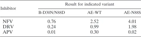
Vitality values were calculated to determine if the protease variants had a selective advantage over NFV, DRV, and APV. AE-WT and AE-N88S protease had calculated vitality values of 2.52 and 4.01 for NFV, respectively, compared with 0.76 for B-WT (Table 4). However, vitality values for DRV were not
significantly different from that of B-WT protease. Vitality values for APV were significantly lower for all variants than for B-WT protease. These results indicate that AE-WT may have a selective advantage over NFV compared to B-WT but that the AE variants may not have a significant selective advantage against DRV or APV relative to B-WT.
DISCUSSION
Although the majority of HIV-1 patients are infected with non-B forms of the virus, molecular studies have been carried out predominantly with clade B variants. The AE protease has several polymorphisms that are associated with inhibitor resis-tance in clade B. AE also shows altered patterns of drug re-sistance to NFV. We have performed detailed studies to de-termine the effects of sequence polymorphisms on enzyme structure, activity, and inhibitor binding. These analyses led to a structural rationalization for the altered pathways for drug resistance.
[image:6.585.301.541.90.153.2]AE-WT protease has an inherently weaker affinity for NFV and DRV than that of B-WT, as is evident from the thermo-dynamic data (Table 2). The weaker affinity observed for NFV is consistent with previously published data for another AE protease variant (3), as well as for clade A protease (42), which is closely related. The inherent weaker affinity for NFV likely allows the AE protease to gain resistance to NFV through a single nonactive-site substitution, N88S. The clade B protease, in contrast, which has a relatively stronger affinity for NFV, requires a combination of an active-site mutation (D30N) and a nonactive-site mutation (N88D) to gain NFV resistance. The ability of the AE-N88S protease to maintain affinity for sub-strates is evident from our enzyme kinetics data (Table 4), in which theKmvalue for AE-N88S was comparable to that of AE-WT and B-WT protease. TheKmvalue for clade B-D30N/ N88D, on the other hand, was significantly worse than that of the B-WT, likely reflecting the effect of the altered active site. As an active-site residue, Asp30 plays a key role in substrate recognition by interacting with substrates through side chain-mediated hydrogen bonds with the MA-CA, CA-p2, p1-p6, and p2-NC cleavage sites (36). Therefore, as is evident from our enzyme kinetics data, the D30N/N88D mutations in clade B will likely affect substrate binding and processing. Several stud-ies have observed substrate coevolution in instances in which the protease mutates active-site residues in order to confer inhibitor resistance (22, 23). However, since the AE-N88S pro-tease variant has no active-site mutations, the enzyme retains the ability to effectively recognize substrates while conferring NFV resistance. Therefore, the presence of the N88S sub-stitution in AE protease is unlikely to induce coevolution of
TABLE 3. Enzyme kinetics parameters for clade B and AE-WT and NFV-resistant variants
Parameter Result for indicated variant
B-WT B-D30N/N88D AE-WT AE-N88S
Km(M) 16.7⫾6.0 35.9⫾0.1 17.5⫾4.0 19.0⫾0.8
kcat(s⫺
1) 1.79⫾0.28 0.13⫾0.09 0.70⫾0.08 0.20⫾0.02
kcat/Km (s⫺1M⫺1)
[image:6.585.42.284.91.163.2]0.11⫾0.04 0.004⫾0.002 0.04⫾0.01 0.010⫾0.001
TABLE 4. Vitality values for clade B and AE WT and NFV-resistant variants
Inhibitor Result for indicated variant
B-D30N/N88D AE-WT AE-N88S
NFV 0.76 2.52 4.01
DRV 0.24 0.99 1.98
APV 0.01 0.30 0.02
on November 8, 2019 by guest
http://jvi.asm.org/
the viral substrates in order to maintain effective enzymatic activity.
Despite havingKmvalues that were comparable to that of
B-WT protease, both AE-WT and AE-N88S had significantly lower catalytic turnover rates (kcat) than that of the B-WT
protease (Table 3). As a result, the catalytic efficiency of the AE variants is lower than that of the B-WT protease. The lower turnover rates of the AE variants could be a direct result of the reduced flexibility of the flap hinge (residues 33 to 39) and core regions (residues 16 to 22) of the protein. Molecular dynamics studies have revealed that hydrophobic sliding of the core region facilitates substrate binding through the opening of the active site (9). The unique hydrogen bonds observed be-tween the flap hinge and the core in the AE variants alter movement of the core, thus impacting the ability of the active site to open up for substrate binding and product release. Based on our enzyme kinetics data, this altered flexibility of the flap hinges in the AE variants has little effect on substrate binding but rather affects the catalytic step of the reaction by slowing down product release.
The higher vitality value observed for AE-WT with NFV provides supporting evidence for the reduction in the efficacy of NFV against the AE protease compared with that of clade B (Table 4). This result is consistent with previous vitality calculations for the clade A protease (42). In addition, these results further highlight the idea that background polymorphic sequence variations in the AE protease can affect the potency of NFV. The suboptimal efficacy of NFV against the AE-WT protease likely permits a nonactive-site variant, AE-N88S, to emerge over variants with active-site mutations to effectively confer resistance to NFV.
The impact on other inhibitors, however, is complex. APV and DRV are chemically very closely related compounds, and similar susceptibility and resistance patterns have been ob-served for these two inhibitors (31). However, this pattern is not evident for this series of resistant variants. Both the N88S mutation in the AE and the D30N/N88D mutations in the clade B proteases result in hypersusceptibility to APV. Similar results have been observed also for a B-N88S protease variant (24, 45). In contrast, the same substitutions in the protease give rise to even greater resistance to DRV. However, since DRV presents a greater genetic barrier to resistance than APV (33),
thein vivoimplications of weaker affinity for DRV in the AE
variants are likely negligible. Indeed, our calculated vitality values indicate that DRV maintains its potency against the AE variants despite having a weaker affinity for WT and AE-N88S relative to clade B protease.
A close look at the NFVB-WTprotease complex reveals an important interaction between the Asp30 residue side chain and the inhibitor bound in the active site. (PDB code 3EKX) (Fig. 4A and B). One of the side chain oxygen atoms of Asp30 forms a direct hydrogen bond with the O38 atom of NFV. Our crystal structures of the NFV-resistant variants show that N88S in AE and N88D in clade B have the ability to interact with residue 30 and orient it away from the active site (Fig. 3B and D) and thereby disrupt the interaction between residue 30 and the inhibitor. These structural observations are similar to in-terpretations made in previous molecular dynamics studies involving NFV-protease complexes (27, 28). Thus, NFV resis-tance is likely caused in large part due to the loss of this interaction in the NFV-resistant variants.
Overall, mutations that emerge in response to inhibitor
ther-FIG. 4. Hydrogen bond network involving residue 88. (A) Asn88 bridges the terminal helix with Asp30 from the active site and Thr74 from one of the outer beta strands. The red box indicates the region of the protease molecule highlighted in panels B to D. (B) Asn88 in NFVB-WT(PDB
code 3EKX). (C) Asp88 in DRVB-D30N/N88D. (D) Ser88 in DRVN88S-WT. Hydrogen bond interactions are indicated by red dashed lines.
VOL. 84, 2010 HIV-1 PROTEASE, POLYMORPHISMS, AND RESISTANCE PATHWAYS 10001
on November 8, 2019 by guest
http://jvi.asm.org/
apy need to have a minimal impact on protease structure and activity to maintain the enzyme’s function. The D30N substi-tution, which is associated with NFV resistance, is one of the few drug-resistant mutations that involve a change in charge. The additional substitution of N88D likely helps preserve the net charge on the protein. In AE, resistance to NFV occurs indirectly with the N88S mutation. Likewise, the sole NFV-resistant alteration, N88S, in the AE protease does not change the overall electrostatics. Thus, in both clade B and AE, NFV resistance is attained with no change to the net charge of the enzyme. In the wild-type variants, Asn88 is one of the few internal hydrogen bonding side chains in the core of the pro-tease monomer. The side chain of Asn88 has a key role in the protease structure bridging the terminal helix, with residues 30 and 31 coming from the active site to the backbone of Thr74 in the center of one of the outer beta strands (Fig. 4A and B). With the substitutions of Asp in clade B and Ser in AE for Asn at position 88 in the NFV-resistant protease variants, the hy-drogen bonding network is preserved through the coordination of some key water molecules in the core of the protease mono-mer (Fig. 4B to D). Thus, mutations confer resistance to NFV through a series of interdependent changes that preserve the structural and electrostatic properties of HIV-1 protease.
In conclusion, protease activity and the response to protease inhibitors can be affected by clade-specific sequence differ-ences. Our findings likely extend beyond HIV-1 protease to other drug targets within HIV and underscore the need to consider clade-specific polymorphisms when developing new drugs and formulating treatment plans. Furthermore, drug re-sistance pathways observed in the context of clade B viruses cannot be assumed to hold true for other HIV-1 clades.
ACKNOWLEDGMENTS
This work was supported by grants from the National Institutes of Health (P01-GM66524) and Tibotec, Inc., to C.A.S. Additionally, this study was supported by a Grant-in-Aid for AIDS research from the Ministry of Health, Labor, and Welfare of Japan (H19-AIDS-007) to W.S.
We thank William Royer, Moses Prabu-Jayabalan, and Madhavi Nalam for helpful discussions and Christina Ng and Brendan Hilbert for assistance with data collection.
We gratefully acknowledge the Mail-In Data Collection Program of the National Synchrotron Light Source, Brookhaven National Labo-ratory, for collecting X-ray data at the X29A beamline, for which financial support comes principally from the Offices of Biological and Environmental Research and of Basic Energy Sciences of the U.S. Department of Energy and from the National Center for Research Resources of the National Institutes of Health. Use of the Advanced Photon Source for X-ray data collection was supported by the U.S. Department of Energy, Basic Energy Sciences, Office of Science, under contract DE-AC02-06CH11357. Use of the BioCARS Sector 14 was supported by the National Institutes of Health, National Center for Research Resources, under grant RR007707.
The protease inhibitors used in this study were obtained through the NIH AIDS Research and Reference Reagent Program, Division of AIDS, National Institute of Allergy and Infectious Diseases, NIH.
REFERENCES
1.Ariyoshi, K., M. Matsuda, H. Miura, S. Tateishi, K. Yamada, and W. Sug-iura.2003. Patterns of point mutations associated with antiretroviral drug treatment failure in CRF01_AE (subtype E) infection differ from subtype B infection. J. Acquir. Immune Defic. Syndr.33:336–342.
2.Carr, J. K., M. O. Salminen, C. Koch, D. Gotte, A. W. Artenstein, P. A. Hegerich, D. St. Louis, D. S. Burke, and F. E. McCutchan.1996. Full-length sequence and mosaic structure of a human immunodeficiency virus type 1 isolate from Thailand. J. Virol.70:5935–5943.
3.Clemente, J. C., R. M. Coman, M. M. Thiaville, L. K. Janka, J. A. Jeung, S. Nukoolkarn, L. Govindasamy, M. Agbandje-McKenna, R. McKenna, W. Leelamanit, M. M. Goodenow, and B. M. Dunn.2006. Analysis of HIV-1 CRF_01 A/E protease inhibitor resistance: structural determinants for main-taining sensitivity and developing resistance to atazanavir. Biochemistry45:
5468–5477.
4.Collaborative Computational Project, Number 4.1994. The CCP4 suite: programs for protein crystallography. Acta Crystallogr. D Biol. Crystallogr.
50:760–763.
5.Copeland, R. A.1996. Enzymes: a practical introduction to structure, mech-anism, and data analysis. Wiley-VCH, New York, NY.
6.DeLano, W. L.2002. The PyMol molecular graphics system. DeLano Scien-tific, San Carlos, CA.
7.de Oliveira, T., S. Engelbrecht, E. Janse van Rensburg, M. Gordon, K. Bishop, J. zur Megede, S. W. Barnett, and S. Cassol.2003. Variability at human immunodeficiency virus type 1 subtype C protease cleavage sites: an indication of viral fitness? J. Virol.77:9422–9430.
8.Emsley, P., and K. Cowtan.2004. Coot: model-building tools for molecular graphics. Acta Crystallogr. D Biol. Crystallogr.60:2126–2132.
9.Foulkes-Murzycki, J. E., W. R. Scott, and C. A. Schiffer.2007. Hydrophobic sliding: a possible mechanism for drug resistance in human immunodefi-ciency virus type 1 protease. Structure15:225–233.
10.Gao, F., D. L. Robertson, S. G. Morrison, H. Hui, S. Craig, J. Decker, P. N. Fultz, M. Girard, G. M. Shaw, B. H. Hahn, and P. M. Sharp.1996. The heterosexual human immunodeficiency virus type 1 epidemic in Thailand is caused by an intersubtype (A/E) recombinant of African origin. J. Virol.
70:7013–7029.
11.Geretti, A. M.2006. HIV-1 subtypes: epidemiology and significance for HIV management. Curr. Opin. Infect. Dis.19:1–7.
12.Gomes, P., I. Diogo, M. F. Gonc¸alves, P. Carvalho, J. Cabanas, M. C. Lobo, and R. Camacho.2002. Different pathways to nelfinavir genotypic resistance in HIV-1 subtypes B and G. Conference on Retroviruses and Opportunistic Infections, abstract 46. Conference on Retroviruses and Opportunistic In-fections, Seattle, WA.
13.Grossman, Z., E. E. Paxinos, D. Averbuch, S. Maayan, N. T. Parkin, D. Engelhard, M. Lorber, V. Istomin, Y. Shaked, E. Mendelson, D. Ram, C. J. Petropoulos, and J. M. Schapiro.2004. Mutation D30N is not preferentially selected by human immunodeficiency virus type 1 subtype C in the develop-ment of resistance to nelfinavir. Antimicrob. Agents Chemother.48:2159– 2165.
14.Gulnik, S. V., L. I. Suvorov, B. Liu, B. Yu, B. Anderson, H. Mitsuya, and J. W. Erickson.1995. Kinetic characterization and cross-resistance patterns of HIV-1 protease mutants selected under drug pressure. Biochemistry34:
9282–9287.
15.Hemelaar, J., E. Gouws, P. D. Ghys, and S. Osmanov.2006. Global and regional distribution of HIV-1 genetic subtypes and recombinants in 2004. AIDS20:W13–23.
16.Hu, D. J., A. Buve, J. Baggs, G. van der Groen, and T. J. Dondero.1999. What role does HIV-1 subtype play in transmission and pathogenesis?: an epidemiological perspective. AIDS13:873–881.
17.Kaleebu, P., N. French, C. Mahe, D. Yirrell, C. Watera, F. Lyagoba, J. Nakiyingi, A. Rutebemberwa, D. Morgan, J. Weber, C. Gilks, and J. Whit-worth.2002. Effect of human immunodeficiency virus (HIV) type 1 envelope subtypes A and D on disease progression in a large cohort of HIV-1-positive persons in Uganda. J. Infect. Dis.185:1244–1250.
18.Kanki, P. J., D. J. Hamel, J. L. Sankale, C. Hsieh, I. Thior, F. Barin, S. A. Woodcock, A. Gueye-Ndiaye, E. Zhang, M. Montano, T. Siby, R. Marlink, I. NDoi, M. E. Essex, and S. MBoup.1999. Human immunodeficiency virus type 1 subtypes differ in disease progression. J. Infect. Dis.179:68–73. 19.Kantor, R., D. A. Katzenstein, B. Efron, A. P. Carvalho, B. Wynhoven, P.
Cane, J. Clarke, S. Sirivichayakul, M. A. Soares, J. Snoeck, C. Pillay, H. Rudich, R. Rodrigues, A. Holguin, K. Ariyoshi, M. B. Bouzas, P. Cahn, W. Sugiura, V. Soriano, L. F. Brigido, Z. Grossman, L. Morris, A. M. Van-damme, A. Tanuri, P. Phanuphak, J. N. Weber, D. Pillay, P. R. Harrigan, R. Camacho, J. M. Schapiro, and R. W. Shafer.2005. Impact of HIV-1 subtype and antiretroviral therapy on protease and reverse transcriptase genotype: results of a global collaboration. PLoS Med.2:e112.
20.King, N. M., L. Melnick, M. Prabu-Jeyabalan, E. A. Nalivaika, S.-S. Yang, Y. Gao, X. Nie, C. Zepp, D. L. Heefner, and C. A. Schiffer.2002. Lack of synergy for inhibitors targeting a multi-drug-resistant HIV-1 protease. Protein Sci.
11:418–429.
21.King, N. M., M. Prabu-Jeyabalan, P. Wigerinck, M.-P. de Be´thune, and C. A. Schiffer. 2004. Structural and thermodynamic basis for the binding of TMC114, a next-generation human immunodeficiency virus type 1 protease inhibitor. J. Virol.78:12012–12021.
22.Kolli, M., S. Lastere, and C. A. Schiffer.2006. Co-evolution of nelfinavir-resistant HIV-1 protease and the p1-p6 substrate. Virology347:405–409. 23.Kolli, M., E. Stawiski, C. Chappey, and C. A. Schiffer.2009. Human
immu-nodeficiency virus type 1 protease-correlated cleavage site mutations en-hance inhibitor resistance. J. Virol.83:11027–11042.
24.Masquelier, B., K. L. Assoumou, D. Descamps, L. Bocket, J. Cottalorda, A. Ruffault, A. G. Marcelin, L. Morand-Joubert, C. Tamalet, C. Charpentier,
on November 8, 2019 by guest
http://jvi.asm.org/
G. Peytavin, Z. Antoun, F. Brun-Vezinet, and D. Costagliola.2008. Clinically validated mutation scores for HIV-1 resistance to fosamprenavir/ritonavir. J. Antimicrob. Chemother.61:1362–1368.
25.Murphy, E., B. Korber, M. C. Georges-Courbot, B. You, A. Pinter, D. Cook, M. P. Kieny, A. Georges, C. Mathiot, F. Barre-Sinoussi, et al.1993. Diversity of V3 region sequences of human immunodeficiency viruses type 1 from the Central African Republic. AIDS Res. Hum. Retroviruses9:997–1006. 26.Nunez, M., C. de Mendoza, L. Valer, E. Casas, S. Lopez-Calvo, A. Castro, B.
Roson, D. Podzamczer, A. Rubio, J. Berenguer, and V. Soriano.2002. Re-sistance mutations in HIV-infected patients experiencing early failure with nelfinavir-containing triple combinations. Med. Sci. Monit8:CR620–CR623. 27.Ode, H., S. Matsuyama, M. Hata, T. Hoshino, J. Kakizawa, and W. Sugiura.
2007. Mechanism of drug resistance due to N88S in CRF01_AE HIV-1 protease, analyzed by molecular dynamics simulations. J. Med. Chem.50:
1768–1777.
28.Ode, H., M. Ota, S. Neya, M. Hata, W. Sugiura, and T. Hoshino.2005. Resistant mechanism against nelfinavir of human immunodeficiency virus type 1 proteases. J. Phys. Chem. B109:565–574.
29.Otwinowski, Z., and W. Minor.1997. Processing of X-ray diffraction data collected in oscillation mode. Methods Enzymol.276:307–326.
30.Painter, J., and E. A. Merritt.2006. Optimal description of a protein struc-ture in terms of multiple groups undergoing TLS motion. Acta Crystallogr. D Biol. Crystallogr.62:439–450.
31.Parkin, N., E. Stawiski, C. Chappey, and E. Coakley. 2007. Darunavir/ amprenavir cross-resistance in clinical samples submitted for phenotype/ genotype combination resistance testing. Conference on Retroviruses and Opportunistic Infections, abstract 607. Conference on Retroviruses and Op-portunistic Infections, San Francisco, CA.
32.Plantier, J. C., M. Leoz, J. E. Dickerson, F. De Oliveira, F. Cordonnier, V. Lemee, F. Damond, D. L. Robertson, and F. Simon.2009. A new human immunodeficiency virus derived from gorillas. Nat. Med.15:871–872. 33.Poveda, E., C. de Mendoza, L. Martin-Carbonero, A. Corral, V. Briz, J.
Gonzalez-Lahoz, and V. Soriano.2007. Prevalence of darunavir resistance mutations in HIV-1-infected patients failing other protease inhibitors. J. Antimicrob. Chemother.60:885–888.
34.Prabu-Jeyabalan, M., E. Nalivaika, N. M. King, and C. A. Schiffer.2004. Structural basis for coevolution of the human immunodeficiency virus type 1 nucleocapsid-p1 cleavage site with a V82A drug-resistant mutation in viral protease. J. Virol.78:12446–12454.
35.Prabu-Jeyabalan, M., E. A. Nalivaika, K. Romano, and C. A. Schiffer.2006. Mechanism of substrate recognition by drug-resistant human immunodefi-ciency virus type 1 protease variants revealed by a novel structural interme-diate. J. Virol.80:3607–3616.
36.Prabu-Jeyabalan, M., E. A. Nalivaika, and C. A. Schiffer.2002. Substrate shape determines specificity of recognition for HIV-1 protease: analysis of crystal structures of six substrate complexes. Structure10:369–381. 37.Robertson, D. L., J. P. Anderson, J. A. Bradac, J. K. Carr, B. Foley, R. K.
Funkhouser, F. Gao, B. H. Hahn, M. L. Kalish, C. Kuiken, G. H. Learn, T. Leitner, F. McCutchan, S. Osmanov, M. Peeters, D. Pieniazek, M. Salminen, P. M. Sharp, S. Wolinsky, and B. Korber.2000. HIV-1 nomenclature pro-posal. Science288:55–56.
38.Rose, J. R., R. Salto, and C. S. Craik.1993. Regulation of autoproteolysis of the HIV-1 and HIV-2 proteases with engineered amino acid substitutions. J. Biol. Chem.268:11939–11945.
39.Spira, S., M. A. Wainberg, H. Loemba, D. Turner, and B. G. Brenner.2003. Impact of clade diversity on HIV-1 virulence, antiretroviral drug sensitivity and drug resistance. J. Antimicrob. Chemother.51:229–240.
40.Surleraux, D. L., A. Tahri, W. G. Verschueren, G. M. Pille, H. A. de Kock, T. H. Jonckers, A. Peeters, S. De Meyer, H. Azijn, R. Pauwels, M. P. de Bethune, N. M. King, M. Prabu-Jeyabalan, C. A. Schiffer, and P. B. Wiger-inck.2005. Discovery and selection of TMC114, a next generation HIV-1 protease inhibitor. J. Med. Chem.48:1813–1822.
41.Tebit, D. M., I. Nankya, E. J. Arts, and Y. Gao.2007. HIV diversity, recom-bination and disease progression: how does fitness “fit” into the puzzle? AIDS Rev.9:75–87.
42.Velazquez-Campoy, A., M. J. Todd, S. Vega, and E. Freire.2001. Catalytic efficiency and vitality of HIV-1 proteases from African viral subtypes. Proc. Natl. Acad. Sci. U. S. A.98:6062–6067.
43.Velazquez-Campoy, A., S. Vega, E. Fleming, U. Bacha, Y. Sayed, H. W. Dirr, and E. Freire.2003. Protease inhibition in African subtypes of HIV-1. AIDS Rev.5:165–171.
44.Williams, T., and C. Kelley.Accessed 28 January 2009. gnuplot, version 4.2. http://www.gnuplot.info/.
45.Ziermann, R., K. Limoli, K. Das, E. Arnold, C. J. Petropoulos, and N. T. Parkin.2000. A mutation in human immunodeficiency virus type 1 protease, N88S, that causes in vitro hypersensitivity to amprenavir. J. Virol.74:4414– 4419.
VOL. 84, 2010 HIV-1 PROTEASE, POLYMORPHISMS, AND RESISTANCE PATHWAYS 10003