Copyright © 2009, American Society for Microbiology. All Rights Reserved.
3
⬘
cis
-Acting Elements That Contribute to the Competence and
Efficiency of Japanese Encephalitis Virus Genome Replication:
Functional Importance of Sequence Duplications, Deletions,
and Substitutions
䌤
Sang-Im Yun,† Yu-Jeong Choi,† Byung-Hak Song, and Young-Min Lee*
Department of Microbiology, College of Medicine and Medical Research Institute, Chungbuk National University, Cheongju, South Korea
Received 10 December 2008/Accepted 26 May 2009
The positive-strand RNA genome of Japanese encephalitis virus (JEV) terminates in a highly conserved 3ⴕ-noncoding region (3ⴕNCR) of six domains (V, X, I, II-1, II-2, and III in the 5ⴕ-to-3ⴕ direction). By manip-ulating the JEV genomic RNA, we have identified important roles for RNA elements present within the 574-nucleotide 3ⴕNCR in viral replication. The two 3ⴕ-proximal domains (II-2 and III) were sufficient for RNA replication and virus production, whereas the remaining four (V, X, I, and II-1) were dispensable for RNA replication competence but required for maximal replication efficiency. Surprisingly, a lethal mutant lacking all of the 3ⴕNCR except domain III regained viability through pseudoreversion by duplicating an 83-nucleotide sequence from the 3ⴕ-terminal region of the viral open reading frame. Also, two viable mutants displayed severe genetic instability; these two mutants rapidly developed 12 point mutations in domain II-2 in the mutant lacking domains V, X, I, and II-1 and showed the duplication of seven upstream sequences of various sizes at the junction between domains II-1 and II-2 in the mutant lacking domains V, X, and I. In all cases, the introduction of these spontaneous mutations led to an increase in RNA production that paralleled the level of protein accumulation and virus yield. Interestingly, the mutant lacking domains V, X, I, and II-1 was able to replicate in hamster BHK-21 and human neuroblastoma SH-SY5Y cells but not in mosquito C6/36 cells, indicating a cell type-specific restriction of its viral replication. Thus, our findings provide the basis for a detailed map of the 3ⴕcis-acting elements in JEV genomic RNA, which play an essential role in viral replication. They also provide experimental evidence for the function of 3ⴕdirect repeat sequences and suggest possible mechanisms for the emergence of these sequences in the 3ⴕNCR of JEV and perhaps in other flaviviruses.
Japanese encephalitis virus (JEV), a mosquito-borne flavi-virus of the familyFlaviviridae, is serologically related to sev-eral significant human pathogens, including West Nile virus (WNV), Kunjin virus (KUNV), St. Louis encephalitis virus, and Murray Valley encephalitis virus. It is also phylogenetically close to other clinically important human pathogens, including yellow fever virus (YFV) and dengue virus (DENV) (11, 67). JEV is the leading cause of viral encephalitis in Southeast Asia, including China, Japan, Korea, the Philippines, Thailand, and India, and it has begun to expand throughout the Indonesian archipelago and as far as Australia (21, 43). Despite the fact that JEV is generally asymptomatic, ⬃50,000 cases are re-ported annually, and the disease has a mortality rate of⬃25%, mainly in children and young adults (29, 63). Thus, the geo-graphic expansion and clinical importance of JEV infection have drawn increasing attention from the international public health community (44, 71).
Like other flaviviruses, JEV is a spherical enveloped virus (⬃50 nm diameter) with a single-stranded positive-sense RNA
genome that contains a 5⬘ cap structure but lacks a 3⬘ poly-adenylated tail. Its genomic RNA of⬃11,000 nucleotides (nt) consists of a single long open reading frame (ORF) with two noncoding regions (NCRs) at the 5⬘and 3⬘ends (41, 84). The ORF is translated into an⬃3,400-amino acid polyprotein pre-cursor, which is co- or posttranslationally cleaved by a cellular protease(s) or a viral protease complex into 10 mature pro-teins: (i) three structural proteins, the capsid (C), premem-brane (prM; which is further processed into pr and M), and envelope (E) proteins; and (ii) seven nonstructural proteins, NS1, NS2A, NS2B, NS3, NS4A, NS4B, and NS5, as arranged in the genome (13, 41, 84). The nonstructural proteins, to-gether with cellular factors, form a viral replicase complex that directs the replication of the genomic RNA in the cytoplasm of the host cell in association with perinuclear membranes (40, 74). For the synthesis of the genomic RNA to take place, this replicase complex must specifically recognize viral cis-acting RNA elements, defined by primary sequences or secondary/ tertiary structures. These RNA elements are found in various locations within the genome but most frequently are located in the 5⬘- and 3⬘NCRs (23, 47). The identification and character-ization of thesecis-acting RNA elements is critical for under-standing the complete cycle of JEV genome replication.
The availability of the complete nucleotide sequence of YFV genomic RNA (57) has led to the identification of three major conserved elements in the 5⬘- and 3⬘-terminal regions of * Corresponding author. Mailing address: Department of
Microbi-ology, College of Medicine & Medical Research Institute, Chungbuk National University, 12 Gaeshin-Dong, Heungduk-Ku, Cheongju-Si, South Korea. Phone: 82-43-261-2863. Fax: 82-43-272-1603. E-mail: [email protected].
† These authors contributed equally to this work.
䌤Published ahead of print on 3 June 2009.
7909
on November 8, 2019 by guest
http://jvi.asm.org/
the genomic RNA that contain the short primary sequences and secondary structures required for flavivirus RNA replica-tion. (i) Both ends of the genomic RNA terminate with the conserved dinucleotides 5⬘-AG and CU-3⬘(9, 10, 32, 45, 57, 72, 73) in all flaviviruses except an insect cell fusing agent virus (12). Mutations substituting another nucleotide for one of these four nucleotides in KUNV or WNV replicon RNA are known to abolish or compromise RNA replication (35, 69). (ii) A 3⬘ stem-loop structure (3⬘SL) has been recognized in all flaviviruses within the ⬃90-nt 3⬘-terminal region of the genomic RNA (9, 45, 57). The structural and functional im-portance of this 3⬘SL in RNA replication has been demon-strated in several flaviviruses (9, 18, 49, 50, 61, 70, 82, 86). (iii) The presence of short 5⬘and 3⬘cyclization sequences (5⬘CYC and 3⬘CYC, respectively) in all mosquito-borne flaviviruses suggests that flavivirus genomes can cyclize via 5⬘-3⬘long-range base-pairing interaction, since the 3⬘CYC upstream of the 3⬘SL is complementary to the 5⬘CYC in the 5⬘coding region of the C protein (30). The role of these CYC motifs in RNA repli-cation has been well characterized via cell-based assays in many mosquito-borne flaviviruses, including KUNV (34), WNV (42), YFV (8, 14), and DENV (2, 22, 49), and in cell-free systems in the case of WNV (51) and DENV (1, 3, 79, 80). Other RNA elements that have recently been shown to be important for RNA replication in DENV and WNV include an additional pair of complementary sequences (designated 5⬘ -and 3⬘UARs) that participate in genome cyclization (3, 4, 17, 87) and a 5⬘ stem-loop structure (designated 5⬘SLA) present within the 5⬘NCR that promotes RNA synthesis in association with the 3⬘NCR (22).
In all flaviviruses, the 3⬘NCR of the genomic RNA is rela-tively long (⬃400 to ⬃800 nt), with an array of conserved primary sequences and secondary structures. Although signif-icant progress has been made in identifyingcis-acting elements within the 3⬘NCRs that are essential for RNA replication, most of these elements (i.e., the 3⬘CYC, 3⬘SL, and CU-3⬘) are lim-ited to the⬃100-nt 3⬘-terminal region that is highly conserved in these viruses (see recent reviews in references 23 and 47). However, the functional importance of the remaining 5⬘ -prox-imal region of the 3⬘NCR, which differs in sequence between the various serological groups, is poorly understood. In partic-ular, comparative sequence analyses and genetic algorithm-based computer modeling have suggested that in addition to the well-studied ⬃100-nt 3⬘-proximal region, the remaining ⬃474-nt 5⬘-proximal region of the 574-nt JEV 3⬘NCR also contains several RNA elements that may play critical roles in the viral life cycle (52, 55, 56, 68). To date, however, experi-mental evidence for the functional importance of these poten-tial RNA elements in JEV genomic RNA replication is lacking. In the present study, we have identified and characterized the 3⬘ cis-acting RNA elements within the JEV 3⬘NCR and shown that they play an essential and/or regulatory role in genomic RNA replication. In particular, we have constructed and functionally characterized genome-length JEV mutant cDNAs with a series of 5⬘-to-3⬘ or 3⬘-to-5⬘ progressive dele-tions within the 3⬘NCR. In addition to identifying particular mutations within this region that affect either the competence or efficiency of genomic RNA replication, we found that the serial passaging of these mutants in susceptible BHK-21 cells produced a large number of pseudorevertants bearing a wide
variety of spontaneous point mutations and sequence duplica-tions, some of which were capable of restoring the replication competence of the defective mutants or enhancing replication efficiency. In addition, we assessed the replication of these mutants in three different cell types (BHK-21, SH-SY5Y, and C6/36 cells). Collectively, these data offer new insights into the functional importance of 3⬘cis-acting RNA elements that reg-ulate the cell type-dependent replication of JEV and perhaps other closely related mosquito-borne flaviviruses. Our findings also provide experimental evidence for the emergence of func-tional 3⬘direct repeat sequences that are duplicated from the coding region and 3⬘NCR of JEV genomic RNA.
MATERIALS AND METHODS
Cells.The three cell lines used in this study, baby hamster kidney (BHK-21), human neuroblastoma (SH-SY5Y), and mosquitoAedes albopictus(C6/36), were cultivated as described previously (36). All reagents used for cell culture were purchased from Invitrogen-Gibco, Carlsbad, CA.
Antibodies.A mouse polyclonal JEV-specific antiserum was purchased from the American Type Culture Collection (36). A rabbit polyclonal antiserum spe-cific for JEV NS1 (166 amino acids, corresponding to nt 2,478 to 2,975) was produced by immunizing New Zealand White rabbits with a glutathioneS -transferase-tagged recombinant protein consisting of the defined coding region of NS1 fused to the C terminus of glutathioneS-transferase (64). Nucleotide positions refer to the complete genome sequence of the JEV CNU/LP2 strain (GenBank accession no. AY585243). A rabbit polyclonal anti-glyceraldehyde-3-phosphate dehydrogenase (GAPDH) antiserum was obtained from LabFrontier, Seoul, South Korea (36). Horseradish peroxidase (HRP)-conjugated goat anti-mouse immunoglobulin G (IgG) and alkaline phosphatase (AP)-conjugated goat anti-mouse and anti-rabbit IgGs were purchased from Jackson ImmunoResearch Laboratories, West Grove, PA.
Mutagenesis of the full-length infectious JEV cDNA.Standard recombinant DNA techniques were used to construct the recombinant molecular clones for JEV (59). All JEV 3⬘NCR mutations were introduced into the full-length infec-tious JEV cDNA molecular clone, pBACSP6
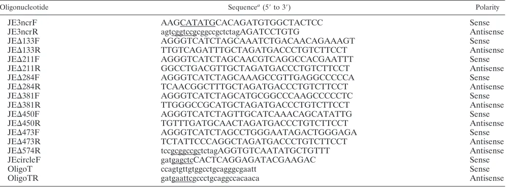
/JVFLx/XbaI (83), designated the wild type (WT) in this report; the complete genome sequence of WT-derived JEV (CNU/LP2x) has been deposited under GenBank accession no. GQ199609. Mutations were generated by PCR-based mutagenesis, and all PCR-derived DNA fragments were sequenced to confirm the presence of the intended muta-tions and the absence of undesired mutamuta-tions. The recombinant plasmids were purified by standard CsCl/ethidium bromide density centrifugation; the integrity of all plasmids was checked by extensive restriction endonuclease mapping and sequencing. The oligonucleotides used for cDNA synthesis, PCR amplification, and mutagenesis in this study are summarized in Table 1.
(i) Construction of JEV 3ⴕNCR mutant cDNAs.We constructed a total of seven JEV 3⬘NCR mutant cDNA clones, each carrying a deletion of a different size within the 3⬘NCR of the full-length WT JEV cDNA. The first six deletion mutants (MUT⌬1-133, MUT⌬1-211, MUT⌬1-284, MUT⌬1-381, MUT⌬1-450, and MUT⌬1-473) were generated by overlapping extension PCR with the WT as the template. Initially, two primary overlapping fragments were amplified by a first round of PCR with two pairs of primers: (i) JE3ncrF⫹JE⌬133R and JE⌬ 133F⫹JE3ncrR for MUT⌬1-133, (ii) JE3ncrF⫹JE⌬211R and JE⌬211F⫹JE3 ncrR for MUT⌬1-211, (iii) JE3ncrF⫹JE⌬284R and JE⌬284F⫹JE3ncrR for MUT⌬1-284, (iv) JE3ncrF⫹JE⌬381R and JE⌬381F⫹JE3ncrR for MUT⌬1-381, (v) JE3ncrF⫹JE⌬450R and JE⌬450F⫹JE3ncrR for MUT⌬1-450, and (vi) JE3 ncrF⫹JE⌬473R and JE⌬473F⫹JE3ncrR for MUT⌬1-473. In all cases, two pri-mary overlapping PCR products were gel purified and subsequently fused by a second round of PCR with the 5⬘outside primer JE3ncrF and the 3⬘outside primer JE3ncrR. The resulting amplicons were digested with NdeI and RsrII, generating fragments of the expected sizes: 887 bp for MUT⌬1-133 (i), 809 bp for MUT⌬1-211 (ii), 736 bp for MUT⌬1-284 (iii), 639 bp for MUT⌬1-381 (iv), 570 bp for MUT⌬1-450 (v), and 547 bp for MUT⌬1-473 (vi). Finally, each of the NdeI-RsrII fragments was cloned into the corresponding site in WT cDNA by a three-piece ligation, i.e., the 887-bp NdeI-RsrII fragment created to construct MUT⌬1-133 was ligated to the 7,449-bp RsrII-PacI and 10,096-bp PacI-NdeI fragments of the WT. In the case of the last (single-deletion) mutant, MUT⌬ 477-574, a fragment of the WT was first amplified by PCR with a pair of primers, JE3ncrF and JE⌬574R, and the 915-bp NdeI-NotI fragment of the resulting
7910 YUN ET AL. J. VIROL.
on November 8, 2019 by guest
http://jvi.asm.org/
amplicons was then ligated with the 7,456-bp NotI-PacI and 10,096-bp PacI-NdeI fragments of the WT.
(ii) Reconstruction of MUT⌬1-450/83ntIns.MUT⌬1-450/83ntIns, a derivative of MUT⌬1-450, contained an 83-nt sequence duplication that was discovered at the 3⬘-terminal region of the replicating genomic RNAs recovered from MUT⌬1-450-derived pseudorevertants. To construct MUT⌬1-450/83ntIns
, a bac-terial plasmid that we used for sequencing (see below) was utilized as a template for PCR with a pair of primers, JE3ncrF and JE3ncrR, to amplify a fragment containing the 83-nt sequence duplication. The 653-bp NdeI-RsrII fragment of the resulting amplicons was then ligated with the 7,449-bp RsrII-PacI and 10,096-bp PacI-NdeI fragments of MUT⌬1-450.
(iii) Reconstruction of MUT⌬1-381 derivatives.A panel of 12 MUT⌬1-381 derivatives was constructed; each of these carried 1 of 12 point mutations that were identified at the 3⬘-terminal region of the genomic RNAs extracted from MUT⌬1-381-derived pseudorevertants. To reproduce these 12 derivatives of MUT⌬1-381, 12 fragments containing 1 of the 12 genetic changes each were PCR-amplified with primers JE3ncrF and JE3ncrR from the corresponding individual bacterial plasmids that we cloned for sequencing analysis (see below). The amplicons were digested with NdeI and RsrII, and the resulting NdeI-RsrII fragments each were ligated with two fragments of MUT⌬1-381 (the 7,449-bp RsrII-PacI and 10,096-bp PacI-NdeI fragments).
(iv) Reconstruction of MUT⌬1-284 derivatives.A set of seven MUT⌬1-284
derivatives was constructed, each harboring one of seven sequence duplications of various sizes that were detected at the 3⬘-terminal region of the genomic RNAs recovered from MUT⌬1-284-derived pseudorevertants. In each case, a fragment containing one of the seven sequence duplications was PCR amplified with primers JE3ncrF and JE3ncrR from the corresponding individual clone that was used for sequencing (see below). The NdeI-RsrII fragment of the resulting variably sized amplicons was substituted for the corresponding region of MUT⌬1-284 by a three-piece ligation, as described above for the MUT⌬1-381 derivatives.
RNA transcription.Template cDNA plasmids were linearized by initial diges-tion with XbaI and subsequent treatment with mung bean nuclease, and linear-ized plasmids were purified by phenol-chloroform extraction and ethanol pre-cipitation. Runoff RNA transcripts were synthesized from linearized plasmid templates in vitro with SP6 RNA polymerase in a 25-l reaction mixture con-taining⬃200 ng of template cDNA, 0.6 mM of cap analog [m7G(5⬘)ppp(5⬘)A; New England Biolabs, Beverly, MA], 1 mM each of the four nucleoside triphos-phates (GE Healthcare, Piscataway, NJ), 0.5M of [3H]UTP (1.0 mCi/ml and 50 Ci/mmol; PerkinElmer, Waltham, MA), 15 U of SP6 RNA polymerase (New England Biolabs), 40 U of RNaseOUT (Invitrogen), 10 mM dithiothreitol (DTT), and the buffer supplied by the manufacturer. The reactions were carried out at 37°C for 1 h. Transcript yields were quantified on the basis of [3 H]UTP-incorporated RNA adsorption to DE-81 (Whatman, Maidstone, United King-dom) filter paper (59), and transcript integrity was verified by agarose gel
elec-trophoresis and ethidium bromide staining. RNA transcripts were used for transfection without any additional purification.
RNA transfection and quantitation of RNA infectivity.For RNA transfection, subconfluent BHK-21 cells were trypsinized, washed three times with cold phos-phate-buffered saline (PBS), and resuspended at 2⫻107cells/ml in PBS. Cell suspensions (400l each) were then transfected with 2g of RNA transcripts by electroporation under previously optimized conditions (980 V, 99-s pulse length, and five pulses) with a model ECM 830 electroporator (BTX, San Diego, CA) (83) and then transferred to 10 ml of the fresh complete medium.
The specific infectivity of RNA transcripts was determined by infectious center assays. The RNA-transfected cells were serially diluted in 10-fold steps and plated onto monolayers of naïve BHK-21 cells (3⫻105cells/well) in a 6-well plate. After incubation at 37°C for 4 to 6 h, the attached cells were overlaid with semisolid minimal essential medium (MEM) containing 0.5% SeaKem LE aga-rose (Lonza, Rockland, ME) and 10% fetal bovine serum (FBS) and then incubated for 4 days at 37°C under 5% CO2. The cells were overlaid with 2 ml of 7% formaldehyde per well and kept at room temperature for⬎4 h. After three 10-min washes with PBS, the cells were permeabilized with 0.25% Triton X-100 in PBS for 10 min and then incubated with the mouse anti-JEV antiserum (diluted 1:500 in PBS with 0.25% Triton X-100) at room temperature for 2 h. After three additional washes with PBS, the cells were incubated with HRP-conjugated goat anti-mouse IgG (1:1,000 dilution) at room temperature for 2 h. The cells were washed again with PBS, and the infectious centers of foci were stained by the addition of 3,3⬘-diaminobenzidine (DAB; Vector Laboratories, Burlingame, CA) according to the manufacturer’s instructions.
Immunoblotting.Total cell lysates were prepared by direct lysis of cell mono-layers with 1⫻sample loading buffer (80 mM Tri-HCl [pH 6.8], 2.0% sodium dodecyl sulfate [SDS], 10% glycerol, 0.1 M DTT, and 0.2% bromophenol blue). An equal amount of cell lysates was boiled for 5 min, separated on an SDS–12% polyacrylamide gel under reducing conditions, and electrotransferred to a meth-anol-activated polyvinylidene difluoride membrane (Bio-Rad Laboratories, Her-cules, CA). The membrane was treated with 5% (wt/vol) nonfat dried milk in TBS-T buffer (20 mM Tris [pH 8.8], 137 mM NaCl, and 0.1% Tween 20) at room temperature for 1 h, followed by three 10-min washes with TBS-T buffer. The membrane was then incubated in TBS-T buffer containing 0.5% nonfat dried milk at room temperature for 2 h with a mouse anti-JEV (1:1,000 dilution), rabbit anti-JEV NS1 (1:1,000 dilution), or rabbit anti-GAPDH antiserum (1: 10,000 dilution). The primary antibody-decorated membrane again was washed three times with TBS-T buffer and incubated with an AP-conjugated goat anti-mouse or anti-rabbit IgG, as appropriate, at a 1:5,000 dilution in TBS-T buffer containing 0.5% nonfat dried milk at room temperature for 2 h. Following three 10-min washes with TBS-T buffer, the membrane was stained with a mixture of 5-bromo-4-chloro-3-indolyl-phosphate (BCIP) and nitroblue tetrazolium (NBT).
Real-time quantitative RT-PCR.Total cellular RNAs were purified by direct lysis of cell monolayers with TRIzol reagent (Invitrogen) according to the man-TABLE 1. Oligonucleotides used for cDNA synthesis, PCR amplification, and mutagenesis
Oligonucleotide Sequencea(5⬘to 3⬘) Polarity
JE3ncrF AAGCATATGCACAGATGTGGCTACTCC Sense
JE3ncrR agtcggtccgcggccgctctagAGATCCTGTG Antisense
JE⌬133F AGGGTCATCTAGCAAATCTGACAACAGAAAGT Sense
JE⌬133R TTGTCAGATTTGCTAGATGACCCTGTCTTCCT Antisense
JE⌬211F AGGGTCATCTAGCAACGTCAGGCCACGAATTT Sense
JE⌬211R GGCCTGACGTTGCTAGATGACCCTGTCTTCCT Antisense
JE⌬284F AGGGTCATCTAGCAAAGCCGTTGAGGCCCCCA Sense
JE⌬284R TCAACGGCTTTGCTAGATGACCCTGTCTTCCT Antisense
JE⌬381F AGGGTCATCTAGCATGCGGCCCAAGCCCCCTC Sense
JE⌬381R TTGGGCCGCATGCTAGATGACCCTGTCTTCCT Antisense
JE⌬450F AGGGTCATCTAGTTGCATCAAACAGCATATTG Sense
JE⌬450R TGTTTGATGCAACTAGATGACCCTGTCTTCCT Antisense
JE⌬473F AGGGTCATCTAGCCTGGGAATAGACTGGGAGA Sense
JE⌬473R TCTATTCCCAGGCTAGATGACCCTGTCTTCCT Antisense
JE⌬574R tccgcggccgctctagAGGTGTCAATATGCTGTTT Antisense
JEcircleF gatgagctcCACTCAGGAGATACGAAGAC Sense
OligoT ccagtgttgtggcctgcagggcgaatt Sense
OligoTR gatgaattcgccctgcaggccacaaca Antisense
aJEV-specific sequences are shown in uppercase letters, and sequences unrelated to JEV are indicated in lowercase letters. Restriction endonuclease recognition
sites used for cDNA cloning are underlined.
on November 8, 2019 by guest
http://jvi.asm.org/
[image:3.585.44.546.81.266.2]ufacturer’s instructions and used for the first-strand cDNA synthesis with two reverse primers (listed below), one specific for the JEV NS3 protein-coding region and the other specific for BHK-21-actin RNA (for use in normalizing the JEV RNA values). Each 20-l reverse transcription (RT) reaction mixture contained 50 ng of template RNA, 5 pmol of the primers, 10 mM of deoxynucleo-side triphosphate mix, 100 U of Superscript II reverse transcriptase (Invitrogen), 40 U of RNaseOUT (Invitrogen), 0.1 mM DTT, and the buffer supplied by the manufacturer. The RT reaction was carried out at 45°C for 30 min, followed by PCR amplification of 2-l aliquots of the RT reaction mixture using the iQ Supermix Quantitative PCR system (Bio-Rad Laboratories) and the iCycler iQ Multicolor Real-Time PCR Detection system (Bio-Rad Laboratories). The re-actions were carried out under the following conditions: 95°C for 10 min, fol-lowed by 45 cycles of 95°C for 15 s and 60°C for 1 min. The target sequences were amplified by using the following primer pairs and fluorogenic TaqMan probes (85): JEV RNA forward (5⬘-ATCCAACTCAACCGCAAGTC-3⬘, correspond-ing to nt 5,757 to 5,766), reverse (5⬘-TCTAAGATGGTGGGTTTCACG-3⬘, corresponding to nt 5,894 to 5,914), and probe (5⬘-6-carboxyfluorescein-CATC TCTGAAATGGGGGCTA-black hole quencher 1-3⬘, corresponding to nt 5,837 to 5,856);-actin RNA forward (5⬘-ACTGGCATTGTGATGGACTC-3⬘), re-verse (5⬘-CATGAGGTAGTCTGTCAGGTC-3⬘), and probe (5⬘-HEX-CCAGC CAGGTCCAGACGCAGG-black hole quencher 2-3⬘). Samples were run in duplicate; a reaction without an aliquot of the RT reaction mixture was used to establish baseline fluorescence levels. Data were based on a threshold cycle (CT)
in which the signal was higher than that of the background. Relative changes in JEV RNA levels were estimated by using the 2⫺⌬⌬CT
method (60, 75).
Analysis of viral growth kinetics.Naïve BHK-21, SH-SY5Y, or C6/36 cells were preseeded in 35-mm dishes at 3⫻105
cells/dish for 12 to 18 h before infection with RNA transcript-derived viruses at a multiplicity of infection (MOI) of 1. After incubation for 1 h at 37°C (for BHK-21 and SH-SY5Y) or 28°C (C6/36), the cells were washed twice and incubated with fresh complete medium for 4 days (BHK-21 and SH-SY5Y) or 6 days (C6/36). At the indicated time points, equal volumes of culture supernatants were collected and used for titra-tion on naïve BHK-21 cells. Cells were preseeded onto 6-well plates at 3⫻105 cells/well for 12 to 18 h and then infected with serial 10-fold dilutions of the culture supernatants containing virus for 1 h at 37°C with frequent shaking. The cell monolayers were overlaid with semisolid MEM containing 0.5% SeaKem LE agarose (Lonza) and 10% FBS. After incubation for 4 days at 37°C with 5% CO2, the infectious centers of foci were immunostained with mouse JEV anti-serum and HRP-conjugated goat anti-mouse IgG and then visualized with DAB as described above. Virus yields were calculated and are presented as focus-forming units (FFU) per milliliter.
Serial passage of viruses.WT virus and five mutant viruses (MUT⌬1-133,
MUT⌬1-211, MUT⌬1-284, MUT⌬1-381, and MUT⌬1-450/83ntIns ) obtained from the corresponding RNA-transfected BHK-21 cells were serially passaged in naïve BHK-21 cells for three consecutive rounds of infection. For each round, a monolayer of naïve BHK-21 cells (3⫻105) preseeded in a 35-mm dish was infected with 1 ml of virus suspension at an MOI of 1 and then gently agitated for 1 h at 37°C. After a rinse with 3 ml of serum-free MEM, the cells were incubated in 3 ml of the fresh complete medium containing 10% FBS at 37°C with 5% CO2. Once any JEV-induced cytopathic effects were noted, the culture supernatants containing progeny virions were clarified by centrifugation at 2,000 rpm for 10 min and kept at⫺80°C until the next round of infection.
Sequence analysis of the 3ⴕ-terminal region of JEV genomic RNAs. Viral genomic RNAs were isolated from 200l of virus-containing culture superna-tants with 600l of TRIzol LS reagent (Invitrogen), as recommended by the manufacturer, and resuspended in 20l of RNafree distilled water. To se-quence the 3⬘-terminal region of JEV genomic RNAs, we employed the 3⬘rapid amplification of cDNA ends (3⬘RACE), as described previously (83). Synthetic oligonucleotide T (OligoT) was ligated to the 3⬘end of the genomic RNA to serve as a specific primer binding site for cDNA synthesis and subsequent PCR amplification. OligoT was 5⬘ phosphorylated with T4 polynucleotide kinase (Takara, Shiga, Japan) and 3⬘blocked by incorporating ddATP with terminal deoxynucleotidyltransferase (Takara) to prevent the intra- and intermolecular ligation of OligoT. The ligation of the modified OligoT to the 3⬘end of the genomic RNA was carried out at 16°C for 12 h using T4 RNA ligase in a 20-l reaction mixture containing 10l of extracted genomic RNAs, 10 pmol of OligoT, 10 U of T4 RNA ligase (New England Biolabs), 40 U of RNaseOUT (Invitrogen), and the buffer supplied by the manufacturer. The OligoT-ligated RNA was extracted with phenol-chloroform, precipitated with ethanol, and re-suspended in 20l of RNase-free distilled water. Half of the ligated RNA was used for cDNA synthesis with oligonucleotide TR (OligoTR), which is comple-mentary to OligoT. RT was carried out for 1 h at 45°C in a 20-l reaction mixture containing 10l of the ligated RNA, 5 pmol of primer, 10 mM of
deoxynucleo-side triphosphate mix, 100 U of Superscript II reverse transcriptase (Invitrogen), 40 U of RNaseOUT (Invitrogen), 0.1 mM DTT, and the buffer supplied by the manufacturer. One-quarter of the RT reaction mixture was subsequently used for PCR amplification withPfu-derived Pyrobest DNA polymerase (Takara) and a pair of primers, OligoTR and JEcircleF (complementary to nt 10,345 to 10,364). The PCRs consisted of 30 cycles of 30 s at 94°C, 30 s at 60°C, and 1 min at 72°C, followed by a final extension of 10 min at 72°C. cDNA amplicons were digested with SacI and EcoRI and subsequently cloned into the respective sites of pRS2 cloning vector. Approximately 20 randomly picked individual clones were sequenced to characterize the genetic changes in the 3⬘-terminal region of the viral genomes.
Secondary structure modeling.Optimal RNA secondary structures were pre-dicted by using the mfold, version 3.2, web server (http://frontend.bioinfo.rpi.edu /applications/mfold/cgi-bin/rna-form1.cgi) under default folding conditions (37°C, 1 M NaCl, no divalent ions, and no limit on distance between paired bases) (48, 88).
RESULTS
Mapping of the 3ⴕ cis-acting RNA elements required for genomic RNA replication and infectious virus production. Comprehensive sequence comparisons and computer-based modeling previously revealed a pattern of RNA elements in the 3⬘NCR of JEV genomic RNA that are highly conserved among all members of JEV-related mosquito-borne flavivi-ruses (30, 52, 55, 56, 68). Based on these previous reports and our own comparative studies here, we have subdivided the 574-nt JEV 3⬘NCR into six domains (V, X, I, II-1, II-2, and III in the 5⬘-to-3⬘direction). With the exception of the X domain, all of these domains include one of the⬃20- to 30-nt conserved sequences (CSs) or repeated conserved sequences (RCSs) that were originally recognized by Hahn et al. (30); domain V contains RCS3, I contains CS3, II-1 contains RCS2, II-2 con-tains CS2, and III concon-tains CS1, which incorporates the 8-nt core sequence of 3⬘CYC (Fig. 1A). Downstream sequences of the CS1 motif in the 3⬘-terminal domain III correspond to the ⬃90-nt 3⬘SL.
To investigate the functional importance of these six do-mains of JEV 3⬘NCR in viral replication, particularly focusing on the 5⬘-proximal region, we introduced a series of progres-sive deletions into the 3⬘NCR in the context of the viral ge-nome by using a full-length infectious JEV cDNA (WT), gen-erating a total of seven mutant cDNAs. For discussion purposes, the nucleotides of the 574-nt WT 3⬘NCR are num-bered consecutively from 1 to 574 in the 5⬘-to-3⬘direction in this report; the designations of the seven mutants indicate the regions deleted from the corresponding mutant constructs. The first five mutant cDNAs (MUT⌬1-133, MUT⌬1-211, MUT⌬1-284, MUT⌬1-381, and MUT⌬1-450) were generated by progressively deleting from one to five of the 5⬘-proximal five domains in the 5⬘-to-3⬘direction, the sixth (MUT⌬1-473) by removing all five domains plus the 3⬘CYC at the 5⬘end of the 3⬘-proximal domain III, and the seventh (MUT⌬477-574) by eliminating only the 3⬘SL at the 3⬘ end of the genome (Fig. 1B).
We first analyzed the effect of these deletions on the specific infectivity and viability of JEV genomic RNA by transfecting naïve BHK-21 cells with 2g of RNA transcripts synthesized from the WT or one of the seven 3⬘NCR mutant cDNA tem-plates. At 4 days after transfection, monolayers of the RNA-transfected cells were first immunostained with a mouse JEV-specific hyperimmune antiserum to determine the number and size of infectious focus centers; subsequently, the same
mono-7912 YUN ET AL. J. VIROL.
on November 8, 2019 by guest
http://jvi.asm.org/
FIG. 1. Schematic presentation of JEV 3⬘NCR mutants constructed in the context of a full-length infectious JEV cDNA molecular clone. (A) Detailed view of short CSs and predicted secondary structures within the 574-nt 3⬘NCR of JEV genomic RNA (30, 52, 55, 56, 68) from the translation stop codon (UAG; box outlined with a dotted line) of the single ORF to the 3⬘end of the genome. Nucleotides are numbered from the first base of the 3⬘NCR, excluding the translation stop codon, toward its 3⬘end. The 574-nt JEV 3⬘NCR was divided into six domains (55): V, nt 1 to 133; X, nt 134 to 211; I, nt 212 to 284; II-1, nt 285 to 381; II-2, nt 382 to 450; and III, nt 451 to 574, as ordered in the 5⬘to 3⬘direction. Highlighted in color are the five⬃20- to 30-nt CS or RCS motifs originally described by Hahn et al. (30): CS1, CS2, RCS2, CS3, and RCS3. Also indicated at the 3⬘end of the genome are two well-documented RNA motifs, the 3⬘CYC (8-nt core sequence; box outlined with a solid line) embedded in the CS1 motif and the⬃90-nt 3⬘SL. (B) Schematic diagram of JEV 3⬘NCR deletion mutants. Shown at the top is the organization of the⬃11,000-nt JEV genomic RNA; viral proteins are indicated with the thick solid lines at both termini representing the 5⬘NCR and 3⬘NCR of the viral genome. Shown below is an expanded view of the six domains of the 574-nt JEV 3⬘NCR, in conjunction with the relative location of the five CSs (CSs or RCSs), 3⬘CYC, and 3⬘SL. Shown at the bottom are seven JEV 3⬘NCR deletion mutants, representing 5⬘- or 3⬘-truncated versions of various lengths within the 3⬘NCR, compared to full-length infectious JEV cDNA (WT). The designations of the seven mutants indicate the regions deleted from each of the individual constructs (shown by dotted lines).
on November 8, 2019 by guest
http://jvi.asm.org/
layers were restained with crystal violet to visualize the number and size of the infectious plaques. This sequential staining strategy allowed us to accurately quantitate the specific infec-tivity of our 3⬘NCR mutant RNAs, in case any of them failed to form the visible plaques under our experimental conditions, even though they were actually replication competent.
Based on our findings from this assay, we grouped the seven mutants into three classes (Fig. 2A). Class I mutants (MUT⌬ 1-133 and MUT⌬1-211) produced RNAs with specific infectivi-ties similar to that of WT-derived RNA (1.0⫻106to 2.1⫻106
FFU/g or PFU/g versus 1.0⫻106to 1.3⫻106FFU/g or
PFU/g for the WT). The infectious foci and plaques displayed
by these two mutant RNAs were as homogeneous as those of WT-derived RNA. In both cases, however, the average size was invariably⬃25% smaller than that of WT-derived RNA. Class II mutants (MUT⌬1-284 and MUT⌬1-381) generated RNAs with specific infectivities that were similar to or only moder-ately lower than that of WT-derived RNA (0.9⫻106to 1.2⫻
106 FFU/g and⬎0.8⫻ 105 to 4.1 ⫻ 105 FFU/g,
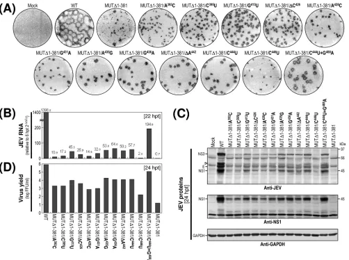
[image:6.585.78.503.69.407.2]respec-tively). However, the average size of their infectious foci varied significantly, ranging from an⬃2.5-fold reduction in the case of the MUT⌬1-284-derived RNA to nearly undetectable in the case of the MUT⌬1-381-derived RNA. Also, only a few or no plaques were seen in cells transfected with either of these two FIG. 2. Functional analysis of JEV 3⬘NCR mutants. Naïve BHK-21 cells were mock transfected (M) or transfected with 2g of the RNA transcripts synthesized from WT cDNA or each of the seven JEV 3⬘NCR mutant cDNAs. (A) Specific infectivity and representative focus (or plaque) morphology. The specific infectivities of RNA transcripts were determined by infectious center assays, as described in Materials and Methods. At 4 days after transfection, the cell monolayers were first immunostained with a mouse anti-JEV antiserum (representative foci) and then restained with crystal violet (representative plaques). RNA infectivities are given as FFU/microgram of RNA. The number of plus signs indicates the magnitude of the cytopathic effect produced by the corresponding mutant RNAs relative to that induced by the WT RNA; a minus sign indicates no cytopathic effect. N.D, not detectable. (B) Production of JEV RNA. Equal amounts of total cellular RNAs extracted at 6 and 22 h posttransfection (hpt) were used for real-time quantitative RT-PCR with a JEV-specific probe complementary to a sequence in the NS3 protein-coding region; a-actin RNA-specific probe was used to normalize total RNA levels. Changes in JEV RNA levels in real-time quantitative RT-PCR assays relative to those detected at 6 hpt were calculated using the 2⫺⌬⌬CTmethod. (C) Accumulation of JEV proteins. Equal volumes
of total cell lysates collected at the indicated time points were separated by SDS-polyacrylamide gel electrophoresis, and the levels of JEV proteins were visualized by immunoblotting with a mouse anti-JEV antiserum (anti-JEV). GAPDH, used as a loading and transfer control, was detected with a rabbit anti-GAPDH antiserum (anti-GAPDH). An asterisk indicates that⬃80% of RNA-transfected cells displayed a cytopathic effect. The positions of the viral proteins (E, NS1, and NS3) and a cleavage-related intermediate (open arrowhead) are shown on the left; molecular size markers (in kilodaltons) are shown on the right. (D) Yield of infectious virions. The amount of the infectious virus particles released into the culture supernatants was monitored by virus titration on naïve BHK-21 cells. The seven JEV 3⬘NCR mutants examined in our study were grouped into three classes (I to III) based on their phenotypes. See the text for detailed descriptions.
7914 YUN ET AL. J. VIROL.
on November 8, 2019 by guest
http://jvi.asm.org/
mutant RNAs. Class III mutants (MUT⌬1-450, MUT⌬1-473, and MUT⌬477-574) produced RNAs that generated no de-tectable infectious foci or plaques; this loss of infectivity was confirmed by the confocal immunofluorescence microscopy of single RNA-transfected cells (data not shown).
We next examined the production levels of JEV RNA and proteins in RNA-transfected cells and assessed the accumula-tion of infectious virus particles released into the culture me-dium. At 22 h posttransfection, the amount of JEV RNA produced was quantified by real-time quantitative RT-PCR using a JEV-specific probe complementary to a sequence in JEV NS3 and compared to those at 6 h posttransfection (Fig. 2B). The first class I and II mutants displayed levels of RNA production that were significantly lower than that of WT-de-rived RNA (⬃3-fold, MUT⌬1-133; ⬃4-fold, MUT⌬1-211; ⬃84-fold, MUT⌬1-284; and ⬃1,958-fold, MUT⌬1-381) and were consistent with the average size of the infectious foci observed (Fig. 2A). As expected, however, the remaining three mutant RNAs (class III; MUT⌬1-450, MUT⌬1-473, and MUT⌬477-574) showed no detectable level of JEV RNA pro-duction. In all cases, the production level of JEV RNA was proportional to the accumulation level of JEV proteins (such as E, NS1, and NS3) during the first 3 days after transfection, as demonstrated by immunoblotting with a JEV-specific hy-perimmune antiserum (Fig. 2C), and to the level of infectious virus particles released into the medium during the same pe-riod (Fig. 2D).
We then analyzed the 3⬘-terminal nucleotide sequences of the viral genomic RNAs recovered from RNA-transfected cells at 72 h posttransfection by 3⬘RACE, followed by cDNA clon-ing into a bacterial plasmid and the sequencclon-ing of⬃20 inde-pendent clones containing inserts. In the case of the class I mutants (MUT⌬1-133 and MUT⌬1-211), all of the recovered genomic RNAs had 3⬘NCRs identical to those of their original transfected RNAs (data not shown). In the case of the class II mutants (MUT⌬1-284 and MUT⌬1-381), however, some of the genomic RNAs acquired a variety of secondary mutations in the vicinity of the corresponding primary deletion sites in their 3⬘NCRs (see Fig. 7 to 11 for a detailed description). The emergence of these secondary mutations was consistent with the heterogeneity in focus morphology observed in the cells transfected with these RNAs. As expected, we obtained no PCR products from the culture supernatants of BHK-21 cells transfected with any of the three class III mutant RNAs (data not shown).
Of the seven JEV 3⬘NCR mutants, four (MUT⌬1-133, MUT⌬1-211, MUT⌬1-284, and MUT⌬1-381) showed little or no loss of RNA infectivity, with a spectrum of average focus sizes that was correlated with their levels of RNA production, protein accumulation, and virus production. In contrast, the other three mutants showed an undetectable level of RNA infectivity: those in which the deletion extended further toward the 3⬘ end of the genome and included the II-2 domain (MUT⌬1-450) or the 3⬘CYC motif embedded in domain III (MUT⌬1-473), or in which only the 3⬘SL at the 3⬘end of the genome was removed (MUT⌬477-574). These results sug-gested that in the BHK-21 cells we used for RNA transfection, the presence of the two 3⬘-proximal domains (II-2 and III) of the JEV 3⬘NCR was sufficient to maintain the replication com-petency of the genomic RNA, whereas the presence of the four
remaining 5⬘-proximal domains (V, X, I, and II-1) was neces-sary to achieve a maximal efficiency of RNA replication. Also, we noted that domain X, which was missing from MUT⌬1-211 but not MUT⌬1-133, had only a minimal effect on RNA rep-lication; this result is consistent with the fact that the X do-main, unlike the other domains, does not contain any recog-nized CS motifs.
Recovery of pseudorevertants originating from the replica-tion-incompetent MUT⌬1-450-derived RNA. Initially, we did not detect any replication-competent pseudorevertants from BHK-21 cells transfected with any of the three class III mutant RNAs (MUT⌬1-450, MUT⌬1-473, and MUT⌬477-574) (Fig. 2). In an attempt to obtain pseudorevertants from these mu-tants, we serially passaged the undiluted culture supernatants from the RNA-transfected cells in naïve BHK-21 cells to am-plify any very small amounts of replication-competent pseu-dorevertants that might have been present. After six passages, 1 ml of each undiluted culture medium was used to infect naïve BHK-21 cells, and the production of JEV-specific proteins and the formation of JEV-induced foci was assessed by immuno-blotting with cell lysates obtained at 72 h postinfection with the mouse anti-JEV antiserum (Fig. 3A) and visualizing the infec-tious foci by the immunostaining of monolayers of the infected cells at 96 h postinfection with the same antiserum (Fig. 3B). In the case of MUT⌬1-450, to our surprise, we noted the production of a significant quantity of JEV-specific proteins (i.e., E, NS1, and NS3), along with a homogeneous population of infectious foci, at passage 2 and thereafter, indicating the emergence of replication-competent pseudorevertants. These foci produced at passages 2 to 6 were invariably homogeneous but consistently⬃4.0-fold smaller than those produced by WT virus. Furthermore, the growth properties of these pseudo-revertants were identical from passages 2 to 6, although they displayed a⬃36-h delay in growth rate and an⬃10-fold re-duction in peak titer compared to those of the WT virus (Fig. 3C). In the case of the other two mutants (MUT⌬1-473 and MUT⌬477-574), neither viral proteins (Fig. 3A) nor infectious foci (Fig. 3B) were detectable in any of the six serial passages of the culture supernatants, indicating no evidence of rever-sion. Overall, these data suggest that although all three mutant RNAs of class III had no detectable infectivity, some replication-competent pseudorevertants could be recovered after the serial passage of the culture supernatants from BHK-21 cells transfected with MUT⌬1-450-derived RNA but not with the mutant RNAs derived from either MUT⌬ 1-473 or MUT⌬477-574.
Acquisition of a novel 83-nt sequence duplication capable of restoring the replicability of MUT⌬1-450-derived RNA. To understand the genetic basis for the reestablishment of RNA replication in the MUT⌬1-450-derived pseudorevertants, we analyzed the 3⬘-terminal nucleotide sequences of the replicat-ing genomic RNAs extracted from the pseudorevertants in the culture medium at passages 2 and 6. In both cases, the 3⬘ -terminal sequence, including the 50-nt 3⬘-terminal region of the viral ORF and the entire 3⬘NCR, was amplified by 3⬘RACE; subsequently, the cDNA amplicons were cloned into a bacterial plasmid, and 20 independent clones containing the insert were sequenced. No differences were found between genomic RNAs prepared from the second and sixth culture passages. All of the clones lacked the 5⬘-proximal 450-nt
on November 8, 2019 by guest
http://jvi.asm.org/
quence that we had originally deleted, and to our surprise, they all had acquired an additional copy of an 83-nt sequence du-plicated from the 3⬘-terminal region of the viral ORF (MUT⌬1-450/Rev) (Fig. 4A). This newly acquired 83-nt se-quence was located 10 nt downstream of the translation stop codon UAG in the forward direction, generating a new direct repeat sequence of two identical 83-nt sequences just 1 nt upstream of the CS1 motif (3 nt upstream of the 8-nt core sequence of the 3⬘CYC motif) in domain III without disturbing the single viral ORF (MUT⌬1-450/Rev) (Fig. 4B).
To address the functional importance of this duplicated 83-nt sequence in MUT⌬1-450-derived RNA replication, we constructed a derivative of MUT⌬1-450 (designated MUT⌬ 1-450/83ntIns) with an additional copy of the 83-nt sequence and
determined the specific infectivity of its RNA transcripts as an indication of its potential replicability after RNA transfection into BHK-21 cells (Fig. 5A). As expected, no infectivity was detectable in cells transfected with the original MUT⌬ 1-450-derived RNA. In contrast, the specific infectivity of the MUT⌬1-450/83ntIns-derived RNA was estimated to be in the
range of 1.4⫻106to 2.1⫻106FFU/g, which is
indistinguish-able from that of WT-derived RNA (1.1 to 1.8⫻106FFU/g),
and its infectious foci were also as homogeneous as those of WT-derived RNA. On the other hand, the average focus size
was dramatically reduced by ⬃4.0-fold compared to that of WT-derived RNA. The foci were detectable only by the im-munostaining of RNA-transfected cells with the anti-JEV an-tiserum and not by staining with crystal violet. Also, the dif-ference in focus size was proportional to their relative accumulation of JEV RNA (Fig. 5B) and JEV proteins (Fig. 5C) in RNA-transfected cells, parallelling the production lev-els of infectious virus particles (Fig. 5D) released over time into their culture supernatants.
[image:8.585.109.473.68.327.2]Taken together, these results indicate that the replicating genomic RNAs that emerged from MUT⌬1-450-derived RNA after amplification in BHK-21 cells acquired an additional copy of the viral 83-nt sequence, duplicated from the 3⬘ -ter-minal region of the viral ORF, just upstream of domain III in its 3⬘NCR. The duplication of this 83-nt sequence was suffi-cient to restore the replication competence of the MUT⌬ 1-450-derived RNA to a level similar to that of WT-derived RNA, although the replication efficiency was significantly com-promised. In addition, we noted that the duplication of this 83-nt sequence did not disrupt the virus’s single ORF, suggest-ing that the reestablishment of the replication competence was attributable to an RNA element(s) of the primary sequences and/or secondary structures embedded in the duplicated 83-nt sequence, acting alone or in combination with other RNA FIG. 3. Recovery of MUT⌬1-450-derived pseudorevertants and their phenotypic characteristics. Undiluted culture supernatants of BHK-21 cells that had been transfected with 2g of the RNA synthesized from one of three mutant cDNAs (MUT⌬1-450, MUT⌬1-473, or MUT⌬477-574) were passaged six times in naïve BHK-21 cells. (A and B) Emergence of MUT⌬1-450-derived pseudorevertants. Culture medium (1 ml) collected at passage 0, 2, 4, and/or 6, as indicated, was used to infect naïve BHK-21 cells. (A) Equal amounts of the total cell lysates were analyzed for JEV protein expression at 72 h postinfection by immunoblotting with the mouse anti-JEV antiserum; the lysate from WT virus-infected cells at 24 h postinfection (WT*) was used as a reference to show the viral proteins E, NS1, and NS3 and a cleavage intermediate (open arrowhead). (B) To visualize representative foci (or plaques), cell monolayers at 4 days postinfection were immunostained first with the anti-JEV antiserum and then with crystal violet. The cytopathic effect produced by the individual mutant viruses relative to the WT level is indicated by plus or minus signs as described in the legend to Fig. 2. (C) Growth of MUT⌬1-450-derived pseudorevertants. Naïve BHK-21 cells were infected at an MOI of 0.1 with the WT or one of two MUT⌬1-450-originated pseudorevertants obtained at passages 2 and 6. Culture supernatants were harvested at the indicated hour postinfection (hpi) and used for virus titration on naïve BHK-21 cells. The data shown are from one of two independent experiments yielding similar results.
7916 YUN ET AL. J. VIROL.
on November 8, 2019 by guest
http://jvi.asm.org/
elements present within the genome. We found that, consistent with this notion, a single copy of the 83-nt sequence was pre-dicted to form two alternative stem-loop structures that are identical to each other except for their upper parts, which can assume either a cruciform-like structure (83ntIns-I) or extend
the basal stem region further (83ntIns-II) (Fig. 6A). Also, a
tandem repeat of the 83-nt sequence, identical to the one identified in MUT⌬1-450/83ntIns-derived RNA, was predicted
to form two other alternative structures (designated 2⫻ 83ntIns-I and 2⫻83ntIns-II), both of which are arranged in a
head-to-head configuration of 83ntIns-I and 83ntIns-II,
respec-tively (Fig. 6B). The molecular basis for the role of this dupli-cated 83-nt sequence in MUT⌬1-450/83ntIns-derived RNA
replication remains to be determined.
Genetic instability of progeny virions derived from class II mutants MUT⌬1-284 and MUT⌬1-381.Of the seven 3⬘NCR mutant cDNAs constructed in this study, five produced mutant viruses that were competent for propagation in BHK-21 cells: two from class I (MUT⌬1-133 and MUT⌬1-211), two from class II (MUT⌬1-284 and MUT⌬1-381), and MUT⌬1-450/ 83ntInscDNA (a MUT⌬1-450 derivative). These five mutant
viruses produced infectious foci (or plaques) that were
pheno-typically distinct from each other, in terms of the magnitude of their reduction in focus size and the extent of their heteroge-neity in focus morphology, compared to those of the WT virus. To further characterize their phenotypic differences, we exam-ined the growth properties of these five mutant viruses during multiple cycles of viral replication, comparing not only their ability to grow and develop a productive infection but also the focus morphology produced by their progeny virions over time, as an indication of genetic stability. For this purpose, mono-layers of naïve BHK-21 cells were infected at an MOI of 0.1 with the WT and each of the five mutant viruses in parallel. The yield of progeny virions released into the culture medium during the course of the 4-day incubation was monitored by titration on naïve BHK-21 cells.
The five mutant viruses revealed three different patterns of growth rate and virus yield, which is consistent with the phe-notypic classification of the seven 3⬘NCR mutant cDNAs into three groups (Fig. 2). The first pattern was exhibited by the two mutant viruses derived from class I mutant cDNAs (MUT⌬ 1-133 and MUT⌬1-211); their growth property was essentially identical to that of the WT virus (peak titer of 1.8 ⫻ 106
[image:9.585.44.533.68.378.2]FFU/ml at 36 h postinfection) (Fig. 7A); they achieved a peak FIG. 4. Identification of a novel 83-nt sequence duplication in the 3⬘-terminal region of the genomic RNAs extracted from MUT⌬1-450-derived pseudorevertants (MUT⌬1-450/Rev). (A) Relative location of a newly acquired 83-nt sequence (83-nt Ins; gray box) duplicated from the 3⬘-terminal region of the viral ORF in the 3⬘-terminal region of the MUT⌬1-450/Rev RNAs. (B) Nucleotide sequences of the 3⬘-terminal region of the original MUT⌬1-450 RNA (left) and its pseudorevertant MUT⌬1-450/Rev RNA (right). The uppercase sequences indicate the 3⬘-terminal sequences of the viral ORF; the lowercase sequences represent the entire 3⬘NCR of the corresponding RNAs. The 5⬘-end nucleotide of the 3⬘NCRs is numbered nt 451, and bases extending downstream are assigned consecutively according to the WT sequence. Also indicated are the translation stop codons UAG (open arrowheads), the 25-nt CS1 motif (curved lines), and the 8-nt core sequence of the 3⬘CYC motif (solid arrowheads). Highlighted only in MUT⌬1-450/Rev RNA is the newly discovered 83-nt sequence (double straight lines) originating from the corresponding 3⬘-terminal region (single straight lines) of the viral ORF.
on November 8, 2019 by guest
http://jvi.asm.org/
titer comparable to that of the WT virus, albeit with an⬃12-h delay, peaking at 48 h postinfection. The second pattern was exhibited by the viruses derived from the other two class II mutant cDNAs (MUT⌬1-284 and MUT⌬1-381); this pattern consisted of two distinct stages, a noticeable eclipse-like stage during the first 18 h postinfection and a sharp exponential stage thereafter. The growth of MUT⌬1-284-derived mutant virus lagged behind that of the WT and two class I-derived mutant viruses for the first 36 h postinfection, but by 48 h postinfection and thereafter, this mutant achieved maximal titers slightly lower than those of WT virus, peaking at 48 to 72 h postinfection. Similarly, the growth of MUT⌬ 1-381-de-rived mutant virus was by far the most delayed of all four mutant viruses during the entire incubation period, but it yielded maximal titers that were⬃10-fold lower than those of WT virus at 72 h postinfection. The third pattern was exem-plified by MUT⌬1-450/83ntIns-derived recombinant virus; the
growth rate and virus yield were similar to those of the MUT⌬1-381-derived mutant at all time points, but the eclipse-like stage observed during the first 18 h postinfection was not as noteworthy as that seen for the MUT⌬1-381-derived virus during the same period of time.
The focus morphologies (size and heterogeneity) observed during multiple cycles of viral replication for the five viruses
were consistent with three patterns of viral growth (Fig. 7B): (i) a homogeneous population of medium-sized foci, with the two class I-derived mutant viruses MUT⌬1-133 and MUT⌬ 1-211 representing the upper end of the size range; (ii) a heter-ogeneous population of medium-to-small foci, exemplified by the two class II-derived mutant viruses MUT⌬1-284 and MUT⌬1-381; and (iii) a homogeneous population of small foci, exemplified by the MUT⌬1-450/83ntIns-derived recombinant
virus. Thus, the diversity in focus morphology and growth property described above provided additional evidence for the functional importance of the 5⬘-proximal four domains of JEV 3⬘NCR in terms of achieving a maximal level of replication efficiency. Of particular note was the fact that two of the class II-derived mutant viruses, unlike the other three, formed in-fectious foci of rather heterogeneous sizes; furthermore, this heterogeneity became more evident at the later time points during this 4-day experiment (Fig. 7B, compare the focus mor-phologies at 6 and 96 hpi), suggesting a rapid emergence of pseudorevertants.
[image:10.585.108.476.64.315.2]To further investigate the genetic stability of the five mutant viruses, we serially passaged each of these viruses three times in naïve BHK-21 cells at an MOI of 0.1 and then determined the 3⬘-terminal sequences of the genomic RNAs extracted from progeny virions at passages 0, 1, and 3 by 3⬘RACE, cDNA FIG. 5. Functional importance of the 83-nt sequence duplication in MUT⌬1-450 RNA replication. Naïve BHK-21 cells were mock transfected (M) or transfected with 2g of the synthetic RNA transcripts derived from the WT cDNA, the original MUT⌬1-450 cDNA, or its derivative MUT⌬1-450/83ntInscontaining the 83-nt sequence duplication that was originally identified from MUT⌬1-450/Rev. (A) Specific infectivity,
representative focus (or plaque) morphology, and cytopathic effect. At 4 days after transfection, the specific infectivity and representative focus morphology were assessed by immunostaining the cell monolayers with the anti-JEV antiserum; plaque morphology was then visualized by restaining with crystal violet. The cytopathic effect induced by MUT⌬1-450/83ntInsRNA replication is indicated relative to the WT level. N.D, not
detectable. (B) Production of JEV RNA. The levels of JEV RNA production at 22 hpt relative to those at 6 hpt were evaluated by real-time quantitative RT-PCR using a JEV-specific probe. (C) Accumulation of JEV proteins. The levels of JEV protein accumulation at 24, 48, and 72 hpt were analyzed by immunoblotting with the anti-JEV antiserum (anti-JEV). GAPDH was used as a loading and transfer control (anti-GAPDH). An asterisk indicates that⬃80% of RNA-transfected cells displayed a cytopathic effect. The viral proteins E, NS1, and NS3 and a cleavage-related intermediate (open arrowhead) are shown on the left; on the right are molecular size markers in kilodaltons. (D) Production of infectious virions. Virus titers accumulated in the culture supernatants at the indicated time points were estimated on naïve BHK-21 cells. Experiments were carried out as described for Fig. 2.
7918 YUN ET AL. J. VIROL.
on November 8, 2019 by guest
http://jvi.asm.org/
cloning, and the sequencing of⬃20 independent clones for each mutant (Fig. 7C). Viruses derived from two class I mutant cDNAs (MUT⌬1-133 and MUT⌬1-211) and from MUT⌬ 1-450/83ntInscDNA yielded genomic RNAs whose 3⬘NCR
[image:11.585.82.514.62.612.2]se-quences were identical to their original transfected RNAs at passages 0 and 3. However, in the case of the remaining two viruses derived from class II mutant cDNAs (MUT⌬1-284 and MUT⌬1-381), a wide variety of spontaneous mutations FIG. 6. Predicted RNA secondary structures of the 83-nt sequence duplicated in the 3⬘-terminal region of the genomic RNAs of MUT⌬ 1-450-derived pseudorevertants. Shown are two pairs of four predicted stem-loop structures: one for a single copy of the 83-nt sequence (A) and the other for a tandem repeat of the 83-nt sequence (B). Also indicated are the initial thermodynamic free energies (⌬G, in kilocalories/mole) of all four potential structures, as determined by the RNA folding program mfold.
on November 8, 2019 by guest
http://jvi.asm.org/
emerged in their 3⬘NCRs with different ratios depending on the passage number (Fig. 8 to 11). Again, these findings sup-ported our premise that the replicating genomic RNAs derived from two class II mutant cDNAs, MUT⌬1-284 and MUT⌬ 1-381, are genetically unstable.
Acquisition of a collection of 12 point mutations capable of increasing the replication efficiency of MUT⌬1-381-derived RNA.The sequence analysis of the 3⬘-terminal region of the MUT⌬1-381-derived viruses at passages 0, 1, and 3 identified a total of 12 point mutations predominantly within the 3⬘ -penul-timate domain II-2 of its 3⬘NCR, with the remaining 3⬘ -termi-nal domain III left intact (Fig. 8A and B). At passage 0, 12 of 21 independent clones had the same sequence as the original MUT⌬1-381, whereas the remaining nine clones each con-tained a unique point mutation within the II-2 domain. Of these nine point mutations, seven were single-nucleotide sub-stitutions (viz. A3923C, C3953U, A4283C, G4313A,
G4363A, C4443U, and C4463U); the remaining two were
single-nucleotide deletions (⌬C426and ⌬A442). At passage 1,
none had a sequence identical to that of the original MUT⌬ 1-381, whereas a majority of the sequenced clones (18/20 clones) had the single C4443U substitution that had been found at
passage 0 in 1 of 21 clones; the remaining two clones had a single G4133U or A4353G substitution in domain II-2. Of
note was the finding that the single C4443U substitution
ap-peared to be predominant at passage 1. This observation be-came more evident at passage 3, when all the sequenced clones had the same single C4443U substitution either alone (19/20
clones) or in combination with a single-nucleotide substitution of G4883A (1/20 clones) immediately downstream of the CS1
motif in the 5⬘ portion of domain III. In all, ⬃60% of the MUT⌬1-381-derived viruses directly recovered from RNA-transfected cells acquired a group of single point mutations exclusively within domain II-2 of their 3⬘NCRs. Of these mu-tations, the single C4443U substitution was predominant, most
likely because it provided a growth advantage for MUT⌬ 1-381-derived virus.
To address the functional significance of these 12 point mutations in MUT⌬1-381-derived RNA replication, we recon-structed 12 derivatives of MUT⌬1-381 with each of these ge-netic changes and then characterized the RNAs synthesized from these reconstructed cDNAs after transfection into BHK-21 cells with respect to (i) specific infectivity, (ii) focus morphology, (iii) viral RNA production, (iv) viral protein ac-cumulation, and (v) virus yield. In all 12 cases, the specific infectivities were in the range of 1.0⫻106to 2.1⫻106FFU/
g, which is similar to that of WT-derived RNA (1.1⫻106to
1.9⫻106FFU/g) and slightly higher than that of the original
MUT⌬1-381-derived RNA (⬎5.6⫻105to 0.5⫻105FFU/g).
[image:12.585.110.472.65.332.2]Thus, the RNAs generated from each of the 12 MUT⌬1-381 FIG. 7. Characterization of recombinant viruses generated from five replication-competent JEV 3⬘NCR mutant cDNAs. (A and B) Viral growth and focus morphology. Subconfluent monolayers of BHK-21 cells were infected at an MOI of 0.1 with the WT or one of the five 3⬘NCR mutant viruses derived from the respective cDNAs, as indicated. (A) At the indicated hour postinfection (hpi), aliquots of culture supernatants were collected during a period of 4 days and used for virus titration on naïve BHK-21 cells. (B) The same monolayers were immunostained at 6 and 96 hpi (indicated by arrows) for infectious foci with the anti-JEV antiserum. The data shown are from one of two independent experiments, which produced similar results. M, mock infected. (C) Acquisition of spontaneous secondary mutations during three consecutive rounds of viral passage in BHK-21 cells. To examine the genetic stability of the five mutant viruses, each of the viruses collected from RNA-transfected cells (passage 0) was passaged three times in naïve BHK-21 cells at an MOI of 0.1. At the indicated passages, the 3⬘-terminal sequences of the replicating genomic RNAs were determined by 3⬘RACE, the cloning of the cDNA amplicons, and the sequencing of⬃20 independent clones containing the insert. Indicated is the presence (Yes) or absence (No) of secondary mutations for each mutant virus. N.T, not tested.
7920 YUN ET AL. J. VIROL.
on November 8, 2019 by guest
http://jvi.asm.org/
derivative cDNAs were as infectious as those produced from WT cDNA (data not shown). However, the foci formed in RNA-transfected cells at 4 days posttransfection, while consis-tently homogeneous, showed a noticeable variation in size compared to those in WT-derived RNA-transfected cells (Fig. 9A). The differences in average focus sizes were proportional to the relative levels of the accumulation of JEV RNA (Fig. 9B) and proteins (Fig. 9C) in RNA-transfected cells during the first 22- to 24-h incubation period and largely paralleled the levels of infectious virions released into the culture superna-tants during the same period (Fig. 9D).
Collectively, these results indicate that the acquisition of these 12 point mutations within or close to domain II-2 in MUT⌬1-381 is functionally important for increasing RNA rep-lication efficiency (by⬃3-fold to⬃90-fold). In particular, we noted that a higher level of RNA replication was associated with the acquisition of four point mutations, A4353G,
G4363A, ⌬A442, and C4443U; all of these mutations
con-verged on the⬃10-nt 3⬘-proximal region of the II-2 domain in
MUT⌬1-381, corresponding to the 3⬘ half of the CS2 motif. Also, the enhanced replication efficiency of the MUT⌬ 1-381-derived RNA with a single C4443U substitution was increased
further by the acquisition of an additional G4883A
substitu-tion just downstream of the CS1 motif at the 5⬘-proximal re-gion of domain III. Thus, our findings demonstrated that al-though the original MUT⌬1-381-derived RNA was replication competent, the replication efficiency was severely impaired (near the limit of detection); nevertheless, various single point mutations acquired within the II-2 domain, particularly the CS2 motif in this domain, were able to enhance the RNA replication efficiency significantly, to the point of generating readily detectable levels of viral RNA and proteins, in parallel with infectious virus particles.
[image:13.585.55.541.69.385.2]Acquisition of a selection of seven sequence duplications capable of enhancing the replication efficiency of MUT⌬ 1-284-derived RNA.We also determined the 3⬘-terminal sequences of MUT⌬1-284-derived viruses that had been collected directly from RNA-transfected cells (passage 0), in parallel with those FIG. 8. Identification of 12 point mutations acquired within the 3⬘-terminal region of the genomic RNAs of MUT⌬1-381-derived pseudor-evertants. The original recombinant viral stock was generated by transfecting BHK-21 cells with RNA transcripts derived from MUT⌬1-381 cDNA (passage 0); this viral stock was serially passaged three times in naïve BHK-21 cells at an MOI of 0.1 (passages 1 to 3). The 3⬘-terminal sequences of the genomic RNAs at passages 0, 1, and 3 were determined as described in Materials and Methods. The relative locations (A) and nucleotide sequences (B) acquired at the 3⬘-terminal region of the genomic RNAs of MUT⌬1-381-derived pseudorevertants are shown. (A) Illustrated at the top is the organization of the genomic RNA derived from the MUT⌬1-381 cDNA; shown below is an expanded view of the 193-nt 3⬘NCR, including only the two 3⬘-proximal domains (II-2 and III), with the CS1 and CS2 motifs and the 3⬘SL indicated by gray boxes. (B) Twelve point mutations (single-nucleotide substitutions or deletions are in boldface type) acquired in MUT⌬1-381-derived pseudorevertants. Dots indicate the conserved nucleotide sequences in adapted pseudorevertants, and hyphens indicate the deleted nucleotide sequences. The number of clones containing each identified nucleotide sequence is given over the total number of clones containing the insert that were sequenced. Open arrowheads indicate the translation stop codon UAG. The 5⬘-end nucleotide of the 3⬘NCR is numbered nt 382, and downstream sequences are consecutively assigned according to the WT sequence.
on November 8, 2019 by guest
http://jvi.asm.org/
from the first (passage 1) and third (passage 3) passages. Re-markably, we found a total of seven nucleotide insertions of various sizes (designated Ins1 to Ins7), ranging from 40 to 168 bases; all of these insertions appeared to be located at the junction between domains II-1 and II-2 (Fig. 10A and B). Whereas 18 out of 20 independent clones had no insertions at passage 0, the remaining two clones had insertions of 155 and 168 nt (Ins2 and Ins3, respectively). At passage 1, the size of the inserted sequences became more heterogeneous. We saw no insertion in 11/21 clones; however, in addition to Ins2 in 2/21 clones and Ins3 in 1/21 clones, we also identified four other insertions of various sizes: 162 nt (Ins1, 4/21 clones), 145 nt (Ins4, 1/21 clones), another of 168 nt (Ins5, 1/21 clones), and 149 nt (Ins7, 1/21 clones). At passage 3, the 162-nt Ins1 became predominant, being found in 16/19 clones; we also saw Ins2 (2/19 clones) and a new 40-nt insertion (Ins6) in 1/19 clones. Of
the seven inserted sequences, six (Ins1 to Ins5 and Ins7) cor-responded to the 3⬘-terminal region of the viral ORF followed by the entire (or nearly entire) domain II-1; the remaining sequence (Ins6) corresponded to the 3⬘ half of domain II-1, including the complete RCS2 motif (Fig. 10C). More impor-tantly, in all cases the newly inserted sequences perfectly matched a sequence immediately upstream of their respective insertion sites (Fig. 10C), indicating that these sequences were introduced by the duplication of the corresponding individual sequences just upstream of domain II-2.
[image:14.585.44.546.66.440.2]To assess the functional importance of these duplicated se-quences in MUT⌬1-284-derived RNA replication, we con-structed seven derivatives of MUT⌬1-284 with all of these duplicated sequences and examined their phenotypes after the transfection of BHK-21 cells with the RNAs synthesized from the corresponding cDNAs. In all cases, the specific infectivities FIG. 9. Effects on RNA replication efficiency of 12 point mutations discovered in MUT⌬1-381-derived pseudorevertants. Naïve BHK-21 cells were mock transfected (Mock) or transfected with a synthetic RNA transcribed from the WT cDNA or one of the reconstructed MUT⌬1-381 derivative cDNAs, as indicated. (A) Representative focus morphology. Infectious foci were visualized at 4 days posttransfection by immunostaining with the anti-JEV antiserum. (B) Production of JEV RNA. The levels of JEV RNA production at 22 hpt relative to those at 6 hpt were estimated by real-time quantitative RT-PCR using a JEV-specific probe. (C) Accumulation of JEV proteins. The levels of JEV protein accumulation at 24 hpt were examined by immunoblotting with the anti-JEV antiserum (anti-JEV) or a rabbit antiserum specific for JEV NS1 (anti-NS1). The positions of the viral proteins E, NS1, and NS3 and a cleavage-related intermediate (open arrowhead) are shown on the left; molecular size markers are shown on the right. (D) Production of infectious virions. Virus yields in the culture supernatants at 24 hpt were determined by virus titration on naïve BHK-21 cells.
7922 YUN ET AL. J. VIROL.
on November 8, 2019 by guest
http://jvi.asm.org/
were essentially the same as those estimated for the original MUT⌬1-284-derived RNA, which were comparable to those of the WT-derived RNA (1.3⫻106to 2.2⫻106FFU/g; data
not shown). There were, however, significant differences be-tween the phenotypes of the seven MUT⌬1-284 derivatives and that of the original MUT⌬1-284. At 4 days posttransfec-tion, all seven derivatives always produced homogeneous foci/ plaques that were essentially indistinguishable from those pro-duced by WT-derived RNA. These foci/plaques were, on average,⬃2.5-fold larger than the rather heterogeneous foci produced by the original MUT⌬1-284-derived RNA (Fig. 11A). Consistent with these results, the levels of JEV RNA produced by the cells transfected with any of the seven MUT⌬1-284 derivative RNAs during the first 22 h of the incubation period were invariably⬃50 to 70-fold higher than that of the original MUT⌬1-284-derived RNA, ranging from ⬃50% of the WT level to nearly the same as that of the WT (Fig. 11B). This increase in JEV RNA production was re-flected in their levels of viral proteins (Fig. 11C) and virus yield (Fig. 11D).
Taken together, our findings indicate that MUT⌬ 1-284-de-rived RNA duplicated a viral sequence of various sizes (40 to 168 nt) just upstream of domain II-2 to generate a direct repeat sequence. All of these duplications dramatically increased the RNA replication efficiency of the MUT⌬1-284-derived RNAs to a level close to that of WT-derived RNA. Of the seven duplicated sequences, the shortest was surprisingly only 40 nt long, corresponding to the 3⬘ half of domain II-1, which in-cludes the complete RCS2 motif. Therefore, our data suggest that the duplication of a sequence as short as the RCS2 motif in MUT⌬1-284 is sufficient to achieve RNA replication levels that are nearly indistinguishable from WT levels.
Cell type-specific replication of MUT⌬1-381-derived viruses, which are capable of replicating in BHK-21 and SH-SY5Y cells but not in C6/36 cells. The five 3⬘NCR mutant viruses (MUT⌬1-133, MUT⌬1-211, MUT⌬1-284, MUT⌬1-381, and MUT⌬1-450/83ntIns) that were competent for replication in
BHK-21 cells were examined further for their growth proper-ties in two other cell types, human neuroblastoma SH-SY5Y and mosquito C6/36 cells, which are potentially important for FIG. 10. Identification of seven sequence insertions (duplications) of various sizes at the junction between domains II-1 and II-2 in the genomic RNAs of MUT⌬1-284-derived pseudorevertants. The original recombinant viral stock was obtained after transfection of BHK-21 cells with the RNA transcripts derived from MUT⌬1-284 cDNA (passage 0); this virus stock was then passaged three times in naïve BHK-21 cells at an MOI of 0.1 (passages 1 to 3). The 3⬘-terminal sequences of the genomic RNAs at passages 0, 1, and 3 were determined as described in Materials and Methods. (A) Relative locations of the seven sequence insertions (Ins1 to Ins7) duplicated from upstream sequences of domain II-2 in the genomic RNAs of MUT⌬1-284-derived pseudorevertants. At the top is a schematic presentation of the genomic RNA derived from the MUT⌬1-284 cDNA; given below is an expanded view of the 290-nt 3⬘NCR containing only the three 3⬘-proximal domains (II-1, II-2, and III), with the three CSs (CS1, CS2, and RCS2) and the 3⬘SL indicated. (B and C) Nucleotide sequences of the seven sequence insertions (Ins1 to Ins7) of various sizes. (B) The number of clones containing each identified nucleotide sequence/the total number of clones containing the insert that we examined is indicated. (C) The nucleotides of the seven sequence insertions (duplications) were aligned; the RCS2 motif is highlighted with a gray box. Open arrowheads indicate the translation stop codon UAG.
on November 8, 2019 by guest
http://jvi.asm.org/
JEV pathogenesis and transmission. Monolayers of each of these two cell lines were infected at an MOI of 1 with the WT or each of the five mutant viruses, and during an incubation period of 4 days (SH-SY5Y) or 6 days (C6/36), equal volumes of the culture fluids were harvested for progeny virions at the indicated time points. The production levels of the progeny virio