Human Choline Kinase-
␣
Promotes Hepatitis C Virus RNA
Replication through Modulation of Membranous Viral Replication
Complex Formation
Mun-Teng Wong, Steve S. Chen
Institute of Biomedical Sciences, Academia Sinica, Taipei, Taiwan, Republic of China
ABSTRACT
Hepatitis C virus (HCV) infection reorganizes cellular membranes to create an active viral replication site named the
membra-nous web (MW). The role that human choline kinase-
␣
(hCK
␣
) plays in HCV replication remains elusive. Here, we first showed
that hCK
␣
activity, not the CDP-choline pathway, promoted viral RNA replication. Confocal microscopy and subcellular
frac-tionation of HCV-infected cells revealed that a small fraction of hCK
␣
colocalized with the viral replication complex (RC) on the
endoplasmic reticulum (ER) and that HCV infection increased hCK
␣
localization to the ER. In the pTM-NS5B model,
NS3-NS5B expression increased the localization of the wild-type, not the inactive D288A mutant, hCK
␣
on the ER, and hCK
␣
activity
was required for effective trafficking of hCK
␣
and NS5A to the ER. Coimmunoprecipitation showed that hCK
␣
was recruited
onto the viral RC presumably through its binding to NS5A domain 1 (D1). hCK
␣
silencing or treatment with CK37, an hCK
␣
activity inhibitor, abolished HCV-induced MW formation. In addition, hCK
␣
depletion hindered NS5A localization on the ER,
interfered with NS5A and NS5B colocalization, and mitigated NS5B interactions but had no apparent effect on
NS5A-NS4B and NS5A-NS4B-NS5B interactions. Nevertheless, hCK
␣
activity was not essential for the binding of NS5A to hCK
␣
or NS5B.
These findings demonstrate that hCK
␣
forms a complex with NS5A and that hCK
␣
activity enhances the targeting of the
com-plex to the ER, where hCK
␣
protein, not activity, mediates NS5A binding to NS5B, thereby promoting functional membranous
viral RC assembly and viral RNA replication.
IMPORTANCE
HCV infection reorganizes the cellular membrane to create an active viral replication site named the membranous web (MW).
Here, we report that human choline kinase-
␣
(hCK
␣
) acts as an essential host factor for HCV RNA replication. A fraction of
hCK
␣
colocalizes with the viral replication complex (RC) on the endoplasmic reticulum (ER) in HCV-infected cells. NS3-NS5B
expression increases ER localization of wild-type, but not D288A mutant, hCK
␣
, and hCK
␣
activity facilitates the transport of
itself and NS5A to the ER. Silencing or inactivation of hCK
␣
abrogates MW formation. Moreover, hCK
␣
is recruited by NS5A
independent of hCK
␣
activity, presumably through binding to NS5A D1. hCK
␣
activity then mediates the ER targeting of the
hCK
␣
-NS5A complex. On the ER membrane, hCK
␣
protein,
per se
, induces NS5A binding to NS5B, thereby promoting
mem-branous RC formation and viral RNA replication. Our study may benefit the development of hCK
␣
-targeted anti-HCV
thera-peutics.
M
ore than 170 million people worldwide are infected with
hepatitis C virus (HCV), and persistent HCV infection may
result in progression to life-threatening diseases, including
cirrho-sis and hepatocellular carcinoma. HCV is an enveloped virus
clas-sified in the genus
Hepacivirus
within the family
Flaviviridae
(
1
,
2
).
This virus has a 9.6-kb single-stranded RNA genome with positive
polarity flanked by 5=
and 3=
untranslated regions (UTRs) (
2
).
Translation of the HCV genomic RNA produces a polyprotein
that undergoes further processing by cellular and viral proteases
into structural proteins (core, E1, and E2) and nonstructural (NS)
proteins (p7, NS2, NS3, NS4A, NS4B, NS5A, and NS5B) (
1
,
2
).
The structural proteins assemble into the viral particle, whereas
the NS proteins play crucial roles in genome RNA replication and
virion assembly (
1
,
2
).
Similar to many other positive-sense RNA viruses, HCV
hi-jacks host lipids and remodels the endomembrane system to
cre-ate a lipid-rich environment necessary for viral replication (
3
).
The viral replication complex (RC), also called the replicase, is
composed of viral proteins NS3 to NS5B and the replicating viral
RNA (
4
). These viral RCs are housed on altered endoplasmic
membranes and form distinct organelle-like structures termed
membranous webs (MWs) (
5–8
). These MWs are characterized
by their unique multivesiculated membrane vesicles, which have
heterogeneous sizes, ranging between 100 to 300 nm in diameter,
and morphologies and which are embedded within a subcellular
membrane structure (
9
,
10
). Immunogold electron microscopy
(EM) showed that all viral proteins formed a complex that
asso-ciated with the NS4B-induced MW (
5
). The MW serves as a
plat-form for compartmentalizing and concentrating the HCV RC,
Received17 May 2016Accepted20 July 2016
Accepted manuscript posted online3 August 2016
CitationWong M-T, Chen SS. 2016. Human choline kinase-␣promotes hepatitis C virus RNA replication through modulation of membranous viral replication complex formation. J Virol 90:9075–9095.doi:10.1128/JVI.00960-16.
Editor:J.-H. J. Ou, University of Southern California
Address correspondence to Steve S. Chen, [email protected]. Copyright © 2016 Wong and Chen. This is an open-access article distributed under the terms of theCreative Commons Attribution 4.0 International license.
on November 7, 2019 by guest
http://jvi.asm.org/
viral products, and host factors to ensure efficient viral replication
and assembly (
2
,
11
).
Among the NS proteins, NS3 is a bifunctional protein that has
serine-type protease, NTPase, and helicase activities, whereas
NS4A acts as a cofactor for NS3 protease. NS4B, an integral
mem-brane protein, is thought to serve as the scaffold for viral RC
as-sembly and is able to induce MW formation (
12
,
13
). Within the
RC, the viral RNA-dependent RNA polymerase NS5B transcribes
viral genome RNA (
2
). NS5A is a multitasking viral protein that is
present as two phosphorylated forms: hypophosphorylated p56
and hyperphosphorylated p58 (
14
). Possessing an RNA-binding
ability (
15
), NS5A contains an N-terminal amphipathic helix
(AH) that tethers the protein to the membrane (
16
), three
do-mains, i.e., D1, D2, and D3, and two low-complexity sequences,
LCS1 and LCS2, which are located in between the domains (
12
,
17
,
18
). D1 functions in RNA replication and is associated with lipid
droplet (LD) and NS5A dimerization (
19
,
20
). LCS1 and D2
func-tion in RNA replicafunc-tion (
12
), while D3 plays a critical role in the
NS5A-core protein interaction and virion assembly (
21
,
22
).
LD serves as not only a host lipid storage site but also a dynamic
organelle in HCV replication and pathogenesis (
23
,
24
). NS5A is
thought to facilitate the transport of viral RNA from the MW
replication site to LDs to interact with core protein, thereby
pro-moting viral RNA encapsidation, nucleocapsid formation, and
virus assembly (
24–26
). In addition, LDs are tightly associated
with the E1- and E2-localizing ER membrane within the MW,
where viral RNA replication occurs (
24
,
27
). Thus, the close
prox-imity of LDs, the MW, and the endoplasmic reticulum (ER)
mem-brane effectively couples viral RNA replication to virion assembly.
Human choline kinase-
␣
(hCK
␣
), the first enzyme in the
CDP-choline (or Kennedy) pathway (
28
), catalyzes the
phosphor-ylation of choline into phosphocholine (
Fig. 1A
). Phosphocholine
is converted to CDP-choline by CTP:phosphocholine
cytidyl-transferase (CCT) (
Fig. 1A
); this conversion is the rate-limiting
step in the CDP-choline pathway. CDP-choline is then processed
by CDP-choline:1,2-diacylglycerol cholinephosphotransferase
(CPT) to phosphatidylcholine (PC) (
Fig. 1A
), the most abundant
phospholipid in eukaryotic cell membranes (
29
). As essential
components of the cell membrane, PC and
phosphatidylethano-lamine (PE) serve as precursors of the lipid second messengers
involved in cell survival and growth (
30
). CK
␣
induces the G
1-to-S
phase transition of the cell cycle and affects many genes involved
in cell proliferation, transformation, and signaling transduction
(
31
). CK
␣
overexpression is oncogenic (
32
). Tissues derived from
most common human tumors show higher levels of CK
␣
expres-sion and enzymatic activity than their normal counterparts, and
high levels of CK
␣
are associated with high histological tumor
grades and poor clinical outcomes (
33
,
34
). Silencing of hCK
␣
by small interfering RNA (siRNA) or treatment with
N
-(3,5-dimethylphenyl)-2-{[5-(4-ethylphenyl)-1H-1,2,4-triazol-3-yl]
sulfanyl} acetamide (CK37), a specific inhibitor of hCK
␣
activity,
leads to decreased phosphocholine and PC levels in transformed and
neoplastic human cells and prevents tumor formation in mice (
35
,
36
) and in a human breast cancer xenograft (
37
).
In an siRNA screen of the human kinome to identify host
fac-tor requirements for HCV replication, Reiss et al. noticed that
silencing of CK
␣
exhibited the strongest inhibitory effect on HCV
replication among the 13 kinases screened (
38
). Li et al. also
per-formed screening experiments and found that CK
␣
is a critical
factor for HCV entry and viral RNA replication (
39
).
Neverthe-less, whether hCK
␣
itself or the CDP-choline pathway is
impor-tant for HCV replication in hepatoma cells and how hCK
␣
en-gages with HCV to regulate viral replication remain elusive.
In the present study, we show that hCK
␣
itself, through its
enzymatic activity, not the CDP-choline pathway, positively
mod-ulates HCV RNA replication. We also show that hCK
␣
is recruited
by NS5A, likely through the binding of hCK
␣
to D1 of NS5A. In
addition, hCK
␣
acts concertedly with NS5A to cooperatively
fa-cilitate targeting of the hCK
␣
-NS5A complex to the ER in an
hCK
␣
activity-dependent manner. On the ER membrane, the
hCK
␣
protein, not hCK
␣
activity, functions as a mediator for
NS5A-NS5B binding, thereby promoting functional viral RC
as-sembly and MW formation crucial for viral RNA replication.
MATERIALS AND METHODS
Cell cultures and antibodies.Human hepatoma cell lines Huh7 and Huh7.5-1 and human embryonic kidney 293T cells (ATCC CRL-3216) were cultured as previously reported (40). The following mouse mono-clonal antibodies (MAbs) were purchased: anti-core protein (sc-57800; Santa Cruz); anti-NS3 (MAB8691 [Merck Millipore] and ab65407 [Ab-cam]); anti-NS5A (HCM-131-5; Austral Biologicals); anti-NS5B (HCV-4B8; BioFront Technologies); anti-Flag M2, anti-Myc, and anti--actin (F1804, M4439, and A5441, respectively; Sigma-Aldrich); anti-green fluo-rescent protein (GFP) and anti-hemagglutinin (HA) (Gm0001-01 and M0003, respectively; Abomics); and anti-human cluster differentiation 81 (CD81) and anti-calnexin (555675 and 610523, respectively; BD Biosci-ences). The following rabbit polyclonal antibodies were purchased: anti-hCK␣(ab88053; Abcam); anti-tail-interacting protein of 47 kDa (TIP47) (sc-14726; Santa Cruz); anti-phosphatidylethanolamineN -methyltransferase (PEMT), anti-Flag, and anti-HA (SAB1401522, F7425, and H6908, respectively; Sigma-Aldrich); anti-glyceraldehyde 3-phosphate dehydrogenase (GAPDH), anti-CCT␣, anti-Myc, and an-ti-green fluorescent protein (GFP) (GTX100118, GTX62359, GTX115046, and GTX113617, respectively; GeneTex); anti-binding im-munoglobulin protein (BiP) (3177S; Cell Signaling Technology); and normal IgG (12-370; Merck Millipore). The purchased goat antibodies included anti-calregulin (CALR) and anti-adipose differentiation-related protein (ADRP) (sc-6467 and sc-32450, respectively; Santa Cruz). The MAb 9E10 was directed against NS5A (41). Alexa Fluor-conjugated sec-ondary antibodies were obtained from Invitrogen.
Reagents and siRNAs.OptiPrep (1114542) was purchased from Axis-Shield. CK37 (CAS1001478-90-5) was purchased from Merck Millipore. Hexadecyltrimethylammonium bromide (HDTAB; H5882), hexade-cylphosphocholine (HePC; M9198), and 5-aminoimidazole-4-car-boxamide 1--D-ribofuranoside (AICAR; A9978) were obtained from Sigma-Aldrich. Small interfering RNAs (siRNAs) targeting hCK␣ (HSS140690 and HSS140691), CCT␣ (HSS107689, HSS107690, and HSS107691), and CD81 (HSS101629, HSS101630, and HSS101631) were purchased from Invitrogen, while a nontargeting control siRNA (29551) was obtained from Santa Cruz.
Plasmids and construction.The plasmids pUC-JFH1 and pFL-Luc-JFH1 were as previously described (40,42). pSGR-Luc-JFH1 (where SGR is subgenomic replicon) (43), pWPI-T7-BLR, and pTM-NS3-5B (44) were as previously described. A set of pCDNA3.1(⫺)/Myc-His-based plasmids expressing full-length NS5A or D1, D2, or D3 of NS5A was as previously described (45). Plasmids expressing HA-tagged genotype 1b Con1 strain NS proteins and the plasmid expressing HA-tagged glutathi-oneS-transferase (GST) were as previously described (46). Plasmids pCMV⌬R8.91 and pMD.G and the pLKO-based short hairpin RNA (shRNA) constructs TRC0005 and TRCN0000284352 were obtained from the National RNAi Core Facility at Academia Sinica. pCMV6-hCK␣ (RC207209), which encodes hCK␣tagged with Myc and DDDK (or Flag) tags at the C terminus, was purchased from Origene, while pEGFP-C1-hCK␣encodes hCK␣fused with EGFP at the N terminus.
Wong and Chen
on November 7, 2019 by guest
http://jvi.asm.org/
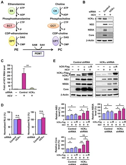
FIG 1Involvement of hCK␣in HCV replication. (A) Biosynthesis of phosphatidylcholine (PC) and phosphatidylethanolamine (PE). PC is mainly synthesized via the CDP-choline and PE methyltransferase (PEMT) pathways. In the CDP-choline pathway, choline kinase (CK), CTP:phosphocholine cytidylyltransferase (CCT), and choline phosphotransferase (CPT) catalyze consecutive reactions to produce PC. In the PEMT pathway, PE undergoes three methylation reactions catalyzed by PEMT to synthesize PC. In the CDP-ethanolamine pathway, ethanolamine is taken up into the cytoplasm and catalyzed by ethanolamine kinase (EK), CTP:phosphoethanolamine cytidylyltransferase (ECT), and ethanolamine phosphotransferase (EPT) to yield PE. (B and C) Huh7 cells remained untrans-fected (⫺) or were transfected with an untargeted control siRNA or hCK␣-specific siRNA. Cells were then infected with HCVcc at an MOI of 1 or received parallel treatment without HCVcc (⫺). Cells were then subjected to Western blot analysis (B) and RT-PCR (C). (D) Huh7 cells transfected with control or hCK␣siRNAs (left panel) and Huh7 cells stably transduced with control or hCK␣shRNA lentiviral vectors (right panel) were assessed for cell viability. (E) The control and hCK␣shRNA Huh7 stable cells were transfected with plasmids encoding the Flag-tagged normal hCK␣(␣) or shRNA-resistant hCK␣(␣-R) followed by infection with HCV. The cells were analyzed by Western blotting (top panel). The levels of NS3, NS5A, and core proteins in HCV-infected control or hCK␣stable knockdown cells overexpressing hCK␣or hCK␣-R are expressed as a percentage relative to that detected in vector plasmid transfected cells, which was designated 100% (bottom panel). *,P⬍0.05; **,P⬍0.01; ns, nonsignificant.
on November 7, 2019 by guest
http://jvi.asm.org/
[image:3.585.83.497.66.606.2]The pCMV6-hCK␣-R plasmid that encoded authentic hCK␣whose mRNA was not restricted by the hCK␣shRNA (TRCN0000284352) was generated by introducing mutations into the shRNA target region of the hCK␣open reading frame using a set of mutagenic forward and reverse primers: 5=-GGGGGCTGAGGCCATGGTACTCGAAAGTGTAATG TTTGCCATTCTCGCAG-3=and 5=-CTGCGAGAATGGCAAACATTAC ACTTTCGAGTACCATGGCCTCAGCCCCC-3=, respectively. pCMV6-hCK␣-D288A-R was generated from pCMV6-hCK␣-R using the muta-genic forward and reverse primers 5=-CCAGTTGTATTTTGTCATAATG CCTGTCAAGAAGG-3=and 5=-CCTTCTTGACAGGCATTATGACAAA ATACAACTGG-3=, respectively. The amplicons were subcloned into pCMV6 using SgfI and MluI. Both pCMV6-hCK␣-R and pCMV-hCK␣ -D288A-R were sequenced using T7 primers to confirm the authentic wild-type and D288A mutant hCK␣open reading frames.
In vitroRNA transcription, viral genome RNA transfection, and siRNA silencing.JFH1, JFH1-Luc, and SGR-Luc-JFH1 RNAs were gen-erated as described previously (22,40,47). Thein vitro-transcribed RNAs were electroporated into Huh7 cells based on a previously described pro-cedure (48). For RNA interference (RNAi) silencing studies, 2⫻105
Huh7 cells seeded in six-well plates overnight were transfected with 100 pmol of siRNAs using Lipofectamine RNAi Max (Invitrogen) according to the manufacturer’s protocol.
HCV production, titration, infection, and UV irradiation.The cell culture-derived infectious HCV (HCVcc) was produced by electroporat-ing JFH1 RNA into Huh7 cells as described above, and then culture su-pernatants were collected, centrifuged, filtered through 0.45- m-pore-size cellulose acetate discs, and stored at⫺80°C. Virus titration was determined as previously described (40,49). Huh7 cells were infected with HCVcc at a multiplicity of infection (MOI) of 1 according to a previously described procedure (40). At 24 h post-siRNA transfection, cells were left uninfected or infected with HCVcc, and cells were collected at 72 h postin-fection for Western blotting, real-time (RT)-PCR, or confocal micro-scopic analyses. UV inactivation of HCVcc was performed as previously described (50).
HCV reporter RNA replication and inhibitor assays.HCV reporter replication was performed using a pipette-type electroporator (Micropo-rator MP-100; Digital Pro), as previously described (40), at 24 h post-siRNA transfection. Transfected cells were harvested at different times after RNA transfection as indicated on the figure, and cell lysates were monitored for firefly luciferase activity. The luciferase activity at 4 h after RNA transfection was used to normalize the transfection efficiency. In inhibitor studies, inhibitors were added at 48 h post-viral RNA transfec-tion and incubated for 24 h before cells were harvested for luciferase assays.
RT-PCR analysis.Total RNAs were isolated, reverse-transcribed into cDNAs, and quantified for viral RNA level by RT-PCR using TaqMan Universal master mix II (4440038, Applied Biosystems) and an ABI 7500 real-time PCR system (Applied Biosystems) based on a previously de-scribed procedure (40). The quantitative PCR (qPCR) probes used were as follows: Hs99999905 for GAPDH, Hs00957878_m1 for hCK␣, Hs00540979_m1 for PEMT, and 6-carboxyfluorescein (6-FAM)-5=-TTC CCGGCAATTCC-3=-minor groove binder and nonfluorescent quencher (MGBNFQ) for HCV. All probes of genes were obtained using TaqMan Gene Expression Assays (Applied Biosystems). The relative amounts of cellular mRNAs and HCV viral RNA were calculated by the comparative threshold cycle (CT) method (⌬⌬CT) and normalized to the level of
GAPDH. The viral RNA levels in different experimental settings relative to the level detected in mock-infected cells, which was arbitrarily set as 1, were expressed as the fold change in the HCV RNA level. The mRNA levels of cellular genes in knockdown cells relative to the level detected in control knockdown cells, which was arbitrarily set as 1, were expressed as the fold change in the mRNA level.
Plasmid DNA transfection and coimmunoprecipitation.Huh7 cells or the paired control and hCK␣stable knockdown Huh7 cells were trans-fected with the plasmids indicated on the figure using Lipofectamine 2000
based on the procedure provided by the manufacturer (Invitrogen). In each transfection analysis, appropriate vector plasmids were added into transfection reaction mixtures to ensure that the total DNA amount in all transfections was identical. In some cases, transfected cells remained mock infected or infected with HCVcc at an MOI of 1 at 48 h post-DNA transfection. Alternatively, transfected cells were transfected with pTM-NS3-NS5B and pWPT-T7-BLR at 48 h post-plasmid transfection. All DNA-transfected cells were harvested at 48 h posttransfection for Western blotting, confocal microscopy, coimmunoprecipitation (co-IP), and transmission electron microscopy (TEM) analyses.
For co-IP analyses, Huh7 or the paired control and hCK␣ stable knockdown Huh7 cells were cotransfected with the two plasmids indi-cated on the figure, while HEK293T cells were cotransfected by a standard calcium phosphate coprecipitation method. Cells expressing hCK␣were lysed with Tris-HCl buffer containing 1% CHAPS (3-[(3-cholamidopro-pyl)-dimethylammonio]-1-propanesulfonate). To assess NS protein-protein interactions, transfected cells were lysed with Tris-HCl buffer containing 1% Triton X-100. For co-IP analysis of NS5A produced by the pTM-NS3-NS5B system, Huh7 cells or the control or hCK␣stable knock-down Huh7 cells were cotransfected with pWPI-T7-BLR and pTM-NS3-NS5B, and then transfected cells were lysed with Tris-HCl buffer contain-ing 1% CHAPS. For co-IP analysis of NS5A in CK37-treated cells, Huh7 cells stably transduced with the T7 RNA polymerase lentiviral vector (T7 Pol/Huh7) were transfected with pTM-NS3-NS5B. At 24 h post-DNA transfection, cells were treated with or without 100M CK37 for 24 h prior to cell harvest. To assess the effect of hCK␣activity on the binding of NS5A to hCK␣and NS5B, control and hCK␣stably knocked down cells that were stably transduced with T7 RNA polymerase lentiviral vector were transfected with wild-type hCK␣or hCK␣-R or with D288A mutant hCK␣-R. At 24 h after transfection, the cells were cotransfected with pTM-NS5A and pTM-NS5B.
For immunoprecipitation, 1g of the antibody indicated on the figure was first incubated with protein A Mag Sepharose (28-9670-62; GE Healthcare) at 4°C for 2 h, followed by incubation with cell lysates at 4°C for 6 h. After the beads were washed five times in phosphate-buffered saline (PBS), they were boiled with sample loading buffer and subjected to SDS-PAGE and immunoblot analyses.
HCV and VSV pseudoparticle package and entry assay.To produce HCV pseudotyped particles (HCVpp) and vesicular stomatitis virus pseu-dotyped particles (VSVpp), 293T cells grown in 10-cm dishes were trans-fected with pNL-Luc-R⫺E⫺ plus pCDNA3-kozakE1E2 (H77 strain), pCX-E1E2-Flag (JFH1 strain), or pMD.G, which encodes the envelope glycoproteins of HCV genotype 1 or 2 or the G protein of vesicular sto-matitis virus, using the calcium phosphate coprecipitation method. Two days after transfection, the culture supernatants were harvested, filtered, and used to inoculate siRNA-transfected Huh7 cells for 48 h. The cells were collected 48 h after pseudotyped particle challenge, washed in PBS, and lysed with 1⫻passive lysis buffer (Promega). Firefly luciferase activity was measured using a Sirus Single Tube Luminometer (Bet-11040010; Berthold Detection Systems). Another set of siRNA-transfected Huh7 cells was harvested at 48 h post-siRNA transfection and analyzed by im-munoblotting to assess the knockdown effects of siRNAs transfected.
Generation of Huh7 cells constitutively expressing control or hCK␣ shRNA and/or T7 RNA polymerase.Lentiviral vector stocks encoding control or hCK␣shRNAs or T7 RNA polymerase were prepared and transduced into Huh7 cells as previously described (40,44). The trans-duced Huh7 cells were selected with 5g/ml puromycin (for control and hCK␣shRNA/Huh7) or with 10g/ml blasticidin (for T7 Pol/Huh7). To obtain Huh7 cells stably expressing T7 RNA polymerase and control or hCK␣shRNAs, T7 Pol/Huh7 cells were transduced with lentiviral vectors encoding control or hCK␣shRNAs, respectively, and cells were selected with both antibiotics.
Inhibitor studies and cell viability and PC assays.All inhibitors were prepared in dimethyl sulfoxide (DMSO) and added to HCVcc-infected or viral RNA-transfected Huh7 cells at 24 h prior to cell harvest. Cells treated
Wong and Chen
on November 7, 2019 by guest
http://jvi.asm.org/
with DMSO (vehicle) at the same concentration as that in the inhibitor treatment were used controls. Unless otherwise indicated, the following concentrations of inhibitors were used: CK37, 100M; HDTAB, 10M; HePC, 10M; and AICAR, 250M. Cell viability was determined at 2 days post-hCK␣siRNA transfection or 24 h after drug treatment using a CellTiter-Glo luminescent cell viability assay kit (G7571; Promega). The amount of PC was measured using a PC assay kit (MAK049; Sigma-Aldrich).
Membrane flotation, SDS-PAGE, and immunoblot analyses.
Mock- or HCV-infected Huh7 cells were resuspended in PBS containing 0.25 M sucrose (PBS-sucrose) plus a protease inhibitor cocktail (000000004693132001; Roche), and the cells were homogenized using an Omni THQ digital tissue homogenizer. Aliquots containing equal pro-teins from each postnuclear supernatant were subjected to a 10%/20%/ 30% discontinuous iodixanol gradient as previously described (51). After the samples were ultracentrifuged, the gradients were fractionated into 22 fractions (500l each). The fractions were precipitated with a final con-centration of 10% cold trichloroacetic acid prior to Western immuno-blotting. SDS-PAGE, Western blotting, and immunoblot visualization were performed as previously indicated (40,48). The levels of the proteins in the immunoblots were quantified using ImageQuant TL software (GE Healthcare), and the relative protein levels were determined.
Confocal laser scanning and electron microscopy.For confocal mi-croscopy, the cells were processed based on previously described pro-cedures (52). Cell samples were mounted with 4=,6= -diamidino-2-phe-nylindole (DAPI) Fluoromount G (0100-20; Southern Biotechnology Associates), and the specimens were examined using a Zeiss LSM 510 confocal laser scanning microscope (Carl Zeiss, Germany). Images were edited using Zen (version 2011) software. Quantification of colocalization of the proteins was performed using ImageJ or MBF ImageJ software as previously described (53). The cell outline was first drawn by freehand selection, and the region of interest was determined by thresholding each signal intensity. Then, the minimum pixel ratio of channel 1 to channel 2 was set to at least 50% using the colocalization functions provided by the software. In the colocalization images, the percentage of the area that harbored the two-color channels relative to the entire cell area was deter-mined using the measurement function. For three-color fluorescence co-localization, the colocalization image between the first two colors was first generated, and the newly generated image was used as channel 1 to deter-mine colocalization with the third color (as channel 2) as described above. For TEM analysis, 2⫻104mock- or HCV-infected cells were reseeded
on ACLAR film (50425-10; EMS). At 24 h after the cells were reseeded, the cells on the film were washed three times with PBS and fixed for 30 min in prewarmed 2.5% glutaraldehyde prepared in 50 mM sodium cacodylate buffer (pH 7.2) containing 1 M KCl, 0.1 M MgCl2, 0.1 M CaCl2, and 2%
sucrose. The cells were washed thoroughly five times with 100 mM caco-dylate buffer and postfixed in 1% OsO4prepared in 50 mM cacodylate
buffer on ice in the dark for 2 h. After the cells were thoroughly washed five times with 50 mM cacodylate buffer, they were successively dehydrated in a graded series of increasing concentrations of ethanol and were finally immersed in LR White resin (62661; Sigma-Aldrich). The embedded cells were sectioned to a thickness of 90 nm using a Leica Ultracut UCT micro-tome and a diamond knife. The sections were then counterstained with 1% uranyl acetate for 1 h, followed by 2% lead citrate in H2O for 5 min, and examined using a JEM-1200EX transmission electron microscope (JEOL USA, Inc.).
Analyses of data and statistical significance.The results from RT-PCR, viral RNA replication as determined by luciferase activity, measure-ment of the PC level, Western blot analysis, and quantification of the relative protein levels in immunoblots were obtained from three indepen-dent studies. Western blot data of a representative set are shown. The confocal microscopy and TEM data were obtained from 30 randomly picked cells in three analyses. All data are presented as the means⫾ stan-dard deviations (SD). Statistical analysis was performed with a two-tailed, unpaired Student’sttest available in GraphPad Prism software.
Statisti-cally significant differences between the two settings shown on the figure are given in the legend.
RESULTS
hCK
␣
is required for HCV expression.
To explore whether
hCK
␣
plays a role in HCV infection, we conducted
siRNA-based hCK
␣
silencing and assessed its effect on HCV
expres-sion. Transfection of hCK
␣
siRNA into HCV-infected cells
effec-tively knocked down hCK
␣
expression (
Fig. 1B
). This knockdown
of hCK
␣
expression was concomitant with the reduced expression
of HCV proteins, such as NS3, NS5A, and core proteins,
com-pared to that detected in control siRNA-transfected cells (
Fig. 1B
).
Of note, HCV infection did not obviously affect the level of hCK
␣
expression (
Fig. 1B
, compare lanes 1 and 2). In parallel,
transfec-tion of hCK
␣
siRNA into HCV-infected cells not only reduced the
hCK
␣
mRNA level (data not shown) but also profoundly
dimin-ished the HCV RNA level (
Fig. 1C
).
To determine whether hCK
␣
is required for cell growth under
our experimental conditions, Huh7 cells transfected with control
or hCK
␣
siRNA were assessed for cell viability. Cells transfected
with hCK
␣
siRNA exhibited cell viability comparable to that of
cells transfected with control siRNA (
Fig. 1D
, left panel). Thus, the
decreased expression of viral RNA and proteins in hCK
␣
siRNA-treated cells (
Fig. 1B
and
C
) was not due to growth retardation
caused by hCK
␣
knockdown but, rather, to the specific effect of
hCK
␣
depletion on HCV expression.
hCK
␣
promotes HCV protein expression.
To rule out the
nontargeted effect of hCK
␣
siRNA on inhibited viral protein
ex-pression, we determined whether this kinase could directly
pro-mote HCV replication by performing a rescue assay. We first
gen-erated untargeted control and hCK
␣
stable knockdown Huh7
cells by lentiviral transduction. Immunoblot analysis revealed that
the hCK
␣
protein level was markedly lower in the cells expressing
the hCK
␣
shRNA than that detected in cells expressing control
shRNA (
Fig. 1E
). These paired stable knockdown Huh7 cells also
showed comparable cell viability (
Fig. 1D
, right panel). We then
constructed an hCK
␣
-R plasmid that encoded authentic hCK
␣
whose mRNA was insensitive to the restriction effect caused by the
hCK
␣
-specific shRNA used to construct the hCK
␣
stable
knock-down cells. In the HCV-infected control shRNA stable cells,
over-expression of both normal hCK
␣
and hCK
␣
-R increased NS3,
NS5A, and core protein expression compared with levels in
in-fected cells without hCK
␣
overexpression (
Fig. 1E
, upper panel).
Additionally, overexpression of hCK
␣
or hCK
␣
-R in
HCV-in-fected cells stably expressing hCK
␣
shRNA partially rescued HCV
expression (
Fig. 1E
, upper panel). Protein quantitation showed
that hCK
␣
or hCK
␣
-R overexpression increased the NS3, NS5A,
and core protein levels in both control and hCK
␣
stable
knock-down cells (
Fig. 1E
, lower panel). These results collectively
dem-onstrate that hCK
␣
positively modulates HCV protein expression.
The enzymatic activity of hCK
␣
is required for HCV RNA
replication.
Next, we determined whether hCK
␣
participated in
HCV entry into host cells using the HCV pseudotyped particle
(HCVpp) entry assay (
54
). As a positive control for the inhibitory
effect of siRNA transfection on viral entry, transfection with
siRNA specific for CD81, a critical viral entry coreceptor (
55
,
56
),
was used as a control. Transfection with CD81 siRNA reduced the
CD81 protein by 52% compared to that detected in control
siRNA-transfected cells (
Fig. 2A
). This partial reduction in the
CD81 level produced a significant inhibitory effect on the entry of
on November 7, 2019 by guest
http://jvi.asm.org/
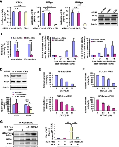
FIG 2Requirement of hCK␣for HCV RNA replication. (A) Huh7 cells transfected with control or hCK␣- or CD81-specific siRNAs for 48 h were separately infected with HCVpp generated from the genotype 1a H77 strain and genotype 2a JFH1 strain or with VSVpp, and luciferase activities were measured. In parallel, a set of cells harvested at 48 h post-siRNA transfection was analyzed by Western blotting; a representative data set is shown. (B) Huh7 cells transfected with control or hCK␣-specific siRNAs were infected with HCV, and the infectivities of the extracellular and intracellular viruses were assessed. This analysis was performed by inoculating culture medium that contained extracellular virions and cell extracts that contained intracellular virions, respectively, into naive Huh7.5-1 cells. The infected cells were harvested, and isolated total RNA was analyzed for the HCV RNA level by RT-PCR. (C) Huh7 cells were transfected with FL-Luc-JFH1 RNA or SGR-Luc-JFH1 RNA, as indicated. HCV RNA replication was measured by determining the fold change in firefly luciferase activity at different time points over that determined at 4 h after RNA transfection. (D) Huh7 cells stably replicating genotype 1b Con1 strain SGR remained untransfected or were transfected with the indicated siRNAs, and cell lysates were analyzed by Western blotting (top panel). The level of the indicated proteins in different settings relative to that detected in siRNA-untreated SGR-replicating cells, which was arbitrarily designated 100%, was expressed as indicated (bottom panel). (E and F) FL-Luc-JFH1 or SGR-Luc-JFH1 RNA-transfected cells were treated with different concentrations of CK37 or HDTAB, as indicated, and the luciferase activity was measured and expressed as the percentage of that detected in cells that received vehicle treatment. (G) Huh7 cells constitutively expressing hCK␣shRNA were transfected with control vector (⫺) or wild-type (␣-R) or D288A (D288A-R) hCK␣resistant to hCK␣shRNA, and cells were left uninfected or infected with HCV. Duplicate sets of cells were analyzed by Western blotting for protein expression (left panel) and by RT-PCR for the viral RNA level (right panel). *,P⬍0.05; **,P⬍0.01; ***,P⬍0.001; ****,P⬍0.0001; ns, nonsignificant.
Wong and Chen
on November 7, 2019 by guest
http://jvi.asm.org/
[image:6.585.90.499.68.576.2]genotype 1a H77 strain and genotype 2a JFH1 strain HCVpp, but
not that of VSVpp (
Fig. 2A
). In contrast, transfection with hCK
␣
siRNA reduced the hCK
␣
protein by 90% but neither affected the
entry of HCVpp derived from both genotypes nor altered the
en-try of VSVpp into Huh7 cells (
Fig. 2A
). These observations
ex-cluded possible effects of hCK
␣
on HCV entry. We also compared
the infectivity of intracellular and extracellular viruses obtained
from control and hCK
␣
-silenced cells infected with HCV by
in-oculating the viruses into naive Huh7 cells, and the total RNA
isolated was quantified to determine the viral RNA level by
RT-PCR. hCK
␣
knockdown reduced both the intracellular and
extra-cellular viral infectivity in parallel (
Fig. 2B
), indicating that this
kinase does not play an obvious role in the assembly or release of
infectious HCV particles.
To assess the contribution of hCK
␣
to HCV RNA replication,
we studied the effect of hCK
␣
knockdown on the HCV RNA
rep-lication kinetics of a bicistronic, full-length (FL) Luc-JFH1
ge-nome that also encodes a firefly luciferase gene as a reporter (
57
).
In the untargeted knockdown cells, luciferase activity decreased
from 4 to 24 h post-viral RNA transfection and then gradually
increased with time (
Fig. 2C
, left panel), exhibiting a lag between
translation of the initial incoming viral genome before active RNA
replication. Nevertheless, luciferase activity was greatly reduced at
different time points post-viral RNA transfection in the hCK
␣
knockdown cells in contrast to their control knockdown
counter-parts (
Fig. 2C
, left panel). Likewise, the replication kinetics of a
subgenomic replicon (SGR) Luc-JFH1 (
43
) were also substantially
attenuated in the hCK
␣
knockdown cells compared to those
de-tected in the control knockdown cells (
Fig. 2C
, right panel).
Fur-thermore, silencing of hCK
␣
in Huh7 cells stably expressing the
genotype 1b Con1 SGR, which did not produce infectious virus
particles, also curtailed NS5A expression (
Fig. 2D
), indicating that
transfection with hCK
␣
siRNA still confers an inhibitory effect on
HCV RNA replication although the virus has established viral
rep-lication in host cells. Taken together, these observations indicate
that the inhibitory effect of hCK
␣
knockdown on viral RNA
rep-lication is not dependent on the entire virus life cycle or on the
virus genotype examined.
To test whether hCK
␣
activity is required for HCV RNA
rep-lication, the effect of CK37, a specific inhibitor of hCK
␣
enzymatic
activity (
36
), on viral RNA replication was examined. The viral
RNA replication of FL and SGR Luc-JFH1 was inhibited by CK37
in a dose-dependent manner (
Fig. 2E
). Similarly, treatment with
HDTAB, another hCK
␣
activity inhibitor (
58
), also interfered
with viral RNA replication of the FL and SGR Luc-JFH1 genomes
proportional to the concentration of HDTAB used (
Fig. 2F
).
Generally, eight highly conserved domains exist among CKs
from different species, such as rat, mouse,
Drosophila
,
Saccharo-myces cerevisiae
, and human (
59
). The main catalytic sites of CK
have been reported to be located in domains 6 and 7. Domain 6,
also known as the Brenner phosphotransferase motif, has the
con-served sequence HXDhXXXNhhh. . . ..D, in which “X” stands for
any amino acid, “h” stands for large hydrophobic amino acids
(such as F, L, I, M, V, W, or Y), and the ellipses represent the
variable lengths of amino acids in different species (
59
). Residues
in the Brenner phosphotransferase motif in CK participate in
maintaining the structure of the ATP binding site and the
orien-tation of choline (
60
). The negative charge of Asp-306 in hCK
␣
2,
equivalent to Asp-288 in hCK
␣
1, deprotonates the hydroxyl
group of the ATP substrate, maintains the proper active site, and
increases the electrophilicity of ATP
␥
-phosphate through
hydro-gen bonding (
61
). In CKs from other species, mutations
intro-duced into the Asp residue analogous to the mutation in hCK
␣
1
Asp-288, for instance, mutation of Asp-255 in
Caenorhabditis
el-egans
CK to Ala or Asn, impair the enzymatic activity (
62
).
To confirm that hCK
␣
activity is essential for viral replication,
we examined the ability of the Asp-to-Ala mutation introduced at
residue 288 of the hCK
␣
active site to rescue HCV replication in
hCK
␣
stable knockdown cells. Toward this end, we constructed a
D288A-R mutant based on the backbone of hCK
␣
-R. Unlike
over-expression of the wild-type hCK
␣
-R in hCK
␣
stable knockdown
cells, which substantially enhanced viral protein expression,
over-expression of the D288A-R mutant did not effectively increase
HCV protein expression despite comparable expression of
wild-type and mutant hCK
␣
-R proteins (
Fig. 2G
, left panel). In
accor-dance, overexpression of wild-type hCK
␣
-R, not the D288A-R
mutant, upregulated the viral RNA level in the HCV-infected
hCK
␣
stable knockdown cells (
Fig. 2G
, right panel). These
find-ings clearly indicate that the enzymatic activity of hCK
␣
is
re-quired for upregulating HCV RNA replication.
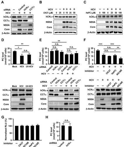
The CDP-choline pathway is not essential for HCV
replica-tion.
To understand the specificity of hCK
␣
for HCV replication,
the effect of knocking down CCT isoform
␣
(CCT
␣
), the
rate-limiting enzyme in the CDP-choline pathway, on HCV expression
was studied. In contrast to the effect of hCK
␣
knockdown on
reduced core and NS5A protein expression, CCT
␣
knockdown
did not affect HCV expression (
Fig. 3A
). Additionally, in contrast
to the dose-dependent inhibitory effect of CK37 on viral protein
expression (
Fig. 3B
), up to 100
M HePC, which was shown to
inhibit PC biosynthesis in HepG2 cells through inhibiting CCT
activity (
63
), did not apparently affect HCV expression in Huh7
cells (
Fig. 3C
). These results emphasize that hCK
␣
activity is
spe-cifically required for HCV replication.
We then determined whether HCV infection might affect the
PC level. Unexpectedly, HCV infection decreased the intracellular
PC level (
Fig. 3D
). Huh7 cells inoculated with UV-irradiated HCV
particles also reduced the PC level (
Fig. 3D
). Moreover, we found
that neither silencing of hCK
␣
nor knockdown of CCT
␣
in
mock-infected cells or HCV-mock-infected cells affected the PC level (
Fig. 3E
).
Consistent with this observation, treatment with CK37 or HePC
did not affect the PC level in the HCV-infected cells (
Fig. 3F
). As a
positive control for the altered PC level in this assay, treatment of
HCV-infected cells with AICAR, a commonly used indirect
acti-vator of AMP-activated protein kinase (
64
) that is known to
at-tenuate PC production in freshly isolated mouse hepatocytes (
65
),
decreased the PC level in infected cells compared to that in
in-fected cells treated with vehicle (
Fig. 3F
, top panel). Nonetheless,
the AICAR-induced reduction in the PC level did not affect HCV
expression (
Fig. 3F
, bottom). Of note, treatment of Huh7 cells
with CK37, HDTAB, HePC, or AICAR at the concentrations
in-dicated in Materials and Methods did not noticeably affect cell
viability (
Fig. 3G
). Additionally, the control and hCK
␣
stable
knockdown cells expressed comparable levels of PC (
Fig. 3H
),
consistent with the comparable cell viability observed with the
paired control and hCK
␣
stable knockdown cells (
Fig. 1D
, right
panel). Collectively, these results indicate that Huh7 cells with
transient knockdown or inactivation of hCK
␣
or CCT
␣
or cells
with constitutive hCK
␣
knockdown still preserve PC
biosynthe-sis and that the PC level does not directly influence viral
on November 7, 2019 by guest
http://jvi.asm.org/
cation. Thus, we conclude that hCK
␣
activity, not the
CDP-choline pathway, is required for HCV replication.
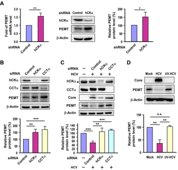
Impairment of the CDP-choline pathway upregulates PEMT
expression.
As the PC level was preserved despite impairment
of the CDP-choline pathway by hCK
␣
or CCT
␣
knockdown
(
Fig. 3E
,
F
, and
H
), we then examined whether the alternative
route that synthesizes PC from PE by the PEMT pathway (
Fig.
1A
) was induced when the CDP-choline pathway was disrupted
in Huh7 cells. RT-PCR and Western blot analyses showed that
the PEMT mRNA and protein levels were higher in the hCK
␣
FIG 3No apparent role of the CDP-choline pathway in HCV replication. (A) Huh7 cells remained untransfected or were transfected with the indicated siRNAs and then mock infected or infected with HCV. Cell lysates were subjected to immunoblot analysis. (B and C) Huh7 cells were mock infected or infected with HCV and then treated with different concentrations of CK37 or HePC, as indicated. Cells lysates were analyzed by immunoblotting. (D) Huh7 cells remained mock infected or were inoculated with untreated or UV-irradiated HCV. (E) Huh7 cells were first transfected with the indicated siRNAs and then mock infected or infected with HCV. (F) Huh7 cells were mock infected or infected with HCV and then treated with DMSO (⫺), CK37, HePC, or AICAR, as indicated, for 24 h. For the experiments shown in panels D to E, a set of 106cells was used to determine the PC amount (top panel), while another set was analyzed by Western blotting(bottom panel). (G) The viability of Huh7 cells treated with vehicle or the indicated inhibitors was assessed. (H) The PC levels in control and hCK␣stable knockdown cells were measured. *,P⬍0.05; **,P⬍0.01; ***,P⬍0.001; ns, nonsignificant.
Wong and Chen
on November 7, 2019 by guest
http://jvi.asm.org/
[image:8.585.78.502.66.567.2]stable knockdown cells than in the control stable knockdown
cells (
Fig. 4A
).
Next, we determined whether hCK
␣
or CCT
␣
transient
knock-down in uninfected cells might upregulate PEMT expression. A
higher level of PEMT protein was detected in hCK
␣
or CCT
␣
transient knockdown cells than that detected in control
knock-down cells (
Fig. 4B
). We then assessed whether depletion of hCK
␣
or CCT
␣
in the HCV-infected cells also induced PEMT protein
expression. To our surprise, HCV infection resulted in a decrease
in PEMT protein expression compared to the level in mock
infec-tion (
Fig. 4C
). Notably, silencing of hCK
␣
or CCT
␣
restored
PEMT protein expression back to the same level as that detected in
mock-infected cells (
Fig. 4C
). To examine whether the reduced
PEMT expression in HCV-infected cells was specific to HCV
in-fection, PEMT protein levels were compared in cells inoculated
with untreated or UV-irradiated HCV. The PEMT protein level
was decreased in HCV-infected cells but not in cells challenged
with UV-irradiated HCV (
Fig. 4D
). Taken together, these results
demonstrate that the PEMT pathway is activated in response to
impairment of the CDP-choline pathway in both uninfected and
HCV-infected Huh7 cells.
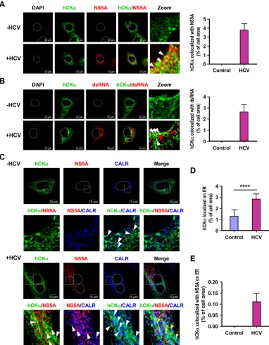
HCV infection increases hCK
␣
localization on the ER.
In
ac-cordance with the indispensable role of hCK
␣
in HCV RNA
rep-lication, confocal microscopy showed that a small fraction of the
hCK
␣
molecules colocalized with NS5A in HCV-infected cells (
Fig.
5A
, left panel). Likewise, in HCV-infected cells, a small portion of
hCK
␣
could colocalize with double-stranded RNA (dsRNA), which
is used to denote the nascent viral dsRNA intermediate newly
synthe-sized during viral replication (
Fig. 5B
). The percentage of the
colocal-ization area of hCK
␣
and NS5A or dsRNA relative to the entire cell
area, which was calculated as previously indicated (
53
), is shown in
the right panels of
Fig. 5A
and
B
, respectively.
Because the HCV polyprotein precursor is synthesized and
proteolytically processed on the ER membrane and because the
ER-derived altered membrane serves as a critical organelle that
houses the viral replication factories (
66
,
67
), we sought to
exam-ine whether HCV infection affected the intracellular localization
of hCK
␣
to the ER using an antibody against CALR, an ER marker.
We noted that a small fraction of hCK
␣
localized on the ER, as
shown by the presence of cyan dots in both mock- and
HCV-infected cells (
Fig. 5C
). Nevertheless, the HCV-infected cells
ex-hibited a greater number of cyan puncta than the mock-infected
FIG 4Effect of disruption of the CDP-choline pathway on PEMT expression. (A) PEMT mRNA and protein expression levels were determined in control and hCK␣stable knockdown cells by RT-PCR (left panel) and Western blot (middle and right panels) analyses, respectively. (B) Huh7 cells were transfected with the indicated siRNAs, and cell lysates were subjected to immunoblot analysis to determine the expression of the indicated proteins. (C) Huh7 cells were transfected with the indicated siRNAs and then mock infected or infected with HCV. Cell lysates were then analyzed by Western blotting. (D) Huh7 cells were mock infected or challenged with untreated HCV or UV-irradiated HCV, and then cell lysates were subjected to immunoblot analysis. *,P⬍0.05; **,P⬍0.01; ***,P⬍0.001; ns, nonsignificant.on November 7, 2019 by guest
http://jvi.asm.org/
[image:9.585.112.475.66.414.2]cells (
Fig. 5C
). In accordance, quantification analysis showed that
HCV infection increased the colocalization of hCK
␣
with CALR
compared to that with mock infection (
Fig. 5D
). We also noted
that a small portion of viral RCs bearing both hCK
␣
and NS5A
signals was detected on the ER, as judged by the appearance of
white puncta in infected cells (
Fig. 5C
, hCK
␣
/NS5A/CALR, yellow
arrows), and approximately 0.11% of the cell area harbored all
three fluorescence signals (
Fig. 5E
).
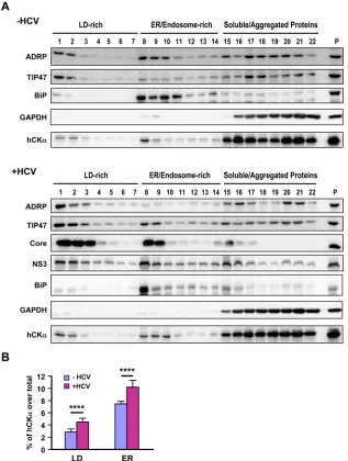
HCV infection increases the cofractionation of hCK
␣
with
viral proteins in the ER membrane.
To provide biochemical
evi-FIG 5Intracellular localization of hCK␣and the hCK␣-associated viral RC on the ER. (A and B) Mock- and HCV-infected Huh7 cells were fixed and processed by confocal microscopy to examine the intracellular localization of hCK␣along with NS5A (A) or dsRNA (B), and the two fluorescence profiles were merged (left panels). The boxed areas in the hCK␣/NS5A and hCK␣/dsRNA images were enlarged (Zoom) to show colocalization (white arrowheads) of hCK␣with NS5A or dsRNA. The degree of colocalization of hCK␣with NS5A or dsRNA was calculated as the percentage of the cell area that harbored the indicated fluorescence signal relative to the entire cell area, as described in Materials and Methods (right panels). (C to E) Mock- and HCV-infected Huh7 cells were examined by confocal microscopy to determine the intracellular localization of the indicated proteins (C). Merged images show the overlay of the three fluorescence signals. The second row of images shows enlargements of the boxed areas in the top row. The white arrowheads indicate the colocalization of the two indicated fluorescence signals, whereas the yellow arrows indicate the colocalization of the three fluorescent signals. The degrees of colocalization of hCK␣and CALR and of hCK␣, NS5, and CALR were quantified (D and E, respectively). ****,P⬍0.0001.Wong and Chen
on November 7, 2019 by guest
http://jvi.asm.org/
[image:10.585.101.483.67.556.2]dence for the association of hCK
␣
with the ER, we performed
iodixanol-based density gradient subcellular fractionation (
51
).
In the context of mock infection or HCV infection, BiP, a
chaper-one located in the lumen of the ER, and GAPDH, a cytosolic
pro-tein, were predominantly fractionated into fractions 8 and 9 and
fractions 15 to 22, representing the ER/endosome and soluble/
aggregated protein fractions, respectively (
Fig. 6A
). The top two
light-density fractions included the LD-associated membranes, as
the two LD markers ADRP and TIP47 were distributed in these
fractions (
Fig. 6A
). Quantification analyses from three
indepen-dent studies showed that HCV infection increased the distribution
of ADRP and TIP47 in LD-rich membranes (
P
⬍
0.05; data not
shown). In this regard, Vogt et al. showed that HCV infection or
viral RNA replication augments the distribution of TIP47, but not
ADRP, in the LD fractions and that TIP47 interacts with NS5A
and recruits LD-rich membranes to the MW for efficient viral
RNA replication (
51
). In HCV-infected cells, a fraction of hCK
␣
cofractionated with the HCV core and NS3 proteins, which are
recruited to LD-rich compartments during HCV viral assembly
(
Fig. 6A
, bottom panel). Furthermore, hCK
␣
also cofractionated
with core and NS3 proteins into the ER/endosome-rich
mem-brane fractions (
Fig. 6A
, bottom panel). As NS3 is a component of
the HCV RC, these findings support a model in which hCK
␣
as-sociates with the HCV RC on the ER and LD-rich membranes.
Quantitative analysis revealed that HCV infection increased
hCK
␣
distribution into LD and ER fractions (
Fig. 6B
), suggesting
that hCK
␣
enrichment in these two compartments is crucial for
viral replication.
FIG 6HCV-induced increase in the distribution of hCK␣into ER membranes by HCV infection. (A) The postnuclear fractions from mock- and HCV-infected Huh7 cells were subjected to iodixanol-based OptiPrep density gradient ultracentrifugation. Proteins in each fraction were precipitated with 10% cold trichlo-roacetic acid and then analyzed by Western blotting for the indicated proteins. Extracts from mock- and HCV-infected cell lysates were separately loaded as controls for the detection of the indicated proteins (P lanes). (B) The intensity of hCK␣in each fraction was quantified by ImageQuant, and the percentage of hCK␣fractionated into LDs (fractions 1 to 7) and the ER (fractions 8 to 14) over the total hCK␣level obtained from mock- and HCV-infected cells was quantified. ****,P⬍0.0001.
on November 7, 2019 by guest
http://jvi.asm.org/
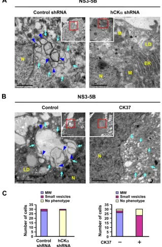
[image:11.585.135.452.67.487.2]Knockdown or inactivation of hCK
␣
abrogates
HCV-in-duced MW formation.
Because hCK
␣
is a critical regulator of
HCV RNA replication (
Fig. 1
and
2
) and because MW is critical
for viral RNA replication (
38
,
68
,
69
), we then examined whether
hCK
␣
participates in MW formation by employing the pTM
tran-sient expression system, which was shown to mediate formation of
the viral RC complex and MW independent of viral RNA
replica-tion (
44
,
70
,
71
).
TEM analysis revealed that expression of NS3 to NS5B in the
control shRNA knockdown Huh7 cells resulted in significant MW
formation, as indicated by the presence of membrane matrix in
the perinuclear region, inside which vesicles of heterogeneous
FIG 7Abrogation of HCV-induced MW formation by hCK␣depletion or CK37 treatment. (A) Control shRNA and hCK␣shRNA stable expression cells were transfected with pTM-NS3-5B and pWPI-T7-BLR, fixed, and processed for TEM. (B) Huh7 cells were first cotransfected with plasmids expressing T7 polymerase and NS3-NS5B. At 24 h post-DNA transfection, cells were left untreated or treated with 100M CK37 for 18 h, and then cells were analyzed by TEM for MW formation. The area boxed in red was enlarged as shown. Light blue arrows indicate vesicles of heterogeneous sizes (left panels of A and B) and small vesicles in CK37-treated Huh7 cells (right panel of B); blue arrowheads indicate double-membrane vesicles (DMVs) (left panels of A and B). N, nucleus; LD, lipid droplet; ER, endoplasmic reticulum; M, mitochondria. (C) Thirty cells randomly picked from each experimental setting were examined by TEM, and the numbers of cells showing three different phenotypes were quantified. MW, vesicles 130 to 270 nm in diameter; small vesicles, vesicles 40 to 100 nm in diameter.Wong and Chen
on November 7, 2019 by guest
http://jvi.asm.org/
[image:12.585.132.451.73.559.2]sizes between 130 and 270 nm in diameter were enclosed (
Fig. 7A
,
left panel, light blue arrows). Among these vesicles,
double-mem-braned vesicles (DMVs) were noted (
Fig. 7A
, left panel, blue
ar-rowheads). These DMV structures were shown to be viral
replica-tion factories because the kinetics of their appearance correlated
with the viral RNA replication rate (
9
) and because purified DMVs
contained HCV replicase proteins such as NS3 and NS5A (
10
).
Small round vesicles with a diameter ranging from 40 to 100 nm
were sometimes observed scattered around the perinuclear
re-gion. In the hCK
␣
stable knockdown cells, intact ER and
mitochon-dria were evident, and no typical MW structures were observed (
Fig.
7A
, right panel). The quantitative results regarding MW formation,
small-vesicle formation, or absence of phenotype for 30 randomly
picked cells is summarized in the left panel of
Fig. 7C
.
To understand whether hCK
␣
activity is essential for MW
for-mation, Huh7 cells expressing NS3-NS5B were treated with or
without CK37. Unlike the vesicles enclosed within the MW
struc-ture (
Fig. 7B
, left panel), no typical MW structure was observed in
CK37-treated cells. Instead, small vesicles, sometimes with an
elongated shape, were observed scattered around the perinuclear
region (
Fig. 7B
, right panel, light blue arrows). These vesicles were
smaller than those vesicles enclosed within the MW structure.
Additionally, a portion of CK37-treated cells exhibited no MW or
small-vesicle phenotype. The right panel of
Fig. 7C
summarizes
the quantitative results regarding the three phenotypes observed
for cells expressing NS3-NS5B that were treated with vehicle or
CK37. These observations demonstrate a correlation between the
inhibitory effects of CK37 on MW formation and viral RNA
rep-lication, implying that hCK
␣
activity contributes to MW
forma-tion.
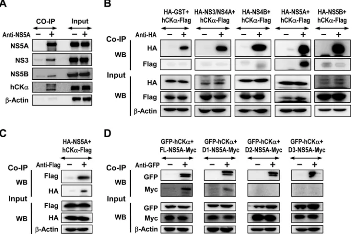
hCK
␣
is recruited onto the viral RC, presumably through its
interaction with D1 of NS5A.
To understand the function of
hCK
␣
in viral RC and MW formation, we performed co-IP assays
to examine whether hCK
␣
was recruited to the viral RC that
formed in the context of the pTM expression system. Precipitation
of lysates expressing proteins NS3 to NS5B with an NS5A MAb not
only precipitated NS5A but also specifically cocaptured NS3 and
NS5B (
Fig. 8A
), indicating viral RC formation. In addition, hCK
␣
was coprecipitated with the viral RC using NS5A MAb but not
using control IgG (
Fig. 8A
), showing that hCK
␣
is recruited to the
viral RC.
To determine the mechanism by which hCK
␣
is recruited by
the viral RC, we studied which NS protein interacts with hCK
␣
by
co-IP. Lysates from cells coexpressing a series of HA-tagged NS
proteins derived from the genotype 1b Con1 strain and
Flag-tagged hCK
␣
were precipitated with rabbit anti-HA, and the
pre-cipitated proteins were analyzed by Western blotting. We found
that hCK
␣
-Flag was predominantly associated with NS5A but not
with other NS proteins (
Fig. 8B
). Conversely, precipitating hCK
␣
-Flag with anti--Flag also pulled down HA-NS5A (
Fig. 8C
). Domain
mapping using GFP-hCK
␣
and a set of constructs encoding
dif-ferent domains of the JFH1 strain NS5A with a Myc tag at their C
termini (
45
) showed that D1 of NS5A mediated the NS5A-hCK
␣
interaction, as anti-GFP coprecipitated FL and D1, but not D2 and
D3, of NS5A (
Fig. 8D
). The results also demonstrated that hCK
␣
interacts with NS5A in a genotype-independent manner.
FIG 8Assessment of the interaction of hCK␣with NS5A. (A) Huh7 cells coexpressing T7 RNA polymerase and NS3-NS5B were lysed and subjected to co-IP with an anti-NS5 MAb or an isotype-matched control mouse IgG. The precipitates were analyzed by SDS-PAGE, followed by immunoblotting analysis to detect each of the indicated proteins. A portion of aliquots containing 5% of the total proteins in the lysates used for precipitation was loaded as the input control. (B) 293T cells were cotransfected with plasmids encoding each of HCV genotype 1b Con1 strain NS proteins, as indicated, with a pCMV6 plasmid encoding hCK␣-Flag. Cell lysates were pulled down with rabbit anti-HA, and the precipitated proteins were subjected to immunoblot analysis with the indicated MAb antibodies. (C) Lysates from Huh7 cells expressing the indicated proteins were precipitated with rabbit anti-Flag, and the coprecipitated proteins were subjected to immunoblot detection with the indicated MAbs. (D) Lysates from Huh7 cells coexpressing GFP-tagged hCK␣and different Myc-tagged NS5A domains were pulled down with rabbit anti-GFP, followed by immunoblot analysis using the indicated MAbs.on November 7, 2019 by guest
http://jvi.asm.org/
[image:13.585.113.474.69.308.2]hCK
␣
activity mediates the enhanced colocalization of
hCK
␣
and NS5A on the ER.
As hCK
␣
is a critical
NS5A-interact-ing partner, we then assessed whether hCK
␣
contributed to the
localization of NS5A on the ER. hCK
␣
stable knockdown cells
showed decreased localization of NS5A on the ER compared with
the level of the control stable knockdown cells (
Fig. 9A
). In
con-trast, stable hCK
␣
knockdown did not affect the localization of
NS3 or NS5B on the ER (
Fig. 9B
and
C
, respectively). These results
indicate that hCK
␣
enhances the localization of NS5A to the ER
and suggest that hCK
␣
may stabilize the association of NS5A with
other NS proteins.
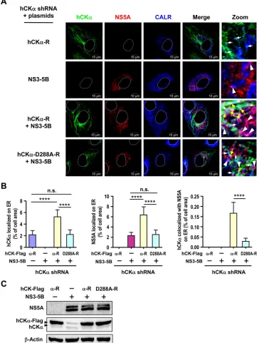
To explore the mechanism by which hCK
␣
targets NS5A to the
ER, we examined whether hCK
␣
activity affected the ER
localiza-tion of NS5A and hCK
␣
and whether the expression of NS
pro-teins enhanced hCK
␣
localization to the ER. To this end, we
ex-amined the intracellular localization of NS5A and hCK
␣
in hCK
␣
stable knockdown Huh7 cells overexpressing wild-type or D288A
mutant hCK
␣
-R, with or without NS3-NS5B expression, by
con-focal microscopy (
Fig. 10A
). A basal level of overexpressed
wild-type hCK
␣
was localized on the ER in the absence of NS3-NS5B
expression (
Fig. 10B
, left panel). Expression of NS3-NS5B
aug-mented ER localization of wild-type hCK
␣
but not of
overex-pressed D288A mutant hCK
␣
(
Fig. 10B
, left panel). This
observa-tion was reminiscent of the earlier observaobserva-tion that HCV infecobserva-tion
increased hCK
␣
localization on the ER (
Fig. 5C
and
D
). In
con-trast, a basal level of NS5A was detected on the ER in the absence of
wild-type hCK
␣
expression (
Fig. 10B
, middle panel).
Interest-ingly, overexpression of wild-type, but not D288A mutant,
hCK
␣
-R resulted in greater accumulation of NS5A on the ER (
Fig.
10B
, middle panel). Moreover, overexpression of wild-type hCK
␣
enhanced hCK
␣
colocalization with NS5A on the ER; however,
overexpression of the D288A mutant resulted in the colocalization
of only a basal level of hCK
␣
with NS5A on the ER (
Fig. 10B
, right
panel). These results collectively indicate that NS3-NS5B
expres-sion promotes hCK
␣
trafficking to the ER and that hCK
␣
activity
is required for effective targeting of NS5A, hCK
␣
, and the hCK
␣
-NS5A complex to the ER.
hCK
␣
is necessary for the interaction of NS5A with NS5B in
the viral RC.
To support the function of hCK
␣
in the association
of NS5A with the viral RC, we first determined the effect of hCK
␣
knockdown on NS5A colocalization with overexpressed
Flag-tagged NS5B by confocal microscopy (
Fig. 11A
, left panel).
Deple-tion of hCK
␣
mitigated the colocalization of NS5A with
Flag-tagged NS5B (
Fig. 11A
, right panel), suggesting the requirement of
hCK
␣
for the association of NS5A with NS5B in the viral RC.
To gain insight into the mechanism of hCK
␣
in viral RC
for-mation, we assessed the binding of NS5A with NS5B or NS3 in
control and hCK
␣
stable knockdown Huh7 cells expressing
NS3-NS5B by co-IP with NS5A MAb. Silencing of hCK
␣
strikingly
abolished the interaction of NS5A with NS5B (
Fig. 11B
).
How-FIG 9The effect of hCK␣knockdown on ER localization of NS5A, NS5B, and NS3. The control and hCK␣shRNA Huh7 stable cells were transfected with pTM-NS3-5B and pWPI-T7-BLR, and the cells were examined by confocal microscopy for ER localization of NS5A (A), NS5B (B), and NS3 (C) (left panels). The white boxed areas in the NS/CALR images were enlarged (Zoom), and white arrowheads indicate the ER localization of each indicated NS protein. The extent of colocalization of each NS protein with CALR in control and hCK␣shRNA cells was quantified (right panel). ****,P⬍0.0001; ns, nonsignificant.Wong and Chen
on November 7, 2019 by guest
http://jvi.asm.org/
[image:14.585.113.476.71.382.2]ever, hCK
␣
depletion only partially inhibited the binding of NS5A
with NS3 as the level of NS3 cocaptured with NS5A was reduced to
43% of that detected in control knockdown cells (
Fig. 11B
). To
dissect the effects of hCK
␣
on NS5A-NS5B and NS5A-NS3
inter-actions in the absence of other NS proteins, we performed co-IP
assays on control and hCK
␣
shRNA stable cells overexpressing
NS5A-Myc and Flag-NS5B and overexpressing NS5A-Myc and
Flag-NS3, respectively. hCK
␣
depletion greatly interfered with the
binding of NS5A to NS5B (
Fig. 11C
). Nonetheless, the ratio of
NS3 to NS5A cocaptured by NS5A MAb was reduced to 0.5 in
hCK
␣
-depleted cells compared with that detected in control
knockdown cells (
Fig. 11D
).
Moreover, we performed co-IP using the paired hCK
␣
stable
knockdown cells overexpressing NS5A-Myc and Flag-NS4B to
understand the involvement of hCK
␣
in the association of NS5A
with NS4B. The result showed that hCK
␣
knockdown did not
FIG 10Analysis of the effect of hCK␣activity on ER localization of hCK␣and NS5A. (A) The hCK␣stable knockdown Huh7 cells were cotransfected with pWPI-T7-BLR along with wild-type or D288A mutant hCK␣-R plasmid in the presence or absence of pTM-NS3-NS5B as indicated. The cells were then analyzed by confocal microscopy for the intracellular localization of hCK␣and NS5A on the ER. The white boxed areas in merged images were enlarged (Zoom) to show the localization of hCK␣and NS5A on the ER (white arrows and white arrowheads, respectively) and the colocalization of hCK␣and NS5A on the ER (yellow arrows). (B) The degree of localization of hCK␣, NS5A, and hCK␣-associated NS5A on the ER was quantified. (C) A representative set of Western blot results for the indicated proteins is shown. ****,P⬍0.0001; ns, nonsignificant.on November 7, 2019 by guest
http://jvi.asm.org/
[image:15.585.111.475.71.552.2]greatly affect the binding of NS5A to NS4B (
Fig. 11E
). We also
examined the effect of hCK
␣
knockdown on the binding of NS5B
to NS4B in the paired Huh7 cells coexpressing Flag-NS4B and
NS5B driven by the T7 promoter. The result showed that hCK
␣
knockdown did not obviously affect the NS5B-NS4B interaction
(
Fig. 11F
). Taken together, these results indicate that hCK
␣
is
indispensable for the stable interaction of NS5A with NS5B and
can enhance the binding of NS5A with NS3. However, the
NS4B-NS5A and NS4B-NS5B interactions occur independent of hCK
␣
.
hCK
␣
activity is not essential for the interaction of NS5A
with hCK
␣
and NS5B.
Next, we studied whether hCK
␣
activity
was crucial for NS5A binding to hCK
␣
and NS5B. T7-Pol/Huh7
cells were cotransfected with pTM-NS5A and pTM-NS5B and
then treated with DMSO or CK37. Cell lysates were precipitated
with anti-NS5A MAb. CK37 treatment neither affected the
NS5A-hCK
␣
interaction nor altered NS5A binding to NS5B (
Fig. 12A
).
To confirm this finding, the wild-type hCK
␣
or wild-type or
D288A mutant hCK
␣
-R plasmid was transfected into T7 RNA
polymerase-expressing control or hCK
␣
stable knockdown Huh7
cells, followed by cotransfection with NS5A and
pTM-NS5B. The binding of NS5A to NS5B was greatly impaired in
hCK
␣
shRNA cells transfected with wild-type hCK
␣
compared to
FIG 11Requirement of hCK␣for the binding of NS5A to NS5B in the viral RC. (A) Huh7 cells were cotransfected with pTM-NS3-5B and pWPI-T7-BLR along with pCMV-Flag-NS5B. The transfected cells were fixed and processed by confocal microscopy using NS5A MAb and rabbit anti-Flag (left panel). The boxed area in the NS5A/Flag-NS5B panel was enlarged (Zoom) to show the colocalization of NS5A with Flag-NS5B (white arrowheads). The degree of colocalization of NS5A with Flag-NS5B was quantified (right panel). (B) Cell lysates from the paired control and hCK␣stable knockdown Huh7 cells cotransfected with pTM-NS3-NS5B and pWPI-T7-BLR were subjected to co-IP analysis with anti-NS5A MAb, and the precipitated proteins were analyzed by Western blotting for the indicated proteins. (C to E) The paired stable knockdown cells were cotransfected with pCMV plasmids encoding NS5A-Myc and Flag-tagged NS5B (C), NS3 (D), or NS4B (E). The cell lysates were subjected to co-IP with anti-Myc to determine the binding of NS5A to each of the NS proteins. (F) The paired knockdown Huh7 cells were transfected with plasmids encoding pCMV-Flag-NS4B, pWPI-T7-BLR, and pTM-NS5B. Cell lysates were precipitated with anti-Flag to determine the binding of NS4B to NS5B. ****,P⬍0.0001.Wong and Chen
on November 7, 2019 by guest
http://jvi.asm.org/
[image:16.585.99.487.71.496.2]that detected in control shRNA cells transfected with wild-type
hCK
␣
(
Fig. 12B
), confirming t