Function of MicroRNA-146a and NF-
κ
B in Physiologic
and Pathologic Hematopoiesis
Thesis by
Jimmy Liu Zhao
In Partial Fulfillment of the Requirements for the Degree of
Doctor of Philosophy
CALIFORNIA INSTITUTE OF TECHNOLOGY
Pasadena, California
2013
2013
Jimmy Liu Zhao
I want to thank Professor David Baltimore for providing great mentorship and training over the years. I
also want to thank Dr. Dinesh Rao for excellent mentorship and Drs. Ryan O’Connell, Alejandro
Balazs, Devdoot Majumdar, Parameswaran Ramakrishnanand, Lili Yang, Shengli Hao, Aadel
Chaudhuri, Arnav Mehta, Yvette Garcia, and other members of the Baltimore Lab for tremendous help
and stimulating discussion over the years. I also want to thank members of my thesis committee, Paul
Patterson, Sarkis Mazmanian, David Baltimore and Dinesh Rao, as well as UCLA MSTP directors,
Steve Smale and Kelsey Martin, for guidance and mentorship. Lastly, I’d like to acknowledge the staff
at the California Institute of Technology animal facility, FACS Core sorting facility and genomic core
facility for excellent technical support. NFκB-GFP reporter mice were a generous gift of Dr. Christian
Jobin at the University of North Carolina, Chapel Hill. My training experience and research work were
significantly enriched and enhanced by wonderful collaborations with Chao Ma of the Heath Lab and
Beverly Lu of the Tirrell Lab at Caltech, and many external collaborators from other institutions. The
work was supported by research grant R01AI079243 (D.B.), National Research Service Award
F30HL110691 and the UCLA/Caltech Joint Medical Scientist Training Program (J.L.Z.), from the
During inflammation and infection, hematopoietic stem and progenitor cells (HSPCs) are stimulated to
proliferate and differentiate into mature immune cells, especially of the myeloid lineage.
MicroRNA-146a (MicroRNA-146a) is a critical negative regulator of inflammation. Deletion of the gene encoding
miR-146a—expressed in all blood cell types—produces effects that appear as dysregulated inflammatory
hematopoiesis, leading to a decline in the number and quality of hematopoietic stem cells (HSCs),
excessive myeloproliferation, and, ultimately, to exhaustion of the HSCs and hematopoietic
neoplasms. Six-week-old deleted mice are normal, with no effect on cell numbers, but by 4 months
bone marrow hypercellularity can be seen, and by 8 months marrow exhaustion is becoming evident.
The ability of HSCs to replenish the entire hematopoietic repertoire in a myelo-ablated mouse also
declines precipitously as miR-146a-deficient mice age. In the absence of miR-146a, LPS-mediated
serial inflammatory stimulation accelerates the effects of aging. This chronic inflammatory stress on
HSCs in deleted mice involves a molecular axis consisting of upregulation of the signaling protein
TRAF6 leading to excessive activity of the transcription factor NF-κB and overproduction of the
cytokine IL-6. At the cellular level, transplant studies show that the defects are attributable to both an
intrinsic problem in the miR-146a-deficient HSCs and extrinsic effects of miR-146a-deficient
lymphocytes and non-hematopoietic cells. This study has identified a microRNA, miR-146a, to be a
critical regulator of HSC homeostasis during chronic inflammatory challenge in mice and has provided
a molecular connection between chronic inflammation and the development of bone marrow failure
and myeloproliferative neoplasms. This may have implications for human hematopoietic malignancies,
Overview and Scientific Background 1
Chapter One
Introduction 20
Results 22
Figures 28
Discussion 37
Experimental Procedures 40
Chapter Two
Introduction 42
Results 44
Figures 60
Discussion 74
Experimental Procedures 79
References 81
OVERVIEW and SCIENTIFIC BACKGROUND
MicroRNA
Initially discovered by the Ambros and Ruvkun labs in the early 1990s, microRNAs (miRNAs) have
emerged as important regulators of gene expression 1-2. In the past several years, significant advances have been made with regards to understanding their integration into molecular networks that control
cellular development and function. In the hematopoietic system, several individual miRNAs have
emerged as important regulators of physiological and pathologic myeloid development and function.
MiRNAs are encoded within exons of unique non-coding genes, or sometimes within introns of
protein-coding genes, and are most often transcribed from the genome by RNA polymerase II. Some
miRNAs exist as clusters and are encoded on the same primary transcript. The primary transcript
(pri-miRNA) is processed by the ribonuclease Drosha/DGCR8 in the nucleus and then transported into the
cytoplasm as a pre-miRNA, which contains the miRNA stem-loop structure. For most miRNAs, this
serves as a substrate for the endoribonuclease Dicer, which creates a double-stranded short RNA that is
unwound and then loaded into the RNA-induced silencing complex (RISC) (reviewed in 3-4). This “classical” description of miRNA biogenesis was true for all known miRNA until mid-2010, when two
seminal papers reported that Dicer independent production of miRNAs could occur via the “slicer”
RNAse function of Argonaute 2 (Ago2), a component of the RNA induced silencing complex (RISC) 5-6. The mature miRNA is loaded onto the RISC, a multi-protein complex that includes members of the Argonaute protein family 7. Once in the RISC, the mature miRNA interacts with a target mRNA via the latter’s 3’UTR. Structural, biochemical and bioinformatics analyses indicate that a 6-nucleotide
seed sequence at the 5’ end of the miRNA is important in mediating the interaction with its target 8. In most cases, miRNAs repress their targets and this change is detectable at the RNA level, and it is now
there are scattered reports of miRNAs that interact with their targets in a non-3’UTR-dependent or
non-seed-dependent fashion, and miRNAs that cause upregulation of “targets” 11-12. These studies suggest that there are aspects to miRNA function that remain elusive. Initial studies revealed the global
importance of miRNAs in hematopoiesis. Dicer deletionin the hematopoietic lineage leads to marked
defect in competitive repopulation assays and the deletion of Ago2 leads to severe problems with
erythroid and B-cell development 13-14. However, the main focus of research in the field has been on examining how individual miRNAs are integrated into various regulatory pathways in myelopoiesis.
miRNAs are induced by transcription factors and other genes involved in myelopoiesis. For example,
PU.1 induces expression of miR-223 and NF-kB induces expression of miR-155 and miR-146a.
miRNAs, in turn, repress the expression of their targets, for example, miR-155 regulates PU.1 and
C/EBP-β, thereby changing the transcriptional profile of the cell 15. In this search for targets, global RNA expression profiling followed by correlation with algorithmic target prediction has been fruitful
15. Confirmation of the targets on a case-by-case basis can then be followed by functional analyses of the regulated pathways.
MicroRNA-146a
MicroRNA-146a (miR-146a) along with miR-146b form a highly conserved two-member miRNA
family. MiR-146a and miR-146b are located on chromosome 5 and 10, respectively, in the human
genome and on chromosome 11 and 19, respectively, in the mouse genome. In 2006, miR-146a first
came to our attention in a systemic microarray screen to identify miRNAs that are involved in innate
immune activation 16. Stimulating THP1 cells, a human monocytic cell line, with lipopolysaccharide (LPS) was found to selectively upregulate the expression of three miRNAs, miR-155, miR-146 and
the immune system. In the hematopoietic system, the expression of miR-146a is relatively low in
progenitor cells and increases modestly with maturation. However, in certain specialized cell types,
such as Ly-6Clo monocytes and epidermal Langerhans cells, miR-146a is constitutively expressed at a
high level (17-18 and immunological genome project http://www.immgen.org/). In addition, miR-146a expression is up-regulated upon stimulation of myeloid cells with microbial components and
pro-inflammatory cytokines. MiR-146a is also stimulated upon activation of T cells with T-cell receptor
antigens 19-21. Viral and fungal infection also induces miR-146a expression 22-24. Basal and induced expression of miR-146a is regulated by several important transcription factors in immune cells. Our
initial characterization of miR-146a promoter locus identified two consensus NF-κB binding sites that
were shown to be important in transcriptional activation of miR-146a in myeloid cells in response to
inflammatory stimulation; mutations of these consensus sequences abolished the promoter activity in a
luciferase reporter system 16. Subsequent studies showed that downregulation of NF-κB activity chemically or genetically inhibits basal and induced expression of miR-146a in macrophages and T
cells, confirming the role of NF-κB in regulating miR-146a expression 20,22-23. In addition to regulation by NF-κB, expression of miR-146a is also regulated by lineage-dependent transcription factors. During
myeloid differentiation, PU.1 works together with NF-κB to control the basal expression of miR-146a
through dynamic occupancy at the promoter region 18,25. In lymphocytes, the lineage-specific regulation of miR-146a may be fulfilled by other transcriptional factors, such as c-ETS 24,26. In addition to transcriptional activation, miR-146a expression may be repressed in a lineage specific
manner. This was shown in the example of megakaryocyte differentiation, where PLZF upregulation
interacts with the miR-146a promoter to inhibit its expression 27. A more extreme case involves global miRNA repression, including miR-146a, by the myc oncogenic transcriptional factor during
miR-146a, but not miR-146b, in mouse splenic lymphocytes. However, it is not clear from the study
whether the effect of estrogen on miR-146a expression is direct 29. Overall, basal miR-146a expression is regulated by a set of lineage-dependent transcription factors in a cell-lineage specific manner. This
basal level can be further regulated in an activation-dependent manner by inducible transcription
factors, such as NF-κB (Figure i). In comparison to miR-146a, the regulation of miR-146b expression
is less understood. Some previous reports on miR-146b expression were performed primarily with
hybridization-based methods, which may have not been specific enough to separate the mature
sequences of 146a and 146b because they only differ by one nucleotide. Nevertheless,
miR-146b seems also to be induced by pro-inflammatory cytokines and dysregulated miR-miR-146b expression
is associated with several human non-hematologic cancers 30-35. In addition, in unpublished work, we have shown that miR-146b expression can be clearly detected by RT-qPCR in miR-146a-deficient
bone marrow cells, indicating that it is not a pseudogene or undetectable in adult hematopoietic cells as
NF-κB
p65
LPS IL-1β
TNF-α IL-6
IRAK1
TRAF6 TRAFs
Ag
p50 p52 RelB
miR-146a
c-ETS PU.1
PLZF MYC
Canonical NF-κB Non-canonical NF-κB
TF Pol II
INF-γ
Other NF-κB-responsive genes: TNFα, IL1-β, IL-6, IFN-γ
STAT1
STATs
STAT3
STAT3
Negatively regulates myeloid development, macrophage/monocyte activation, and effector T-cell activation
Positively regulates regulator T-cell function Down-regulates/terminates inflammation
Suppresses the development of cancer and autoimmunity
NF-κB
TNFR TCR IL-1R TLR
[image:10.612.80.477.72.371.2]4 IFN g R IL-6R
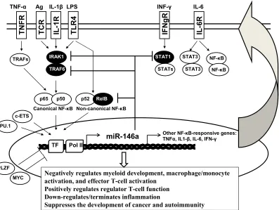
Figure i. Expression of miR-146a is regulated by a set of transcriptional factors in a cell-lineage specific manner, involving such factors as PU.1, c-ETS, and PLZF, and in an activation-dependent manner, involving such factors as NF-κB. Validated targets of miR-146a include Irak1, Traf6, Stat1 and RelB, which are all involved in the NF-κB and STAT pathways, highlighting the role of miR-146a as a critical negative regulator of inflammatory and interferon signaling.
MiR-146a Functions in Immunity
Our initial report showed that miR-146a is highly up-regulated in human monocytes in a NF-κ
B-dependent manner following toll-like receptor (TLR) stimulation 16. Initial sequence complementation-based algorithms (e.g. TargetScan) identified TRAF6 and IRAK1 as targets of miR-146a. The
regulation of these two proteins by miR-146a in monocyte/macrophages, T- and B- lymphocytes was
then confirmed by 3’UTR luciferase reporter assays and gene expression analyses 16,19-20,22,37. Because TRAF6 and IRAK1 are known feedback regulators of the NF-κB signal pathway, a molecular circuitry
involving miR-146a, TRAF6/IRAK1 and NF-κB is proposed to be important in the regulation of both
from the study of miR-146a-deficient mice generated in our laboratory 19. Consistent with the proposed hypothesis, miR-146a-/- mice were found to be hyper-responsive to LPS challenge. Stimulation with
sublethal level of LPS in vivo induced a higher serum level of pro-inflammatory cytokines— including
TNFα and IL-6 and IL-1β— in miR-146a-/- mice than in wild type mice. In addition, miR-146a-/-
mice experienced a higher mortality rate in response to minimal lethal LPS challenge. The
hyper-inflammatory response was also observed in bone marrow derived macrophages (BMDMs) from
146a-/- mice in vitro. To complement the study on 146a-/- mice, enforced overexpression of
miR-146a in THP-1 cells resulted in an attenuated inflammatory response 19. Work of others has confirmed these general observations. These studies provide further support that miR-146a is an important
negative regulator of innate immune activation, potentially by targeting TRAF6 and IRAK1 and by
orchestrating the silencing of TNFα gene in human monocytic cell lines in the context of
endotoxin-induced tolerance 39-41. In addition to regulating pro-inflammatory cytokine production, miR-146a negatively regulates type I interferon (IFN) production in vesicular stomatitis virus-infected mouse
peritoneal macrophages by targeting TRAF6, IRAK1 and IRAK2 through a RIG-I/NF-κB-dependent
but TLR4/MyD88-independent pathway 22. A study in human peripheral blood mononuclear cells confirmed a role for miR-146a as a negative regulator of the type I IFN pathway 42. In a more elaborate example of cell-type-specific regulation by miR-146a in the innate immune system, miR-146a
specifically controls the Ly-6Chi, but not Ly-6Clo, monocyte response during inflammatory challenge
17. The regulation by miR-146a in this context is cell-intrinsic, apparently through directly targeting Relb, thus also connecting miR-146a to the non-canonical pathway of NF-κB activation. This study
further substantiates the role of miR-146a in innate immunity by controlling monocyte functional
heterogeneity during an inflammatory response. Given the hyper-inflammation and the exaggerated
to bacterial infection than wild type animals. Indeed, miR-146a-/- mice displayed a lower bacterial
load upon infection with Listeria monocytogenes 17. The role of miR-146a in adaptive immunity is particularly well studied in T cells. Aging miR-146a-/- mice develop an autoimmune disorder with
lymphadenopathy, lymphocyte infiltration in various organs and an activated T cell phenotype 19. We carried out detailed analysis in a subsequent study to understand the physiological role of miR-146a in
regulating the T cell response to antigen stimulation 20. We showed that miR-146a-deficient CD4 and CD8 T cells are hyper-responsive following T-cell receptor (TCR) stimulation, as indicated by
increased proliferation and survival, exaggerated activation phenotype and enhanced effector cytokine
production. The regulation of cell-intrinsic T cell response upon TCR stimulation by miR-146a was
shown to again be through the regulation of TRAF6/IRAK1 and NF-κB signaling. This study adds
miR-146a to a list of well-known negative regulators of NF-κB, including IκBα and A20, acting
following TCR activation and suggests that these feedback regulators function collaboratively to limit
the extent and timing of activation, with each apparently fulfilling a crucial non-redundant role. These
mice studies were supported by studies in human T cells showing that miR-146a overexpression
impairs IL-2 production. However, contrary to our observation that TCR-induced apoptosis was
reduced in miR-146a-deficient T cells and enhanced in T cells with miR-146a overexpression,
activation-induced cell death seems to be inhibited by miR-146a overexpression in human Jurkat T cell
cancer line 26. The discrepancy may be due to the differences between mouse primary T cells and human cancer cell lines or the level of miR-146a overexpression achieved in the two experimental
systems. In addition to functioning in effector T cells in a cell-intrinsic manner, miR-146a also controls
the regulation of Th1 responses by affecting regulatory T cell (Treg)-function 43. Treg cells maintain immune homeostasis by suppressing the inflammatory response and preventing an over-reaction. In
turn, miR-146a-deficient Treg cells are functionally defective in their ability to restrain effector T cells,
leading to overproduction of IFNγ and autoimmunity. Stat1 appears to be a responsible target for
miR-146a in regulating Treg cells. This study revealed an important regulatory function of miR-miR-146a in
Treg cells, suggesting upregulation of miR-146a is required for Treg cells to properly control the
IFNγ-mediated Th1 response. The results imply an interesting phenomenon, which is that excessive
effector cytokine signaling in Treg cells can lead to their failure to suppress the corresponding cytokine
response in effector T cells. Further studies are required to elucidate the mechanistic implications
behind this type of Treg cells/effector T cells regulation. Our knowledge of the function of miR-146a
in B cells is limited. One study has provided evidence for miR-146a induction in a NF-κB-dependent
manner by Epstein-Barr virus Latent Membrane Protein 1 (LMP1) in human B lymphocytes 24. Study from miR-146a-deficient mice suggested that B cells in these mice are also hyper-activated and
autoreactive, because we can detect anti-dsDNA antibodies in the serum and a small percentage of
B-cell lymphomas in aging miR-146a-/- mice 19,44. We have also seen de-repression of TRAF6 and IRAK1 by Western blot in splenic B cells from miR-146a-/- mice 19. Further studies will be required to understand the function of miR-146a in B cells.
Functions of miR-146a in Hematopoiesis
In addition to the functional role of miR-146a within immune cells, miR-146a also is important for
regulating the development of hematopoietic cells (Figure ii). Consistent with its expression pattern,
miR-146a plays a role in the development of a wide spectrum of hematopoietic cells. An early in vitro
study using human leukemic cell lines and CD34+ progenitor cell culture suggested a role for
146a in megakaryopoiesis, during which upregulation of the transcription factor PLZF represses
In this study, overexpression of miR-146a led to impaired megakaryocytic proliferation, differentiation
and maturation 27. However, the cell-autonomous regulation of miR-146a in megakaryopoiesis is challenged by a later study, which showed no detectable changes in megakaryocyte development and
platelet activation properties by overexpression of miR-146a in all hematopoietic cells in mice 45. This discrepancy may be a consequence of differences between the two experimental systems, an in vitro
study with human cells versus a mouse in vivo system. Nevertheless, the study done with an
overexpression system does not exclude a role for miR-146a in megakaryopoiesis because miR-146a
expression is upregulated during megakaryopoiesis and the endogenous level of miR-146a may be
sufficient to downregulate the relevant target genes to ensure normal megakaryocyte development. On
the other hand, knocking down miR-146a and miR-145 concurrently with a “sponge” in mouse
hematopoietic stem and progenitor cells (HSPCs) leads to megakaryocyte expansion and
thrombocytosis in vivo. However, this effect may be indirectly mediated by increased production of
the positive acting cytokine IL-6 from miR-146a-deficient myeloid and lymphoid cells 37. In contrast, significant reduction in platelet counts is frequently seen in aging, but not in young, miR-146-/- mice
19,44. The observed thrombocytopenia in aging miR-146a-/- is a manifestation of bone marrow failure and fibrosis or chronic dysregulation of inflammatory cytokine production. Overall, miR-146a has a
regulatory function in megakaryopoiesis in a cell-intrinsic manner and/or by controlling the
inflammatory environment. The involvement of miR-146a in myeloid development is particularly
interesting. Stable knock-down of both miR-145 and miR-146a concurrently in mouse HSPCs results
in variable neutropenia and decreased colony-forming ability of bone marrow cells 37. In support of 146a as the dominant miRNA in this effect, over-expression of TRAF6, a validated target of
miR-146a, also leads to mild neutropenia, with a subset of mice progressing to marrow failure or acute
MiR-146a-/- mice are born at the expected Mendalian frequency and show no detectable phenotype
during the first 2 months of their life in the absence of an inflammatory or infectious challenge. This
suggests that miR-146a is not an essential gene for the proper development of blood lineages under
steady state in young mice. However, with natural aging, miR-146a-/- mice develop a progressive
myeloproliferative phenotype in both spleen and bone marrow. Specifically, there is a significant
increase in the percentage of CD11b+ cells in their spleens and bone marrows. It can readily be
detected by FACS analysis of 6-month-old miR-146a-/- mice. The myeloid expansion becomes
progressively larger by 12 months, as indicated by more than a 3-fold increase in the CD11b+
percentage and more than a 10-fold increase in the total number of CD11b+ cells in spleens. Bone
marrows of these mice are also dominated by myeloid cells, representing close to 80% of all bone
marrow cells 19,44. Bone marrow-derived macrophages (BMDM) developed from young knockout mice also proliferate faster in response to macrophage colony-stimulating factor (M-CSF). Consistent
with this observation, increased surface expression of CSF1R, the receptor for M-CSF, is also detected
in the knockout spleen, bone marrow and peripheral blood myeloid cells 19,44. In addition, miR-146a-deficiency also confers a proliferative advantage on Ly-6Chi monocytes in bone marrow, spleen and
the peritoneal cavity in inflammatory conditions 17. Mechanistically, miR-146a-/- spleen and bone marrow cells exhibit increased transcription of NF-κB-responsive genes. Increased NF-κB p65
translocation to the nucleus is also evident in miR-146a-/- spleen cells. The increased
myeloproliferation is dependent on the activation of NF-κB because genetic ablation of an important
subunit of NF-κB, p50, suppresses myeloproliferation 44. Study pursuing the mechanism of NF-κ B-driven myeloproliferation will help shed further insight on the molecular link between chronic
inflammation and hematopoietic malignancy. Enforced expression of miR-146a in bone marrow
miR-146a in 5-fluorouracil (5-FU) treated bone marrow cells results in a transient myeloid expansion
that subsequently returns to normal 21. Another report showed no detectable change in circulating granulocytes, T cells and B cells by overexpressing miR-146a in lineage negative bone marrow cells
45. Lastly, one other group showed that ectopic expression of miR-146a in sorted LKS (Lineage-cKit+Sca1+) cells directs the selective differentiation of HSCs into peritoneal macrophages in mice.
Surprisingly, other than the peritoneal cavity, this study failed to detect any transplanted
miR-146a-expressing donor cells in the peripheral blood, spleen and bone marrow, precluding comparison in
these hematopoietic compartments with the other overexpression studies. The same report also showed
that inhibition of miR-146a impairs macrophage formation during early zebrafish development 25. In addition to different investigators overexpressing miR-146a in different fractions of bone marrow cells,
it is also unclear whether the levels of miR-146a overexpression achieved were comparable between
these studies. Despite some inconsistent findings, it appears that enforced expression of miR-146a in
mouse bone marrow cells has an overall minor effect on hematopoiesis, at least in the major
HSC
CMP
granulocytes MPP
CLP
Pro-B B cell
Naïve T cells Effector T cells
Monocytes/ macrophages
activated macrophages
Megakaryocytes
Regulatory T cells miR-146a
miR-146a miR-146a miR-146a A B C E miR-146a D HSC CMP granulocytes MPP CLP
Pro-B B cell
Naïve T cells Effector T cells
Monocytes/ macrophages
activated macrophages
Megakaryocytes
Regulatory T cells miR-146a
[image:17.612.91.438.74.389.2]miR-146a miR-146a miR-146a A B C E miR-146a D
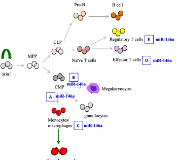
Figure ii. A simplified schematic depiction of hematopoietic tree highlighting the role of miR-146a in hematopoiesis and immune cell function. A. miR-146a negatively regulates myelopoiesis and myeloproliferation during inflammation and aging. In the absence of miR-146a, increased myeloprolifreation is observed. B. miR-146a negatively regulates megakaryopoiesis. Overexpression of 146a inhibits megakaryocyte proliferation and differentiation while down-regulation of miR-146a promotes megakaryocyte expansion and platelet production. However, in aging miR- miR-146a-deficient mice with bone marrow fibrosis, thrombocytopenia is observed. MiR-146a is a negative regulator of monocyte and macrophage activation (C) and effector T cell activation (D). E. In regulatory T cells, 146a positively controls Treg functional competence. In the absence of miR-146a, Treg cells are defective in suppressing effector T cell activation.
Overview of MiRNA-146a in Pathology
The importance of miR-146a is further exemplified by its extensive involvement in immunological
pathologies, including autoimmunity and hematologic malignancy. Loss-of-function studies in mice
have provided some important insight in the role of miR-146a in the pathogenesis of autoimmunity.
splenomegaly, lymphadenopathy, lymphocyte-infiltration in liver, kidney and lung, and the
development anti-dsDNA antibodies 19. The contribution of miR-146a to autoimmunity occurs in at least 3 separate ways: (1) regulation of miR-146a in effector T cells; (2) regulation of miR-146a in
Treg cells; (3) regulation of myeloid cell-mediated inflammation. MiR-146a-deficient T effector cells
contribute to autoimmunity in a cell-intrinsic manner. MiR-146a-deficeint T cells remain
hyper-responsive to antigen stimulation when adoptively transferred into wild type mice and they induce a
spontaneous autoimmune pathology when transferred into Rag1-/- mice. Mechanistically, the
enhanced proliferation, impaired activation-induced apoptosis, and exaggerated effector cytokine
production seen in miR-146a-deficient CD4 and CD8 T cells derive from uncontrolled NF-κB
activation as a result of de-repressed TRAF6 and IRAK1 20. Whether the multi-organ autoimmune pathology is caused by a general over-activation of miR-146a-deficient T cells or by a more specific
autoreactivity against a particular cell type or self-antigen remains an interesting unanswered question.
In addition, miR-146a-deficient Treg cells are also functionally defective in suppressing excessive Th1
response. In the absence of miR-146a, upregulation of STAT1 in Treg cells is responsible for the
failure to suppress wildtype effector T cells, leading to fatal IFNγ-mediated autoimmunity in a variety
of organs 43. The chronic inflammatory environment conditioned by miR-146a-deficient myeloid cells also contributes to autoimmune pathology by further stimulating T-cell and promoting tissue damage
19,44. Lastly, anti-dsDNA antibodies are present in the serum of aging miR-146a-/- mice 19. Future studies examining of the role of miR-146a in B cells may yield additional insight into the regulation of
autoantibody production. Given the widespread expression of miR-146a outside of hematopoietic
cells, miR-146a may exert protective function in non-hematopoietic tissues. Evidence supporting this
miR-146a expression seems to be critical to mediate innate immune tolerance by repressing IRAK1 46. In light of this, we speculate that miR-146a-deficiency in intestinal epithelial cells may lead to intestinal
inflammation, contributing to the overall autoimmune pathology in miR-146a-/- mice. Corroborating
the in vivo studies in mice, miR-146a has been found to be extensively dysregulated in human patients
with autoimmune diseases, including systemic lupus erythematosus (SLE) and rheumatoid arthritis
(RA). In lupus patients, miR-146a expression is downregulated in peripheral blood mononuclear cells
(PBMCs). In addition, the level of miR-146a expression is negatively correlated with disease severity,
with lower expression correlating with more active disease and proteinuria symptoms 42. Type I IFN is suggested to be the responsible pathway upregulated in lupus and additional targets of miR-146a, IRF5
and STAT1, have been identified based on 3’UTR luciferase reporter assay and Western blot analysis
in 293T cells overexpressiing miR-146a. Interestingly, enforced expression of miR-146a in PBMCs of
lupus patients can downregulate a subset of IFN-responsive genes 42. Additional evidence comes from genome-wide association studies that identified SNPs (single-nucleotide polymorphism) in the
regulatory region of miR-146a that are associated with lupus disease risk 48-49. One such SNP (rs57095329) in the promoter region of miR-146a is associated with the binding strength of
transcription factor Ets-1, another lupus-susceptibility gene. The risk-associated G allele in this SNP
correlates with reduced Ets-1 binding and decreased miR-146a expression 49. In contrast to SLE where miR-146a expression is shown to be down-regulated, over-expression of miR-146a is frequently
detected in rheumatoid arthritis (RA) patients 50-52. MiR-146a also seems to be highly expressed in osteoarthritis cartilage 53. Despite that both SLE and RA are autoimmune diseases with some shared characteristics, the underlying etiologies may be distinct. For example, chronic inflammation is a
shared pathology, but the dysregulated cytokine profiles are different between SLE and RA. The IFN
production are more prominent features of RA 50. Because pro-inflammatory cytokines can significantly upregulate miR-146a expression in various cell types, increased miR-146a expression
may simply reflect the hyper-inflammatory environment and may be a useful marker for disease
severity in RA. On the other hand, miR-146a-deficiency may be causally related to the pathogenesis of
SLE. It will be interesting to further investigate the involvement of miR-146a-deficiency in the
development and progression of SLE and a number of other autoimmune diseases and whether
upregulation of miR-146a may have therapeutic benefit in mouse models and human clinical samples.
In the addition to the role of miR-146a in Th1 cells, Treg cells and monocyte/macrophages,
understanding the function of miR-146a in T17 cells 54, a key cell type in a number of autoimmune diseases, may provide additional insight on this miRNA’s involvement in autoimmunity. There is also
some evidence suggesting a role of miR-146a in atherosclerosis, a less studied aspect of miR-146a
biology. Two reports suggested that upregulation of miR-146a may inhibit atherosclerosis by
suppressing TLR-4 and CD40L expression in low-density lipoprotein (LDL)-induced macrophages
and dendritic cells 55-56.
MiRNA-146a in Hematologic Malignancy
Recent studies have also revealed an important role of miR-146a in hematologic pre-malignancy and
malignancy, most notably myelodysplastic syndromes (MDS), myeloproliferative disease, myeloid
cancer and bone marrow failure. MDS represent a heterogeneous group of clonal diseases of HSC
origin characterized by ineffective hematopoietic differentiation, peripheral blood cytopenias, bone
marrow dysplasia, and a propensity to progress to leukemia and bone marrow failure. Recent studies
have made significant advances on the molecular pathogenesis of MDS, including dysregulated
miR-146a, are located in or near the commonly deleted region of 5q- syndrome, a common subtype of MDS
37. Accordingly, expression of these two miRNAs is reduced in patients of 5q- syndrome. The same study also showed that the stable knockdown of miR-145 and miR-146a concurrently in mouse HSPCs
recapitulates many of the key features of human 5q- syndrome, including thrombocytosis,
megakaryocytic dysplasia and mild neutropenia. In addition, overexpression of TRAF6, a target of
146a, was sufficient to phenocopy many of the features of knocking down 145 and
miR-146a, suggesting that miR-146a may be a more dominant miRNA. Studies with miR-146a knockout
mouse are reaffirming. With increasing age, miR-146a-/- mice develop myeloid expansion in spleen
and bone marrow, pancytopenia in the periphery and a propensity to progress to bone marrow failure
and myeloid cancer, a constellation of features reminiscent of a mixed myelodysplastic syndrome and
myeloproliferative neoplasm (MDS/MPN) 19,44. Mechanistically, increased NF-κB activation is a main driver of the myeloproliferative and bone marrow failure phenotype because ablation of the p50
subunit greatly reduces MDS/MPN symptoms. It is now increasingly appreciated that a distinct
hematopoietic developmental program, termed inflammatory hematopoiesis, is activated in bone
marrow during inflammation to promote myelopoiesis at the expense of lymphopoiesis and
erythropoiesis 59. We speculate that uncontrolled and persistent inflammatory hematopoiesis may promote pathological features of MDS and MPN, and over time, the pathologic hematopoietic program
becomes permanent and irreversible through additional genetic mutations and/or epigenetic changes.
Because chronic inflammation is a prominent feature of miR-146a-/- mice during stimulation and
aging, it will be interesting to investigate whether miR-146a may serve as a molecular link between
chronic inflammation and hematopoietic malignancy, such as MDS and MPN. In addition to myeloid
cancers, about 15% of miR-146a-/- mice also develop B-cell or mixed B and T-cell lymphomas by 18
miR-146a-deficient mice is distinct from the one driving myeloid oncogenesis. Many groups have
extensively profiled miRNA expression in human leukemia in an effort to identify microRNA
signatures associated with leukemia diagnosis, prognosis, and pathogenesis. Reports from different
groups have identified miR-146a downregulation in CD34+ cells or total bone marrow cells from
patients with 5q- syndrome 37,58,60. However, miR-146a downregulation is not consistently observed in leukemia studies. One expression profiling study showed that miR-146a is downregulated in bone
marrow cells of AML patients compared to normal CD34+ cells 61. However, in another study that compared miRNA expression between AML and ALL samples, increased expression of miR-146a in
both ALL and AML was shown to correlate with poor survival 62. MiR-146a was also shown to function as a tumor-suppressor in natural killer/T cell lymphoma and reduced expression of miR-146a
was identified as a poor prognostic factor 63. It seems miR-146a-downregulation is a consistent feature of 5q- syndrome or even all MDS and may be involved in the pathogenesis of MDS. The role of
miR-146a in leukemia and lymphoma requires further clarification.
Relationship between MiR-146a and MiR-155
MicroRNA-155 (miR-155) is another miRNAs identified from our original screen for miRNAs
induced upon NF-κB activation 16. However, in contrast to miR-146a, miR-155 plays an almost exact opposite role in immunity and hematopoiesis. For more detailed reviews of miR-155 biology, we refer
you to these recent reviews elsewhere 3,36,59,64-65. Here we will only focus on the aspect of miR-155 function opposing that of miR-146a. NF-κB activates an elaborate and potent transcription program
central to inflammation and immunity. Because of the potency of this pathway, proper regulation of
driven by NF-κB activation. The opposing roles of miR-146a and miR-155 have been most clearly
demonstrated in the function and development of myeloid cells. While basal expression of miR-155 is
low in myeloid cells, many of the inflammatory stimuli that can induce miR-146a expression,
including TLR ligands and pro-inflammatory cytokines, also upregulate miR-155 expression
significantly 15,69. In regulating the functional capacity of myeloid cells, upon induction miR-155 promotes expression of pro-inflammatory cytokines and the IFN response. This regulation is most
likely through repression of SOCS1 and SHIP1, both negative regulators of the pro-inflammatory
pathways 70-71. Consistent with this, inhibition of miR-155 in macrophages confers tolerance to endotoxin shock in mice while mice with miR-155 overexpression become hypersensitive 71-72. The exactly opposite effect has been observed with miR-146a inhibition and overexpression 19. In addition to pro-inflammatory cytokine production, miR-146a and miR-155 also regulate immunity in the
opposite manner. In general, mice with targeted miR-155 deletion are immuno-compromised as
indicated by attenuated immune response to immunization and infection 73-74. On the other hand, inhibiting miR-146a may result in a heightened immune response as shown by enhanced protection
against Listeria infection 17. MiR-146a and miR-155 also have an opposite effect in regulating certain aspects of T cell function. As discussed above, miR-146a-deficient CD4 T cells display a
hyper-activated response upon TCR stimulation while miR-155-deficient CD4 T cells are attenuated in IL-2
and IFNγ cytokine production upon TCR stimulation 20,73. Similarly, miR-155 and miR-146a also function in an opposite direction in autoimmunity. Contrary to miR-146a-/- mice that are prone to
developing autoimmune disease, miR-155-/- mice were shown to be significantly resistant to an
induced model of experimental autoimmune encephalomyelitis (EAE) because of a deficit in
miR-146a, miR-155, or both miRNAs were implanted with subcutaneous solid tumors and were
shown to have differential ability to control tumor growth 76. This study showed again that in the context of antitumor immunity, miR-146a and miR-155 play opposing roles with miR-155 promotes,
while miR-146a represses antitumor immunity, correlating their functions to regulate IFNγ-producing
CD4+ T cells development. As described above, miR-146a and miR-155 are concurrently up-regulated
in response to pro-inflammatory cues. Data suggest that miR-146a and miR-155 may counterbalance
each other during inflammatory hematopoiesis. Sustained expression of miR-155 in mouse HSPCs
results in a myeloproliferative disorder, characterized by profound myeloproliferation with dysplastic
changes in the bone marrow, splenomegaly as a result of extramedullary hematopoiesis, peripheral
anemia, lymphopenia, and thrombocytopenia with increased myeloid cells 15. The constellation of hematologic abnormalities seen in miR-155-overexpressing mice is strikingly similar to the one
observed in aging miR-146a-deficient mice 44. MiR-155-induced myeloproliferative disorder is thought to be primarily through repressing SHIP1 70. In addition, over-expression of miR-155 in a B-cell-specific manner in a transgenic mouse model with E(mu)-promoter-driven miR-155 expression
results in B-cell leukemia and lymphoma 77. Similar B-cell lymphoma pathology has also been observed in a subset of aging miR-146a-/- mice 44. Given the above evidence, it is tempting to speculate that miR-146a and miR-155 are evolutionarily selected to function in a tug-of-war manner to
properly control the magnitude and duration of NF-κB activation. Balanced level and proper timing of
miR-146a and miR-155 expression represents one critical layer of regulation to ensure that NF-κB is
CHAPTER ONE
NF-
κ
B Dysregulation in MicroRNA-146a-deficient Mice Drives the Development
of Myeloid Malignancies
INTRODUCTION
MicroRNAs (miRNA) are a group of ~19-23 nucleotide-long non-coding RNAs that repress target
gene expression by a combination of mRNA degradation and translation inhibition 8. Recent studies have revealed important physiological roles of miRNAs in many aspects of mammalian hematopoiesis
and immune cell function, and their altered expression has been linked to pathological conditions of
the immune system, such as hematologic cancers and autoimmunity 3,78-79.
Chronic inflammation contributes to cancer initiation and progression. Among a myriad of proposed
mechanisms linking inflammation to cancer, NF-κB has been identified as a key mediator of
inflammation-induced carcinogenesis 80. Moreover, constitutive NF-κB activation is frequently detected in many types of lymphoid and myeloid malignancies 81-82. Hence understanding how NF-κB activity is downregulated has been a focus of study with important advances in recent years 67. In particular, NF-κB regulation by non-coding RNAs has recently begun to be characterized. A few years
ago, we carried out a microarray screen to identify miRNAs induced by NF-κB activation and
miR-146a was discovered to be one of the miRNAs induced by lipopolysacharide (LPS) in a human
monocytic cell line. The induction of miR-146a was shown to be NF-κB-dependent, and upon
induction, miR-146a functioned to directly down-regulate TNF receptor-associated factor 6 (TRAF6)
and Interleukin-1 receptor-associated kinase 1 (IRAK1), two of the signal transducers in the NF-κB
terminating an inflammatory response via a negative feedback loop. To definitively characterize the
function of miR-146a in vivo and directly test the hypothesis that miR-146a is a negative regulator of
the NF-κB pathway, we generated two independent mouse strains with a targeted germline deletion of
miR-146a, one on the mixed C57BL/6x129/sv background and one on the pure C57BL/6 background.
Initial study done primarily with the mixed background miR-146a-/- mice showed that miR-146a-/-
mice were hypersensitive to LPS challenge, and aging miR-146a-/- mice developed autoimmune-like
disease, myeloid proliferation in their spleens, and hematopoietic tumors 84. However, the cellular lineage of the tumors and the mechanistic basis of miR-146a-deficiency mediated myeloproliferation
remained important unanswered questions. In addition, the relationship of the tumor phenotype in
miR-146a-/- mice and NF-κB dysregulation was uncertain due to the multiple potential targets of
miR-146a in different molecular pathways. Here, we focus on characterizing the incidence, cellular lineage,
and transplantability of the tumors and to understand the molecular basis of oncogenesis.
We have found that when they are allowed to age naturally, miR-146a-/- mice on a pure
C57BL/6 background develop a chronic inflammatory phenotype with progressive myeloproliferation
involving both the spleen and the bone marrow, which eventually progresses to splenic myeloid
sarcoma and bone marrow failure at about 18 months of age. Lymphomas of either a B-cell lineage or
a mixed T-and-B-cell lineage are also observed in older miR-146a-/- mice at a much higher frequency
than in wild-type animals. Myeloproliferation in miR-146a-deficient mice are driven primarily by the
action of NF-κB because reduction in the NF-κB level by deletion of the NF-κB subunit p50
effectively suppresses the pathology. Thus, we have provided genetic evidence that miR-146a
functions as a tumor suppressor in both myeloid and lymphoid cells and that chronic NF-κB activation
as a result of miR-146a-deficiency is responsible for driving the myeloproliferative disease, which can
RESULTS
miR-146a knockout mice develop myeloid and lymphoid malignancies.
The miR-146a-deficient pure C57BL/6 mouse was made by deleting about 300 base pairs of genomic
DNA fragment containing the miR-146a precursor 84. The miR-146a-deficient mice (homozygous knockouts, designated as miR-146a KO) were born at the expected Mendelian frequency and appeared
to be normal at birth. But starting at about 5-6 month of age, they developed progressively enlarged
spleens. By 18-22 months of age, 80% of the KO mice were moribund and were culled for analysis.
However, the entire cohort of age-matched littermate C57BL/6 (WT) control mice, except for one case
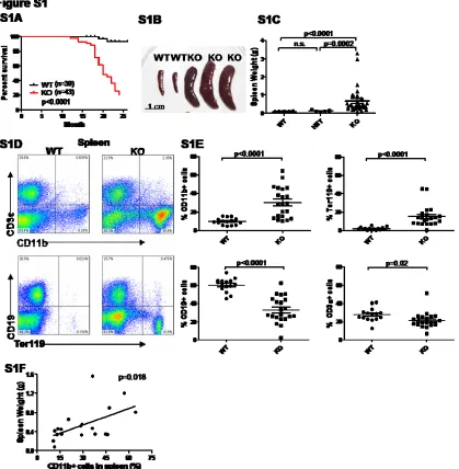
of thymoma and one case of seizure, were still alive and healthy (Figure S1A). On necropsy, KO mice
demonstrated various degrees of splenomegaly, with KO spleens on average weighing 6-7 times more
than WT spleens (Figure S1B and S1C). By FACS analysis, splenomegaly was correlated with a
massive expansion of the CD11b+ myeloid population (Figure S1D, S1E, and S1F). Expanded splenic
hematopoiesis was also noted based on the increased Ter119+ erythroid precursor population in the
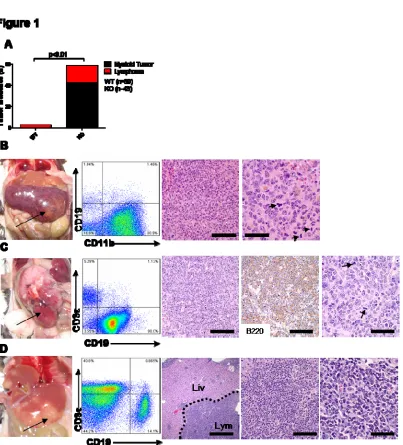
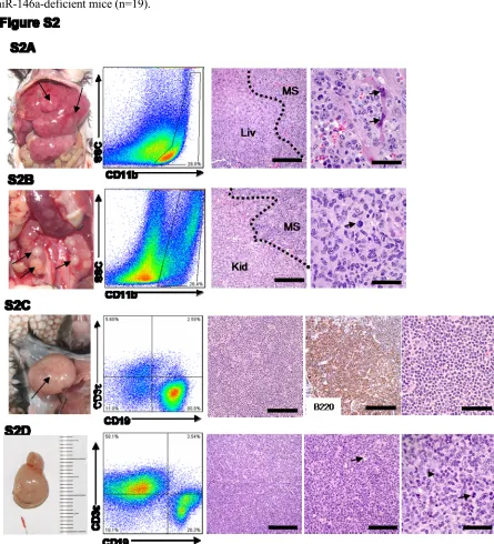
spleen. About 40% of the mice demonstrated distinct splenic tumors (Figure 1A and 1B). The majority
of these tumors displayed the histologic appearance of a myeloid sarcoma (Figure 1B) 85. FACS analysis of carefully dissected tumors showed that the cells derived from these tumors expressed the
pan-myeloid antigen, CD11b (Figure 1B). Occasionally, liver and kidney were also heavily infiltrated
by myeloid sarcoma (Figure S2A and S2B). A general role of miR-146a as a tumor-suppressor
miRNA in the hematolymphoid system was demonstrated by the development of lymphomas of a
B-cell or a mixed T-and-B B-cell lineage in cervical lymph node, gastrointestinal tract, liver, and kidney, in
about 20% of the KO mice (Figure 1A). Similar lymphoid tumors were not identified in wild-type
mice. The lineage of these tumors was confirmed by FACS as well as immunohistochemical stains
ranged from low-grade follicular lymphoma (Figure S2C) to high-grade diffuse large B-cell lymphoma
with apoptotic bodies and atypical mitosis (Figure 1C and Figure S2D).
Myeloid tumors are transplantable into immunocompromised recipient mice.
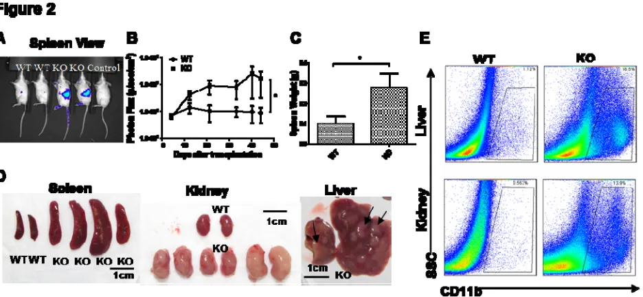
The ability of a mass of cells to form tumors upon transplantation to a secondary mouse can
distinguish a malignant growth from a reactive inflammatory process. KO splenic tumor cells dissected
from the tumor nodules in the spleen and wild-type (WT) control spleen cells were transplanted into
immunocompromised Rag2-/-γC-/- Balb/c recipient mice intravenously (i.v.), with or without
transduction by a retrovirus expressing enhanced green fluorescence protein (eGFP) and luciferase
protein. For experiments with retroviral transduction, bioluminescent imaging was utilized as a
sensitive method to track luciferase expression by the transplanted cells in vivo in recipient mice.
Starting one to two weeks after intravenous injection, KO splenocytes showed markedly increased
proliferation compared to wild-type splenocytes in recipient mice (Figure 2A). When whole body
bioluminiscence intensity was quantified over the course of eight weeks, mice receiving KO
splenocytes showed a progressive increase in signal culminating in an approximately 10-20-fold higher
signal, indicating a proliferation of the transplanted cells, while WT cells did not (Fig 2B).
Interestingly, recipient spleens, kidneys, and livers showed the strongest biolumniscence signal,
indicating the proclivity of transplanted splenic tumor cells to localize to the same organs where the
most significant myeloid tumor pathology was observed in the original KO mice. By two months
following transplantation, most KO recipient mice became cachectic and moribund, while WT
recipient mice showed some mild weight loss. In mice receiving KO spleen cells, but not in those
receiving WT spleen cells, necropsy revealed significant myeloid pathologies, including enlarged
Interestingly, not every KO spleen was transplantable. Transplantation of age-matched KO splenocytes
from mice without overt myeloid sarcoma did not result in significant myeloid pathology in recipient
mice, suggesting there is a qualitative difference (e.g. additional mutations) between KO spleens with
myeloproliferation and those with overt myeloid sarcoma.
Chronic myeloproliferation and myelofibrosis in miR-146a deficient bone marrow.
In addition to splenic myeloproliferation and/or myeloid sarcoma, the bone marrow of miR-146a KO
mice showed significant myeloproliferative disease. By 18-22 months of age, the CD11b+ population
accounted for 80% of all nucleated bone marrow cells, while the percentages of CD19+ B cell dropped
to below 10% in miR-146a KO mice (Figure 3A and 3B). In line with the myeloid-dominated bone
marrow, peripheral blood showed significant lymphopenia, anemia, and thrombocytopenia, but
preserved granulocyte numbers (Figure 3C and S3A). In addition, the expanded CD11b+ population in
both bone marrow and spleen was also predominantly Gr1+. An increased expression of macrophage
colony stimulating factor receptor (CSF1R) was also noted in the myeloid population in both spleen
and peripheral blood (Figure S3B). By 18-22 months of age, WT bone marrow showed partial
replacement of bone marrow cells by adipose tissue as a result of aging, while KO bone marrow
showed end-stage fibrosis or a hypercellular bone marrow. The end-stage KO bone marrow was
markedly pale by gross analysis, and histologic sections demonstrated paucicellular marrow with
thickened bone and fibrosis (Figure 3D and S3C). This chronic myeloproliferation resembles in some
ways human myeloproliferative diseases that result in an end stage of marrow fibrosis.
Spleen and bone marrow cells from miR-146a-deficient mice show increased activation of
We and others have previously identified TRAF6 and IRAK1 as targets of miR-146a in various cell
types, including monocytes and macrophages 16,22. Because TRAF6 and IRAK1 are signal transducers upstream of NF-κB activation, their de-repression in miR-146a deficient mice could result in increased
activation of NF-κB. In fact, we have recently demonstrated that TRAF6 and IRAK1 are derepressed
in miR-146a KO mice 84. Therefore, we investigated whether increased NF-κB activation might be a feature of the myeloproliferation and/or myeloid sarcoma in the miR-146a-deficient mice. We
extracted RNA from total nucleated spleen cells and total nucleated bone marrow cells and examined
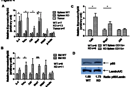
RNA expression levels for some genes well-known to be NF-κB-responsive, including interleukin-6
(IL-6), tumor-necrosis factor α (Tnfα), monocyte-chemotatic protein 1 (Mcp1), A20, and the NF-κB
subunit p50 (Nfkb1). In the absence of ex vivo stimulation, KO splenocytes and bone marrow cells
showed basally up-regulated expression of a subset of NF-κB-responsive genes compared to the WT
control, suggesting the presence of constitutively activated NF-κB (Figure 4A and 4B). For some of
the genes, such as Tnfα and Mcp1, the expression was even higher in tumor cells isolated from the KO
spleen (Figure 4A). To exclude the possibility that a different cellular composition is responsible for
the difference, especially in spleens where KO spleens showed a 3-fold increase in CD11b+ myeloid
population, we purified the CD11b+ population from WT and KO spleens by magnetic bead labeled
cell separation (MACS). When the enriched CD11b+ splenocyte population was subjected to the same
gene expression analysis, NF-κB-activated genes, including Tnfα and Mcp1, were still up-regulated in
the KO population compared to the WT control (Figure 4C). To further confirm the status of NF-κB
activation, we assessed the level of nuclear translocation of the NF-κB subunit, RelA (p65), by western
blot analysis of the nuclear protein extract from total nucleated splenocytes. KO splenocytes showed a
1.5- to 2-fold higher level of nuclear p65 than WT splenocytes, confirming the activated status of
nuclear p65 protein. Increased nuclear p65 correlated well with the presence of myeloid sarcoma
(Figure S4), suggesting that the activation of the NF-κB becomes constitutive only when the KO
spleen progresses from myeloproliferation to myeloid sarcoma.
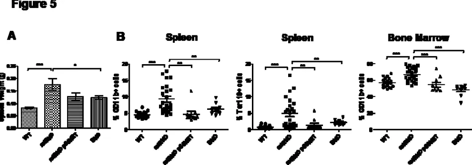
Reduction in the NF-κB level rescues the myeloproliferation in spleen and bone marrow.
Both gene expression and nuclear protein analysis indicated that the KO spleen and bone marrow cells
showed increased NF-κB activation. To investigate whether activated NF-κB is a causal factor in the
observed myeloproliferation, we bred the miR-146a KO strain with an Nfkb1 (p50) KO strain to
generate the double transgenic strain with homozygous deletion in both miR-146a and the p50 subunit.
p50-/- mice show no developmental abnormality in the immune system and elsewhere but display
defective B cell proliferation and specific antibody production. Cytokine production, including IL-6,
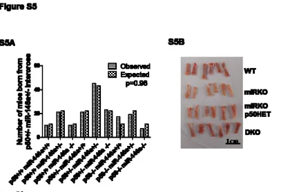
TNFα, and IL-1α, from LPS-stimulated macrophage is also impaired 86-87. When we intercrossed double heterozygotes for the two knockout genes, the various genotypes were born at the expected
Mendelian frequency (Figure S5A) and showed no overt abnormalities. Littermates from different
genotype groups, miR-146a+/+ p50+/+ (WT), miR-146a-/- p50+/+ (miRKO), miR-146a-/- p50+/-
(miRKO p50HET), and miR-146a-/- p50-/- (DKO), were aged to 6-7 month-old and then were
sacrificed for analysis for the development of myeloproliferation in spleen and bone marrow. As
expected, by 6-7 months of age, miR-146a KO mice started to develop splenomegaly and
myeloproliferation. Importantly, splenomegaly and myeloproliferative disease were significantly
reduced in the miR-146a-/- p50+/- group, with the exception of a few outliers. The rescue from the
myeloproliferative phenotype was more consistent in the miR-146a-/- p50-/- double knockout (DKO)
group, as indicated by the reduction in spleen weight and the lack of myeloid cell expansion in the
miRKO femurs, the bone marrow in DKO mice was a vibrant red color, similar to what was observed
routinely in WT mice (Figure S5B). In the spleens of DKO mice, there was also significant reduction
in Ter119+ cells relative to miRKO mice, suggesting that expanded splenic hematopoiesis was also
rescued by the deletion of p50 in DKO mice (Figure 5B). We conclude that activated NF-κB as a result
of miR-146a deletion is the primary factor driving myeloproliferation in miR-146a-deficient mice,
because reduction in the NF-κB level by deleting the NF-κB subunit p50 effectively rescues the
FIGURE
Figure 1. miR-146a-deficient mice develop myeloid and lymphoid malignancies. Mice were18-22 month-old miR-146a-/- mice (KO) and sex and age-matched C57BL/6 control mice (WT).
(A) Incidences of myeloid and lymphoid malignancies observed in WT (n=39) and KO (n=43) mice. (B) Photograph, FACS plot, and histological analysis of a representative myeloid tumor from a KO spleen. Panel 3 and 4, Hematoxylin and eosin (H&E) stained spleen section. Left scale bar, 100 microns; right scale bar, 40 microns. Arrows, mitotic figures.
(C) Photograph, FACS plot, and histological analysis of a representative B-cell lymphoma from a KO gastrointestinal tract. Panel 3 and 5, Hematoxylin and eosin (H&E) stained tumor section; Panel 4: Positive immunohistochemical staining (IHC) for B220. L to R scale bars are 100 microns in Panel 3, 4 and 40 microns in Panel 5.
(D) Photograph, FACS plot, and histological analysis of a representative mixed T-and-B-cell
Figure 2. Myeloid sarcoma is transplantable into immunocompromised Rag2-/-γC-/- recipient mice, causing lethal myeloid pathology. WT designates Rag2-/-γC-/- mice transplanted with WT splenocytes; KO designates Rag2-/-γC-/- mice transplanted with miR-146a KO splenocytes. (n=4 for WT and n=4 for KO; data are representative of three independent experiments. Student t-test, * designates p < 0.05)
(A) Representative bioluminiscence images of Rag2-/-γC-/- recipient mice splenic side view. (B) Quantification of whole body bioluminiscence intensity from splenic side view of one
representative experiment. Vertical axis is in logarithmic scale. Transduction efficiency is determined by flow cytometric analysis of GFP+ cells prior to injection. The bioluminescence intensity is normalized to the percentage of initially transduced cells.
(C) Spleen weight of Rag2-/-γC-/- recipient mice.
(D) Photographs of spleens, kidneys, and livers from representative Rag2-/-γC-/- recipient mice. (E) Flow cytometric analysis of myeloid cells (defined as CD11b+) in representative recipient kidneys and livers. SSC, side scatter.
Figure 3. Chronic myeloproliferation and myelofibrosis occur in miR-146a-deficient bone marrow. Mice were 18-22 month-old miR-146a-/- mice (KO) and sex and age-matched wild type control mice (WT). Data are shown as Mean +/- SEM. Each individual dot represents one individual mouse.
(A) Flow cytometric analysis of nucleated bone marrow cells from one representative WT mouse and one representative KO mouse for B cells (defined as CD19+) and myeloid cells (defined as CD11b+).
(B) Percentage of B cells (defined as CD19+) and myeloid cells (defined as CD11b+) in nucleated bone marrow cells by flow cytometric analysis (n=12 for WT and n=17 for KO from at least three independent experiments).
(C) Absolute numbers of total white blood cells, lymphocytes, red blood cells, and platelets by complete blood count (CBC) analysis (n=8 for WT and n=8 for KO).
Figure 4. Spleen and bone marrow cells from miR-146a-deficient mice (KO) show increased activation of the NF-κB-mediated transcription. Mice were 18-22 month-old miR-146a-/- mice (KO) and sex and age-matched wild type control mice (WT). Data are shown as Mean +/- SEM. n equals to the number of mice analyzed from at least two independent experiments. Student t-test, * designates p<0.05, *** designates p<0.005.
(A) Gene expression analysis of NF-κB-responsive genes in WT (n=7) nucleated splenocytes, KO (n=13) nucleated splenocytes, and myeloid tumor cells (Tumor, n=7) isolated from KO spleen by gross dissection.
(B) Gene expression analysis of NF-κB-responsive genes in WT (n=9) and KO (n=11) bone marrow cells.
(C) Gene expression analysis of NF-κB-responsive genes in CD11b+ population purified with MACS beads (WT n=9 and KO n=6).
Figure 5. Reduction in the NF-κB level by deleting the p50 subunit of NF-κB effectively rescues the myeloproliferative phenotype in miR-146a-deficient mice. All mice were 6-7 month-old miR-146a+/+, p50+/+ (WT), miR-146a-/-, p50+/+ (miRKO), miR-146a-/-, p50+/- (miRKO p50HET), or miR-146a-/-, p50-/- (DKO) mice (n=17 for WT, n=24 miRKO, n=10 for miRKO p50HET, and n=9 for DKO. Data are shown as Mean +/- SEM from at least three independent experiments. Student t-test, * designates p<0.05, ** designates p<0.01, and *** designates p<0.005.)
(A) Spleen weight of WT, miRKO, miRKO p50HET, and DKO mice.
Figure S1. 146a-deficient mice develop myeloproliferation in spleens. Mice were 18-22 month-old miR-146a-/- mice (KO), miR-146a+/- mice (HET), and sex and age-matched C57BL/6 control mice (WT). Data are shown as Mean +/- SEM. Each individual dot represents one individual mouse. At least 3 independent experiments were performed.
(A) Kaplan-Meier survival curve of aging miR-146a KO and WT mice starting at 1 year of age (n=39 for WT and n=43 for KO).
(B) Photographs of spleens isolated from KO and WT mice.
(C) Weight of spleens isolated from KO, HET, and WT mice (n=26 for WT, n=5 for HET, and n=33 for KO).
(D) Flow cytometric analysis of nucleated splenocytes from one representative KO mouse and one representative WT mouse for T cells (defined as CD3ε+), B cells (defined as CD19+), myeloid cells (defined as CD11b+), and erythroid cells (defined as Ter119+).
(F) Correlation between spleen weight and percentage CD11b+ cells in spleen from 18-22 month-old miR-146a-deficient mice (n=19).
Figure S2. Additional examples of myeloid and lymphoid malignancies in miR-146a-deficient mice.
(A) Photograph, FACS plot, and histological analysis of a representative myeloid tumor infiltrating a KO liver. Panel 3 and 4, Hematoxylin and eosin (H&E) stained liver section. Liv, residual liver, MS, myeloid sarcoma infiltration. Scale bar on Panel 3, 200 microns and on Panel 4, 40 microns. Arrows, apoptotic hepatocytes at the edge of the infiltrate.
(C) Photograph, FACS plot, and histological analysis of a representative B-cell lymphoma from a KO cervical lymph node. Panel 3 and 5, Hematoxylin and eosin (H&E) stained spleen section; Panel 4: Positive immunohistochemical staining (IHC) for B220. Scale bars are 100 microns in Panel 3, 4 and 40 microns in Panel 5.
(D) Photograph, FACS plot, and histological analysis of a representative mixed T-and-B-cell lymphoma from a KO gastrointestinal tract. Panel 3, 4, and 5, Hematoxylin and eosin (H&E) stained tumor section. Scale bars are 200 microns, 100 microns, 40 microns from left to right. Arrow on Panel 4, apoptotic body (“starry sky appearance”), arrowhead on Panel 5, large immunoblastic cell (there are many more in this field); arrow on Panel 5, atypical mitotic figure.
(A) Complete blood count (CBC) of peripheral blood from 18-22 month-old miR-146a-/- mice (KO, n=8) and sex and age-matched wild type control mice (WT, n=8). Data are shown as Mean +/- SEM. Each individual dot represents one individual mouse. n.s., not significant.
(B) Percentage of CD11b/Gr1 double positive cells and CD11b/CSF1R double positive cells in spleen, bone marrow (BM), and peripheral blood (PB) of 18-22 month-old miR-146a-/- mice (KO) and sex and age-matched wild type control mice (WT). Data are shown as Mean +/- SEM. Each individual dot represents one individual mouse. Total number of mice ranges from 6 to 11 from 3 independent experiments. n.s., not significant.
[image:41.612.83.457.235.325.2](C) Photograph of representative tibias and fibulas isolated from WT or KO mice.
Figure S4. Western blot analysis of the nuclear protein extracts from spleens of 18-22 month-old miR-146a-/- mice (KO) and sex and age-matched wild type control. KO (T), KO spleen with myeloid sarcoma; KO, KO spleen without myeloid sarcoma.
Figure S5.
[image:41.612.80.487.393.672.2]DISCUSSION
These results and our previous paper 84 demonstrate that miR-146a plays an important role as a tumor suppressor miRNA in hematopoietic lineages because chronic miR-146a-deficiency in mice leads to
myeloid sarcomas in spleens and lymphomas in various organs. miR-146a controls myeloproliferation
in both the spleen and bone marrow compartments primarily through negatively regulating NF-κB.
NF-κB is known to activate many genes involved in inflammation so that this study provides direct
evidence correlating the chronic inflammation caused by activated NF-κB and the development of
progressive myeloproliferative disease.
Consistent with the initial observation of miR-146a KO mice on the mixed C57BL/6x129/sv genetic
background 84, miR-146a KO mice on the pure C57BL/6 background developed progressive myeloproliferation in their spleens, although the onset was delayed compared with the mixed
background mice. miR-146a KO on the pure C57BL/6 background also developed significant
myeloproliferative disease in the bone marrow, which was not observed in the mixed background
strain. Moreover, miR-146a KO mice on the pure C57BL/6 background displayed less severe
autoimmune-like disease but developed a higher incidence of tumor later on in life. All these indicate
that the genetic background can significantly influence the phenotypic manifestation of deleting the
miR-146a KO gene.
It is interesting that miR-146a-deficient mice showed no obvious abnormality early on when
left unchallenged but gradually developed myeloproliferation in both spleen and bone marrow
compartments starting at about 5-6 months of age. Eventually, more than 50% of the mice developed
myeloid sarcomas and lymphomas at about 18-22 months of age. miR-146a normally down-regulates
TRAF6 and IRAK1, and in the miR-146a-deficient mice, derepression of these important signal
TRAF6 or IRAK1 in the 293 human embryonic kidney cell line is able to activate NF-κB 88-89, suggesting a possibility of cell-intrinsic activation of NF-κB when TRAF6 or IRAK1 is overexpressed
at a high level. However, this is unlikely to be the case in miR-146a KO mice. We believe that the
NF-κB activation in miR-146a KO mice likely involves external stimulation from other cells (autoimmune
stimuli) or from environmental pathogens/commensal bacteria for TRAF6/IRAK1 to amplify the
signal and that the mechanism of myeloproliferation and oncogenesis requires repeated episodes of
activation of NF-κB due to external stimulation. In support of this notion, our previous paper reports
an increased expression of both TRAF6 and IRAK1 at the protein level in bone marrow-derived
macrophages (BMDM) from young miR-146a KO mice. But there was no difference in the serum
levels of TNFα and IL-6 inflammatory cytokine between young WT and KO mice without LPS
challenge 84. In addition, constitutive NF-κB activity detected by conventional biochemical methods was only consistently noted in spleens with overt myeloid sarcoma (Figure S4). Based on this
evidence, we suggest that the activation of the NF-κB may only become constitutive and significant
when KO spleens transition from the pre-malignant myeloproliferative state to malignant myeloid
tumor. Given the long latency and the partial penetrance of the tumor phenotype, there are likely
secondary mutations cooperating in the oncogenesis, either complementing the NF-κB activity or
amplifying it, resulting in malignant tumors. It will be interesting to identify secondary mutations
driving this progression and malignant transformation.
NF-κB family transcription factors are homo-or heterodimers of 5 subunits, RelA (p65), Nfkb1
(p50), Nfkb2 (p52), RelB, and c-Rel. Among them, p65 and p50 are thought to play an important role
in the induction of many of the inflammatory genes 90. In addition, nfkb1ΔCT/ΔCT mice, with elevated p50 activity as a result of targeted deletion of the C-terminal ankyrin repeats, display phenotypes
lymph nodes 91. Moreover, deletion of p50 is able to partially rescue IkBα-deficienct mice, which display constitutive NF-κB activation, increased granulopoiesis, and neonatal lethality 92. Given the importance of the p50 subunit to NF-κB-driven inflammation, we bred miR-146a-/- mice with p50-/-
mice to investigate the role of NF-κB activation in the pathogenesis of myeloproliferation in
146a-deficient mice. We showed that p50-deficiency effectively rescued myeloproliferation in
miR-146a-deficient mice. The rescue seen in miRKO p50HET mice was not consistent and complete, but
merely a delay of symptoms, because by about one-year-old, mice in the mIRKO p50HET group
started to show increased myeloid cells while the DKO mice continued to show consistent rescue of
the myeloproliferative phenotype. The 50% increase in spleen weight and the marginal increase in
CD11b+ and Ter119+ cells in the spleen of DKO mice, compared to WT (Figure 5B and 5C), suggest
that the other pathways and factors other than p50 may also be involved. In addition, p50-deficiency
may not rescue all the phenotypes in miR-146a KO mice, such as autoimmunity as a result of defective
regulatory T-cells and/or activated effector T-cells, which has been shown to be due to a different
pathway, the Signal Transducer and Activator of Transcription 1 (STAT1)/interferon pathway 43. Autoimmune inflammation may contribute to the modestly increased spleen weight in the DKO mice.
Constitutive NF-κB activation is a well recognized phenomenon in lymphoid and plasma cell
malignancies, but the status of NF-κB activation in myeloid malignancies is less well characterized.
Some recent studies have demonstrated constitutive NF-κB activity in cells from high-risk MDS, some
AML cases, and CML with blast crisis, suggesting that constitutive NF-κB activation may be
associated with more advanced myeloid malignancies 81,93-94. In many of these cases, the mechanisms for constitutive NF-κB activation remain to be established. It is likely that some of these myeloid