Reduces the Levels of Viral RNA Replication
Amanda J. Chase, Sarah Daijogo, Bert L. Semler
Department of Microbiology and Molecular Genetics, School of Medicine, University of California, Irvine, California, USA
ABSTRACT
Due to their small genome size, picornaviruses must utilize host proteins to mediate cap-independent translation and viral RNA replication. The host RNA-binding protein poly(rC) binding protein 2 (PCBP2) is involved in both processes in poliovirus in-fected cells. It has been shown that the viral proteinase 3CD cleaves PCBP2 and contributes to viral translation inhibition. How-ever, cleaved PCBP2 remains active in viral RNA replication. This would suggest that both cleaved and intact forms of PCBP2 have a role in the viral RNA replication cycle. The picornavirus genome must act as a template for both translation and RNA rep-lication. However, a template that is actively being translated cannot function as a template for RNA replication, suggesting that there is a switch in template usage from translation to RNA replication. We demonstrate that the cleavage of PCBP2 by the polio-virus 3CD proteinase is a necessary step for efficient viral RNA replication and, as such, may be important for mediating a switch in template usage from translation to RNA replication.
IMPORTANCE
Poliovirus, like all positive-strand RNA viruses that replicate in the cytoplasm of eukaryotic cells, uses its genomic RNA as a tem-plate for both viral protein synthesis and RNA replication. Given that these processes cannot occur simultaneously on the same template, poliovirus has evolved a mechanism(s) to facilitate the switch from using templates for translation to using them for RNA synthesis. This study explores one possible scenario for how the virus alters the functions of a host cell RNA binding pro-tein to mediate, in part, this important transition.
T
he picornavirus family is made up of small, single-stranded,positive-sense RNA viruses that cause a range of diseases. Poliovirus, coxsackievirus, and human rhinovirus (HRV) are some of the more well-studied members of this family, causing poliomyelitis, myocarditis, and the common cold, respectively. All picornaviruses have a similar genomic RNA structure that
lacks a methyl guanosine cap on the 5=terminus. Additionally, the
5=noncoding region (NCR) is highly structured, with six RNA
stem-loop structures that preclude canonical ribosome scanning
from the 5=end of the template to the initiating start codon (1).
Therefore, translation of the picornavirus genome is initiated in a cap-independent manner via an internal ribosome entry site (IRES). For poliovirus, coxsackievirus, and HRV, the IRES is
com-prised of stem-loop structures II to VI of the 5=NCR (2,3).
Ribo-somes must be recruited to the IRES by a mechanism distinct from the canonical cap-dependent recruitment, although the mecha-nism is not yet understood. Canonical and noncanonical cellular translation factors aid in ribosome recruitment and translation initiation to translate the picornavirus genome into a single poly-protein that is subsequently processed to produce mature viral proteins. Among those proteins produced are the RNA-depen-dent RNA polymerase 3D and the viral proteinases 2A and 3C, as well as the 3C precursor 3CD, which cleave the polyprotein into precursor and mature viral proteins important for RNA replica-tion, packaging, and release.
As translation and processing of the polyprotein proceeds, there is an accumulation of viral proteins important for RNA syn-thesis, leading to the formation of replication complexes and a switch from translation to RNA replication. As positive-sense RNAs, picornavirus genomes are able to serve as templates for both translation and RNA replication. Translation proceeds along
the template in the 5=-to-3=direction, whereas RNA replication is
initiated at the 3=end and traverses the template to the 5=end.
However, previous studies have shown that the viral polymerase
cannot dislodge translating ribosomes (4). Furthermore, it has
been shown that templates that are actively being translated can-not function as a template for RNA replication, suggesting that there must be a switch in template usage from translation to RNA
replication (4,5). Initiation of RNA synthesis produces
negative-strand templates that are used for multiple rounds of positive-strand RNA synthesis. The resulting positive-positive-strand RNAs are then used for subsequent rounds of translation and RNA replica-tion or are packaged and released as virion RNA.
Due to their small genome size and limited coding capacity, picornaviruses must usurp the functions of host cell proteins for viral translation and RNA replication, directly benefiting the vi-rus. Examples include poly(rC) binding protein 2 (PCBP2), poly-pyrimidine tract binding protein (PTB), and lupus autoantigen (La). PTB is a member of the hnRNP family and is known to
shuttle from the nucleus to the cytoplasm (6). Previous studies
Received29 August 2013 Accepted23 December 2013
Published ahead of print26 December 2013
Editor:K. Kirkegaard
Address correspondence to Bert L. Semler, blsemler@uci.edu. A.J.C. and S.D. contributed equally to this work.
Present address: Amanda J. Chase, Department of Infectious Diseases, Virology, University Hospital Heidelberg, Germany; Sarah Daijogo, Focus Diagnostics, Inc., Cypress, California, USA.
Copyright © 2014, American Society for Microbiology. All Rights Reserved.
doi:10.1128/JVI.02503-13
on November 7, 2019 by guest
http://jvi.asm.org/
have shown that PTB binds to stem-loop IV of the poliovirus 5=
NCR and stimulates poliovirus translation (7–10). PCBP2 is
in-volved in host cell mRNA stability and translational control of cellular mRNAs. During poliovirus infection, PCBP2 is important for both translation and RNA replication. PCBP2 binds to stem-loop IV of the poliovirus IRES and forms a ribonucleoprotein (RNP) complex necessary for synthesis of the viral polyprotein (11–14). Additionally, PCBP2 binds to stem-loop I of the 5=NCR, along with the viral proteinase 3CD, to form a ternary complex that is necessary for initiation of negative-strand RNA synthesis (13,15–18).
PCBP2 is comprised of three hnRNP K homologous domains (called KH domains), which are common to a subset of a class of RNA binding proteins. Domains KH1 and KH3 have been shown to mediate the poly(rC) binding activity, with domain KH1 being
the major RNA binding determinant (14,19,20). Domain KH2 is
important for dimerization of the protein (21). Additionally,
do-main KH3 has been shown to be an important dodo-main for the binding of PCBP2 to stem-loop IV to form the RNP complex
necessary for translation (14). The KH3 domain is also required
for binding to cellular protein SRp20, forming a complex that is proposed to play a role in the recruitment of translation initiation
factors and the ribosome for viral translation (22). SRp20 belongs
to the SR protein family and plays a role in host cell mRNA
splic-ing and nucleo-cytoplasmic RNA export (23,24). The function of
SRp20 is usurped to benefit the virus by helping mediate efficient
IRES-dependent translation (22).
Between domains KH2 and KH3, there is a linker region that has been shown to be an important determinant for binding of
PCBP2 to stem-loop IV of poliovirus RNA (25). Previous studies
have shown that PCBP2 is cleaved by the viral proteinase 3CD in the linker region between domains KH2 and KH3, removing the third KH domain. PCBP2 that lacks domain KH3 is unable to bind to stem-loop IV, leading to the inhibition of viral translation. However, the cleaved form of PCBP2 is still able to bind to stem-loop I to form a functional ternary complex with 3CD proteinase
and thus remains active in RNA replication (26). Since RNA
syn-thesis occurs in replication complexes, cleaved PCBP2 would be concentrated in these specified complexes, rather than occurring throughout the cell, to allow a switch to RNA replication. PCBP1, an isoform of PCBP, is thought to be generated from a retrotrans-position of a fully processed PCBP2 mRNA and differs in the
length of the linker region between domains KH2 and KH3 (25,
27). Previous work has shown that PCBP1 is able to interact with
stem-loop I of the 5=NCR but cannot bind to stem-loop IV (14,
25). Additionally, when recombinant PCBP1 is incubated with
purified poliovirus 3C or 3CD, the proteolysis of PCBP1 by 3CD proteinase occurs with an efficiency similar to that of PCBP2
cleavage (26). While PCBP1 can rescue RNA replication, it is not
needed for viral translation as it cannot bind efficiently to
stem-loop IV and is unable to rescue translationin vitro(14). Therefore,
the focus of this paper is on PCBP2.
PCBP2, unlike PCBP1, is involved in both translation and RNA replication. We hypothesize that the cleavage of PCBP2 by viral proteinase 3CD helps, in part, to mediate the switch in tem-plate usage from translation to RNA replication. Cleavage of PCBP2 precludes stem-loop IV binding and, as this cleaved form is unable to bind stem-loop IV, inhibits translation, allowing the template to be cleared of ribosomes. The cleaved form of PCBP2 is still able to bind to stem-loop I and form a ternary complex with
3CD proteinase, initiating negative-strand RNA synthesis on the newly cleared template. In this paper, we describe experiments to test our hypothesis about the role of 3CD-mediated cleavage of PCBP2 in poliovirus replication. We report the generation of an uncleavable form of PCBP2 that is able to bind to both stem-loop I and stem-loop IV and function in translation. This genetically altered form of PCBP2 was used to demonstrate that cleavage of PCBP2 is important for poliovirus RNA replication.
MATERIALS AND METHODS
Infection of HeLa cells and lysate preparation.HeLa cells were grown as monolayers in Dulbecco’s modified Eagle’s medium (DMEM) supple-mented with 8% newborn calf serum (NCS). Cells were infected with poliovirus or coxsackievirus B3 (CVB3) at a multiplicity of infection (MOI) of 20, and adsorption was carried out at room temperature for 30 min. DMEM with 8% NCS was added, and the cells were incubated at 37°C until the indicated time points. At specific times postinfection (0 to 7 h), cells were washed twice with phosphate-buffered saline (PBS) before the cells were scraped and collected to generate whole-cell lysate. Cells were lysed in NP-40 lysis buffer (50 mM Tris-HCl [pH 8.0], 5 mM EDTA [pH 8.0], 150 mM NaCl, 0.5% NP-40) on ice for 30 min. After incubation on ice, cell debris was pelleted, the resulting supernatant was collected, and total protein concentration of the whole-cell lysate was determined by Bradford assay. Total protein (100g) from lysates was analyzed by West-ern blotting.
Western blot analysis.Following SDS-PAGE, proteins were electro-blotted to a polyvinylidene difluoride (PVDF) membrane. The mem-branes were blocked in 5% milk in PBS with 0.1% Tween 20 for 1 h at room temperature. Mouse monoclonal antibody against PCBP2 (1:2,000 in PBS with 0.1% Tween 20), as described by Sean and coworkers, was used to detect full-length and cleaved forms of PCBP2 (25). The PVDF membrane was incubated with anti-PCBP2 antibody (1:2,000) for 1 h, followed by incubation with goat anti-mouse secondary antibody conju-gated to horseradish peroxidase (HRP) (Millipore) for 1 h. Protein bands were visualized using chemiluminescence (Thermo Scientific).
HRV16 3CD construct.Synthetic oligonucleotide primers used were as follows: HRV16-3CD-Nde(⫹) (5=-AGCCATATGGGTCCAGA AGAAGAATTT-3=), HRV16-3CD-BamHI(⫺) (5=-GCCGGATCCCT ATTAAGAATTTTTCATACATT-3=), HRV16-3CD10(⫹) (5=-ATCAT ACTTCACTGAACAAGCAGCCCAAATTCAAATCTCTAAAC-3=), HRV16-3CD10(⫺) (5=-GTTTAGAGATTTGAATTTGGGCTGCTTGTTCAGT GAAGTATGAT-3=). Synthetic oligonucleotide primers HRV16-3CD-Nde (⫹) and HRV16-3CD-BamHI(⫺) were used to generate PCR frag-ments containing HRV type 16 (HRV16) amino acids 5155 to 7085, flanked by NdeI or BamHI upstream or downstream, respectively. The restriction sites were then used to clone HRV16 3CD into the pET15b vector through ligation. Site-directed mutagenesis of the newly synthe-sized pET15b HRV16 3CD plasmid was then used as a template for site-directed mutagenesis to alter the 3C/3D self-cleavage site such that recom-binant 3CD could not be self-cleaved into 3C and 3D. The synthetic oligonucleotide primers used were HRV16-3CD10(⫹) and the reverse complement, HRV16-3CD10(⫺). The resulting plasmid, pET15b HRV16 3CD10, was used for protein expression.
Protein purification.Purification of hexa-histidine-tagged pET22b PCBP2, wild-type PCBP2, or uncleavable PCBP2 (QS¡ID) (i.e., an un-cleavable form of the PCBP2 protein with the glutamine-serine changed to an isoleucine-aspartic acid), was carried out as previously described (17,26). Briefly, PCBP2 was expressed in BL21(DE3) (Rosetta) cells. The cells were grown at 37°C to an absorbance at 600 nm (A600) of 0.6, and then protein expression was induced with isopropyl--D -thiogalacto-pyranoside (IPTG; 1 mM) for 3 h at 25°C. Cells were resuspended in lysozyme buffer without lysozyme (10 mM EDTA, 25 mM Tris-HCl [pH 8.0], 4.5% glucose) and lysed by sonication for five cycles consisting of 15 s followed by 1 min on ice. The soluble fraction following sonication was precipitated with ammonium sulfate (20% [wt/vol]). The resulting pellet
on November 7, 2019 by guest
http://jvi.asm.org/
fraction was dialyzed overnight in I-60 buffer (20 mM Tris-HCl [pH 8.0], 250 mM NaCl, 60 mM imidazole, 10% glycerol) with 0.5% NP-40. The fraction was then subjected to Ni2⫹ion-based affinity chromatography
(GE Healthcare). The column was washed with I-60 buffer, and the pro-tein was eluted with I-200 buffer (20 mM Tris-HCl [pH 8.0], 250 mM NaCl, 200 mM imidazole, 10% glycerol). Hexa-histidine-tagged 3CD vi-ral proteinase (poliovirus or HRV16) was purified as previously described (17, 28), with a few modifications. Briefly, 3CD was expressed in BL21(DE3) (Rosetta) cells, and the cells were grown at 37°C to anA600of 0.4. Recombinant protein expression was induced by the addition of IPTG (1 mM) for 3 h at 25°C. Cells were resuspended in buffer A (20 mM Tris-HCl [pH 8.0], 25 mM NaCl, 5% glycerol, 1 mM dithiothreitol [DTT]) and lysed by one passage through a French pressure system (Ami-con) at 8,000 lb/in2. The lysate was then subjected to centrifugation for 20 min at 13,800⫻gusing a Beckman Coulter centrifuge and JA-17 rotor, and the resulting supernatant was discarded. The pellet was washed three times in buffer A, omitting DTT for the last wash. To extract protein from the pellet fraction, the pellet was resuspended in high-salt I-30 buffer (20 mM Tris-HCl [pH 8.0], 1 M NaCl, 30 mM imidazole, 10% glycerol) and incubated on ice for 30 min. The resuspended pellet was then subjected to centrifugation for 20 min at 13,800⫻g, as described above. The resulting supernatant was subjected to Ni2⫹ion-based affinity chromatography
(GE Healthcare). The column was washed with high-salt I-30 buffer, and the protein was eluted with high-salt I-200 buffer (20 mM Tris-HCl [pH 8.0], 1 M NaCl, 200 mM imidazole, 10% glycerol).
PCBP2in vitrocleavage assays.The PCBP2in vitrocleavage assays were carried out as previously described (26), with some minor modifi-cations. Briefly, recombinant purified PCBP2 and 3CD proteinase were incubated at a 1:1 molar ratio in cleavage buffer (20 mM HEPES [pH 7.4], 1 mM DTT, 150 mM potassium acetate [KOAc]) for 4 h at 30°C. The cleavage products were analyzed by SDS-PAGE and Western blotting.
HeLa cell S10 preparation.Preparation of HeLa cell S10 cytoplasmic extract was carried out by following the procedure described previously (14). Depletion of PCBP from HeLa cell S10 cytoplasmic extracts using a poly(rC) column was performed as previously described (29). To seques-ter PCBP2 from S10 cytoplasmic extracts, 400 nM poly(rC) RNA (Thermo Scientific) was incubated with HeLa cell S10 cytoplasmic extract for 30 min at 30°C.
In vitrotranslation and RNA replication.The coupledin vitro trans-lation and RNA replication assays were carried out essentially as described previously (14), with some alterations. Briefly, the translation-replication mixture (50l) contained 60% HeLa S10 cells (by volume), 250 ng po-liovirus virion RNA, and all-4 buffer lacking CTP (1 mM ATP [Pharmacia Biotech], 0.25 mM GTP, 0.25 mM, 30 mM creatine phosphate, 60 mM KOAc, 20 mM HEPES [pH 7.4], 4 mg/ml creatine kinase). The negative control contained 2 mM guanidine hydrochloride (GuHCl), which is known to inhibit poliovirus negative-strand RNA synthesis (30, 31). Poly(rC) and recombinant wild-type PCBP2 or PCBP2 (QS¡ID) were added to the indicated concentrations (seeFig. 5 and 6). Following poly(rC) RNA incubation with HeLa cell cytoplasmic extract, virion RNA, buffer, and recombinant protein were added to the reaction mixture. The reaction mixtures were then split into two portions, with 10l for trans-lation and the remaining 40l for RNA replication. TenCi [35 S]methio-nine (PerkinElmer) was added to the translation reaction, and it was in-cubated for 4 h at 30°C, supplemented with 2⫻Laemmli sample buffer (LSB), boiled, and subjected to SDS-PAGE followed by fluorography. The replication mixture was incubated at 30°C for 4 h before 25Ci [32P]CTP (PerkinElmer) was added, and the reaction mixture was incubated for an additional 2 h at 34°C. The RNA replication product was then purified using an RNeasy kit (Qiagen) and separated on a 1.1% agarose gel. Images of both translation and RNA replication were based on scans of phosphor screens performed using a Personal Molecular Im-ager FX Rad) and analyzed with Quantity One software (Bio-Rad). Quantity One software was used to determine the optical density per mm2for the band of interest. The VP3 band was analyzed for
translation, and the single-stranded RNA (ssRNA) band was evaluated for RNA synthesis. The fold change with the addition of wild-type PCBP2 or PCBP2 (QS¡ID) was determined as a ratio relative to the optical density per mm2for poly(rC), with poly(rC) set to 1.
Electrophoretic mobility shift assays.Mobility shift assays were car-ried out by following previously described methods (14, 32). Briefly, [32P]UTP-labeled RNA corresponding to the stem-loop I or stem-loop IV probe was transcribed from linearized plasmid. Increasing amounts of recombinant PCBP2 and/or 3CD proteinase were incubated in RNA binding buffer (25 mM KCl, 0.05 mM HEPES-KOH [pH 7.4], 2 mM MgCl2, 0.1 mM EDTA, 2 mM DTT, 3.7% glycerol [vol/vol]), with 1 mg/ml tRNA, 0.5 mg/ml bovine serum albumin (BSA; New England Bio-Labs), 8 U RNasin (Promega), and diluted probe (0.1 nM final concen-tration for the 10-l reaction). After incubating for 10 min at 30°C, 50% glycerol was added for a 12.5% final concentration and the reaction mix-tures were subjected to nondenaturing electrophoresis on a 5% polyacryl-amide gel containing glycerol in 0.5⫻Tris-borate-EDTA (TBE). Images were generated from scans of phosphor screens performed using a Per-sonal Molecular Imager FX and analyzed with Quantity One software.
RESULTS
Cleavage of PCBP2 during poliovirus and coxsackievirus infec-tion.Previous studies have shown that PCBP2 cleavage products
accumulate over the course of poliovirus infection (26). To
deter-mine if PCBP2 cleavage could be observed in cells infected by other picornaviruses, HeLa cell lysates were collected over the course of infection by poliovirus or coxsackievirus, and PCBP2 cleavage was analyzed by Western blotting, probing with a mono-clonal antibody that is specific for PCBP2. During mock infection, only full-length PCBP2 was detected, migrating around 39 kDa (Fig. 1A). There is also a faster-migrating PCBP2-related species that could be an alternative form of PCBP2, perhaps with different
posttranslational modifications. During either poliovirus (Fig.
1B) or coxsackievirus B3 (Fig. 1C) infection, cleaved PCBP2 is
detected by 4 h postinfection. We estimate that the cleavage prod-uct has a molecular mass of approximately 28 kDa and that it
FIG 1Cleavage of PCBP2 during poliovirus and coxsackievirus infection. HeLa cells were mock infected (A), infected with poliovirus (B), or infected with coxsackievirus B3 (C) at an MOI of 20. NP-40 lysate was collected from 0 to 7 h postinfection, and equal amounts of total protein were analyzed for PCBP2 cleavage by Western blotting, probing with a monoclonal antibody against PCBP2. Arrows to the right of the gel image indicate full-length and cleaved PCBP2.
on November 7, 2019 by guest
http://jvi.asm.org/
[image:3.585.300.541.64.250.2]represents PCBP2 lacking the KH3 domain. The appearance of cleaved PCBP2 at 4 h postinfection corresponds with an increase in titer, as determined by plaque assay, suggesting that PCBP2 cleavage may be important for more efficient RNA replication (data not shown). Therefore, these data suggest that PCBP2 cleav-age is important for infection with both poliovirus and coxsacki-evirus.
PCBP2 cleavage by viral proteinase 3CD.Previous work has demonstrated that poliovirus 3CD proteinase is responsible for cleaving PCBP2 during infection and that while full-length PCBP2
can bind to stem-loop IV, cleaved PCBP2 cannot (26). PCBP2 is
cleaved by the viral proteinase 3CD when bound to stem-loop IV
or when unbound with a similar efficiency (data not shown) (33).
This suggests that PCBP2 can be cleaved when bound to stem-loop IV, when bound to stem-stem-loop I, or when unbound, leaving the question of how cleavage of PCBP2 exerts its effect on the viral RNA replication cycle.
To further characterize the importance of the observed cleav-age during the course of viral infection, a PCBP2 construct with a cleavage site mutation was utilized. Previous studies have shown that poliovirus 3CD preferentially cleaves at glutamine-glycine
sites, with an aliphatic residue in the P4 position (34,35). 3CD also
cleaves at glutamine-serine sites, as found in the linker region of
PCBP2 (26). Therefore, an uncleavable form of the PCBP2 protein
with a double-amino-acid substitution at the cleavage site within the linker region was generated by changing the glutamine-serine
to an isoleucine-aspartic acid (QS¡ID) at amino acid positions
253 and 254, respectively, as shown inFig. 2A(26). This mutated
form of PCBP2 was then utilized in a cleavage assay to ensure that it was unable to be cleaved by the viral proteinase 3CD. The ability of recombinant 3CD derived from poliovirus or HRV16, a closely related picornavirus, to cleave the purified, recombinant mutated
PCBP2 (QS¡ID) was tested in anin vitrocleavage assay.
Recom-binant wild-type PCBP2 or PCBP2 (QS¡ID) was incubated with
poliovirus or HRV16 3CD proteinase, and cleavage was analyzed by Western blotting. In the absence of viral proteinase, wild-type
PCBP2 or PCBP2 (QS¡ID) is not cleaved and the full-length
39-kDa band is observed (Fig. 2B, lanes 1 and 4, respectively).
There is also a faster-migrating PCBP2 with an apparent approx-imate molecular mass of 35 kDa that is present with wild-type
PCBP2 or PCBP2 (QS¡ID) alone (lanes 1 and 4), suggesting that
it could be a truncated form of PCBP2 produced during
expres-sion in Escherichia coli, as no posttranslational modifications
could be occurring. This form of PCBP2 is present when incu-bated with poliovirus 3CD proteinase (lanes 2 and 5) but is absent in the presence of HRV16 3CD proteinase (lanes 3 and 6). It is known that HRV16 3CD proteinase has less stringent require-ments for cleavage site recognition, suggesting that the altered form of PCBP2 has a secondary cleavage site that is recognized by
HRV16 3CD but not by poliovirus 3CD (34). With the addition of
either poliovirus or HRV16 3CD, the 28-kDa PCBP2 cleavage
product is observed (Fig. 2B, lanes 2 and 3). However, when
PCBP2 (QS¡ID) was incubated with either poliovirus or HRV16
3CD proteinase, only full-length PCBP2 was observed (Fig. 2B,
lanes 5 and 6). Our results demonstrate that both poliovirus and HRV16 3CD have the ability to cleave wild-type PCBP2 and that
the mutated version of PCBP2 (QS¡ID) is uncleavable by the
viral proteinase 3CD.
Functional analysis of uncleavable PCBP2.To be functional
in translation or RNA synthesis, PCBP2 (QS¡ID) must efficiently
form the RNP complexes required for translation or RNA
repli-cation. Therefore, the ability of PCBP2 (QS¡ID) to bind
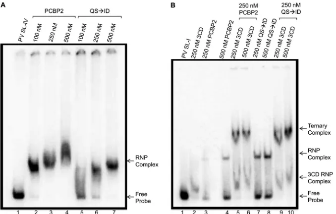
stem-loop I and stem-stem-loop IV RNA, which is required for RNA replica-tion and translareplica-tion, respectively, was tested. The ability of PCBP2
(QS¡ID) to form an RNP complex with stem-loop IV was
ana-lyzed by incubating radiolabeled poliovirus stem-loop IV RNA
with increasing amounts of recombinant wild-type PCBP2 (Fig.
3A, lanes 2 to 4) or PCBP2 (QS¡ID) (Fig. 3A, lanes 5 to 7). As
expected, wild-type PCBP2 could efficiently shift the electropho-retic mobility of stem-loop IV RNA, indicating RNP complex
for-mation. PCBP2 (QS¡ID) was also seen to bind to stem-loop IV
RNA, as indicated by the shifted probe. The shifted complex
ob-served with PCBP2 (QS¡ID) had a slightly different
electropho-retic mobility than the complex formed with wild-type PCBP2 (Fig. 3A, compare lanes 4 and 7). The slightly altered electropho-retic mobility could be due to altered folding of the mutated pro-tein, contributing to a different migration pattern.
To analyze the ability of the uncleavable PCBP (QS¡ID) to
function in RNA replication and form a complex with stem-loop I, radiolabeled poliovirus stem-loop I RNA was incubated with
recombinant wild-type PCBP2 or PCBP2 (QS¡ID). An
electro-phoretic mobility shift was observed with both the wild-type or uncleavable forms of PCBP2, indicating RNP complex formation (Fig. 3B, lanes 3 and 4 and lanes 7 and 8, respectively). Increasing amounts of recombinant poliovirus 3CD were then added to an-alyze ternary complex formation, which is required for initiation
E. coli aberrant product (?)
KH1 KH2 KH3
AAR Q R QQ SQ SH FH F AR Q I DH F
PCBP2 (wild type) PCBP2 (QSID) A
PCBP2 PCBP2 QSID
Cleaved PCBP2 Full-length PCBP2 72 kDa
25 kDa 35 kDa 55 kDa B
FIG 2PCBP2 cleavage site mutations and cleavage by picornavirus proteinase 3CD. (A) To generate an uncleavable version of PCBP2 (QS¡ID), the P1 and P1=positions at the cleavage site were mutated from the wild-type glutamine-serine (Q-S; red type) to an isoleucine-aspartic acid (I-D; green type). The K-homologous domains (KH1, KH2, and KH3) of PCBP2 are depicted as colored rectangles. (B) The cleavage of wild-type and mutated PCBP2 (2M) was tested in anin vitrocleavage assay with recombinant poliovirus or HRV16 3CD (2M) and analyzed by Western blotting, probing with a monoclonal antibody against PCBP2. Lane 1 is wild-type PCBP2, with added poliovirus and HRV16 3CD in lanes 2 and 3, respectively. Uncleavable PCBP2 (QS¡ID) was incubated alone (lane 4), or with poliovirus (lane 5) or HRV16 (lane 6) 3CD. Arrows to the right of the gel image indicate full-length and cleaved PCBP2, and molecular mass markers are indicated on the left of the gel image. The unmarked species migrating around 35 kDa is an altered species of PCBP2 that can be recognized and cleaved by HRV16 3CD but not poliovirus 3CD.
on November 7, 2019 by guest
http://jvi.asm.org/
[image:4.585.302.539.71.273.2]of negative-strand RNA synthesis. A supershifted product, indi-cating ternary complex formation, was observed with the PCBP2
wild type (Fig. 3B, lanes 5 and 6) and with uncleavable PCBP2
(QS¡ID) (Fig. 3B, lanes 9 and 10). Importantly, the two forms of
the protein form a ternary complex with a similar affinity. There-fore, the uncleavable form of PCBP2 was able to form an RNP complex with stem-loop IV and a ternary complex with stem-loop I and 3CD proteinase. This suggests that uncleavable PCBP2 can form the complexes necessary for both viral translation and RNA replication.
To analyze the ability of uncleavable PCBP2 (QS¡ID) to
res-cue IRES-dependent translation, HeLa cell cytoplasmic extracts
were depleted of PCBP by a poly(rC) affinity column (29).
Trans-lation experiments were carried out utilizing purified poliovirus
virion RNA and [35S]methionine labeling of the synthesized viral
proteins (Fig. 4). As a positive control for the level of viral
trans-lation, HeLa cell cytoplasmic extract was mock depleted (lane 1), and a high level of viral translation was observed. When HeLa cytoplasmic extracts were PCBP depleted, the level of viral trans-lation was significantly decreased (lane 2). As expected, the addi-tion of wild-type PCBP2 was able to rescue translaaddi-tion in PCBP-depleted HeLa cytoplasmic extracts to near wild-type levels of translation (lanes 3 and 4). In agreement with the ability of PCBP2
(QS¡ID) to bind to stem-loop IV, uncleavable PCBP2 was able to
rescue translation in PCBP-depleted HeLa cytoplasmic extracts (Fig. 4, lanes 5 and 6). From these experiments, we conclude that the uncleavable version of PCBP2 is able to function similarly to
wild-type PCBP2 in poliovirus translation, as shown with bothin
vitrotranslation assays and mobility shift assays with stem-loop
IV. Additionally, we predict that PCBP2 (QS¡ID) is functional in
viral RNA replication because it is able to bind stem-loop I and form a ternary complex with an affinity similar to that of wild-type PCBP2.
In vitrotranslation and RNA replication in the presence of wild-type or uncleavable PCBP2.Since the uncleavable PCBP2
(QS¡ID) can function in both translation and RNA replication,
the mutated protein can be used to address what role PCBP2 cleavage plays in the poliovirus replication cycle. To answer this
question, anin vitrotranslation/RNA replication assay was
uti-lized. To circumvent potential complications inherent with de-pleting cytoplasmic extracts of PCBP, HeLa cell S10 cytoplasmic extract was incubated with poly(rC) RNA to bind and sequester PCBP2 present in the extract, thereby inhibiting translation. Fol-lowing the preincubation, recombinant PCBP2 or uncleavable
PCBP2 (QS¡ID) was added in increasing amounts, along with
poliovirus virion RNA. Translation was analyzed by the
incorpo-ration of [35S]methionine to label synthesized viral proteins. As
shown inFig. 5A, translation of poliovirus virion RNA (lane 1)
produces viral proteins, both mature and precursor forms, labeled
by the incorporation of [35S]methionine. In the presence of
poly(rC) RNA, levels of translation are decreased (lane 3) as ex-pected because PCBP2 is sequestered in the cytoplasmic extract and unable to form an RNP complex with stem-loop IV. With the addition of either wild-type PCBP2 (lanes 4 to 6) or uncleavable
PCBP2 (QS¡ID) (lanes 7 to 9), levels of translation are increased
above translation levels with poly(rC) alone. Additionally, the lev-els of translation with added wild-type PCBP2 or PCBP2
(QS¡ID) are similar, as seen by the equal amounts of viral
pro-teins synthesized. Translation can be quantified and compared based on levels of viral proteins produced, such as the structural
protein VP3 (Fig. 5A; quantified inFig. 5B). The data shown are
representative of results of two independent experiments. Nor-malization of translation levels with the addition of recombinant protein to levels of translation with poly(rC) RNA, but without added protein, allows a more direct comparison of levels of viral
translation (Fig. 5B). With the addition of increasing amounts of
FIG 3RNA binding of cleavage-resistant PCBP2. Radiolabeled poliovirus stem-loop RNA was used in mobility shift assays to determine the ability of the cleavage-resistant PCBP2 to bind to stem-loop I or IV derived from the 5=NCR. (A) Radiolabeled poliovirus stem-loop IV (SL-IV) was incubated with increasing amounts of PCBP2 (lanes 2 to 4) or uncleavable PCBP2 (QS¡ID) (lanes 5 to 7). (B) Radiolabeled poliovirus stem-loop I (SL-I) was incubated with poliovirus 3CD alone (lane 2) or with PCBP2 alone (lanes 3 and 4) or PCBP2 (QS¡ID) alone (lanes 7 and 8). To analyze ternary complex formation, increasing amounts of poliovirus 3CD were added to wild-type (lanes 5 and 6) or uncleavable (lanes 9 and 10) PCBP2. The arrows to the right of the gel image indicate probe alone (Free Probe), RNP complex formation, and ternary complex formation.
on November 7, 2019 by guest
http://jvi.asm.org/
[image:5.585.124.463.66.282.2]PCBP2, either the wild type or PCBP2 (QS¡ID), the levels of translation were enhanced above levels seen with poly(rC) deple-tion of PCBP. Importantly, levels of transladeple-tion appear to be sim-ilar with the addition of either wild-type PCBP2 or PCBP2
(QS¡ID), indicating that there are similar levels of viral proteins
present to be used for RNA synthesis, allowing for a direct com-parison of the levels of negative-strand RNA synthesis.
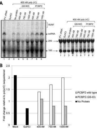
RNA replication was analyzed by the incorporation of
[32P]CMP into newly synthesized single-stranded RNA or the
par-tially double-stranded replicative intermediate. Labeled RNAs
produced in vitro were analyzed on an agarose gel, and the
ethidium bromide-stained gel was utilized to indicate levels of
rRNA as a loading control (Fig. 6A, right panel). Negative-strand
RNA synthesis depends on levels of newly synthesized viral pro-teins, and RNA replication will be less efficient if there are fewer viral proteins synthesized. Since there are equivalent levels of
rRNA (Fig. 6A, right panel) and newly synthesized viral proteins
(Fig. 5), RNA replication can be directly compared in the presence
of wild-type PCBP2 or uncleavable PCBP2 (QS¡ID). As seen in
Fig. 6A(left panel), poliovirus virion RNA replicates to a high level in mock-sequestered extracts, as indicated by the single-stranded
RNA band and the partially double-stranded replicative interme-diate (RI/RF) band (lane 1). In the presence of guanidine hydro-chloride (GuHCl), there is no RNA replication (lane 2), as GuHCl
inhibits negative-strand RNA synthesis (30,31). In the presence of
poly(rC) RNA (lane 3), levels of RNA replication are significantly reduced, as expected, due to both the decreased levels of transla-tion and the absence of available PCBP2 for ternary complex for-mation. However, with the addition of wild-type PCBP2 (lanes 7 to 9), there is a rescue of RNA replication that is not observed
when uncleavable PCBP2 (QS¡ID) is added (lanes 4 to 6). To
directly compare levels of RNA synthesis with added wild-type
PCBP2 or PCBP2 (QS¡ID), the ssRNA band was quantified and
relative levels of RNA synthesis were normalized to the level of RNA replication with poly(rC) RNA in the absence of added
pro-tein (Fig. 6B). The data shown are representative of results of two
independent experiments. Taken together, these data show that there is a pronounced rescue of RNA replication with the addition
of wild-type PCBP2 compared to uncleavable PCBP2 (QS¡ID).
Therefore, the data suggest that the cleavage of PCBP2 is impor-tant for efficient poliovirus RNA replication.
DISCUSSION
The picornavirus growth and replication cycle utilizes the genomic RNA as a template for both translation and RNA repli-cation. Translation necessarily precedes RNA replication because several viral proteins are required for initiation and elongation of RNA synthesis. However, it has been shown that a template that is actively being translated cannot function as a template for RNA replication, indicating a switch in template usage from translation
to RNA replication (4,5). Host proteins, including PCBP2,
medi-ate viral translation by binding to stem-loop IV of the IRES (11–
14). PCBP2 is also involved in RNA replication and binds to
stem-loop I of the 5= NCR to form a ternary complex with 3CD
proteinase that is needed for initiation of negative-strand RNA
synthesis (13,15–17). Previously, Perera and coworkers showed
that PCBP2 is cleaved by the viral proteinase 3CD during the
course of poliovirus infection (26). This cleavage event removes
the KH3 domain of the protein, disrupting the interaction with stem-loop IV, thereby inhibiting translation initiation. The cleaved version of PCBP2 can also no longer interact with SRp20, further inhibiting translation by disrupting ribosome recruit-ment. However, the shortened form of PCBP2 is functional in RNA replication, as it can efficiently bind to stem-loop I to form the ternary complex during negative-strand RNA synthesis
initi-ation (26). Therefore, the cleavage of PCBP2 could, in part,
me-diate a switch in template usage from translation to RNA replica-tion.
PCBP2 cleavage occurs in HeLa cells during infection by
po-liovirus or coxsackievirus, two closely related picornaviruses (Fig.
1).In vitrocleavage assays further show that poliovirus 3CD, as
well as HRV16 3CD proteinase, can cleave PCBP2 (Fig. 2).
Know-ing that PCBP2 can be cleaved by the 3CD proteinase of several different picornaviruses raises key questions: is the cleavage of PCBP2 important for efficient RNA replication and production of progeny virus, and is it involved in the switch in template usage? To understand how PCBP2 functions in mediating a possible switch in template usage, an uncleavable version of PCBP2
(QS¡ID) was utilized that retained the ability to function like
wild-type PCBP2 in binding to stem-loop IV or stem-loop I, forming a ternary complex with stem-loop I and 3CD, and also to
FIG 4Functional analysis of uncleavable PCBP2 (QS¡ID) during poliovirus translation. Poliovirus virion RNA was translatedin vitroin PCBP-depleted HeLa cytoplasmic extracts in the presence of [35S]methionine. Poliovirus pro-teins were resolved by SDS-PAGE and detected by autoradiography. Transla-tion reacTransla-tions were carried out in mock-depleted (lane 1) or PCBP-depleted (lanes 2 to 6) HeLa cytoplasmic extracts. No added recombinant protein (lane 2), 100 nM or 500 nM recombinant wild-type PCBP2 (lanes 3 and 4), and 100 nM or 500 nM purified recombinant PCBP2 (QS¡ID) (lanes 5 and 6) were analyzed for rescue of viral translation levels. Viral proteins are indicated to the left of the gel. WT, wild type.
on November 7, 2019 by guest
http://jvi.asm.org/
[image:6.585.81.248.66.370.2]retain the ability to rescue translation similarly to wild-type
PCBP2 (as shown inFig. 3and4). Therefore, the importance of
PCBP2 cleavage on RNA replication could be analyzed utilizing
this uncleavable form of PCBP2. Anin vitrotranslation and RNA
replication assay showed that when poly(rC) RNA sequestered PCBP2 present in the cytoplasmic extract, translation, and there-fore RNA replication, was decreased, as expected. When
recom-binant wild-type PCBP2 or PCBP2 (QS¡ID) was added
follow-ing PCBP2 sequestration with poly(rC) RNA, translation levels
were increased relative to translation with just poly(rC) RNA (Fig.
5). Importantly, levels of translation were similar with added
wild-type PCBP2 or PCBP2 (QS¡ID), allowing for a direct
compari-son of levels of negative-strand RNA synthesis. In addition,
re-combinant PCBP2 (QS¡ID) bound stem-loop I RNA with an
affinity similar to that of wild-type PCBP2, further indicating that the two proteins could potentially function in RNA replication
with a similar efficiency (Fig. 3B). Therefore, we concluded that
the significantly lower level of viral RNA synthesis observed with
added PCBP2 (QS¡ID) than with wild-type PCBP2 was evidence
that RNA replication could not be rescued by the addition of
un-cleavable PCBP2 (Fig. 6).
Our results suggest that PCBP2 cleavage is important for effi-cient poliovirus RNA replication. Although we have not provided direct evidence, our data support a role for this cleavage in facili-tating a switch in template usage from translation to RNA repli-cation. We propose an updated model in which full-length PCBP2
binds to stem-loop IV to form an RNP complex necessary for
initiation of viral translation (Fig. 7). PCBP2 also binds to cellular
protein SRp20 to form a complex that has been proposed to help
recruit ribosomes for translation (22). In addition, host protein
PTB is known to bind to stem-loop IV and stimulate poliovirus
translation (7–10). As translation of the polyprotein proceeds, the
concentration of 3CD proteinase, along with that of other viral proteins, increases and the concentration of cleaved PCBP2 accu-mulates. Also, PTB is cleaved by 3C proteinase, which has also been proposed to play a role in meditating a switch in template
usage from translation to RNA replication (9,36). Although not
shown as a cleaved polypeptide inFig. 7, host cell poly(A) binding
protein (PABP) has also been shown to be cleaved by poliovirus 3C or 3CD proteinase, contributing to the shutdown of
cap-inde-pendent viral translation (37). With the increased concentration
of viral proteins, RNA replication complexes are formed, leading to cytoplasmic foci with higher concentrations of cleaved PCBP2 than of full-length PCBP2. Under these conditions, cleaved PCBP2 no longer binds to stem-loop IV or SRp20 and translation initiation is inhibited, allowing the template to be cleared of trans-lating ribosomes. The cleaved PCBP2 is instead bound to stem-loop I, along with 3CD, to form the ternary complex necessary for initiation of negative-strand RNA synthesis.
The switch from translation to RNA replication is a necessary
part of the poliovirus replication cycle. As shown inFig. 1, PCBP2
is also cleaved during the course of coxsackievirus infection. In
FIG 5In vitrotranslation of poliovirus RNA in the presence of wild-type or uncleavable PCBP2. HeLa cytoplasmic extract was preincubated with poly(rC) RNA to bind and sequester PCBP2. Poliovirus virion RNA was then added with or without increasing amounts of recombinant wild-type PCBP2 or uncleavable PCBP2 (QS¡ID). (A) Translation of [35S]methionine-labeled viral proteins was analyzed by SDS-PAGE. Viral proteins are indicated to the left of the gel image. Lane 1 is the positive control with cytoplasmic extract mock depleted and incubated with poliovirus virion RNA (vRNA). The addition of guanidine hydrochloride (GuHCl) in lane 2 has no effect on translation. Poly(rC) is added in lanes 3 through 9, with increasing concentrations of added PCBP2 wild type (lanes 4 to 6) or uncleavable PCBP2 (QS¡ID) (lanes 7 to 9). (B) Quantitation of viral translation was performed using Quantity One software to determine the concentration of the VP3 band (optical density per mm2). A ratio of the determined optical density with the addition of wild-type PCPB2 (white) or PCBP2 (QS
¡ID) (hatched) to the optical density for poly(rC) was calculated to determine the fold change. The positive controls indicating levels of translation in the absence of poly(rC) RNA or recombinant protein (no protein) are depicted as black columns. Results shown are representative of two independent experiments.
on November 7, 2019 by guest
http://jvi.asm.org/
[image:7.585.43.540.70.328.2]addition, PCBP2 has been shown to be cleaved during human
rhinovirus infection of HeLa cells (41), suggesting that PCBP2
cleavage could be a mechanism for template switching utilized by multiple picornaviruses. However, our recent data suggest that a switch in template usage during human rhinovirus infection is mediated by a different mechanism (Chase and Semler, submit-ted). Thus, there may be redundant functions involved in medi-ating the switch in template usage for picornaviruses. PCBP2 is a
known IRES trans-acting factor (ITAF) involved in both RNA
replication and translation, but other ITAFs involved only in en-hancing translation could also be important in mediating the
switch. PTB is an ITAF that is known to enhance translation and has also been shown to be cleaved by poliovirus 3C proteinase during infection, suggesting that this protein could also be
in-volved in mediating a switch in template usage (36,38).
Interest-ingly, the La autoantigen (the first ITAF discovered for poliovirus
[39]) is cleaved during poliovirus infection; however, the
trun-cated form is still able to stimulate poliovirus translation in rabbit
reticulocyte lysates (40). Further work is necessary to elucidate
which other mechanisms, if any, could be involved in mediating the switch in template usage and to determine if those mecha-nisms act synergistically or redundantly. Additional studies also
FIG 6In vitropoliovirus RNA replication in the presence of added wild-type or uncleavable PCBP2. As inFig. 5, HeLa cytoplasmic extract was preincubated with poly(rC) RNA to bind and sequester PCBP2. Poliovirus virion RNA was then added with or without increasing amounts of recombinant PCBP2 or uncleavable PCBP2 (QS¡ID). (A) RNA replication was analyzed by incorporation of [32P]CMP and the presence of labeled ssRNA and the partially double-stranded RI/RF intermediate, as indicated to the right of the gel image, following agarose gel electrophoresis. Lane 1 is the positive control with poliovirus virion RNA (vRNA) and no poly(rC) RNA. The addition of guanidine hydrochloride (GuHCl) in lane 2 is a negative control for RNA synthesis. Poly(rC) is added in lanes 3 through 9, with increasing concentrations of added PCBP2 wild type (lanes 7 to 9) or uncleavable PCBP2 (QS¡ID) (lanes 4 to 6). The left panel analyzes ssRNA and the RI/RF intermediate based on scans of phosphor screens using Personal Molecular Imager FC. The right panel depicts the ethidium bromide stained agarose gel to analyze levels of rRNA. (B) Quantitation of viral RNA synthesis was analyzed using Quantity One software to determine the concentration of the ssRNA band (optical density per mm2). The fold change with added recombinant protein was then determined as a ratio of the determined the optical density when wild-type PCBP2 (white) or PCBP2 (QS¡ID) (hatched) was added to the optical density for poly(rC). Positive and negative controls in the absence of poly(rC) RNA or recombinant protein (no protein) are depicted in black. Results shown in the figure are representative of those of two independent experiments.
on November 7, 2019 by guest
http://jvi.asm.org/
[image:8.585.138.449.65.477.2]need to be carried out to elucidate whether the cleavage of PCBP2 by 3CD proteinase to mediate a switch in template usage is a mechanism unique to poliovirus or is a mechanism utilized by other picornaviruses.
ACKNOWLEDGMENTS
We are grateful to Eric Baggs and Dylan Flather for critical comments on the manuscript. We are indebted to Hung Nguyen and MyPhuong Tran for expert technical assistance.
This research was supported by Public Health Service grant AI026765 from the National Institutes of Health.
REFERENCES
1.Fitzgerald KD, Semler BL.2009. Bridging IRES elements in mRNAs to the eukaryotic translation apparatus. Biochim. Biophys. Acta1789:518 – 528.http://dx.doi.org/10.1016/j.bbagrm.2009.07.004.
2.Jang SK, Krausslich HG, Nicklin MJ, Duke GM, Palmenberg AC, Wimmer E.1988. A segment of the 5=nontranslated region of encepha-lomyocarditis virus RNA directs internal entry of ribosomes during in vitro translation. J. Virol.62:2636 –2643.
3.Pelletier J, Sonenberg N.1988. Internal initiation of translation of eu-karyotic mRNA directed by a sequence derived from poliovirus RNA. Nature334:320 –325.http://dx.doi.org/10.1038/334320a0.
4.Barton DJ, Morasco BJ, Flanegan JB.1999. Translating ribosomes in-hibit poliovirus negative-strand RNA synthesis. J. Virol.73:10104 –10112. 5.Gamarnik AV, Andino R.1998. Switch from translation to RNA replica-tion in a positive-stranded RNA virus. Genes Dev.12:2293–2304.http: //dx.doi.org/10.1101/gad.12.15.2293.
6.Michael WM, Choi M, Dreyfuss G.1995. A nuclear export signal in hnRNP A1: a signal-mediated, temperature-dependent nuclear protein export pathway. Cell 83:415– 422. http://dx.doi.org/10.1016/0092 -8674(95)90119-1.
7.Gosert R, Chang KH, Rijnbrand R, Yi M, Sangar DV, Lemon SM.2000. Transient expression of cellular polypyrimidine-tract binding protein stimulates cap-independent translation directed by both picornaviral and flaviviral internal ribosome entry sites in vivo. Mol. Cell. Biol.20:1583– 1595.http://dx.doi.org/10.1128/MCB.20.5.1583-1595.2000.
8.Hellen CU, Witherell GW, Schmid M, Shin SH, Pestova TV, Gil A, Wimmer E.1993. A cytoplasmic 57-kDa protein that is required for translation of picornavirus RNA by internal ribosomal entry is identical to the nuclear pyrimidine tract-binding protein. Proc. Natl. Acad. Sci. U. S. A.90:7642–7646.http://dx.doi.org/10.1073/pnas.90.16.7642. 9.Hunt SL, Jackson RJ.1999. Polypyrimidine-tract binding protein (PTB)
is necessary, but not sufficient, for efficient internal initiation of transla-tion of human rhinovirus-2 RNA. RNA5:344 –359.http://dx.doi.org/10 .1017/S1355838299981414.
10. Sawicka K, Bushell M, Spriggs KA, Willis AE.2008. Polypyrimidine-tract-binding protein: a multifunctional RNA-binding protein. Biochem. Soc. Trans.36:641– 647.http://dx.doi.org/10.1042/BST0360641. 11. Blyn LB, Swiderek KM, Richards O, Stahl DC, Semler BL, Ehrenfeld E.
1996. Poly(rC) binding protein 2 binds to stem-loop IV of the poliovirus RNA 5=noncoding region: identification by automated liquid chromatog-raphy-tandem mass spectrometry. Proc. Natl. Acad. Sci. U. S. A.93: 11115–11120.http://dx.doi.org/10.1073/pnas.93.20.11115.
12. Blyn LB, Towner JS, Semler BL, Ehrenfeld E. 1997. Requirement of poly(rC) binding protein 2 for translation of poliovirus RNA. J. Virol. 71:6243– 6246.
13. Gamarnik AV, Andino R.1997. Two functional complexes formed by KH
3D 1. Translation
initiation SL-IV
1
AAAAAA
PABP
PTB
80S
SL-I
2. Translation to generate viral proteins
SL-IV
1
AAAAAA
PABP
PTB
80S
80S
SL-I 3CD 3CD
SL-IV
4. PCBP2 cleavage and initiation of RNA replication
P1
3D
AAAAAAUUU
3 PABP
PTB
3D 3D33D 3D33 SL-I
3CD
PTB Cleaved PTB Cleaved PCBP2
3CD 3CD 3CD
3CD
3. Accumulation of viral proteins
SL-IV
1
AAAAAA
PABP
PTB 80S
SL-I 3CD 3CD
80S
FIG 7Template switching model for poliovirus replication. The switch in template usage based on the cleavage of PCBP2 is described (1). During translation, full-length PCBP2 is bound to stem-loop IV, forming an RNP complex to allow translation. PTB binds to the base of stem-loop IV to stimulate viral translation, and SRp20 forms a complex with PCBP2 on stem-loop IV that is also necessary for translation (2). Translation and processing of the polyprotein produces viral proteins, such as 3CD proteinase (3). As translation proceeds, the concentration of 3CD increases, leading to cleavage of PCBP2 (4). As the concentration of cleaved PCBP2 increases, PCBP2 no longer binds to stem-loop IV and no longer forms a complex with SRp20. Therefore, translation is inhibited and the template is cleared of translating ribosomes. Cleaved PCBP2 can still bind to stem-loop I and forms a functional ternary complex with 3CD viral proteinase, allowing initiation of RNA synthesis and elongation by the viral 3D polymerase. Poly(A) binding protein (PABP) is also depicted in the model. Although not shown as a cleaved polypeptide in the figure, PABP has been shown to be cleaved by poliovirus 3C/3CD proteinase, contributing to the shutdown of cap-independent viral translation (37).
on November 7, 2019 by guest
http://jvi.asm.org/
[image:9.585.115.470.69.338.2]domain containing proteins with the 5=noncoding region of poliovirus RNA. RNA3:882– 892.
14. Walter BL, Parsley TB, Ehrenfeld E, Semler BL.2002. Distinct poly(rC) binding protein KH domain determinants for poliovirus translation ini-tiation and viral RNA replication. J. Virol.76:12008 –12022.http://dx.doi .org/10.1128/JVI.76.23.12008-12022.2002.
15. Andino R, Rieckhof GE, Baltimore D.1990. A functional ribonucleo-protein complex forms around the 5=end of poliovirus RNA. Cell63:369 – 380.http://dx.doi.org/10.1016/0092-8674(90)90170-J.
16. Andino R, Rieckhof GE, Achacoso PL, Baltimore D.1993. Poliovirus RNA synthesis utilizes an RNP complex formed around the 5=-end of viral RNA. EMBO J.12:3587–3598.
17. Parsley TB, Towner JS, Blyn LB, Ehrenfeld E, Semler BL.1997. Poly (rC) binding protein 2 forms a ternary complex with the 5=-terminal se-quences of poliovirus RNA and the viral 3CD proteinase. RNA3:1124 – 1134.
18. Vogt DA, Andino R.2010. An RNA element at the 5=-end of the polio-virus genome functions as a general promoter for RNA synthesis. PLoS Pathog.6:e1000936.http://dx.doi.org/10.1371/journal.ppat.1000936. 19. Dejgaard K, Leffers H. 1996. Characterisation of the
nucleic-acid-binding activity of KH domains. Different properties of different domains. Eur. J. Biochem.241:425– 431.
20. Silvera D, Gamarnik AV, Andino R.1999. The N-terminal K homology domain of the poly(rC)-binding protein is a major determinant for bind-ing to the poliovirus 5=-untranslated region and acts as an inhibitor of viral translation. J. Biol. Chem.274:38163–38170.http://dx.doi.org/10.1074 /jbc.274.53.38163.
21. Bedard KM, Walter BL, Semler BL.2004. Multimerization of poly(rC) binding protein 2 is required for translation initiation mediated by a viral IRES. RNA10:1266 –1276.http://dx.doi.org/10.1261/rna.7070304. 22. Bedard KM, Daijogo S, Semler BL. 2007. A nucleo-cytoplasmic SR
protein functions in viral IRES-mediated translation initiation. EMBO J. 26:459 – 467.http://dx.doi.org/10.1038/sj.emboj.7601494.
23. Zahler AM, Lane WS, Stolk JA, Roth MB.1992. SR proteins: a conserved family of pre-mRNA splicing factors. Genes Dev.6:837– 847.http://dx.doi .org/10.1101/gad.6.5.837.
24. Huang Y, Steitz JA.2001. Splicing factors SRp20 and 9G8 promote the nucleocytoplasmic export of mRNA. Mol. Cell7:899 –905.http://dx.doi .org/10.1016/S1097-2765(01)00233-7.
25. Sean P, Nguyen JH, Semler BL.2008. The linker domain of poly(rC) binding protein 2 is a major determinant in poliovirus cap-independent translation. Virology378:243–253.http://dx.doi.org/10.1016/j.virol.2008 .05.007.
26. Perera R, Daijogo S, Walter BL, Nguyen JH, Semler BL.2007. Cellular protein modification by poliovirus: the two faces of poly(rC)-binding pro-tein. J. Virol.81:8919 – 8932.http://dx.doi.org/10.1128/JVI.01013-07. 27. Makeyev AV, Chkheidze AN, Liebhaber SA.1999. A set of highly
con-served RNA-binding proteins, alphaCP-1 and alphaCP-2, implicated in mRNA stabilization, are coexpressed from an intronless gene and its in-tron-containing paralog. J. Biol. Chem.274:24849 –24857.http://dx.doi .org/10.1074/jbc.274.35.24849.
28. Parsley TB, Cornell CT, Semler BL. 1999. Modulation of the RNA binding and protein processing activities of poliovirus polypeptide 3CD by the viral RNA polymerase domain. J. Biol. Chem.274:12867–12876. http://dx.doi.org/10.1074/jbc.274.18.12867.
29. Walter BL, Nguyen JH, Ehrenfeld E, Semler BL. 1999. Differential utilization of poly(rC) binding protein 2 in translation directed by picor-navirus IRES elements. RNA 5:1570 –1585. http://dx.doi.org/10.1017 /S1355838299991483.
30. Loddo B, Ferrari W, Brotzu G, Spanedda A.1962. In vitro inhibition of infectivity of polio viruses by guanidine. Nature193:97–98.http://dx.doi .org/10.1038/193097a0.
31. Rightsel WA, Dice JR, Mc AR, Timm EA, Mc LI, Jr, Dixon GJ, Schabel FM, Jr.1961. Antiviral effect of guanidine. Science134:558 –559.http://dx .doi.org/10.1126/science.134.3478.558.
32. Blair WS, Nguyen JH, Parsley TB, Semler BL.1996. Mutations in the poliovirus 3CD proteinase S1-specificity pocket affect substrate recogni-tion and RNA binding. Virology218:1–13.http://dx.doi.org/10.1006/viro .1996.0160.
33. Chase AJ.2013. An examination of RNP complex formation and host protein modification during rhinovirus and enterovirus infections. Ph.D. dissertation. University of California, Irvine, CA.
34. Palmenberg AC.1990. Proteolytic processing of picornaviral polyprotein. Annu. Rev. Microbiol. 44:603– 623. http://dx.doi.org/10.1146/annurev .mi.44.100190.003131.
35. Pallai PV, Burkhardt F, Skoog M, Schreiner K, Bax P, Cohen KA, Hansen G, Palladino DE, Harris KS, Nicklin MJ, Wimmer E. 1989. Cleavage of synthetic peptides by purified poliovirus 3C proteinase. J. Biol. Chem.264:9738 –9741.
36. Back SH, Kim YK, Kim WJ, Cho S, Oh HR, Kim JE, Jang SK.2002. Translation of polioviral mRNA is inhibited by cleavage of polypyrimidine tract-binding proteins executed by polioviral 3C(pro). J. Virol.76:2529 – 2542.http://dx.doi.org/10.1128/jvi.76.5.2529-2542.2002.
37. Bonderoff JM, Larey JL, Lloyd RE.2008. Cleavage of poly(A)-binding protein by poliovirus 3C proteinase inhibits viral internal ribosome entry site-mediated translation. J. Virol. 82:9389 –9399. http://dx.doi.org/10 .1128/JVI.00006-08.
38. Kafasla P, Morgner N, Robinson CV, Jackson RJ.2010. Polypyrimidine tract-binding protein stimulates the poliovirus IRES by modulating eIF4G binding. EMBO J.29:3710 –3722.http://dx.doi.org/10.1038/emboj.2010 .231.
39. Meerovitch K, Svitkin YV, Lee HS, Lejbkowicz F, Kenan DJ, Chan EK, Agol VI, Keene JD, Sonenberg N.1993. La autoantigen enhances and corrects aberrant translation of poliovirus RNA in reticulocyte lysate. J. Virol.67:3798 –3807.
40. Shiroki K, Isoyama T, Kuge S, Ishii T, Ohmi S, Hata S, Suzuki K, Takasaki Y, Nomoto A.1999. Intracellular redistribution of truncated La protein produced by poliovirus 3Cpro-mediated cleavage. J. Virol.73: 2193–2200.
41. Chase AJ, Semler BL.2014. Differential cleavage of IRES trans-acting factors (ITAFs) in cells infected by human rhinovirus. Virology449:35– 44.http://dx.doi.org/10.1016/j.virol.2013.10.030.


![FIG 4 Functional analysis of uncleavable PCBP2 (QS¡ID) during poliovirustranslation. Poliovirus virion RNA was translated in vitro in PCBP-depletedHeLa cytoplasmic extracts in the presence of [35S]methionine](https://thumb-us.123doks.com/thumbv2/123dok_us/147001.24145/6.585.81.248.66.370/functional-uncleavable-poliovirustranslation-poliovirus-translated-depletedhela-cytoplasmic-methionine.webp)