Cytoplasmic Relocalization and Colocalization with Viroplasms
of Host Cell Proteins, and Their Role in Rotavirus Infection
Poonam Dhillon,
aVarsha N. Tandra,
aSandip G. Chorghade,
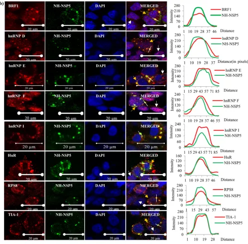
aNima D. Namsa,
aLipika Sahoo,
aC. Durga Rao
aaDepartment of Microbiology and Cell Biology, Indian Institute of Science, Bangalore, India
ABSTRACT
Rotavirus replicates in the cytoplasm of infected cells in unique
virus-induced cytoplasmic inclusion bodies called viroplasms (VMs), which are nucleated
by two essential viral nonstructural proteins, NSP2 and NSP5. However, the precise
composition of the VM, the intracellular localization of host proteins during virus
in-fection, and their association with VMs or role in rotavirus growth remained largely
unexplored. Mass spectrometry analyses revealed the presence of several host
heter-ogeneous nuclear ribonucleoproteins (hnRNPs), AU-rich element-binding proteins
(ARE-BPs), and cytoplasmic proteins from uninfected MA104 cell extracts in the
pull-down (PD) complexes of the purified viroplasmic proteins NSP2 and NSP5.
Immuno-blot analyses of PD complexes from RNase-treated and untreated cell extracts,
analy-ses of coimmunoprecipitation complexes using RNase-treated infected cell lysates,
and direct binding assays using purified recombinant proteins further demonstrated
that the interactions of the majority of the hnRNPs and ARE-BPs with viroplasmic
proteins are RNA independent. Time course immunoblot analysis of the nuclear
and cytoplasmic fractions from rotavirus-infected and mock-infected cells and
immunofluorescence confocal microscopy analyses of virus-infected cells
re-vealed a surprising sequestration of the majority of the relocalized host proteins
in viroplasms. Analyses of ectopic overexpression and small interfering RNA
(siRNA)-mediated downregulation of expression revealed that host proteins
ei-ther promote or inhibit viral protein expression and progeny virus production in
virus-infected cells. This study demonstrates that rotavirus induces the
mic relocalization and sequestration of a large number of nuclear and
cytoplas-mic proteins in viroplasms, subverting essential cellular processes in both
com-partments to promote rapid virus growth, and reveals that the composition of
rotavirus viroplasms is much more complex than is currently understood.
IMPORTANCE
Rotavirus replicates exclusively in the cytoplasm. Knowledge on the
relocalization of nuclear proteins to the cytoplasm or the role(s) of host proteins in
rotavirus infection is very limited. In this study, it is demonstrated that rotavirus
in-fection induces the cytoplasmic relocalization of a large number of nuclear
RNA-binding proteins (hnRNPs and AU-rich element-RNA-binding proteins). Except for a few,
most nuclear hnRNPs and ARE-BPs, nuclear transport proteins, and some cytoplasmic
proteins directly interact with the viroplasmic proteins NSP2 and NSP5 in an
RNA-independent manner and become sequestered in the viroplasms of infected cells.
The host proteins differentially affected viral gene expression and virus growth. This
study demonstrates that rotavirus induces the relocalization and sequestration of a
large number of host proteins in viroplasms, affecting host processes in both
com-partments and generating conditions conducive for virus growth in the cytoplasm of
infected cells.
KEYWORDS
rotavirus, viroplasm, nuclear transport, nuclear-cytoplasmic
relocalization, colocalization, hnRNPs, AU-rich element-binding proteins (ARE-BPs),
pulldown assay, immunofluorescence confocal microscopy, sequestration, immune
confocal microscopy
Received12 April 2018Accepted8 May 2018
Accepted manuscript posted online16 May
2018
CitationDhillon P, Tandra VN, Chorghade SG,
Namsa ND, Sahoo L, Rao CD. 2018. Cytoplasmic relocalization and colocalization with viroplasms of host cell proteins, and their role in rotavirus infection. J Virol 92:e00612-18. https://doi.org/10.1128/JVI.00612-18.
EditorSusana López, Instituto de
Biotecnologia/UNAM
Copyright© 2018 American Society for
Microbiology.All Rights Reserved. Address correspondence to C. Durga Rao, [email protected].
crossm
on November 6, 2019 by guest
http://jvi.asm.org/
R
otaviruses, a major cause of acute gastroenteritis in infants and young children (1),
have a genome composed of 11 segments of double-stranded RNA (dsRNA)
enclosed in an icosahedral triple-layered protein capsid. The viral genome encodes six
structural viral proteins (VP1 to VP4, VP6, and VP7) and six nonstructural proteins (NSP1
to NSP6) (2, 3). During virus entry into the cell, removal of the outer layer from the
triple-layered particle (TLP) activates the synthesis and extrusion of the viral
positive-strand single-positive-stranded RNAs (ssRNAs) through dedicated class I channels of the
double-layered particles (DLPs) into the cytoplasm of the infected cell (4–7). The positive-strand
RNAs function as mRNAs for viral protein synthesis and as pregenomic templates for
negative-strand synthesis.
Key steps in the virus replication cycle, such as viral genome replication and the
assembly of immature DLPs, take place exclusively in the cytoplasm of infected cells in
specialized non-membrane-bound electron-dense cytoplasmic inclusion bodies called
viroplasms (VMs). These bodies are nucleated by two essential nonstructural proteins,
NSP2 and NSP5, and the inner virion capsid protein VP2, with NSP5 being crucial for
both the recruitment of viroplasmic proteins and the architectural assembly of VMs
(8–14). VP2 appears to be associated with the formation of replication complexes
consisting of a positive-strand ssRNA segment, VP1, VP3, and a pentamer of VP2 rather
than with VM formation (7, 15, 16). Viroplasm-like structures (VLSs) are formed in
transfected cells coexpressing NSP5 with either NSP2 or VP2 (9, 10, 12, 14). At early
times (3 to 5 h) postinfection, the number of small VMs increases, but by 8 h
postinfection (hpi), several small VMs join to form large VMs, resulting in a decrease in
the overall number of VMs (17, 18). The temporal transition of small VMs into large VMs
and their concomitant movement to the perinuclear region depend on NSP2 and VP2
and appear to be stabilized by the acetylated microtubular network (19, 20). The
formation of VMs seems to be initiated by a specific interaction of the cytoplasmic
disperse form of NSP2 (dNSP2) with acetylated tubulin and hypophosphorylated NSP5.
The maturation of these nascent VMs is associated with the phosphorylation of NSP5
and the viroplasmic form of NSP2 (vNSP2) (21).
As obligate intracellular parasites with small genomes, many RNA viruses are highly
dependent on cellular factors, particularly the heterogeneous nuclear
ribonucleopro-teins (hnRNPs), for the completion of productive replication, coopting them into new
roles, modulating their functions through the induction of nuclear-cytoplasmic
relocal-izations and/or posttranslational modification and cleavage (22–24). Cellular hnRNPs
(25–30) and AU-rich element-binding proteins (ARE-BPs) (31–33) are predominantly
nuclear, but many, with the exception of hnRNP C1/2 and hnRNP U (25–27), shuttle
between the nucleus and the cytoplasm and often exhibit stress-induced or
stimulus-dependent cytoplasmic relocalization. hnRNPs and ARE-BPs function in both cellular
compartments at multiple levels of mRNA metabolism, encompassing transcription
through translation and stability.
The cytoplasmic relocalization of the nuclear protein RoXaN and the nuclear
accu-mulation of the cytoplasmic poly(A)-binding protein PABPC1 in rotavirus-infected cells
(34, 35) are the only examples reported to date of cellular factors being affected by
rotavirus infection. However, the significance of these changes for virus infection is as
yet not clearly understood (36–38). To date, there have been no studies on the
cytoplasmic relocalization of hnRNPs, ARE-BPs, and other host proteins; their
associa-tion with VMs; or their possible role(s) in virus replicaassocia-tion in infected cells. To investigate
the possible relocalization of nuclear proteins to the cytoplasm during rotavirus
infec-tion and the associainfec-tion of host factors with viroplasmic complexes, the interacinfec-tions of
cellular proteins with the viral VM proteins NSP2 and NSP5 were examined by using
mass spectrometry, pulldown (PD) assays using control cell extracts,
coimmunoprecipi-tation (co-IP) analyses using virus-infected cell extracts, and immunofluorescence
con-focal microscopy (ICM) analyses. The influence of the relocalized host proteins on virus
infection was examined by their ectopic overexpression and small interfering RNA
(siRNA)-mediated downregulation of expression.
Dhillon et al. Journal of Virology
August 2018 Volume 92 Issue 15 e00612-18 jvi.asm.org 2
on November 6, 2019 by guest
http://jvi.asm.org/
RESULTS
Identification of host cell proteins in PD complexes with NSP2 and NSP5 by
mass spectrometry analyses.
Mass spectrometry analyses of PD complexes of
N-terminal hexahistidine (NH)-tagged NSP2 and NSP5 from human rotavirus IS2 (39)
with extracts from uninfected rotavirus-permissive MA104 cells revealed the presence
of several nuclear hnRNPs (see Tables S1 and S2 in the supplemental material). PD
complexes with both NSP2 and NSP5 contained hnRNPs D, I (polypyrimidine
tract-binding protein [PTBP]), K/J, F/H, L, and U. The coat protein complex II (COPII) secretory
vesicle component Sec31A was also detected in both complexes, whereas hnRNPs
A1/3, E1/2 [poly(rC)-binding proteins 1 and 2 (PCBP1/2)], and M; the cytoplasmic
vacuolar protein sorting-associated protein VPS35; and the small-subunit ribosomal
protein RPS8 were detected only in NSP5 complexes, and

-tubulin, which has been
reported to interact with NSP2 (19–21), was detected only in the NSP2 complexes
(Tables S1 and S2).
In addition, pre-mRNA cleavage polyadenylation specificity factor 6 (CPSF6), HSP90,
Hsc70, HSPA1A (Hsp70), and HSPA5 (GRP78/BiP) in NSP2 and NSP5 complexes and
kinesin heavy chain 1 and eIF4A in NSP5 complexes were observed (Tables S1 and S2)
but were not further investigated in this study.
It is important to note that control uninfected cell extracts were deliberately used in
the PD assays, as NSP2 and NSP5 are known to interact with each other as well as with
other viral proteins (9, 10, 12, 14, 40, 41). Consequently, had extracts been made from
virus-infected cells, it would not have been possible to establish whether cellular
proteins captured in the PD complexes were directly interacting with bead-bound NSP2
or NSP5 or being recruited to the PD complex through their interaction with another
viral protein(s) and viroplasm assemblies present in virus-infected cells.
The presence of some cellular proteins in the PD complexes could not be
unequiv-ocally established using mass spectrometry. Consequently, where the necessary
immu-nological reagents were available, the possible binding of hnRNPs and other
well-studied ARE-BPs (31–33), besides hnRNP D, was examined by immunoblotting of the PD
complexes. This revealed that hnRNPs A1, C1/2, and M, which were not detected by
mass spectrometry in the NSP2 PD complexes, and the ARE-BPs human antigen R (HuR),
butyrate response factor 1 (BRF1), T cell-restricted intracellular antigen 1 (TIA1),
TIA1-like 1 (TIAL1), KH-type splicing regulatory protein (KSRP), and Staufen1, which were not
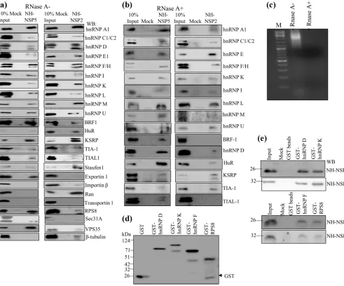
detectable in the PD complexes of both NSP2 and NSP5, were detectable by
immu-noblotting (Fig. 1a).
To both complement and extend the mass spectrometry data, immunoblot assays
were carried out. The MA104 cell extracts used for the mass spectrometry-based
analyses were not treated with RNase in order to not lose the possible RNA-mediated
interactions. Therefore, in the immunoblot-based assays, comparative analyses using
both RNase-treated and untreated cell extracts were performed to distinguish the
possible RNA-mediated interactions from RNA-independent interactions (Fig. 1a and b).
The results shown in Fig. 1a and b independently confirmed the interacting cellular
proteins identified by using mass spectrometry of PD complexes. In addition, while all
the ARE-BPs, including hnRNP D; HuR; the TIS11 family proteins BRF1, TIA1, and TIAL-1;
KSRP; and Staufen1, from RNase-untreated extracts bound to NSP5 and NSP2, the
interactions of BRF1 and TIAL-1 with NSP2 were abolished upon RNase treatment of the
extracts (Fig. 1b and c). Of note, RNase treatment of the extracts resulted in enhanced
binding of HuR to NSP5 but reduced binding to NSP2 (Fig. 1b). These results suggest
that while the interactions of the majority of the host proteins with both NSP5 and
NSP2 are RNA independent, those of BRF1 and TIAL-1 with NSP2 are RNA dependent.
Finally, the results in Fig. 1b show that RNase treatment of cell extracts resulted in a
significant reduction in the binding of hnRNP A1 to NSP2. The results on
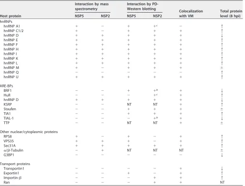
RNA-independent and -dependent interactions are summarized in Table 1.
That the host proteins interact directly with NSP2 and NSP5 was further
demon-strated by a direct binding PD assay using a few purified recombinant host proteins and
the two viral viroplasmic proteins (Fig. 1d and e).
on November 6, 2019 by guest
http://jvi.asm.org/
The proteins listed in Tables S1 and S2 in the supplemental material are only those
that were exclusively detected in the NSP5- or NSP2-bound PD complexes, and those
that were also found in PD complexes generated using control Ni-nitrilotriacetic acid
(NTA)-agarose beads were excluded. However, it is important to note that neither mass
spectrometry nor immunoblot analyses allowed any assessment to be made of the
relative affinities with which cellular proteins bound to control and viral protein-bound
beads; consequently, the proteins listed in Tables S1 and S2 in the supplemental
material are likely to represent only a partial cataloguing of the complete interactome.
FIG 1Interaction of cellular hnRNPs and ARE-BPs in RNase-treated and untreated cell extracts with purified recombinant viral nonstructural proteins NSP2 and NSP5 demonstrated by a pulldown assay. The recombinant NH-tagged viral proteins NSP5 and NSP2 were purified fromE. coliby affinity chromatography using Ni2⫹-NTA-agarose beads. Control Ni2⫹-NTA-agarose beads, which were prepared by passing the lysate fromE. coliharboring the pET22-NH vector lacking the viral gene, were used for mock binding. Both the experimental and control beads were further incubated in binding buffer containing 0.5% BSA to minimize the nonspecific binding of cellular proteins. (a and b) The RNase-treated purified recombinant NSP2 and NSP5 proteins bound to Ni2⫹-NTA-agarose beads, and the control beads (mock binding) were incubated with equal amounts (500g) of control MA104 cell extracts that were either not treated with RNase (a), similar to what was done for mass spectrometry, or treated with RNase (b). The cellular proteins bound to the beads were resolved by SDS-PAGE, and the interacting cellular proteins were detected by immunoblotting. In the lane representing 10% input, 50g of the RNase-treated or untreated cell extracts was loaded. The same blot was used to detect two or three host proteins by sequential deprobing and reprobing depending on clear differences in the molecular weights of the proteins. Each PD assay was repeated at least 3 to 4 times to confirm reproducibility. (c) The cell extracts (1 mg/ml) were incubated with 100g of RNase A for 45 min at room temperature, and 100g of the RNase-treated and untreated cell extracts was resolved by agarose gel electrophoresis and visualized by ethidium bromide staining. Note the complete digestion of cellular RNA in the RNase-treated extract. M, molecular marker. (d) Expression and purification of GST-tagged recombinant host proteins. The bacterial cell extracts were incubated with RNase A (100 mg/ml) prior to purification. (e) Demonstration of direct interactions of purified NH-NSP2 and NH-NSP5 with glutathione bead-bound GST-tagged nuclear proteins. Ten micrograms of purified NH-NSP2 or NH-NSP5 was incubated with approximately 5g of the bead-bound recombinant GST-tagged hnRNPDp40isoform and hnRNP K (top) and hnRNP F and RPS8 (bottom)
treated further with RNase A (10 mg/ml), and the bound viral protein was detected by Western blotting (WB).
Dhillon et al. Journal of Virology
August 2018 Volume 92 Issue 15 e00612-18 jvi.asm.org 4
on November 6, 2019 by guest
http://jvi.asm.org/
[image:4.585.46.537.72.480.2]Cell compartment relocalization of cellular proteins detected in PD complexes
during rotavirus infection.
Rotavirus replication is confined to the cytoplasm of the
virus-infected cell, and neither NSP2 nor NSP5 is known to localize to the nucleus.
Consequently, the detection of a large number of host cell nuclear proteins in the PD
complexes of NSP2 and NSP5 (Fig. 1a and b) suggested the possibility of their
cytoplasmic relocalization following rotavirus infection. As nothing is known about the
localization of hnRNPs in rotavirus-infected cells, the intracellular distribution of
NSP5-and NSP2-interacting hnRNPs NSP5-and ARE-BPs during the course of rotavirus infection was
investigated by immunoblotting.
[image:5.585.42.548.82.465.2]The analysis of the nuclear and cytoplasmic fractions at different time points after
infection of rhesus rotavirus (RRV)-infected MA104 cells (Fig. 2a, right) and
serum-starved control mock-infected cells (Fig. 2a, left) revealed that a number of the hnRNPs
analyzed were specifically enriched in the cytoplasmic fraction of virus-infected cells
during the course of infection. For clarity, the proteins in Fig. 2 are discussed according
to their relative pattern of intracellular localization during infection. The cytoplasmic
relocalizations of hnRNPs A1, K/J, and U (Fig. 2a) and of HuR, hnRNP D, and BRF1
TABLE 1Summary of interactions between the viroplasmic proteins NSP2 and NSP5 and host proteinsa
Host protein
Interaction by mass spectrometry
Interaction by PD-Western blotting
Colocalization with VM
Total protein level (8 hpi)
NSP5 NSP2 NSP5 NSP2
hnRNPs
hnRNP A1 ⫹ ⫺ ⫹ ⫹c ⫺ 1
hnRNP C1/2 ⫹ ⫺ ⫹ ⫹ ⫹ 1
hnRNP D ⫹ ⫹ ⫹ ⫹ ⫹ 2
hnRNP E ⫹ ⫹ ⫹ ⫹ ⫹ 1
hnRNP F ⫹ ⫹ ⫹ ⫹ ⫹ 1
hnRNP H ⫹ ⫹ ⫹ ⫹ ⫹ 1
hnRNP I ⫹ ⫹ ⫹ ⫹ ⫹ 1
hnRNP K ⫹ ⫹ ⫹ ⫹ ⫹ 1
hnRNP L ⫹ ⫹ ⫹ ⫹ ⫹ 1
hnRNP M ⫹ ⫺ ⫹ ⫹ ⫹ ⫾
hnRNP Q ⫺ ⫺ ⫺ ⫺ ⫺ 1
hnRNP U ⫹ ⫹ ⫹ ⫹ ⫹ 1
ARE-BPs
BRF1 ⫺ ⫺ ⫹ ⫹b ⫹ 2
HuR ⫺ ⫺ ⫺ ⫹c ⫹ 1
hnRNP D ⫹ ⫹ ⫹ ⫹ ⫹ 2
KSRP ⫺ ⫺ NT NT ⫹ 1
Staufen ⫺ ⫺ ⫹ ⫹ ⫹ ⫾
TIA1 ⫺ ⫺ ⫹ ⫹ ⫹ 1
TIAL-1 ⫺ ⫺ ⫺ ⫹b ⫹ 2
TTP ⫺ ⫺ NT NT ⫹ ⫾
Other nuclear/cytoplasmic proteins
RPS8 ⫹ ⫺ ⫹ ⫺ ⫹ 1
VPS35 ⫹ ⫹ ⫹ ⫺ ⫹ 1
Sec31A ⫹ ⫹ ⫹ ⫹ ⫹ 1
␣/-Tubulin ⫺ ⫹ NT NT NT ⫾
G3BP1 ⫺ ⫺ ⫺ ⫺ ⫺ 2
Transport proteins
Transportin1 ⫺ ⫺ ⫺ ⫺ ⫹ 2
Exportin1 ⫺ ⫺ ⫹ ⫺ ⫹ 1
Importin- ⫺ ⫺ ⫺ ⫹ ⫹ 1
Ran ⫺ ⫺ ⫺ ⫹ ⫹ NT
aThe interactions identified between the cellular proteins and viral proteins NSP2 and NSP5 by a PD assay followed by immunoblotting and colocalization by ICM (Fig.
1a and b and 4) are summarized.⫹, positive interaction/colocalization;⫺, no interaction/colocalization.1and2indicate increased and decreased host protein levels in virus-infected cells in comparison to those in uninfected cells, respectively;⫾indicates that there was no significant change in the protein levels between virus-infected and uninfected cells. NT, not tested.
bLoss of binding of NSP2 with BRF1 and TIAL-1 in RNase-treated cell extracts. cReduced binding of NSP2 with hnRNP A1 and HuR in RNase-treated cell extracts.
on November 6, 2019 by guest
http://jvi.asm.org/
FIG 2Analysis of nuclear and cytoplasmic distributions of hnRNPs, ARE-BPs, and other proteins in mock-infected and rotavirus RRV-infected MA104 cells during the course of infection. (a) Time course immunoblot analysis of the levels of hnRNPs in the nuclear and cytoplasmic fractions. The samples in the lane from 0 hpi represent nuclear and cytoplasmic fractions from serum-grown control cells. Lanes from 2 to 10 hpi on the left represent mock-infected cells incubated for the indicated time periods in medium lacking serum, similar to the conditions under which virus infections were performed, and those on the right represents lysates prepared from (Continued on next page)
Dhillon et al. Journal of Virology
August 2018 Volume 92 Issue 15 e00612-18 jvi.asm.org 6
on November 6, 2019 by guest
http://jvi.asm.org/
[image:6.585.43.462.71.690.2](Fig. 2b) were specifically induced by virus infection and not by simple serum
starvation-induced stress in mock-infected MA104 cells. In contrast, hnRNPs C1/2, F/H,
L, and M (Fig. 2a) and the ARE-BPs KSRP and TIA1 (Fig. 2b) showed a partial
relocal-ization to the cytoplasm in mock-infected cells following serum starvation-induced
stress, and virus infection caused further increases in their cytoplasmic relocalization. To
date, both hnRNPs C1/C2 and U have been considered to be nonshuttling proteins
(25–27), but rotavirus infection clearly induces their cytoplasmic relocalization. hnRNP
E levels increased in both compartments in both mock-infected and virus-infected cells
(approximately 2.5-fold at 8 hpi), which appeared to be primarily due to serum
starvation but not rotavirus infection. Of note, both isoforms of hnRNP E were observed
in the cytoplasm, but only one was observed in the nucleus.
The increased cytoplasmic accumulation of hnRNPs A1, K/J, L, I, and M and the
ARE-BPs HuR, hnRNP D, and BRF1 (Fig. 2a and b) during virus infection correlated with
concomitant decreases in the nucleus. Note that for hnRNPs A, D, F/H, L, K, Q, and U;
HuR; and BRF1, no signal could be detected in the cytoplasm of serum-grown control
and/or serum-starved cells during the course of the experiment using 50
g of protein
in Western blot analyses (Fig. 2a and b; see also Table S3 in the supplemental material).
The ARE-BPs FXR1, TIAL-1, tristetraprolin (TTP), and Staufen1 were abundant and stable
in the cytoplasm. While there was no change in the levels of TTP and Staufen1 in the
nucleus, those of FXR1 and TIAL-1 decreased during the course of infection (Fig. 2b and
Table S3). Staufen1 showed cleavage during the late period of infection. The relative
increases or decreases in the levels of hnRNPs and ARE-BPs in the nuclear and
cytoplasmic fractions in infected cells (Table 1) correlated with the total protein levels
between control and infected cells (Fig. S1a and S1b).
Immunofluorescence confocal microscopy (ICM) analysis reveals the predominant
nuclear localization of the majority of the hnRNPs (Fig. S2a) and ARE-BPs (Fig. S2b) in
serum-grown control MA104 cells. TTP and TIAL-1 showed significant presences in both
compartments, correlating with the results of the analysis of the nuclear and
cytoplas-mic fractions (Fig. 2b and Fig. S2b).
hnRNP Q showed no change in its localization and was detected only in the nucleus
under all conditions. No change in either the localization or abundance of PCNA and

-actin, used as nuclear and cytoplasmic control proteins, respectively, was observed
under any condition (Fig. 2a and b and Table S3), suggesting that the cytoplasmic
relocalization of nuclear proteins is selective but not global.
The cytoplasmic proteins RPS8 and VPS35, which were detected in the PD
com-plexes with NSP5, and Sec31A, which was detected in comcom-plexes with NSP2 and NSP5,
showed significant increases (4- to 10-fold) in their levels in the cytoplasm of
virus-infected MA104 cells (Fig. 2c). Of note, while RPS8 showed a significant increase in its
level in the nucleus in both mock- and virus-infected cells, a small proportion of Sec31A
was detectable in the nucleus during late stages of infection. In contrast, VPS35 was
detected only in the cytoplasm. The total protein levels of RPS8, Sec31A, and VPS35
showed approximately 13-, 1.4-, and 3.9-fold increases, respectively, correlating with
the changes observed in their cytoplasmic and/or nuclear levels (Fig. 2c and Fig. S1c).
PABPC1, which has been previously shown to relocalize to the nucleus during rotavirus
infection, showed 30% and 90% reductions in the cytoplasm of mock-infected and
FIG 2Legend (Continued)
RRV-infected cells (2 to 10 hpi). NSP5 was detected using protein A-agarose affinity-purified rabbit PAb generated against purified recombinant NSP5, and VP6 was detected using subgroup I MAb 631/9, which are very specific to the viral proteins, with no cross-reactivity to host proteins.-Actin and PCNA were used to determine the purity of the cytoplasmic and nuclear fractions, respectively, and as internal loading controls. At each time point, 50g of the protein was analyzed. C.E., cytoplasmic extract; N.E., nuclear extract. (b) Analysis of the altered nuclear and cytoplasmic distributions of ARE-BPs during rotavirus infection. NSP2 and VP6 were detected using affinity-purified rabbit PAbs generated against purified NSP2 and RRV DLPs, respectively. For other details, see the legend to panel a. (c) Analysis of intracellular levels of host cytoplasmic proteins that interact with NSP5 and/or NSP2. See the legends to panels a and b for details. (d) Nuclear-cytoplasmic levels of nuclear transport proteins during rotavirus infection. Note the enhanced cytoplasmic retention of nuclear transport proteins during rotavirus infection in MA104 cells. ns, not significant. Other details are described in the legend to panel a. See Table S3 in the supplemental material for quantification of the changes in the protein levels in the nucleus and cytoplasm during the course of infection, and see Fig. S1 for changes in total protein levels.
on November 6, 2019 by guest
http://jvi.asm.org/
virus-infected cells but exhibited increases in the nucleus of approximately 5.5- and
9.5-fold, respectively, at 8 hpi (Fig. 2c and Table S3), suggesting that its relocalization
to the nucleus is partly due to serum starvation during virus infection. The total protein
levels of both PABPC1 and G3BP1 were marginally reduced in infected cells (Fig. S1c).
Intracellular distribution of nuclear transport proteins in rotavirus-infected
cells.
The relocalization of some host cell proteins from the nucleus to the
cyto-plasm observed following rotavirus infection raised the possibility that the
nuclear-cytoplasmic transport processes may suffer dysregulation following virus infection.
To explore this possibility, the nuclear-cytoplasmic abundance of a few known
nuclear transport factors (42, 43) was examined by immunoblotting. This revealed
2- to 3-fold increases in the levels of Exportin1, importin-

, and Ran in the
cytoplasm of rotavirus-infected cells compared to those seen in either control or
serum-starved mock-infected MA104 cells, but there was no significant change in the
cytoplasmic levels of Transportin1 (Fig. 2d and Table S3). In contrast, in the nucleus, the
level of Transportin1 decreased by approximately 3-fold following virus infection, while
there was no significant change in the level of either Exportin1 or importin-

. The
steady-state level of the Ran transporter increased to the same extent in both cellular
compartments following virus infection. Comparison of the total protein levels in
control and infected cells revealed a good correlation with the changes observed in the
nuclear and cytoplasmic fractions (Fig. S1d).
Relocalized host proteins are associated with the viroplasmic complexes in
rotavirus-infected cells.
Coimmunoprecipitation (co-IP) studies on RNase-treated
virus-infected cell lysates were undertaken to investigate the possible physical
associ-ation of cytoplasmically relocalized host proteins with the key viral constituents (NSP2,
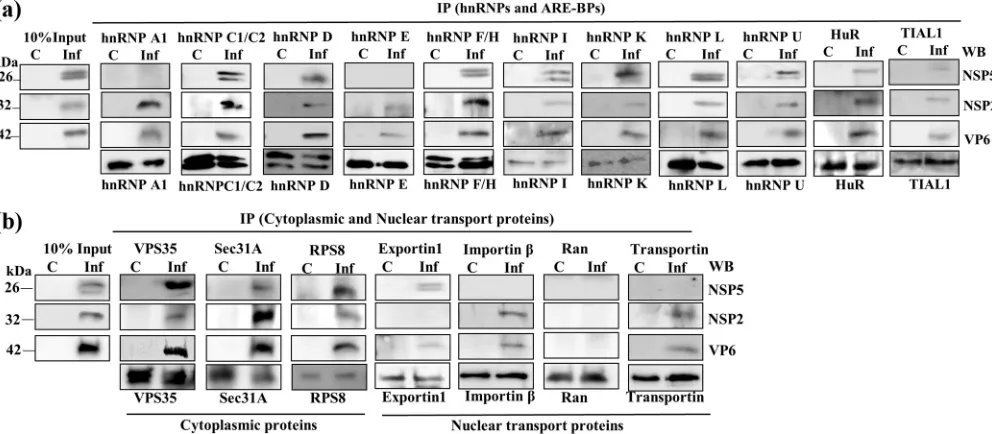
NSP5, and VP6) of viroplasmic complexes. Figures 3a and b reveal that NSP2, NSP5, and
VP6 could be detected in the co-IP complexes with the majority of the relocalized host
proteins examined.
Immunofluorescence confocal microscopy analyses reveal selective
sequestra-tion of relocalized host proteins to VMs in rotavirus-infected cells.
In all the ICM
analyses, the cells were infected at a multiplicity of infection (MOI) of approximately 0.5
FIG 3Co-IP analyses using virus-infected cell extracts demonstrate the association of host proteins with viroplasmic complexes/proteins. RNase-treated lysates from mock-infected and RRV-infected (MOI of 10) MA104 cells were prepared at 8 hpi. A total of 500g of the lysate was used to immunoprecipitate the cellular proteins using specific MAbs/PAbs. The immune complexes from mock-infected (C) and virus-infected (Inf) cell lysates were captured using BSA-saturated protein A-Sepharose beads and were resolved by SDS-PAGE. The lanes under 10% input (C and Inf) contain 50g of the lysates from control and infected cells, respectively. The viral proteins were coimmunoprecipitated using antibodies against the interacting host protein, and the viral proteins were detected by Western blotting using PAbs against NSP2, NSP5, and VP6 or MAb against VP6.
Dhillon et al. Journal of Virology
August 2018 Volume 92 Issue 15 e00612-18 jvi.asm.org 8
on November 6, 2019 by guest
http://jvi.asm.org/
[image:8.585.45.541.73.290.2]so that the localization statuses of the host proteins in both infected and uninfected
cells could be compared in the same field under identical experimental conditions. ICM
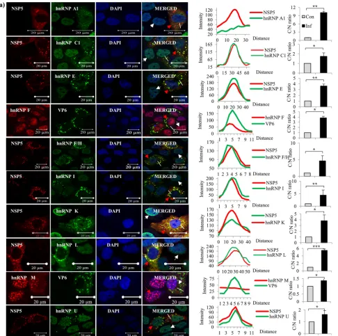
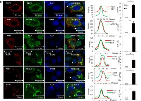
analyses demonstrated the cytoplasmic relocalization of the majority of the hnRNPs
and ARE-BPs, confirming the results of immunoblot analyses of the nuclear and
cytoplasmic fractions of virus-infected cells (Fig. 2a and b). Surprisingly, the majority of
the relocalized hnRNPs (hnRNPs C1/C2, E, F/H, I, K/J, L, and U) and ARE-BPs (hnRNP D,
BRF1, HuR, TIA1, TIAL-1, TTP, Staufen1, and KSRP) showed colocalization with the
viroplasmic proteins in VMs in virus-infected cells (Fig. 4a and b).
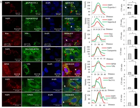
These analyses further confirmed the enhanced levels of the nuclear transport
proteins in the cytoplasm of virus-infected cells in comparison to those in uninfected
cells, as demonstrated by immunoblotting of the nuclear and cytoplasmic fractions (Fig.
2d and 4c). Moreover, all four transport proteins exhibited enrichment/colocalization
with the VMs (Fig. 4c).
The cytoplasmic proteins VPS35, Sec31A, and RPS8 also showed colocalization with
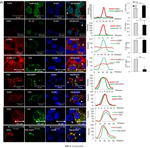
VMs (Fig. 4c). In contrast, no detectable colocalization of hnRNP A1 and hnRNP M with
the VM was observed, and the splicing factor Sc35 and hnRNP Q remained solely in the
nucleus (Fig. 4a and d). In addition, cytoplasmic PABPC1 relocalized to the nucleus in
infected cells, as previously reported (34, 35, 38), and there was consequently no
significant colocalization with VMs (Fig. 4d). The abundant cytoplasmic protein G3BP1,
the best-characterized stress granule (SG)-specific marker, neither colocalized with the
VM nor formed G3BP-specific SGs in infected cells (Fig. 4d), confirming a previous report
that SGs are not formed in rotavirus-infected cells (35).
It is important to note that the colocalization of hnRNPs and ARE-BPs with the VM
is not due to their release into the cytoplasm as a result of nuclear lysis caused by an
ultimately cytocidal viral infection, as 4
=
,6-diamidino-2
=
-phenylindole dihydrochloride
(DAPI)-stained virus-infected cells clearly showed that nuclei remained intact at 8 hpi
(Fig. 4a to d). The cytoplasmic relocalization of host cell hnRNPs and ARE-BPs is a
gradual process that commences at between 2 and 4 hpi, and significant cytoplasmic
relocalization and colocalization of the nuclear proteins with VM could be detected at
approximately 4 hpi, with their enrichment in the VM increasing with time, as shown by
time course confocal image analysis of nuclear TIA1 and cytoplasmic RPS8 (see Fig. S3
in the supplemental material). In addition,

-actin and VPS35 were detected only in the
cytoplasm, and PCNA localized to the nucleus (Fig. 2 and 4). As expected, the viral
proteins NSP2, NSP5, and VP6 and ectopically expressed NH-NSP5 were observed only
in the cytoplasm and colocalized with each other (Fig. 2 and 4).
It may be argued that colocalization with the VM of some of cytoplasmic proteins
such as RPS8, Sec31A, and VPS35 and ARE-BPs such as TIAL-1 and TTP could be
nonspecific due to their high abundance in the cytoplasm. However, the distinct
punctate structures observed at identical positions for both the viral and host proteins
in infected cells suggest that their colocalization is specific (Fig. 4a to d), which was also
verified by Z-stack analysis, shown, for example, for two of the host proteins, hnRNP D
and hnRNP L, in Fig. S4 in the supplemental material.
Ectopically expressed nuclear and cytoplasmic proteins colocalize with
rotavi-rus VMs.
To further confirm the cytoplasmic relocalization and colocalization of host
cell proteins with the VM, viral and selected host cell proteins tagged with a small
amino-terminal NH or Myc tag or a larger fluorescent protein (FP) tag, green FP (GFP),
enhanced cyan FP (ECFP), or mCherry (mCH), were ectopically expressed by transfection
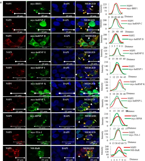
of cDNA constructs into HEK293T cells, followed by virus infection. Figure 5a shows the
colocalization of a number of Myc-tagged hnRNPs and ARE-BPs (hnRNPs D, I, K, and L;
BRF1; and TIA-1), NH-HuR, and Myc-RPS8 with virus-expressed NSP5 and NSP2 in the
VM. Conversely, NH-tagged NSP5 also colocalized with the endogenous host proteins
in the VM (Fig. 5b). As expected, ectopically expressed ECFP-NSP5 also colocalized with
virus-expressed NSP2 and VP6 in the VM (Fig. S5). Ectopically expressed host nuclear
proteins carrying larger fluorescent tags (hnRNP D, hnRNP C1, and
ECFP-hnRNP K) also showed colocalization with viral proteins in the VMs (Fig. S5). However,
unlike the ectopically expressed host proteins carrying the smaller Myc tags, a
on November 6, 2019 by guest
http://jvi.asm.org/
FIG 4Immunofluorescence confocal microscopy analyses of relocalization and colocalization of host proteins with VS in rotavirus RRV-infected MA104 cells. (a) ICM analyses of the nuclear-cytoplasmic distribution and colocalization with VMs of endogenous hnRNPs. MAbs against different hnRNPs (see Table S4 in the supplemental material), affinity-purified PAbs against NSP5 and NSP2, anti-RRV DLP PAbs against VP6, and Cy3-tagged anti-mouse (for cellular proteins) and Cy5-tagged anti-rabbit secondary antibodies (for viral proteins) were used. After mounting in DAPI, the proteins were visualized by fluorescence under a LSM Zeiss 710 immunofluorescence confocal microscope (63⫻oil immersion). Infections of cells grown on coverslips for confocal imaging were done at an MOI of 0.5 for 8 h. In ICM analyses, fields having relatively fewer infected cells were selected to clearly distinguish the localization status of host proteins in uninfected cells. Note the colocalization of hnRNPs with the VM. In all the confocal images, uninfected cells, infected cells, and the plot profile path on the VM are indicated by white and red arrowheads and a white broken line, respectively. The plot profiles and quantification of the cytoplasmic/nuclear (C/N) ratios of the proteins from 50 infected and uninfected cells were done using Image J software. The fluorescence quantification data represent the average C/N ratios for 50 infected cells normalized to the average C/N ratio for 50 control cells from one of three independent experiments and are expressed as averages⫾standard deviations.
*,P⬍0.05;**,P⬍0.01;***,P⬍0.001; ns, nonsignificant (as determined by attest). (b) Cytoplasmic relocalization and colocalization with VMs of ARE-BPs in RRV-infected cells. MAbs against different ARE-BPs (Table S4) were used. See the legend to panel a for more details. See Fig. S2 in the supplemental material for the predominant nuclear localization of hnRNPs and ARE-BPs in the nuclei of control cells. (c) Colocalization of nuclear transport proteins and cytoplasmic proteins in the VMs. MAbs against Exportin1, importin-, and Transportin1 and rabbit PAb against Ran were used. See the colocalization of the transport proteins with viroplasmic NSP5 and VP6. The plots of cytoplasmic/nuclear ratios of the transport proteins are shown. See the legend to panel a for details. (d) Nuclear and cytoplasmic proteins that do not interact or colocalize with viroplasmic proteins. The virus-encoded NSP2, NSP5, and VP6 proteins and the ectopically expressed NH-tagged NSP5 protein colocalize in the VM. The panel showing NH-NSP5 represents HEK293T cells transfected with pc-NH-NSP5 expressing NH-tagged IS2-NSP5. Other panels represent MA104 cells. See Fig. S3 in the supplemental material for time course ICM analysis of the nuclear (TIA1) and cytoplasmic (RPS8) proteins, showing their colocalization with VM starting from 4 hpi, and see Fig. S4 for Z-stack analysis of the colocalization of hnRNP D with NSP2 and of hnRNP L with NSP5.
Dhillon et al. Journal of Virology
August 2018 Volume 92 Issue 15 e00612-18 jvi.asm.org 10
on November 6, 2019 by guest
http://jvi.asm.org/
[image:10.585.46.532.71.554.2]icant proportion of the ECFP-tagged nuclear proteins was retained in the nucleus (Fig.
S5). In contrast, the mCH-tagged cytoplasmic protein VPS35 (mCH-VPS35) was present
only in the cytoplasm and colocalized with the VM (Fig. S5).
To investigate if the simple ectopic expression of the key VM proteins (NSP2, NSP5,
or VP6) alone is able to induce the relocalization of host proteins seen in virus-infected
cells, the subcellular localization of some hnRNPs and ARE-BPs was examined in
HEK293T cells transiently expressing the VM viral proteins. BRF1 was present at
detect-able levels in the cytoplasm of control HEK293T cells, and the ectopic expression of the
viral proteins did not further induce its cytoplasmic localization (Fig. S6a to S6c).
However, the ectopic expression of NSP2 and VP6 induced the partial cytoplasmic
relocalization of hnRNP D, HuR, and hnRNP K but not of the other host cell proteins
examined (Fig. S6a to S6c). These results suggest that the expression of just the key viral
VM proteins (NSP2, NSP5, or VP6) alone does not induce the drastic changes in the
nuclear-cytoplasmic localization of hnRNPs and ARE-BPs seen following rotavirus
infec-tion.
Influence of relocalized host cell proteins on viral gene expression and
infec-tious virus yield.
To assess the influence of host cell proteins relocalized following
rotavirus infection on viral gene expression and progeny virus production, the effect of
the ectopic overexpression and/or siRNA-mediated knockdown of selected host cell
proteins was examined in HEK293T cells. The positive or negative effect of relocalized
host proteins on viral gene expression was assessed by looking at the expression of the
viroplasmic proteins NSP2, NSP5, and VP6. The ectopic expression of BRF1 and TIA1 had
a negative effect on the levels of NSP5 expression but did not affect either NSP2 or VP6
FIG 4(Continued)
on November 6, 2019 by guest
http://jvi.asm.org/
[image:11.585.44.527.72.420.2]expression levels (Fig. 6a). hnRNP C1, G3BP1, and VPS35 affected the expression of all
three viral proteins. While hnRNP C1 exhibited a significant negative effect on the
expression of NSP2, hnRNP E1 affected those of both NSP2 and VP6. The p40 and p42
isoforms of hnRNP D did not support the expression of NSP2, but the p37 and p45
isoforms enhanced the expression of the three viral proteins (Fig. 6a), and NSP5
expression was highly enhanced by hnRNP L (Fig. 6a). The siRNA-mediated
downregu-lation of hnRNP F significantly affected the expression of NSP5 (Fig. 6b). While the
knockdown of hnRNP C1 resulted in a significant increase in the NSP2 level, that of
hnRNP E1 enhanced the expression of both NSP2 and NSP5 (Fig. 6c). The
downregu-lation of G3BP1 markedly increased the levels of not only NSP5 but also NSP2 and VP6
in virus-infected cells (Fig. 6c).
Turning next to the effects of regulating the synthesis of relocalized host cell
proteins on infectious virus yields, Fig. 7 shows that while the siRNA-mediated
down-regulation of HuR, hnRNP D
45, hnRNP I, and hnRNP K decreased progeny virus titers by
almost 2 orders of magnitude, their overexpression resulted in only a 6- to 8-fold
increase in the progeny virus titers, as determined in MA104 cells by an enzyme-linked
immunoperoxidase focus assay (ELIFA) (44–46). In contrast, the downregulation of
G3BP1, TIA1, and hnRNP C1 increased the progeny virus yields by 3, 2.4, and 2.5 orders
of magnitude, respectively, while ectopic overexpression resulted in reductions of virus
yields by 3.2, 1.6, and 2.0 orders of magnitude, respectively (Fig. 7). Of note, the ectopic
expression and siRNA-mediated knockdown of host proteins in HEK293T cells resulted
in only a 1.2- to 2-fold increase or decrease in the number of infected cells, as examined
FIG 4(Continued)
Dhillon et al. Journal of Virology
August 2018 Volume 92 Issue 15 e00612-18 jvi.asm.org 12
on November 6, 2019 by guest
http://jvi.asm.org/
[image:12.585.47.520.75.443.2]by a direct ELIFA on transfected cells, but differences in progeny virus titers of 1.4 to 2.0
orders of magnitude, as assessed in MA104 cells. This suggests that host proteins could
influence either the number of mature viroplasms, the size of the viroplasms, or both,
impacting the infectious progeny virus yield, which was not examined in this study.
DISCUSSION
Since virtually all viruses are totally dependent on host translational machinery for
their protein synthesis, they employ a variety of mechanisms to subvert and hijack the
system to promote the rapid and selective translation of viral mRNAs by manipulating
one or more processes associated with host mRNA metabolism and/or translation
during their short life cycle (22–24, 47, 48). An increasing number of studies in recent
years reported the cytoplasmic relocalization of either one or a few hnRNPs and/or
ARE-BPs during infection by different viruses and their influence on viral gene
expres-sion and/or replication (22–24, 49–55). However, very little is known about the
intra-cellular localization of hnRNPs, ARE-BPs, and other host proteins during rotavirus
FIG 4(Continued)
on November 6, 2019 by guest
http://jvi.asm.org/
[image:13.585.44.525.70.544.2]FIG 5Colocalization of tagged ectopically expressed hnRNPs and ARE-BPs with viral proteins in the VM. (a) Ectopically expressed Myc- and NH-tagged hnRNPs and ARE-BPs colocalize with the VM. HEK293T cells on coverslips were transfected with plasmid DNA constructs expressing Myc- or NH-tagged host proteins for 36 h, followed by RRV infection (MOI of 0.5 for 8 h). Proteins were visualized by using anti-Myc or anti-His tag antibodies (mouse), PAb against NSP5 (rabbit), and the corresponding Cy3-tagged anti-mouse and Cy5-tagged anti-rabbit secondary antibodies. The transfected-infected cells and uninfected cells are indicated with orange and white arrowheads, respectively. The plot profile path on the VM is indicated by a white dashed line. See the legend to Fig. 4a for more details. (b) Ectopically expressed NH-NSP5 colocalizes with endogenous hnRNPs and ARE-BPs in the VM. Ectopically expressed NH-NSP5 was detected using anti-His tag antibody (mouse), and the endogenous cellular proteins were detected using rabbit PAbs, along with the corresponding fluorescent dye-tagged secondary antibodies. Transfected-infected HEK293T cells are shown by orange arrowheads. For other details, see the legend to Fig. 4a. See Fig. S5 in the supplemental material for colocalization of the ectopically expressed fluorescent protein-tagged host proteins and NSP5 in the VM, and see Fig. S6 for a demonstration that ectopic expression of the viroplasmic proteins does not induce the relocalization of host proteins, as observed in infected cells.
Dhillon et al. Journal of Virology
August 2018 Volume 92 Issue 15 e00612-18 jvi.asm.org 14
on November 6, 2019 by guest
http://jvi.asm.org/
[image:14.585.47.544.70.622.2]infection. The results presented here demonstrate that rotavirus infection induces the
relocalization of a large number of nuclear proteins, the majority of which associate
with the viroplasmic proteins NSP2 and NSP5 in the VM.
The most unexpected finding was the direct interaction of large numbers of hnRNPs,
ARE-BPs, and nuclear transport proteins and some cytoplasmic proteins with the
viroplasmic proteins NSP2 and NSP5 (Table 1). It could be argued that the interaction
of several host proteins with both NSP2 and NSP5 could be because of some kind of
nonspecific binding due to an improper folding of the recombinant viral proteins. It is
also possible that most of the host proteins could be recruited through one or two
common proteins that interact with NSP2 and NSP5 or a common motif in the viral
proteins, which requires further investigation by deletion mapping analyses. In this
context, it may be noted that some hnRNPs are known to interact with each other (25,
FIG 5(Continued)
on November 6, 2019 by guest
http://jvi.asm.org/
[image:15.585.46.543.71.560.2]56, 57). The VM contains a large number of NSP2 and NSP5 molecules depending on
the state of maturation. It is unlikely that a single NSP2 or NSP5 molecule interacts with
many host proteins simultaneously. Different NSP2 and NSP5 molecules could be
interacting with one or two host proteins. The lack of one or two viral proteins in the
co-IP complexes (Fig. 6) with a few host proteins could be due to the binding of the
host protein to subviroplasmic complexes or a soluble form of the viral proteins.
Furthermore, NSP2 and NSP5 are known to undergo phosphorylation, glycosylation,
methylation, and/or SUMOylation in virus-infected cells (10, 12, 14, 58–62). Since
proteins expressed in
Escherichia coli
lack these modifications, the interactome of the
native viral proteins could differ from that of the recombinant proteins. It may be
argued that some of the interactions observed in the
in vitro
pulldown analyses could
still be RNA mediated in spite of the use of RNase-treated viral proteins and cell extracts
due to incomplete RNA digestion. Note that hnRNPs and ARE-BPs as well as NSP2 and
NSP5 are RNA-binding proteins, and the majority of the host proteins interacted with
the viral proteins in an RNA-independent manner. However, in spite of the observed
apparent RNA-independent interactions
in vitro
, the association of host proteins with
the VM inside the cell, as revealed by ICM analyses, in reality could be facilitated by RNA
by virtue of their inherent RNA-binding properties, rendering the argument that the
interactions between viral and cellular proteins in the PD and co-IP complexes
in vitro
are nonspecific to be irrelevant and untenable.
FIG 6Effect of ectopic expression and siRNA-mediated knockdown of cellular proteins on viral protein expression. (a) Differential influence of ectopic expression of hnRNPs, ARE-BPs, and other host proteins on viral protein expression. HEK293T cells, transfected for 48 h with the plasmid DNA constructs expressing the cellular proteins, were infected with rotavirus strain RRV (MOI of 5.0), and lysates were prepared at 8 hpi from virus-infected and mock-infected cells. Fifty micrograms of total protein was used for the detection of NSP2 and NSP5 using purified PAbs and for the detection of VP6 using a MAb by Western blotting. Since the tagged ectopically expressed host proteins are detected using antibodies specific to the tag (Myc, GFP, mCH, or His tag), the endogenous protein is not detectable. C refers to serum-grown control cells. The levels of the endogenous host proteins can be seen in lane C for control cells that were treated with Accell nontarget siRNAs in panels b and c. The numbers at the bottom of each blot refer to the fold changes in the protein levels in transfected-infected cells in comparison to the levels in serum-grown control cells. (b and c) Differential effect of siRNA-mediated downregulation of expression of host proteins on viral protein expression. HEK293T cells were transfected with 30 pmol of siRNA against the host protein for 48 h and infected with the RRV strain of rotavirus, and at 8 hpi, 50g of total protein was analyzed by Western blotting. Note that the hnRNP D siRNA targets all four isoforms. Fold changes in the protein levels in siRNA-transfected–virus-infected cells in comparison to the levels in serum-grown control cells are indicated at the bottom of each blot.
Dhillon et al. Journal of Virology
August 2018 Volume 92 Issue 15 e00612-18 jvi.asm.org 16
on November 6, 2019 by guest
http://jvi.asm.org/
[image:16.585.42.543.73.376.2]Rotaviral mRNAs, in general, are 57 to 68% rich in A
⫹
U content (63), with UU, UA,
and AU sequences being uniformly distributed along the length, and could be subject
to AU-rich sequence-mediated translational repression and/or destabilization mediated
by the interacting ARE-BPs (31–33, 64, 65). In contrast, cellular mRNAs generally contain
AREs in the 3
=
untranslated regions but not in the protein-coding regions (31, 66, 67).
It is intriguing to understand why a cytoplasmic virus with an AU-rich genome imports
ARE-BPs on a large scale into the cytoplasm, the site of its replication and protein
synthesis (Fig. 2b and 4b), and how the virus circumvents the ARE-mediated
transla-tional repression and degradation of the AU-rich viral mRNAs to promote its protein
synthesis, genome replication, and productive infection. This aspect needs further
investigation to be better understood.
For productive rotavirus infection, the sustained selective nuclear import and
accu-mulation of specific cellular proteins, such as PABPC1 (34–36, 38), appear to be
important. PABPC1, a nuclear-cytoplasmic shuttling protein (68), is predominantly
cytoplasmic at steady state but accumulates in the nucleus in response to several types
of cellular stress (69–71), the depletion of paxillin (72) and cytoplasmic poly(A) RNA in
the cytoplasm (73), and infection by many viruses, including rotavirus (34, 38, 74–79).
The cytoplasmic depletion and nuclear accumulation of PABPC1 by rotavirus (Fig. 2c
and 4d) and other viruses result in hyperadenylation and nuclear retention of host
mRNAs and their global degradation and/or inhibition of host protein synthesis in the
cytoplasm (38, 73, 75, 78, 79).
Furthermore, the significant increase in hnRNP C1/2, hnRNP U, hnRNP F/H, KSRP, and
exportin1 levels in both cell compartments (Fig. 2a, b, and d and 4a to c) suggests
selective modulation of the expression of these genes at the transcriptional,
posttran-scriptional, translational, and/or protein stabilization level in rotavirus-infected cells.
The molecular basis for the selectively increased levels of these host proteins during
rotavirus infection remains to be addressed. In this context, rotavirus was reported to
antagonize the cellular antiviral response by inhibiting the nuclear accumulation of
FIG 7Effect of ectopic expression and downregulation of expression of selected host proteins on infectious virus yield. HEK293T cells in duplicate wells in a 24-well plate were transfected with the Myc-tagged hnRNP D45isoform, hnRNP C1, hnRNP E1, hnRNP I, hnRNP L, NH-HuR, and ECFP-G3BP1plasmid DNA constructs (1g/well) or the corresponding siRNAs (25 pmol/well) for 48 h, followed by infection with 400 FFU of the RRV strain of rotavirus. Nontarget fluorescent Accell siRNA was used as a control. At 8 hpi, cell lysates were prepared in DMEM by the freeze-thaw method. MA104 cells were infected with serially diluted lysates from the transfected-infected HEK293T cells, and the progeny virus titers in two wells each from two independent experiments were determined by an ELIFA as described in Materials and Methods. MAb 631/9 against VP6 was used for detection, and deep-brown foci were counted under a microscope.
on November 6, 2019 by guest
http://jvi.asm.org/
[image:17.585.102.308.69.282.2]STAT1, STAT2, and NF-
B by a mechanism after STAT1 binding to importin-
␣
(80, 81).
All these findings suggest that the impairment of nuclear transport processes in
rotavirus infection is selective, but not global, and the import pathways involving
different members of the importin families or other karyopherins affected by rotavirus
need to be understood.
RNA-binding proteins undergo posttranslational modifications, such as
phosphoryla-tion, methylaphosphoryla-tion, acetylaphosphoryla-tion, and SUMOylaphosphoryla-tion, in response to environmental stimuli,
stress, and virus infection, which influence their functions associated with
nuclear-cytoplasmic localization, RNA binding, protein-protein interactions, and RNA metabolism
(82–87). In this context, hnRNP A1, which showed interactions with both NSP2 and NSP5 in
the PD assay, showed a diffuse distribution in the cytoplasm and did not show significant
colocalization with VMs, unlike other hnRNPs. Whether posttranslational modifications
affect protein-protein interactions between some host proteins and viral proteins
re-quires further investigations. Since several signaling pathways have been reported to
be activated in rotavirus-infected cells (88–91), it will be of interest to understand how
rotavirus, which is not known to encode a protease, kinase, or phosphatase, affects the
selective subversion of nuclear transport processes.
Recently, VMs were shown to be associated with lipids and components of lipid
droplets (92, 93), and VMs assemble in association with membrane components.
Interruption of lipid/lipid droplet homeostasis was shown to decrease the formation,
number, and size of VMs and the infectivity of rotavirus yields (94). Furthermore, due to
the accumulation of the viral viroporin NSP4 in the endoplasmic reticulum (ER) and
other membrane structures and the maturation of the virus in the ER lumen, the
membrane structures are highly compromised in infected cells (95–97). The present
finding that VPS35 and Sec31A, associated with vesicular traffic, interact with the
cytoplasmic viroplasmic proteins suggests a disruption of cytoplasmic transport
path-ways in rotavirus infection (98).
The dramatic modulation of the intracellular environment in both the nuclear and
cytoplasmic compartments by the relocalization of a large number of host proteins
appears to be a potent viral strategy to subvert nuclear processes, including the
splicing, processing, polyadenylation, and export of host mRNAs to the cytoplasm,
leading to the cytoplasmic depletion of host mRNAs and generating a conducive
environment in the cytoplasm for hijacking the host translational machinery for the
selective translation of the nonpolyadenylated viral mRNAs. Since each of the nuclear
proteins might influence the expression of individual viral proteins differentially, it will
be of interest to investigate the role of each host protein in the expression of different
viral genes in future studies. In conclusion, the present study lays the foundation for
future detailed explorations of the influence, at different levels, of the large number of
nuclear and cytoplasmic proteins on the expression of individual viral proteins, viral
genome replication, the role of posttranslational modifications in intracellular
localiza-tion, functions and association with VMs of host proteins in virus-infected cells, virus
morphogenesis, infectious virus yield, and the architectural organization of viral and
host proteins in viroplasms.
MATERIALS AND METHODS
Cells, viruses, and infection of cells.The African green monkey kidney cell line MA104 (provided by Harry B. Greenberg, Stanford University, USA) and the human embryonic kidney cell line HEK293T (from Stuart A. Aaronson, Mount Sinai School of Medicine, New York, NY, USA) were grown in Dulbecco’s modified Eagle’s medium (DMEM) supplemented with 10% fetal bovine serum (FBS) (Life Technologies– Gibco-BRL). The virus strains used were rhesus monkey rotavirus RRV (G3[P3]; subgroup I [SGI]), simian rotavirus SA11 (G3[P2]; SGI), and human rotaviruses KU (G1[P8]; SGII) (obtained from Harry B. Greenberg) and IS2 (G2[P4]; SGI), an Indian isolate from a diarrheic child (39). The viruses were grown in MA104 cells in the presence of 0.5g/ml trypsin and purified by isopycnic centrifugation in cesium chloride gradients (99).
Enzymes and reagents.Restriction endonucleases, nucleotides, DNA purification kits, T4 DNA ligase, and 2⫻PCR reagent mix were obtained from Fermentas and Roche Applied Science. Laboratory reagents were purchased from Sigma-Aldrich and Promega. Oligonucleotides were obtained from Eurofins and Sigma-Aldrich, India. O’gene ruler DNA ladder mix (catalogue no. SM1173) and protein molecular weight
Dhillon et al. Journal of Virology
August 2018 Volume 92 Issue 15 e00612-18 jvi.asm.org 18
on November 6, 2019 by guest
http://jvi.asm.org/
markers (catalogue no. SM1811) were obtained from Fermentas, Lithuania, or from Genetix, India (catalogue no. PG-PMT2922 or PG-PMT2962).
Plasmid vectors.The protein-coding regions (open reading frames [ORFs]) of the genes encoding NSP2, NSP5, and VP6 from the IS2 strain of rotavirus and that for NSP2 from the SA11 strain were all amplified by one-step reverse transcription-PCR (RT-PCR) (Invitrogen), and the BamHI- and XhoI-digested cDNAs were cloned into the pBS-bluescript KS(⫹) (pBS) vector. The SA11-NSP2 and IS2-NSP5 cDNAs, excised from the recombinants, were cloned into a modified bacterial expression vector, pET22-NH (99), between the NdeI and XhoI sites to generate fusion constructs (pET-NH-NSP5 and pET-NH-NSP2) with a His tag at the N terminus. The IS2-NSP5, IS2-VP6, and SA11-NSP2 ORFs were also cloned into the pcDNA3 mammalian expression vector between the BamHI and XhoI sites, generating pc-NSP5, pc-VP6, and pc-NSP2. ORFs from a number of cellular genes (hnRNP Dp40, hnRNP K, hnRNP F, and RPS8) were cloned
into the pGEX-5X-2 vector (GE Healthcare), and the expressed glutathioneS-transferase (GST) fusion proteins were purified by using glutathione-Sepharose beads and elution with reduced glutathione. Plasmid DNAs containing ORFs of the four isoforms of cellular hnRNP D with a Myc tag fused to the N terminus (100) were kindly provided by Ann-Bin Shyu, University of Texas Health Medical School, Houston, TX, USA. The sequences of all the constructs were verified by nucleotide sequencing at Eurofins or Medauxin, Bangalore, India.
The ECFP and mCherry ORFs (Clontech) lacking the translational termination codon were amplified by PCR and cloned into pcDNA3 between the HindIII and BamHI sites, generating pc-ECFP (pc-EC) and pc-mCH, respectively, or into pc-NSP5 and pc-NSP2 upstream of the rotaviral gene ORF between the HindIII and BamHI sites in translational fusion with the downstream IS2-NSP5 or SA11-NSP2 ORF for the expression of the rotaviral protein with a fluorescent tag, generating pc-EC-NSP5 and pc-EC-NSP2, respectively. The sequence-verified clones containing the genes for hnRNP C1, hnRNP K, hnRNP L, hnRNP U, hnRNP E1, hnRNP I, TIA1, G3BP1, VPS35, and RPS8 were obtained from Thermofisher (Dharmacon). The HuR ORF was amplified by RT-PCR using poly(A) mRNA from HEK293T cells. The ORFs were amplified by PCR and cloned in translational fusions downstream of the fluorescent reporter ORF into pc-EC or pc-mCH or in fusions with the N-terminal Myc or His (NH) tag into the pCMV-Myc and pCMV-His vectors (Clontech or Sigma-Aldrich) between the BamHI and XhoI sites, generating pc-EC-HuR, pc-EC-p40hnRNPD,
pc-EC-hnRNP K, pc-EC-hnRNP L, pc-EC-hnRNP I, pc-EC-hnRNP C1, pc-EC-G3BP1, pc-mCherry-VPS35, pMyc-hnRNP C1, pMyc-hnRNP K, pMyc-HuR, and pNH-HuR, etc. hnRNP E1 was cloned between the EcoRV and XhoI sites. HuR was also cloned into the pET-NH vector (pET-NH-HuR) and purified as an NH-tagged protein. The plasmid clone for BRF1 was kindly provided by C. Moroni, Institute for Medical Microbiology, Basel, Switzerland. pMyc-BRF1 was generated by cloning the PCR fragment into the pCMV-Myc vector between the BamHI and XhoI sites. The pCMV6-myc-TIA-1 (pMyc-TIA1) clone was kindly provided by S. P. Somasekharan, BC Cancer Centre, Vancouver, Canada. The Sec31A expression clone pQCXIH-Sec31 expressing GFP-Sec31 was a kind gift from Ayano Satoh, Okayama University, Okayama, Japan. Plasmid DNAs purified using a Qiagen kit were used for transfection. All the constructs were verified by nucleotide sequencing at Eurofins or Medauxin, Bangalore, India.
Antibodies.Horseradish peroxidase (HRP)-conjugated rabbit and mouse secondary antibodies were obtained from GE Healthcare. Antibodies against purified recombinant NSP2 and NSP5 from the IS2 strain were generated in rabbits and affinity purified using viral proteins immobilized either on an agarose matrix or by using protein A-agarose. SGI- and SGII-specific monoclonal antibodies (MAbs) were kindly provided by Harry B. Greenberg, Stanford University, and anti-NSP5 and -NSP2 antibodies raised in guinea pigs were generously provided by O. Burrone, ICGEB, Trieste, Italy. Information on the large number of antibodies to host cell proteins used in this study is given in Table S4 in the supplemental material. Mouse monoclonal antibodies against the majority of the host proteins were used in this study. When rabbit polyclonal antibodies (PAbs) to cellular proteins had to be used, they were screened for their cross-reactivity with rotaviral proteins by Western blotting and an enzyme-linked immunosorbent assay (ELISA) using recombinant VP6 or infected cell lysates prior to their use. Any PAbs that showed cross-reactivity with viral proteins were not used in this study. The PAbs generated in rabbits against NSP5, NSP2, and RRV DLPs are very specific to viral proteins and did not show detectable cross-reactivity with cellular proteins. These PAbs can detect NSP2 and NSP5 from a large number of human and animal group A rotaviruses, similar to those generated against SA11 and RRV proteins and used extensively by others, as they are essential proteins and their structure and function are conserved among group A rotaviruses.
Oligonucleotide primers.The NSP2 and NSP5 gene-specific oligonucleotide primers used corre-sponded to the 5=and 3=ends of the viral gene ORFs; the corresponding GenBank accession numbers for the two genes from the IS2 strain areEF185863andX94562, and that for SA11 NSP2 isNC_011502. ECFP- and mCherry-specific forward and reverse primers corresponded to the 5=and 3=ends of the ORFs (Clontech), except that the nuclear localization signal sequence at the 3=end of ECFP was not included. The primers for the 5=and 3=ends of the cellular gene ORFs were synthesized based on their GenBank accession numbers, which are U38175 for HuR, NM_002138 for hnRNP D, NM_004926 for BRF1,
NM_022037 for TIA1, NM_006196 for PCBP1, NM_005016 for PCBP2, NM_002140 for hnRNP K,
NM_0031844for hnRNP U,NM_017452for Staufen,NM_031314for hnRNP C1,BC013694for PTBP1,
BC108278 for G3BP1, BC070875 for RPS8, and BC093036 for VPS35. For sequencing, EC3=Forward, mCherry 3=Forward, SP6, and M13 forward and reverse primers were used.
Preparation of nuclear, cytoplasmic, and whole-cell extracts; SDS-PAGE; and Western blotting.
Nuclear and cytoplasmic extracts of control, serum-starved mock-infected, and rotavirus-infected (MOI of 10) MA104 and HEK293T cells were prepared at the desired time points postinfection using NE-PER nuclear and cytoplasmic extraction reagents from Thermoscientific, as described by the supplier.
on November 6, 2019 by guest
http://jvi.asm.org/
Whole-cell extracts were prepared by lysing the cells in ice-cold 1⫻radioimmunoprecipitation assay (RIPA) buffer containing 50 mM Tris-HCl (pH 7.4), 1% NP-40 or Triton X-100, 0.25% sodium deoxycholate, 150 mM NaCl, and a protease inhibitor cocktail (Thermoscientific). After vortexing and centrifugation at 4°C, the protein concentration was estimated by using the Bradford protein assay reagent (Bio-Rad), and the cell extracts at 1 mg/ml were treated with 100g/ml of RNase (GE Healthcare) for 45 min at room temperature. RNA digestion was assessed by analysis of the RNase-treated and untreated cell extracts (100g) by agarose gel electrophoresis followed by ethidium bromide staining.
Enzyme-linked immunoperoxidase focus assay.Virus titers in MA104 and HEK293T cells and the effects of the siRNA-mediated knockdown and overexpression of the cellular proteins on rotavirus replication in HEK293T cells were determined by using an immunoperoxidase-based focus assay or ELIFA (44–46). The virus titers determined by a PFU assay and by an ELIFA (focus-forming units [FFU]) were comparable, with the ELIFA method showing about 2% more foci than the PFU method. The focus assay has been extensively used in the laboratory of C. F. Arias and S. Lopez (44, 45, 101). Recently, this method was successfully adapted for enterovirus detection and enumeration (46). Because of the ease and rapidity of the ELIFA over the plaque assay, the former method has been extensively used during the Indian rotavirus vaccine trials during the last 20 years by us (102). Primary antibodies generated against the purified RRV DLP in rabbits or a MAb against the RRV DLP and secondary anti-rabbit or anti-mouse HRP-conjugated IgG (GE Healthcare) were used in the assay. The cells in each well were infected with 400 FFU (determined in MA104 cells, which corresponds to approximately 200 FFU in HEK293T cells). At 8 hpi, the cells were washed with phosphate-buffered saline (PBS) twice, fixed in 3.7% formaldehyde in a buffer containing 10 mM Tris-HCl (pH 7.4) and 100 mM NaCl for 30 min, permeabilized with 1% Triton X-100 in the same buffer for 3 min, washed, and incubated with a 1:2,000 dilution of the primary antibody solution. After 1 h of incubation at 37°C, the antibody was removed, and cells were washed and incubated for 1 h with HRP-conjugated secondary antibody. Cells were washed and stained for 15 to 30 min, until dark brown foci of infected cells were visible, using an AEC (3-amino-9-ethylcarbazole)-staining kit (Sigma-Aldrich). The AEC reagent was discarded, and the cells were left in PBS containing 0.01% sodium azide. The foci were counted under a microscope.
Coimmunoprecipitation.Co-IP experiments were performed using 0.5 mg per co-IP assay of the RNase-treated total cell extract prepared from RRV-infected cells or control uninfected MA104 cells in a buffer containing 150 mM NaCl, 10 mM Tris-Cl (pH 7.5), and 0.05% NP-40 in a reaction volume of 0.7 to 1.0 ml. The appropriate antibody (2 to 8l per IP reaction depending on the antibody concentration) was incubated with the precleared lysate (cell extract incubated with protein A/G-Sepharose beads, which were presaturated with bovine serum albumin [BSA] [0.5 mg/ml] for 1 h at 4°C) for 3 h at 4°C with continuous mixing. Following incubation, 100l of a washed BSA-saturated protein A/G-Sepharose (3 mg) slurry was added to each IP reaction mixture and incubated for 4 h under the same conditions. The protein-bound beads were extensively washed five times in wash buffer, with 5 min of mixing in between. The bead-bound complexes were resolved by SDS-PAGE. In the control reaction, the uninfected cell lysate was incubated with the antibody.
Immunofluorescence confocal microscopy. Confluent MA104 cells grown on coverslips were infected with the rhesus monkey RRV strain at an MOI of approximately 0.5. At 4 to 8 hpi or at the desired time points, cells were fixed in 3.7% formaldehyde in PBS for 10 min at room temperature, washed twice with PBS– 0.1 M glycine buffer (pH 7.3) for 5 min each, permeabilized with a PBS– 0.5% Triton X-100 solution, and blocked in a 5% BSA solution for 30 min, followed by incubation with the primary antibody overnight in a cold room. In the majority of experiments, an appropriate combination of MAbs against cellular proteins, rabbit or guinea pig PAbs against NSP2 and NSP5, and rabbit PAb or MAb against RRV DLPs was used to visualize the proteins in the same cell. After washing, cells were incubated with the appropriate fluorophore-tagged secondary antibodies for 2 h at room temperature. Anti-rabbit second-ary antibodies tagged with Alexa Fluor 488 or Cy5 (emission, 670 nm), anti-mouse secondsecond-ary antibody conjugated to Cy3 (emission, 570 nm), and anti-guinea pig secondary antibody conjugated to Alexa Fluor 633 were used in this study, at a dilution of 1:200. After washing, the cells were mounted in ProLong Gold antifade mountant with DAPI (Invitrogen). DAPI emission was monitored at 461 nm. Images were taken at a ⫻63 magnification using an LSM Zeiss 710 microscope. Images were processed with Adobe Photoshop (Adobe Systems, Inc., San Jose, CA) and ImageJ freeware (http://rsb.info.nih.gov/ij/index .html). Plotting of the plot profile curves and quantification of band intensities in Western blots were also done using Image J software. Quantification of the ratio of cytoplasmic to nuclear abundance of proteins in the cells was done using Image J software. The corrected total cell fluorescence (CTCF) was calculated using the formula CTCF⫽integrated density⫺(area of selected cell⫻mean fluorescence background reading). The ratio of cytoplasmic to nuclear fluorescence was quantified using Image J software for 50 virus-infected and 50 uninfected cells in each of three independent experiments. The normalized ratios from one experiment were plotted in the graph next to the confocal images to show the relative fold changes. Statistical analyses were performed using Studentttest.
Expression and affinity purification of recombinant proteins.E. coliBL21(DE3) was transformed with pET-NH-NSP5 and pET-NH-NSP2 plasmid DNAs. The cells were grown at 37°C to an optical density at 600 nm (OD600) of 0.8, protein expression was induced with 0.3 mM isopropyl--D
-thiogalactopyranoside (IPTG) for 3 h, and proteins were purified by Ni2⫹-nitrilotriacetic acid (NTA)-agarose affinity chromatography as described previously (99). Since the proteins were found mostly in the insoluble fraction, they were purified after solubilization in 8 M urea. The cells were lysed by sonication in urea buffer (20 mM Tris-Cl [pH 7.4], 150 mM NaCl, 1 mM phenylmethylsulfonyl fluoride [PMSF], and 8 M urea) and incubated at room temperature for 30 min, followed by centrifugation at 17,000 rpm (35,000⫻g) for 45 min to remove cellular debris. The supernatant was passed through
Dhillon et al. Journal of Virology
August 2018 Volume 92 Issue 15 e00612-18 jvi.asm.org 20
on November 6, 2019 by guest
http://jvi.asm.org/
Ni2⫹-NTA-agarose resin (Qiagen). The protein-bound beads were washed with 10 column volumes (approximately 120 ml) of wash buffer containing 20 mM Tris-Cl (pH 7.4), 100 mM NaCl, 40 mM imidazole, and 2 M urea, followed by washing with wash buffer lacking urea. Although the proteins are likely to be free of RNA when solubilized in urea, the matrix-bound recombinant proteins were treated with RNase A for 30 min (10g/ml) before the extensive final wash. The proteins, eluted in 500M imidazole, were renatured by step dialysis, gradually reducing the concentration of urea in the buffer, and final dialysis in the absence of urea. The GST-tagged host proteins were e