of a High-Intensity Laser Pulse with a Laser-Wakefield Accelerated Electron Beam
.
White Rose Research Online URL for this paper:
http://eprints.whiterose.ac.uk/128239/
Version: Published Version
Article:
Cole, J. M., Behm, K. T., Gerstmayr, E. et al. (22 more authors) (2018) Experimental
Evidence of Radiation Reaction in the Collision of a High-Intensity Laser Pulse with a
Laser-Wakefield Accelerated Electron Beam. Physical Review X. 011020. pp. 1-11. ISSN
2160-3308
https://doi.org/10.1103/PhysRevX.8.011020
[email protected] https://eprints.whiterose.ac.uk/ Reuse
This article is distributed under the terms of the Creative Commons Attribution (CC BY) licence. This licence allows you to distribute, remix, tweak, and build upon the work, even commercially, as long as you credit the authors for the original work. More information and the full terms of the licence here:
https://creativecommons.org/licenses/
Takedown
If you consider content in White Rose Research Online to be in breach of UK law, please notify us by
Experimental Evidence of Radiation Reaction in the Collision of a
High-Intensity Laser Pulse with a Laser-Wakefield Accelerated Electron Beam
J. M. Cole,1,* K. T. Behm,2 E. Gerstmayr,1T. G. Blackburn,3 J. C. Wood,1 C. D. Baird,4 M. J. Duff,5 C. Harvey,3A. Ilderton,3,6 A. S. Joglekar,2,7K. Krushelnick,2 S. Kuschel,8 M. Marklund,3P. McKenna,5 C. D. Murphy,4 K. Poder,1 C. P. Ridgers,4 G. M. Samarin,9 G. Sarri,9 D. R. Symes,10A. G. R. Thomas,2,11
J. Warwick,9 M. Zepf,8,9,12Z. Najmudin,1and S. P. D. Mangles1,†
1
The John Adams Institute for Accelerator Science, Imperial College London, London SW7 2AZ, United Kingdom
2
Center for Ultrafast Optical Science, University of Michigan, Ann Arbor, Michigan 48109-2099, USA 3
Department of Physics, Chalmers University of Technology, SE-41296 Gothenburg, Sweden 4
York Plasma Institute, Department of Physics, University of York, York YO10 5DD, United Kingdom 5
SUPA Department of Physics, University of Strathclyde, Glasgow G4 0NG, United Kingdom 6
Centre for Mathematical Sciences, Plymouth University, Plymouth PL4 8AA, United Kingdom 7
University of California, Los Angeles, Los Angeles, California 90095, USA 8
Institut für Optik und Quantenelektronik, Friedrich-Schiller-Universität, 07743 Jena, Germany 9
School of Mathematics and Physics, The Queen’s University of Belfast, BT7 1NN Belfast, United Kingdom 10
Central Laser Facility, Rutherford Appleton Laboratory, Didcot OX11 0QX, United Kingdom 11
Physics Department, Lancaster University, Bailrigg, Lancaster LA1 4YW, United Kingdom 12
Helmholtz Institut Jena, 07743 Jena, Germany
(Received 21 July 2017; revised manuscript received 9 December 2017; published 7 February 2018)
The dynamics of energetic particles in strong electromagnetic fields can be heavily influenced by the energy loss arising from the emission of radiation during acceleration, known as radiation reaction. When interacting with a high-energy electron beam, today’s lasers are sufficiently intense to explore the transition between the classical and quantum radiation reaction regimes. We present evidence of radiation reaction in the collision of an ultrarelativistic electron beam generated by laser-wakefield acceleration (ε>500MeV) with an intense laser pulse (a0>10). We measure an energy loss in the postcollision electron spectrum that
is correlated with the detected signal of hard photons (γrays), consistent with a quantum description of radiation reaction. The generatedγrays have the highest energies yet reported from an all-optical inverse Compton scattering scheme, with critical energyεcrit>30MeV.
DOI:10.1103/PhysRevX.8.011020 Subject Areas: Optics, Particles and Fields, Plasma Physics
I. INTRODUCTION
Accelerating charges radiate and therefore lose energy. The effective force on charged particles resulting from these losses, known as radiation reaction (RR), scales quadratically with both particle energy and applied electro-magnetic field strength. Normally radiation reaction is negligible, but it becomes comparable in magnitude to the Lorentz force on an electron whenγEapproachesEcr,
whereEis the electric field on a particle of Lorentz factorγ,
andEcr¼m2
ec3=ℏe¼1.3×10¹⁸ V m−1is the critical field
of quantum electrodynamics (QED). High electric fields and electron energies are then required to observe radiation reaction, a regime which may occur in astrophysical contexts[1,2]and the laser-plasma interaction physics that will be explored at next-generation, 10 PW class laser facilities [3,4]. In the weak field classical limit there are different formulations of radiation reaction[5,6]; the most widely used is that of Landau and Lifshitz[7], which can be derived from the low-energy limit of QED[8,9]. A notable deficiency of classical models is that the radiation spectrum is unbounded, allowing the emission of photons with more energy than the electron. Classical models therefore over-estimate radiation reaction forces and emitted photon energies compared to quantum-corrected models[6,8–13]. The collision of a high-energy electron bunch with a tightly focused, intense laser pulse provides a suitable configuration for the observation of radiation reaction.
†
Published by the American Physical Society under the terms of the Creative Commons Attribution 4.0 International license. Further distribution of this work must maintain attribution to the author(s) and the published article’s title, journal citation, and DOI.
Experimentally realizing the high intensities required for this necessitates the use of laser pulses of femtosecond duration, and so synchronization between the electron bunch and the colliding laser pulse must also be maintained at the femtosecond level. Laser-wakefield accelerators are plasma-based electron accelerators driven by intense laser pulses[14–17], capable of accelerating electron beams to
the GeV level [18–21]. The high electron beam energy
coupled to the intrinsic synchronization with the driving laser pulse means that wakefield accelerators are uniquely suited to the study of ultrafast laser-electron beam inter-actions, and have been the focus of much recent work
[22–25]. In our scheme, one laser pulse is used to drive a wakefield accelerator while a second, counterpropagating pulse collides with the electron bunch. The electrons oscillate in the fields of the second laser and backscatter radiation boosted in the direction of the bunch, a process known as inverse Compton scattering (ICS).
The spectrum of the scattered photons is determined by the normalized laser amplitudea0¼0.855λ0 ½μmI1=2
½1018
W cm−2
, the laser frequency ω0¼2πc=λ0, and
the electron beam energy. In the lowa0limit the electron motion is simple harmonic and the backscattered photon energy is the Doppler-upshifted laser photon energyℏω¼ ℏω0γð1þβÞ=½γð1−βÞ þ2ℏω0=mec2≃4γ2ℏω0 for γ ≫1
andℏω0≪mec2. All-optical experimental configurations
involving the collision of wakefield accelerated electron beams with laser pulses in this regime have produced scattered x rays with energies in the range of hundreds of keV[22,26,27].
As a0 increases, the scattered photon energy initially decreases as ℏω≃4γ2ℏ
ω0=ð1þa2
0=2Þ, measured
exper-imentally fora0<1[28,29]. The electron motion becomes
anharmonic and it begins to radiate higher harmonics, or equivalently interacts with multiple photons in the nonlinear regime of Compton scattering[24,25,30]. Fora0≫1 the
effective harmonic order increases asa3
0and the spectrum of
the scattered radiation becomes broad, similar to synchro-tron radiation. The characteristic energy of the spectrum εICS¼3γ2a0ℏω0[31]increases witha0. The fraction of the
electron energy lost per photon emission is then of order εICS=γmec2¼3η=2, whereη¼2γa0ℏω0=mec2is the
quan-tum nonlinearity parameter in this geometry[32], the ratio of the laser electric field toEcrin the rest frame of the electron.
[image:3.612.316.557.560.682.2]Strong field quantum effects are present even whenη≪1 [4,33]; asηapproaches unity the impact of radiation reaction on the electron and discrete nature of the photon emission cannot be neglected when calculating the photon spectrum
[10,34], and the scaling ofεICSwithγanda0slows. This is
known as the quantum regime of radiation reaction. Here we describe an experiment which probes radiation reaction by simultaneously measuring the electron and Compton-scattered photon spectra after the collision of a wakefield accelerated electron beam with an intense laser pulse. We observe scattered γ rays at the highest energies
measured to date in a wakefield-driven inverse Compton scattering experiment. Independent measurements of the γ-ray spectrum and the electron energy after the collision are only consistent when radiation reaction is taken into account, and we find that the internal consistency of these measurements is improved when a fully quantum (stochas-tic) description of radiation reaction is used.
II. EXPERIMENT
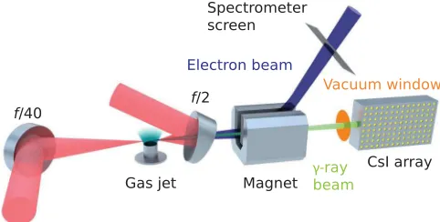
The experiment was conducted using the Gemini laser of the Central Laser Facility, Rutherford Appleton Laboratory, UK. Figure1shows a schematic of the experimental setup. Gemini is a Ti:sapphire laser system delivering two synchronized linearly polarized beams of 800 nm central wavelength and pulse durations of 45 fs FWHM. One of the beams, used to drive a laser wakefield accelerator, was
focused with an f=40 spherical mirror to a focal spot
FWHM size of 37×49μm. The energy delivered to the
target was ð8.60.6ÞJ generating a peak intensity of
ð7.70.4Þ×1018
W cm−2
, corresponding to a peak
nor-malized amplitude of a0¼1.90.1. This pulse was
focused at the leading edge of a 15-mm-diameter supersonic helium gas jet, which produced an approximately trapezoidal density profile with 1.5 mm linear ramps at the leading and trailing edges. Once ionized by the laser, the peak plasma electron densities used here wereð3.70.4Þ×1018
cm−3
. The second Gemini beam was focused at the rear edge of the gas jet, counterpropagating with respect to the first. As the laser-wakefield generated electron beam interacted with the second focused laser pulse, inverse
Compton-scattered γ rays were generated, copropagating with the
electron beam. By colliding close to the rear of the gas jet, the electron bunch did not have time to diverge before the collision and so the overlap between the electron bunch and laser was maximized.
The focusing optic for the second pulse was an off-axis
f=2parabolic mirror with a hole at the center to allow free
passage of thef=40beam, electron beam, and scatteredγ
rays. Accounting for the hole in the optic, the pulse energy on target wasð10.00.6ÞJ. This was focused to a focal
spot FWHM size of 2.4×2.8μm at a peak intensity of
ð1.30.1Þ×1021 W cm−2, corresponding to a peak
nor-malized amplitudea0¼24.70.7.
In order to align the two laser beams onto the same optical axis, a 90° prism with a micrometer-sharp edge was inserted into the beam line at the interaction point. After overlapping the focused pulses on the tip of the prism, half
of each was reflected collinearly onto a CCD [35] and
imaged with a 10× magnification microscope objective.
After reflection the different wave front curvatures of the
f=2andf=40beams caused circular interference fringes to
appear when the pulses overlapped in time. By optimizing the fringe visibility the two pulses were overlapped to a
precision of 30fs, limited by the random optical path
length fluctuations during the measurement.
After passing through the hole in the f=2 mirror, the
electron beam was deflected from the optical axis by a permanent dipole magnet with total magnetic length R
BðxÞdx¼0.4Tm. The electron energy spectrum was
recorded in the range of 0.25–2 GeV on a scintillating Gd2O2S∶Tb (Lanex) screen placed in the path of the
magnetically dispersed beam, and imaged with a cooled 16-bit CCD camera. An exemplary spectrometer image is shown in Fig.2(a), and the calculated electron spectrum in Fig.2(b).
The f=40 laser pulse was blocked at the rear of the
interaction chamber with a 50-μm-thick aluminium foil,
which along with a250-μm-thick Kapton vacuum window
was traversed by theγ-ray beam. Theγ-ray detector consisted of an array of5×5×50mm caesium iodide (CsI) crystals
doped with thallium, which convert deposited energy into optical photons at an efficiency of ≈5×10⁴
MeV−1
. The array was 33 crystals high and 47 crystals in the longitudinal direction, with theγ rays entering through the5×50mm
faces. The crystals were separated by 1-mm-thick aluminium spacers, and the face of the stack exposed to theγbeam was
covered with a 9-mm-thick stainless steel plate. By imaging the5×5mm faces of the CsI crystals from the side and
recording the scintillation light, it was possible to record a vertically resolved map of the energy deposition in the detector; see Fig.2(b). Low-energy photons deposit most of their energy in the first crystal column, with the energy deposited in subsequent crystals decreasing monotonically. High-energy photons create an electromagnetic shower which causes the energy deposition to initially increase with depth before decaying.
III. ELECTRON SPECTRA AND γ-RAY YIELD
The data analyzed here are a sequence of 18 shots where electron spectra andγ-ray signals were recorded simulta-neously. For the first 8 shots thef=2beam was on, then for
the next 10 shots it was switched off. Because of the shot-to-shot variations of the electron beam and laser pointing and timing, we do not expect every collision to be successful (based on the measured fluctuations we expect approximately 1 shot in 3 to occur at an a0 that is large enough to produce a measurable radiation reaction). Before we can proceed in assessing if radiation reaction was occurring in our experiment, we must therefore first identify which collisions were successful. This can be achieved by analyzing the γ-ray signal—successful
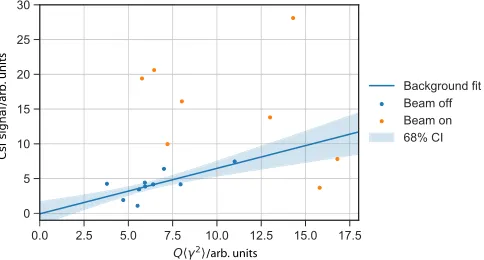
colli-sions will produce a much brighter signal than those which are not. In Fig.3we plot the integrated signal on the CsI detector as a function of the electron beam properties.
It is important to account for any background which could contaminate the CsI detector (such as bremsstrahlung emission), as this will increase with electron beam charge and energy in the same way as the inverse Compton signal. Specifically, for an electron of Lorentz factor γ the total energy of the emitted radiation scales asγ2. An electron
spectrumdNe=dγ will therefore create background signal
with an energy proportional toR γ2
ðdNe=dγÞdγ¼Qhγ2i,
whereQis the total beam charge. The energy radiated into the ICS beam, and therefore the integrated signal on the CsI
[image:4.612.53.292.45.216.2](a) (b) (c) (d)
FIG. 2. (a) Electron spectrometer screen image, transformed onto a linear energy axis. (b) Angularly integrated electron spectrum. (c) Raw image of CsI crystal stack detector. (d) Inte-grated CsI signal as a function of penetration depth into the stack.
[image:4.612.318.560.533.663.2]detector, is approximately proportional to a2
0Qhγ2i for
γa2
0<5.5×10
5
[4], and so the total signal is expected to be
CsI signal¼cBGQhγ2i þcICSa20Qhγ 2
i ð1Þ
for some constantscBG,cICS. In Fig.3a linear estimation ofcBG is performed using the“beam-off”shots, providing
an estimate (with error) of the background signal in the
“beam-on” shots.
The consecutive angularly integrated electron spectra
are plotted in Fig. 4. The spectra were almost always
observed to possess two components—a high-charge,
low-energy feature, and a low-charge, high-low-energy feature. It is possible that this is due to separate injection events caused by density structures in the plasma, observable on trans-verse plasma diagnostics and likely due to fluid shocks in the gas flow. This would imply that the high-energy component was generated by self-injection[36]and that the low-energy component was injected at an abrupt density transition[37]. In Fig.4the energy at which these features become distinct is highlighted, overlaid on the electron spectrum. We refer to this feature as an “edge” in the
spectrum.
The quantity determining the magnitude of the inter-action is, in the terms of Eq. (1), cICSa
2
0. This is a factor
representing the overlap of the electron beam and focal spot, and so the effectivea0of the interaction. The quantity of interest is then calculated from the measured signal by subtracting an estimated background, and dividing by
Qhγ2
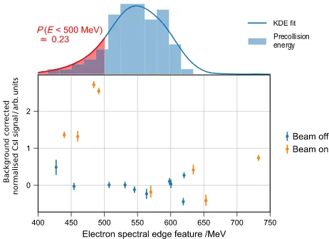
i. This is the quantity plotted in Fig.5as a function of the measured energy of the edge in the electron spectrum.
This corrected signal for the beam-off shots is clustered around zero, as expected, but also for some of the beam-on shots, as expected due to the spatiotemporal jitter between the laser focal spot and the electron beam. There are however four shots with exceptionally large signals, more than four standard deviations above the background level—shots 4, 5,
6, and 8. The chance of observing a signal this large in a sample of 8 shots due to background fluctuations alone is less than 0.1%. These shots are highlighted in red in Fig.4.
Using this signal threshold as an independent criterion for assessing which shots successfully resulted in a strong laser-electron beam interaction, we then consider the electron energies for these collisions. All four electron beam energies are below 500 MeV, while the mean of the
10 beam-off shots was measured to beð55020ÞMeV,
with a standard deviation of ð6314Þ MeV. Assuming
that the electron energies on the shots with thef=2beam
off are sampled from a distribution which is approximately normal, this implies that there would be a∼23%chance of
[image:5.612.53.560.47.177.2]observing an electron energy below 500 MeV on a single successful collision even if the interaction did not result in energy loss. However, the chance of observing an energy less than 500 MeV on all four successful collision shots falls to 0.3% under this null hypothesis. This is sufficiently unlikely that we conclude that the low energies observed on the four successful collision shots are due to the radiation reaction force on the electron bunch. This analysis assumes that the beam-off shots are sampled from a normal
FIG. 4. Consecutive electron spectra. Each spectrum has been normalized to its own maximum.
Precollision energy
[image:5.612.321.552.473.641.2]distribution. To assess this a larger set of 87 shots where no radiation reaction signal could occur was recorded under similar conditions. The distribution of energies from this larger data set is shown as a histogram in
Fig. 5. An Anderson-Darling test confirms that the 10
beam-off shots are consistent with being sampled from a normal distribution, at the 99.9% confidence level. Note also that, under the null hypothesis, the chance of observing four or more electron beams with energies below 500 MeV in a sample of 8 is much higher, approximately 10%, and so taken alone the electron beam energies would not have represented a clear observation. It is only by independently identifying which collisions were successful, based on the
observa-tion of a bright γ-ray signal, that the observed low
energies become statistically significant.
From the observed decrease in electron energy in the shots generating a bright CsI signal, we can estimate the laser intensity required to cause this energy shift under different radiation reaction models. Assuming that the“bright”shots
and beam-off shots are sampled from two different normal distributions with different mean values, we consider the
energy shift between these mean values from εinitial¼
ð55020ÞMeV toεfinal¼ ð47010ÞMeV. If the
elec-tron beam interacts with a pulse of FWHM duration 45 fs, the required peaka0to generate this energy loss is102
for a quantum model, and91for a classical model based
on the Landau-Lifshitz equation. Forεinitial≈550MeV and a0≈10, η≈0.07and in the quantum model the electron
loses energy at a rate≈0.75that of the classical model[38].
In the classical model a slightly lowera0is therefore able to generate the same electron energy loss, though due to the relatively low value ofηthe difference in a0between the models is small compared to the experimental uncertainties when considering the electron beam energy loss alone.
Additionally, the peak a0 of the f=2 focal spot was
calculated to be greater than 20 from measured pulse parameters, which is much larger than that inferred from the electron energy shift.
In practice the effectivea0of the interaction should be expected to be significantly lower than 20, due to the finite size of the electron beam and any timing offset of the collision point, since this will result in the interaction occurring a distance from the laser focus. Post experiment we identified a systematic offset caused by the delay between thef=40laser pulse and the wakefield accelerated
electron bunch. During the alignment of the experiment the two pulses were temporally overlapped at the rear edge of the gas jet, but this is not the same point as the collision
between the electron beam and the f=2 pulse. This is
because the wakefield electron bunch will be trailing the
f=40 pulse by approximately half a plasma wavelength,
and therefore the location of the collision between the electron bunch and thef=2beam will be offset from this
position by
δz¼3d
4 ne
nc
þλ0
4
ffiffiffiffiffi
nc
ne
r
; ð2Þ
wheredis the electron injection point (measured from the front of the gas jet),ne the electron density,nc the critical
density, andλ0the laser wavelength. Here it is assumed that
the laser travels in the plasma at the nonlinear group velocity[39] and that the electrons travel at c from their
injection point. Assuming a uniform distribution for d
between 0 and 10 mm and a normal distribution of30fs
for the timing jitter, the maximum expected interactiona0
at the collision (averaged over an area of10μm2
) is121,
where the laser transverse size is approximately 5μm
FWHM. This figure has been corrected for the measured change in size of the focal spot between the low-power alignment modes and the full-power shot mode of 7%. This is not the peaka0of the spot, but the maximuma0which encloses a contour of area10μm2
, an area of similar size to the electron beam plus shot-to-shot position fluctuations of the focal spot. The variation of a0 near focus under this criterion is slower than the variation of peaka0, and so shot-to-shot timing jitter has less of an impact on the effective interaction a0 than might be expected. It is very difficult to measure this effectivea0, and therefore problematic to distinguish between different radiation reaction models using only the shift in energy of a single feature in the electron spectrum. While we are confident that we have observed radiation reaction effects, it is not possible from our electron spectral measurements alone to investigate this process in more detail, due to the inherent uncertainty ina0. To help assess compatibility with different radiation reaction models, we therefore augment the electron beam measurements with spectral data from theγ-ray beam in the following section.
IV. γ-RAY SPECTRA
A. Measurements
We measure the γ-ray spectrum experimentally by
analyzing the scintillation yield, and thus energy deposited, in the CsI scintillator array. To understand the response of the detector, detailed Monte Carlo modeling of the array
was performed inGEANT4 [40]andMCNP[41]in full 3D,
where the simulation geometry included the large objects inside the vacuum chamber, the electron spectrometer magnets, the vacuum chamber itself, and all of the components of the CsI array. Forγ-ray energies between
2 and 500 MeV, 106
photons were propagated from the electron-laser interaction point into the array. The energy deposited in each crystal element was recorded and the scintillation light output was assumed proportional to the deposited energy, as is the case for high-energy photons
A more detailed description of theγ-ray spectrometer data analysis is presented in Ref.[43]. From simulations of the inverse Compton scattering process (see below), a good parametrized approximation toγ-ray spectrum over a wide photon energy range was observed to be
dNγ
dεγ ∝ε −2=3
γ e−εγ=εcrit; ð3Þ
whereεcritis a parameter controlling the spectral shape. For
this parametrization the mean photon energy isεcrit=3and
49% of the photon energy is radiated belowεcrit, soεcritis a
characteristic energy of the spectrum. In the experimental
measurements we treat εcrit as a free parameter and
minimize the mean-squared deviation between the simu-lated and measured detector light yield. Errors inεcritwere
assigned by forming simulated detector response curves and adding synthetic noise at similar levels to that observed in the experimental data, then averaging the retrieved εcrit
over 50 fits. In this way the1σfractional fit error was found
to be 15%.
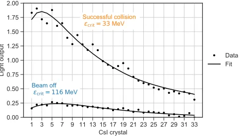
Exemplary fits to data from the γ-ray detector are
shown for a successful collision and a beam-off shot in Fig.6. When bright inverse Compton-scatteredγ rays are observed, the best-fit value ofεcritis in the range of several
tens of MeV. With the colliding beam switched off, the spectrum of the background signal is observed to be much harder, typically with εcrit >100MeV. This is consistent
with a background composed of primarily bremsstrahlung photons produced when the electron beam impacts the walls of the vacuum chamber.
B. Simulations
In order to calculate a theoretical γ-ray spectrum,
Monte Carlo simulations of the laser-electron collision were performed for differenta0and electron beam energies. In the simulations quantum and classical models of
radiation reaction are compared and contrasted against a control in which no radiation reaction is included.
In the quantum description photon emission is a series of discrete events, the locations of which are stochastically determined based on emission probabilities calculated in the locally constant field approximation[32]. This is valid fora30=
η≫1[13], as is the case here; its use is necessary
because the number of photons emitted per electron is much larger than one, making exact calculation of the spectrum from QED intractable at present. While the locally constant field approximation causes the low-energy
part of the photon spectrum (<100keV here) to be
overestimated, these photons do not contribute signifi-cantly to the recoil of the electron or to the total radiated energy[44].
Between individual emission events the electron follows a classical trajectory with motion determined by the Lorentz force. In the standard numerical implementation emission events are determined using Monte Carlo sam-pling as described in Refs.[38,45]. This approach has been used to study the production of photons and radiation reaction effects in the experimental configuration
consid-ered here [12,34,46]. Equivalently one could solve the
kinetic equations for the electron distribution function
[10,47,48]. The approach we use here is based on single particle dynamics in prescribed fields because collective effects are negligible in the considered parameter regime. Simulated spectra were obtained and cross-checked using a suite of codes includingEPOCH [49],SIMLA[50], and that
used in Ref.[51], which confirmed that collective effects were negligible.
Turning to the classical description, the electron trajec-tory is determined by integrating an equation of motion which includes both the Lorentz force and energy loss as described by the Landau-Lifshitz radiation reaction force. For simplicity we take only the dominant term from this force; as the next term is a factor2γ2
smaller, and across the parameter regime we consider1=ð2γ2
Þ≪1, this
approxi-mation is appropriate.
While there are other classical models of radiation reaction, we do not expect there to be any differences between them for the energy and intensity parameters
considered here [6,52]. The emitted γ-ray spectrum is
obtained by sampling the classical synchrotron distribution. We expect this model to overpredict the energy loss, as classical descriptions of radiation reaction unphysically fail to preclude the emission of photons with energy higher than that of the seed electron[11].
Finally, in the “no RR,” or control, model, photon
[image:7.612.55.296.47.184.2]emission is calculated as in the quantum case above (to ensure that photons cannot be emitted with an energy larger than the electron energy), but the recoil of the emitting particle is neglected. This control case will be used to show that neglecting radiation reaction is incompatible with the experimental results obtained, indicating that a regime
FIG. 6. CsI spectrometer measurements recorded for a suc-cessful collision and “beam-off” shots, and associated best-fit response functions from the Monte Carlo model of the detector. The maximum likelihood estimate of the fit parameter εcrit is
where radiation reaction is important has been reached (in contrast to previous experiments of this type[24,25]).
One could also consider a modified classical model[47], in which the energy loss is continuous but scaled by a
correction factor gðηÞ that emerges from the quantum
theory of photon emission in constant crossed fields. However, since the stochastic QED model described above and the modified classical model have been shown to give the same average behavior [53], and as our experimental data are effectively a measure of the mean energy loss, there would be no significant difference between the predictions of a modified classical model and the fully stochastic calculation we have used at the values ofηreached in this experiment. Our simulations indicate that signatures of stochastic emission will be become apparent atη≈0.25.
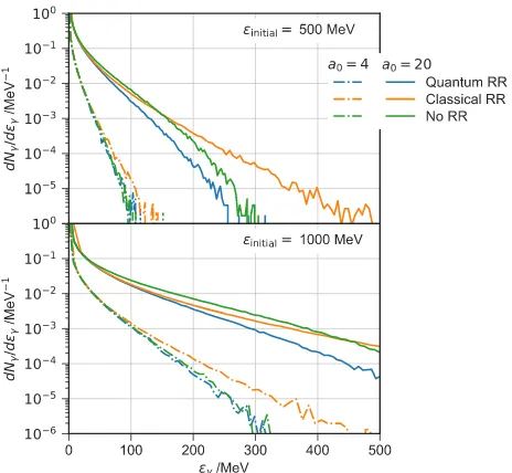
A sample of the simulated spectra are plotted in Fig.7, where in all simulations the scattering pulse was assumed to be a plane wave with FWHM duration 45 fs and wavelength 800 nm.
Because we collide the electron and laser beams very close to the exit of the gas jet, the electron beam is significantly smaller than the laser beam. Assuming that
the electron beam waist is 1μm (based on measurements
on Astra-Gemini and comparable to published results[54]) and that the waist is positioned close to the edge of the gas jet (which is expected due to the focusing forces acting on the beam while it is inside the plasma), we calculate that the electron beam doubles in area over a distance of approx-imately300μm. At the collision point the electron beam
area is therefore still approximately1μm2
, compared to a laser beam area of 20μm2
.
Because the collision occurs between an electron bunch with finite duration and a laser pulse that is going through
focus, different longitudinal slices of the electron bunch actually interact with different peak intensities. However, assuming a bunch length of≈λp=2≈10μm and using the
measured variation of the laser intensity with distance from
focus, we calculate that the difference in the peak a0
experienced by the front and back of the bunch is less than 10%. The critical energy of the radiation spectrum varies as
≈a0.50 , so the effect of this on both the overall energy loss
and radiation spectrum are small compared to the other fluctuations in the measurement.
The small transverse electron beam size and relatively slow longitudinal variation ofa0along the electron bunch mean that radiation reaction is well described by a plane wave model, where one can neglect the variation in laser intensity due to focusing across the electron beam[55].
To assess the discriminatory power of our experiment against the different models, we simulated the full photon generation, measurement, and fitting process for a range of peaka0. Theεcritwhich would be measured with a perfect
noise-free detector are plotted in Fig.8, where the results of the ICS simulations were interpolated over our measured off electron spectra for the RR models, and the beam-on electrbeam-on spectra for the no RR model. For the no RR model the retrievedεcrit varies almost linearly witha0, the
models including radiation reaction predict a lowerεcrit at
higha0 as expected.
V. MODEL COMPARISON
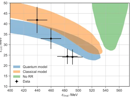
The measured εcrit is plotted as a function of the
measured postcollision energy of the electron beam εfinal
[image:8.612.59.291.46.260.2]in Fig.9for the four shots, where the error bars represent diagnostic uncertainties. The sign of the correlation is significant, in that if we were observing Compton scattering without radiation reaction, then the correlation should be in the positive direction. After taking into account the
FIG. 7. The spectrum of ICS photons radiated by a single electron as simulated for various εinitial and a0 for the three
radiation reaction models described in the text.
FIG. 8. Simulated retrievals ofεcritassuming the collision of a
plane wave of given peaka0 with the experimentally measured
[image:8.612.321.554.46.214.2]measurement uncertainties in the data, we find that there is a >98%probability that the correlation is negative, the
direction expected if radiation reaction is occurring in the successful collisions. Under the null hypothesis that no radiation reaction is occurring, the chance of observing a negative correlation at least this strong simultaneously with the electron energies all being below 500 MeV is lower than 1 in 3000.
An important source of variation here is the interaction
a0, which should be expected to vary significantly between laser shots due primarily to the spatial jitter between the electron and photon beams. If more data were available, one would therefore expect the points to trace out a curve in ðΔε=ε;εcritÞ space, parametrized along its length by a0.
Each radiation reaction model generates a different curve, and so matching the data to a curve is a method for finding the model most consistent with the experiment independ-ently of any knowledge ofa0for a particular datum.
Because of the shot-to-shot jitter these curves are
broadened into the shaded areas plotted in Fig. 9.
To calculate these areas a large number of “numerical
experiments” are conducted. For various values of the
laser strength a0 (uniformly distributed over the range
a0¼4–20), a set of 10 initial electron beam energiesεinitial
are drawn from the measured precollision distribution. From the assumed a0 a final energy after the interaction with the laser is calculated,εfinal, and a set ofεcritfor each
radiation reaction model as would be measured on the detector. The spread of of εcrit takes into account the
measured fluctuations of the electron beam spectral shape. We then calculate the averagesε¯finalandε¯critand place these
onto the (εfinal, εcrit) space. This process is repeated 500
times for each value of a0, equivalent to taking 50 000 successful collision shots. The shaded regions represent the
area in (εfinal, εcrit) space that contains 68% of these
numerical experiment results, i.e., what would be measured 68% of the time under different radiation reaction models if one could repeat the experiment many times.
As a0 tends to zero the γ-ray spectrum would become
monochromatic. Our γ-ray diagnostic would erroneously
measure a finite effective εcrit in this case, and for this
reason the curves in Fig.9do not tend towardsεcrit¼0at
lowa0.
We observe that the data are more consistent with a quantum rather than classical model of radiation reaction, though there is large overlap between models at lowa0, and several data points are consistent with both models. If the electron energy loss is ignored, it could be argued that the data are consistent with the no RR model if the interaction
a0 is lowered to ∼5. However, this situation is unlikely
given the experimental precision of the spatial and temporal alignment between the electron bunch and colliding laser pulse and the observed correlation between electron beam energy,γ-ray yield, andεcrit.
As was discussed for the electron spectral data, it is also possible to estimate the interactiona0independently using theγ-ray spectra by interpolating the measuredεcritonto the
curves in Fig.8. We perform this estimation for each data point, and calculate the ratio of the estimates from theγ-ray data and the electron beam data R¼a0ðεcritÞ=a0ðεfinalÞ.
This is another metric of the model consistency which is independent of any knowledge of the interactiona0. The data are considered fixed soR is a function of the model used to interpret the data, and perfect internal consistency impliesR¼1. Averaged over this data, at the 68% level for
the quantum modelR¼0.8þ0.7
−0.3and for the classical model R¼0.6þ0.3
−0.2. Under this metric the quantum radiation
reaction model is slightly better at bringing the data from both diagnostics into agreement, whereas the classical model appears to systematically underestimatea0 for the γ-ray data compared to the electron beam data.
VI. DISCUSSION
The main evidence for the observation of radiation reaction presented here is the observation of low-energy electron beams on all successful collision shots and the correlation between the postcollision electron beam energy
and the γ-ray yield and spectrum. This observation is
consistent with the measurement of hard photons, of characteristic energyεcrit >30MeV, which carried a
sig-nificant fraction of the initial electron energy, meaning that the electron recoil should be non-negligible. Moreover, this is reinforced by the agreement between the interactiona0
inferred separately from the electron andγ-ray spectra under a quantum radiation reaction model, and that expected experimentally.
[image:9.612.62.291.45.214.2]Simulations of the electron-laser overlap indicate that brightγ-ray beams withεcrit>20MeV would be expected FIG. 9. Experimentally measured εcrit as a function of εfinal
to be produced on 30% of shots. This is in line with our data when the measurements were taken immediately after align-ment (4 out of 8 shots), though subsequent spatial and temporal drifts mean that the chance of later shots showing significant γ signal drops quickly with time, limiting the duration of useful shot runs. In future experiments we plan to more carefully identify and control these drifts, which will aid in the acquisition of a significantly larger data set.
While the observed correlation is encouraging, the number of shots demonstrating significant overlap in the experiment was limited and there are aspects of the laser-particle interaction which could be further investigated by the acquisition of more data. In classical radiation reaction the width of the electron energy spectrum can only decrease, but in quantum radiation reaction it can increase or decrease depending on the strength and duration of the interaction[10,12,53]. This was not distinguishable here due to the value of η achieved at collision, the low number of shots, and the variability in the electron spectra. Direct evidence of the stochastic nature of the photon emission during radiation reaction could be achieved in a similar experimental setup by increasing the electron beam stability and energy as well as the laser intensity at collision (e.g., to ≈1.5GeV anda0≈15).
We have focused our attention on the energy loss of the dominant low-energy feature of the electron spectrum. The high-energy tail did not exhibit significant changes in energy, though removing it entirely from simulations of the inverse Compton scattering process generates photon beams with slightly lowerεcritindicating that it does in part
contribute to theγ-ray beam despite its low charge. Given that the two components of the electron spectra are so different, it is conceivable that each arises from a separate injection event inside the accelerator. In this case it is plausible that the spectral components are separated in space and time inside and subsequently outside the plasma. If so, it is possible that only a portion of the high-energy component experienced a significant interaction, which would diminish radiation reaction effects on that compo-nent of the beam.
VII. CONCLUSION
In summary, we have presented data from a recent laser-wakefield inverse Compton scattering experiment designed to identify the onset of radiation reaction. The electron and γ-ray spectra were simultaneously measured and independ-ently used to infer the laser intensity during the interaction. A fully quantum model of radiation reaction performs best in bringing the measurements from these two diagnostics
into alignment. Furthermore, we have generated γ-ray
beams with the highest energies yet reported from an all-optical inverse Compton scattering scheme, previously limited to below 20 MeV[24,25], and measurable here with a scintillation detector highly sensitive to the electromag-netic shower produced by high-energy photons.
While the results presented here represent statistically significant evidence of radiation reaction occurring during the collision of a intensity laser pulse with a high-energy electron beam, they are not a systematic study of radiation reaction. The QED model currently appears to provide a more self-consistent description of the data than a purely classical one; however, this is only at the1σlevel. A
future study systematically varying the quantum parameter ηthrough either the electron beam energy or laser intensity which extends to higher values ofη than achieved in this experiment will allow detailed comparisons of different models and a proper assessment of their range of validity. Laser-wakefield accelerators have already demonstrated sufficient beam stability, e.g., Refs.[35,56,57], and higher beam energies, e.g., Ref.[20], and higher laser intensities are readily achievable with existing lasers, so that the necessaryηto clearly observe quantum radiation reaction is within reach of existing laser systems. The main remaining challenge is therefore to achieve all of these simultaneously in an experiment which maintains femtosecond and micrometer level alignment over extended periods of time, so as to allow sufficient data to be collected for a systematic study of radiation reaction in the quantum regime.
The authors confirm that all data used in this study are available without restriction. Data can be obtained by contacting the Imperial College Plasma Physics Research Group[58].
ACKNOWLEDGMENTS
We acknowledge funding from EPSRC Grants No. EP/ M018555/1, No. EP/M018091/1, No. EP/M018156/1, No. EP/P010059/1, and No. EP/N027175/1, STFC
Grants No. ST/J002062/1 and No. ST/P000835/1,
Horizon 2020 funding under the Marie Skł
odowska-Curie Grant No. 701676 (A. I.) and the European Research Council (ERC) Grant Agreement No. 682399 (S. P. D. M.), the Knut & Alice Wallenberg Foundation (T. G. B., C. H., A. I., M. M.), the Swedish Research Council, Grants No. 2012-5644 and No. 2013-4248 (M. M.), the U.S. NSF CAREER Grant No. 1054164 (A. G. R. T., K. T. B.), and U.S. DOD under Grant No. W911NF-16-1-0044 (A. G. R. T., K. T. B., K. K.) and U.S. DOE under Grant No. DE-NA0002372. Simulations were performed on
resources provided by the Swedish National
Infrastructure for Computing at the HPC2N. We would like to thank the CLF for their assistance in running the experiment.
[2] C. H. Jaroschek and M. Hoshino, Radiation-Dominated Relativistic Current Sheets,Phys. Rev. Lett. 103, 075002 (2009).
[3] A. Di Piazza, C. Muller, K. Z. Hatsagortsyan, and C. H. Keitel, Extremely High-Intensity Laser Interactions with Fundamental Quantum Systems,Rev. Mod. Phys.84, 1177 (2012).
[4] A. G. R. Thomas, C. P. Ridgers, S. S. Bulanov, B. J. Griffin, and S. P. D. Mangles,Strong Radiation-Damping Effects in a Gamma-Ray Source Generated by the Interaction of a High-Intensity Laser with a Wakefield-Accelerated Electron Beam,Phys. Rev. X2, 041004 (2012).
[5] R. T. Hammond, Radiation Reaction at Ultrahigh Inten-sities,Phys. Rev. A81, 062104 (2010).
[6] D. A. Burton and A. Noble, Aspects of Electromagnetic Radiation Reaction in Strong Fields, Contemp. Phys. 55, 110 (2014).
[7] L. D. Landau and E. M. Lifshitz, The Classical Theory of Fields(Elsevier, Oxford, 1975).
[8] V. S. Krivitski and V. N. Tsytovich, Average Radiation-Reaction Force in Quantum Electrodynamics, Sov. Phys. Usp.34, 250 (1991).
[9] A. Ilderton and G. Torgrimsson, Radiation Reaction from QED: Lightfront Perturbation Theory in a Plane Wave Background,Phys. Rev. D88, 025021 (2013).
[10] N. Neitz and A. Di Piazza,Stochasticity Effects in Quantum Radiation Reaction,Phys. Rev. Lett.111, 054802 (2013). [11] S. R. Yoffe, Y. Kravets, A. Noble, and D. A. Jaroszynski,
Longitudinal and Transverse Cooling of Relativistic Elec-tron Beams in Intense Laser Pulses, New J. Phys. 17, 053025 (2015).
[12] M. Vranic, T. Grismayer, R. A. Fonseca, and L. O. Silva,
Quantum Radiation Reaction in Head-On Laser-Electron Beam Interaction,New J. Phys.18, 073035 (2016). [13] V. Dinu, C. Harvey, A. Ilderton, M. Marklund, and G.
Torgrimsson,Quantum Radiation Reaction: From Interfer-ence to IncoherInterfer-ence,Phys. Rev. Lett.116, 044801 (2016). [14] S. P. D. Mangles, C. D. Murphy, Z. Najmudin, A. G. R. Thomas, J. L. Collier, A. E. Dangor, E. J. Divall, P. S. Foster, J. G. Gallacher, C. J. Hookeret al.,Monoenergetic Beams of Relativistic Electrons from Intense Laser-Plasma Inter-actions,Nature (London)431, 535 (2004).
[15] C. G. R. Geddes, C. S. Toth, J. Van Tilborg, E. Esarey, C. B. Schroeder, D. Bruhwiler, C. Nieter, J. Cary, and W. P. Leemans, High-Quality Electron Beams from a Laser Wakefield Accelerator Using Plasma-Channel Guiding, Nature (London)431, 538 (2004).
[16] J. Faure, Y. Glinec, A. Pukhov, S. Kiselev, S. Gordienko, E. Lefebvre, J.-P. Rousseau, F. Burgy, and V. Malka, A Laser-Plasma Accelerator Producing Monoenergetic Elec-tron Beams,Nature (London)431, 541 (2004).
[17] E. Esarey, C. B. Schroeder, and W. P. Leemans,Physics of Laser-Driven Plasma-Based Electron Accelerators, Rev. Mod. Phys.81, 1229 (2009).
[18] W. P. Leemans, B. Nagler, A. J. Gonsalves, C. Toth, K. Nakamura, C. G. R. Geddes, E. Esarey, C. B. Schroeder, and S. M. Hooker, GeV Electron Beams from a Centimetre-Scale Accelerator,Nat. Phys.2, 696 (2006).
[19] S. Kneip, S. R. Nagel, S. F. Martins, S. P. D. Mangles, C. Bellei, O. Chekhlov, R. J. Clarke, N. Delerue, E. J. Divall,
G. Doucaset al.,Near-GeV Acceleration of Electrons by a Nonlinear Plasma Wave Driven by a Self-Guided Laser Pulse,Phys. Rev. Lett.103, 035002 (2009).
[20] W. P. Leemans, A. J. Gonsalves, H.-S. Mao, K. Nakamura, C. Benedetti, C. B. Schroeder, Cs. Tóth, J. Daniels, D. E. Mittelberger, and S. S. Bulanov,Multi-GeV Electron Beams from Capillary-Discharge-Guided Subpetawatt Laser Pulses in the Self-Trapping Regime,Phys. Rev. Lett.113, 245002 (2014).
[21] X. Wang, R. Zgadzaj, N. Fazel, Z. Li, S. A. Yi, X. Zhang, W. Henderson, Y. Y. Chang, R. Korzekwa, H.-E. Tsai,et al.,
Quasi-Monoenergetic Laser-Plasma Acceleration of Elec-trons to 2 GeV,Nat. Commun. 4, 1988 (2013).
[22] K. Ta Phuoc, S. Corde, C. Thaury, V. Malka, A. Tafzi, J. P. Goddet, R. C. Shah, S. Sebban, and A. Rousse,All-Optical Compton Gamma-Ray Source, Nat. Photonics 6, 308 (2012).
[23] S. Chen, N. D. Powers, I. Ghebregziabher, C. M. Maharjan, C. Liu, G. Golovin, S. Banerjee, J. Zhang, N. Cunningham, A. Moortiet al.,MeV-Energy X Rays from Inverse Compton Scattering with Laser-Wakefield Accelerated Electrons, Phys. Rev. Lett.110, 155003 (2013).
[24] G. Sarri, D. J. Corvan, W. Schumaker, J. M. Cole, A. Di Piazza, H. Ahmed, C. Harvey, C. H. Keitel, K. Krushelnick, S. P. D. Mangles, Z. Najmudinet al.,Ultrahigh Brilliance Multi-MeV γ-Ray Beams from Nonlinear Relativistic Thomson Scattering, Phys. Rev. Lett. 113, 224801 (2014).
[25] W. Yan, C. Fruhling, G. Golovin, D. Haden, J. Luo, P. Zhang, B. Zhao, J. Zhang, C. Liu, M. Chen et al., High-Order Multiphoton Thomson Scattering,Nat. Photonics11, 514 (2017).
[26] N. D. Powers, I. Ghebregziabher, G. Golovin, C. Liu, S. Chen, S. Banerjee, J. Zhang, and D. P. Umstadter, Quasi-Monoenergetic and Tunable X-Rays from a Laser-Driven Compton Light Source,Nat. Photonics8, 28 (2014). [27] H.-E. Tsai, X. Wang, J. Shaw, A. V. Arefiev, Z. Li, X.
Zhang, R. Zgadzaj, W. Henderson, V. Khudik, G. Shvets, and M. C. Downer, Compact Tunable Compton X-Ray Source from Laser Wakefield Accelerator and Plasma Mirror,Phys. Plasmas 22, 023106 (2015).
[28] Y. Sakai, I. Pogorelsky, O. Williams, F. O’Shea, S. Barber, I. Gadjev, J. Duris, P. Musumeci, M. Fedurin, A. Korostyshevsky
et al.,Observation of Redshifting and Harmonic Radiation in Inverse Compton Scattering, Phys. Rev. ST Accel.
Beams 18, 060702 (2015).
[29] K. Khrennikov, J. Wenz, A. Buck, J. Xu, M. Heigoldt, L. Veisz, and S. Karsch,Tunable All-Optical Quasimonochro-matic Thomson X-Ray Source in the Nonlinear Regime, Phys. Rev. Lett.114, 195003 (2015).
[30] C. Bula, K. McDonald, E. Prebys, C. Bamber, S. Boege, T. Kotseroglou, A. Melissinos, D. Meyerhofer, W. Ragg, D. Burkeet al.,Observation of Nonlinear Effects in Compton Scattering,Phys. Rev. Lett.76, 3116 (1996).
[31] E. Esarey, S. K. Ride, and P. Sprangle,Nonlinear Thomson Scattering of Intense Laser Pulses from Beams and Plas-mas,Phys. Rev. E 48, 3003 (1993).
[33] E. N. Nerush and I. Y. Kostyukov, Kinetic Modelling of Quantum Effects in Laser-Beam Interaction,Nucl. Instrum. Methods Phys. Res., Sect. A653, 7 (2011).
[34] T. G. Blackburn, C. P. Ridgers, J. G. Kirk, and A. R. Bell,
Quantum Radiation Reaction in Laser-Electron-Beam Col-lisions,Phys. Rev. Lett.112, 015001 (2014).
[35] J. Faure, C. Rechatin, A. Norlin, A. Lifschitz, Y. Glinec, and V. Malka, Controlled Injection and Acceleration of Elec-trons in Plasma Wakefields by Colliding Laser Pulses, Nature (London)444, 737 (2006).
[36] S. V. Bulanov, F. Pegoraro, A. M. Pukhov, and A. S. Sakharov, Transverse-Wake Wave Breaking, Phys. Rev. Lett.78, 4205 (1997).
[37] K. Schmid, A. Buck, C. M. S. Sears, J. M. Mikhailova, R. Tautz, D. Herrmann, M. Geissler, F. Krausz, and L. Veisz,
Density-Transition Based Electron Injector for Laser Driven Wakefield Accelerators,Phys. Rev. ST Accel. Beams
13, 091301 (2010).
[38] C. P. Ridgers, J. G. Kirk, R. Duclous, T. G. Blackburn, C. S. Brady, K. Bennett, T. D. Arber, and A. R. Bell,Modelling Gamma-Ray Photon Emission and Pair Production in High-Intensity Laser-Matter Interactions,J. Comput. Phys.
260
, 273 )
2014 ).
[39] C. D. Decker, W. B. Mori, K.-C. Tzeng, and T. Katsouleas,
The Evolution of Ultra-Intense, Short-Pulse Lasers in Underdense Plasmas,Phys. Plasmas3, 2047 (1996). [40] S. Agostinelli, J. Allison, K. Amako, J. Apostolakis, H.
Araujo, P. Arce, M. Asai, D. Axen, S. Banerjee, G. Barrand
et al., GEANT4—A Simulation Toolkit, Nucl. Instrum. Methods Phys. Res., Sect. A506, 250 (2003).
[41] T. Goorley, M. James, T. Booth, F. Brown, J. Bull, L. J. Cox, J. Durkee, J. Elson, M. Fensin, R. A. Forsteret al.,Initial MCNP6 Release Overview,Nucl. Technol.180, 298 (2012). [42] E. Frlež, I. Supek, K. A. Assamagan, C. Brönnimann, T. Flügel, B. Krause, D. W. Lawrence, D. Mzavia, D. Počanić, D. Renkeret al.,Cosmic Muon Tomography of Pure Cesium Iodide Calorimeter Crystals,Nucl. Instrum. Methods Phys. Res., Sect. A440, 57 (2000).
[43] K. T. Behm, J. M. Cole, J. C. Wood, E. Gerstmayr, K. Poder, S. P. D. Mangles, Z. Najmudin, A. G. R. Thomas, K. Krushelnick, C. D. Murphyet al.,“Novel Design of a
Gamma Ray Spectrometer Measuring Spectra with Photon Energies Greater than 100 MeV”(unpublished).
[44] C. N. Harvey, A. Ilderton, and B. King,Testing Numerical Implementations of Strong-Field Electrodynamics, Phys. Rev. A91, 013822 (2015).
[45] A. Gonoskov, S. Bastrakov, E. Efimenko, A. Ilderton, M. Marklund, I. Meyerov, A. Muraviev, A. Sergeev, I. Surmin, and E. Wallin,Extended Particle-in-Cell Schemes for Physics in Ultrastrong Laser Fields: Review and Developments,Phys. Rev. E92, 023305 (2015).
[46] S. S. Bulanov, C. B. Schroeder, E. Esarey, and W. P. Leemans, Electromagnetic Cascade in High-Energy Electron, Positron, and Photon Interactions with Intense Laser Pulses,Phys. Rev. A87, 062110 (2013).
[47] I. V. Sokolov, N. M. Naumova, J. A. Nees, and G. A. Mourou,Pair Creation in QED-Strong Pulsed Laser Fields Interacting with Electron Beams, Phys. Rev. Lett. 105, 195005 (2010).
[48] N. V. Elkina, A. M. Fedotov, I. Yu Kostyukov, M. V. Legkov, N. B. Narozhny, E. N. Nerush, and H. Ruhl,
QED Cascades Induced by Circularly Polarized Laser Fields, Phys. Rev. ST Accel. Beams 14, 054401 (2011).
[49] T. D. Arber, K. Bennett, C. S. Brady, M. G. Ramsay, N. J. Sircombe, P. Gillies, R. G. Evans, H. Schmitz, A. R. Bell, and C. P. Ridgers,Contemporary Particle-in-Cell Approach to Laser-Plasma Modelling,Plasma Phys. Controlled Fu-sion57, 113001 (2015).
[50] D. G. Green and C. N. Harvey,SIMLA: Simulating Particle Dynamics in Intense Laser and Other Electromagnetic Fields via Classical and Quantum Electrodynamics, Com-put. Phys. Commun.192, 313 (2015).
[51] T. G. Blackburn,Measuring Quantum Radiation Reaction in Laser-Electron-Beam Collisions, Plasma Phys. Con-trolled Fusion57, 075012 (2015).
[52] M. Vranic, J. L. Martins, R. A. Fonseca, and L. O. Silva,
Classical Radiation Reaction in Particle-in-Cell Simula-tions,Comput. Phys. Commun.204, 141 (2016).
[53] C. P. Ridgers, T. G. Blackburn, D. Del Sorbo, L. E. Bradley, C. Slade-Lowther, C. D. Baird, S. P. D. Mangles, P. McKenna, M. Marklund, C. D. Murphy, and A. G. R. Thomas, Signatures of Quantum Effects on Radiation Reaction in Laser-Electron-Beam Collisions, J. Plasma Phys.83, 715830502 (2017).
[54] M. Schnell, A. Savert, B. Landgraf, M. Reuter, M. Nicolai, O. Jackel, C. Peth, T. Thiele, O. Jansen, A. Pukhovet al.,
Deducing the Electron-Beam Diameter in a Laser-Plasma Accelerator Using X-Ray Betatron Radiation,Phys. Rev. Lett.108, 075001 (2012).
[55] C. Harvey, M. Marklund, and A. R. Holkundkar,Focusing Effects in Laser-Electron Thomson Scattering,Phys. Rev. Accel. Beams19, 094701 (2016).
[56] A. Buck, J. Wenz, J. Xu, K. Khrennikov, K. Schmid, M. Heigoldt, J. M. Mikhailova, M. Geissler, B. Shen, F. Krausz
et al.,Shock-Front Injector for High-Quality Laser-Plasma Acceleration,Phys. Rev. Lett.110, 185006 (2013). [57] A. J. Gonsalves, K. Nakamura, C. Lin, D. Panasenko,
S. Shiraishi, T. Sokollik, C. Benedetti, C. B. Schroeder, C. G. R. Geddes, J. van Tilborget al.,Tunable Laser Plasma Accelerator Based on Longitudinal Density Tailoring,Nat. Phys.7, 862 (2011).