This is a repository copy of Lactose-crosslinked fish gelatin-based porous scaffolds embedded with tetrahydrocurcumin for cartilage regeneration.
White Rose Research Online URL for this paper: http://eprints.whiterose.ac.uk/134384/
Version: Accepted Version
Article:
Etxabide, A., Ribeiro, R.D.C., Guerrero, P. et al. (5 more authors) (2018) Lactose-crosslinked fish gelatin-based porous scaffolds embedded with
tetrahydrocurcumin for cartilage regeneration. International Journal of Biological Macromolecules, 117. pp. 199-208. ISSN 0141-8130
https://doi.org/10.1016/j.ijbiomac.2018.05.154
Article available under the terms of the CC-BY-NC-ND licence (https://creativecommons.org/licenses/by-nc-nd/4.0/).
Reuse
This article is distributed under the terms of the Creative Commons Attribution-NonCommercial-NoDerivs (CC BY-NC-ND) licence. This licence only allows you to download this work and share it with others as long as you credit the authors, but you can’t change the article in any way or use it commercially. More
information and the full terms of the licence here: https://creativecommons.org/licenses/
Takedown
If you consider content in White Rose Research Online to be in breach of UK law, please notify us by
Lactose-crosslinked fish gelatin-based porous scaffolds embedded with 1
tetrahydrocurcumin for cartilage regeneration 2
A. Etxabidea,R.D.C. Ribeirob, P. Guerreroa, A.M. Ferreirab, G.P. Staffordc, K. Dalgarnob, 3
K. de la Cabaa, P. Gentileb* 4
a BIOMAT Research Group, University of the Basque Country (UPV/EHU), Escuela de 5
Ingenier’a de Gipuzkoa, Plaza de Europa 1, 20018 Donostia-San Sebasti‡n, Spain. 6
b School of Engineering, Newcastle University, Claremont Road, Newcastle Upon Tyne, NE1 7
7RU, United Kingdom. 8
c
School of Clinical Dentistry, University of Sheffield, 19 Claremont Crescent, Sheffield S10 2TA, 9
United Kingdom. 10
*Corresponding author email: piergiorgio.gentile@ncl.ac.uk 11
Telephone number: +44 0191 208 3620 12
ABSTRACT 13
Tetrahydrocurcumin (THC) is one of the major colourless metabolites of curcumin and 14
shows even greater pharmacological and physiological benefits. The aim of this work 15
was the manufacturing of porous scaffolds as a carrier of THC under physiological 16
conditions. Fish-derived gelatin scaffolds were prepared by freeze-drying by two 17
solutions concentrations (2.5% and 4% w/v), cross-linked via addition of lactose and 18
heat-treated at 105 ¡C. This cross-linking reaction resulted in more water resistant 19
scaffolds with a water uptake capacity higher than 800%. Along with the cross-linking 20
reaction, the gelatin concentration affected the scaffold morphology, as observed by 21
changes led to a scaffoldÕs strength enhancement from 0.92±0.22MPa to 2.04±0.18MPa 24
when gelatin concentration was increased. THC release slowed down when gelatin 25
concentration increased from 2.5 to 4%w/v, showing a controlled profile within 96 h. 26
Preliminary in vitro test with chondrocytes on scaffolds with 4%w/v gelatin offered higher 27
metabolic activities and cell survival up to 14 days of incubation. Finally the addition of 28
THC did not influence significantly the cytocompatibility and potential antibacterial 29
properties were demonstrated successfully against Staphylococcusaureus. 30
KEYWORDS: Tetrahydrocurcumin; gelatin; lactose; scaffolds; antibacterial. 31
1. Introduction 32
Porous scaffolds are crucial for many biomedical and biological applications [1, 2]. In 33
tissue engineering, compared with synthetic materials, natural polymers have been 34
shown to favourably regulate division, adhesion, differentiation and migration of cells [3]. 35
Gelatin was chosen as a base material because it is biocompatible, biodegradable, non-36
carcinogenic, less antigenic than collagen, and commercially available at relatively low 37
cost [4, 5]. Moreover, it is recognised as safe material by the Food and Drug 38
Administration. Therefore, porous gelatin scaffolds have found many applications in 39
tissue engineering research, e.g. for bone, skin, cartilage and nerve regeneration [6-10]. 40
In particular, tissue engineering of articular cartilage in vitro is a promising strategy for 41
cartilage repair, in order to tackle the difficulties related with the self-repair of articular 42
cartilage due to its avascular tissue nature, low rate of chondrocyte proliferation and 43
matrix turnover [11]. Porous scaffolds are used to support cell adhesion and 44
cartilage often has thickness limitation, heterogeneous cartilage extracellular matrix 46
(ECM) and weak mechanical property which limit their clinical application [12, 13]. 47
In order to preserve structure and provide mechanical support to cells during tissue 48
formation [14], a cross-linking of gelatin is required. Chemical cross-linking methods 49
typically use chemicals such as aldehydes, aspartic and glutamic acids [15]. 50
Additionally, physical cross-linking methods, such as heating, drying, and irradiation, are 51
also commonly applied to proteins [16, 17]. In particular, in this work we considered the 52
effect of heating in the presence of sugars on modifying the conformation and 53
interactions within proteins, leading to a complex cross-linking process known as 54
Maillard reaction or non-enzymatic glycation. The main variables affecting the extension 55
of the Maillard reaction are temperature, time, initial pH, carbonyl/sugar ratio, and water 56
activity [18-20]. So, these factors should be analysed in order to obtain the properties 57
required for biomedical applications. 58
Additionally, gelatin-based scaffolds can be used as a vehicle for the release of 59
bioactive agents such as antioxidants, peptides, growth factors, antimicrobials and 60
drugs [21-24]. In particular, infection is a major problem in orthopaedics leading to 61
implant failure. Sources of infectious bacteria include the environment of the operating 62
room, surgical equipment, clothing worn by medical and paramedical staff, resident 63
bacteria on the patient's skin and bacteria already residing in the patientÕs body [25]. 64
Implant-associated infections are the result of bacteria adhesion to an implant surface 65
and subsequent biofilm formation at the implantation site [26]. In this work, we decided 66
to evaluate the antibacterial properties of the crosslinked gelatin scaffolds with the 67
antioxidant, antidiabetic, anticancer and anti-inflammatory plant-derived compound, that 69
is increasingly being used for pharmaceutical, medical and food applications [27, 28]. 70
Literature reports only studies on the delivery of the hydrophobic compound curcumin to 71
facilitate wound healing with the addition of ethanol in chitosan-alginate sponges [29] or 72
cyclodextrins in slightly hydrated alginate foams in order to improve the distribution of 73
curcumin [30]. 74
Therefore, beyond the state of the art, this is the first work where gelatin-based 75
scaffolds, prepared by freeze-drying, were cross-linked by using lactose. Lactose is a 76
disaccharide occurring almost exclusively in the milk of mammals with important 77
nutritional and probiotic properties and is mainly used in various food, nutrition and 78
pharmaceutical formulations [31]. In this study, lactose was used as a cross-linker in 79
order to promote the Maillard reaction, between the carbonyl group of lactose and the 80
gelatin amino group, and to improve the mechanical stability of the gelatin scaffolds. 81
Furthermore, THC was added to the initial gelatin solution to analyse the release of the 82
bioactive compound from the gelatin scaffold and its antibacterial effect in the presence 83
of the common nosocomial and joint-replacement/ wound infecting organisms 84
Pseudomonas aeuroginosa and Staphylococcus aureus. 85
This work aimed at studying the effect of initial gelatin concentration, cross-linking 86
reaction and THC addition on the physico-chemical, mechanical and biological 87
characteristics of porous gelatin scaffolds as potential medical device in 88
2. Materials and methods 90
2.1 Materials 91
A commercial cod fish gelatin type A was employed in this study. It has bloom 200, 92
11.06% moisture and 0.147% ash. Fish gelatin was kindly supplied by Weishardt 93
International (Liptovsky Mikulas, Slovakia) and meets the quality standard for edible 94
gelatin (1999/724/CE). Glycerol and lactose (Panreac, Barcelona, Spain) were used as 95
plasticizer and cross-linking agent, respectively. Tetrahydrocurcumin was gifted by 96
Sabinsa Corporation (East Windsor, New Jersey, USA) and was used as bioactive 97
agent. 98
2.2 Scaffold preparation 99
2.5 and 4% w/v fish gelatin porous scaffolds were prepared by freeze-drying. Firstly, 100
2.5 g or 4 g gelatin and 20 wt% lactose (on gelatin dry basis) were dissolved in 100 mL 101
distilled water for 30 min at 80 ¡C under continuous stirring to obtain a homogenous 102
blend. After that, a mixture of 10 wt% glycerol and 5 wt% THC (on gelatin dry basis) 103
was added to the solution, pH was adjusted to 10 with 1 N NaOH and the solution was 104
placed in an ultrasonic device (USC300TH, VWR International, USA) at room 105
temperature for 5 min. Subsequently, the solution was maintained at 80 ¼C for 30 min 106
under stirring, 2 mL solution was poured into each well of a 24 multiwell plate (Costar 107
3526, Corning Incorporated, USA) and the plate was kept in the fridge at 4 ¼C to cool 108
down. Once the solution was gelled, the plate was kept in a freezer at -20 ¼C for 48 h 109
and then, freeze-dried for 48 h (Alpha 1-2 LDplus, CHRIST, Germany). Finally, non-110
heated (NH) scaffolds were taken out from the wells and 1 day later some of them were 111
order to promote the Maillard cross-linking reaction [32-34]. As verified in a previous 113
work [27], these conditions do not affect the thermal stability of gelatin and THC. The 114
samples containing THC and heat-treated were coded as HT-THC. 115
2.3 Attenuated total reflection-Fourier transform infrared (ATR-FTIR) spectroscopy 116
FTIR spectra were obtained using a Spectrum Two PE instrument equipped with a 117
horizontal attenuated total reflectance (ATR) crystal (ZnSe) (PerkinElmer Inc., USA). 118
The samples were placed directly onto the ATR crystal and spectra were collected in 119
transmittance mode. Each spectrum was the result of the average of 32 scans at 4 cm−1 120
resolution. Measurements were recorded in the wavelength range of 1800-800 cm−1. All 121
spectra were smoothed using the Savitzky-Golay function. Second-derivative spectra of 122
the amide region were used at bands position guides for the curve fitting procedure, 123
using OriginPro 9.1 software. 124
2.4 Compression test 125
Mechanical tests were performed using a mechanical testing machine (EZ-SX, 126
Shimadzu, Japan) equipped with a 500 N load cell, as described in a previous work 127
[35]. Test specimens were cylinder-shaped scaffolds with a 1.3 cm diameter and an 128
average height of around 1 cm. The crosshead speed was set at 1 mm·min−1, and the 129
load was applied until the specimen was compressed to around 80% of its original 130
height before break. Compression resistance of five dried samples for each composition 131
was evaluated at room temperature and the stress was calculated by dividing the 132
applied force with the initial scaffold surface area, whereas strain was calculated from 133
the displacement of the scaffolds in relation to the original thickness. YoungÕs modulus 134
2.5 Scanning electron microscopy (SEM) 136
SEM (Hitachi TM3030 Tabletop, Germany) equipped with Energy Dispersive 137
Spectroscopy (EDS) was utilised to study the scaffold inner morphology. The samples 138
were cut into small squares, fixed on the aluminium stub using carbon tape. For pore 139
size evaluation SEM images were analysed by ImageJ software. 140
2.6 Water uptake (WU) measurements 141
WU was calculated gravimetrically according to ASTM D570-98 [36] under 142
physiological conditions. Three specimens of each composition were weighed (W0) and 143
immersed in 6 mL of a phosphate buffered saline (PBS) solution at pH 7.0 in order to 144
determine the WU profile at 37¡C in an incubator (INCU-Line, VWR). Then, the samples 145
were removed from the buffer solution at fixed times, wiped with a paper and reweighed 146
(Wt). WU was calculated using the following equation [37]: 147
WU! ! !!!!!!
!! !100 Eq.1
148
A graph depicting WU against time was plotted in order to determine the equilibrium 149
swelling. 150
2.7 Degradation test 151
The degradation degree (DD) was calculated gravimetrically under physiological 152
conditions. Three specimens of each composition were weighed (WI) and immersed in 6 153
mL of a PBS solution at pH 7.0 in order to determine the degradation at 37 ¡C in an 154
incubator. Samples were removed from the buffer solution once swelling test ended, 155
wiped with a paper, left to dry at room temperature for 24 h and reweighed (WF). DD 156
2.8 THC release 159
UV-Vis spectroscopy is one of the methods to determine the release and 160
concentration of bioactives in buffer solutions. Firstly, the wavelength of maximum 161
absorbance for THC in PBS was measured (ʎmax 280 nm) and then, standard solutions 162
of THC were prepared over a concentration range (0.31250-0.00977 mg/mL) to 163
establish a calibration curve (y=0.0314 + 2.2012x, R2=0.9985). 164
THC release was determined by immersion of a quarter of a scaffold in 6 mL of a PBS 165
solution (pH 7.0) at 37 ¡C in an incubator. At particular time intervals (1, 2, 4, 8, 24, 28 166
and 96 h), aliquots of buffer (3 mL) were withdrawn, replaced with fresh buffer and 167
analysed by UVÐVis spectroscopy (Lambda 2S Perkin Elmer) at 280 nm. All tests were 168
carried out in triplicate and the results were expressed as % of released THC with 169
respect to the THC incorporated in the scaffold by employing the calibration curve. 170
2.9 Biological characterisation 171
2.9.1 Cell culture 172
Chondrocytes cells were differentiated from Human Bone Marrow Stromal Cells (Y201) 173
and cultured according the protocol described by Genever et al.[38] at 37 ¡C, 5% CO2, 174
in Chondrocyte Growth Medium ready-to-use (PromoCell, UK). 175
In order to perform biocompatibility assays, the scaffolds were prepared according the 176
same procedure explained in 2.2 section with few modifications. Once gelatin solution 177
was prepared, 1 mL was poured into each well of a 48 multiwell plate in order to get 178
smaller scaffolds, which were subsequently cut in samples of 8 mm diameter and an 179
average height of around 2.5 mm. Each sample was put into a membrane-based cell 180
insertion was carried out in a class 2 laminar flow hood and each insert was placed in a 182
well of a 24 multiwell plate. Afterwards, the plate was placed below the UV light for 30 183
min in order to keep it sterile. A suspension of 20x104 cells in Growth medium was 184
seeded dropwise (in 500 µL) on the top surface of the scaffolds and incubated at 37 ¡C, 185
5% CO2 for 30 min. Then, fresh medium was added up to1 mL volume. 186
2.9.2 Cytocompatibility study 187
The Presto Blue assay was exploited to test the metabolic activity of cells seeded on 188
the scaffolds after 1, 3, 7 and 14 days of culture. A LS-50B Luminescence Spectrometer 189
(Perkin Elmer, Waltham, MA) was used to measure the fluorescence (560 nm excitation 190
and 590 nm emission) after 5 h of incubation with a 10% aliquot of Presto Blue (Thermo 191
Scientific, USA). The obtained values were corrected subtracting the average 192
fluorescence of control wells. Results were expressed as mean ± standard deviation. 193
2.9.3 Cell fixation and probe staining for confocal microscopy 194
Three and seven days after seeding cells on the scaffolds, cells were fixed using 4% 195
paraformaldehyde (Sigma Life Science) for 15 min at room temperature. Cells were 196
washed three times using 0.1% DPBS/Tween 20 (Sigma Life Science), followed by a 197
20-min light-protected incubation period at room temperature with phalloidin (1 mg/mL, 198
Sigma Life Science). After three new washes with 0.1% DPBS/Tween 20, 4′ ,6-199
diamidino-2-phenylindole (DAPI; 1:2500 solution, Vector Laboratories) was added, and 200
the solution was subjected to a 10-min light-protected incubation period at room 201
temperature. Afterwards, cells were washed and resuspended in 500 µL of 0.1% 202
visualised using a Leica TCS SP2 UV AOBS MP (Upright) point scanning confocal 204
microscope (Leica Microsystems) at 20× magnification. 205
2.10 Antibacterial tests 206
Antimicrobial activity of the scaffolds was tested against Staphylococcus aureus (S. 207
aureus) NCTC 8325 and a clinical strain of Pseudomonas aeruginosa (P. aeruginosa) 208
(SOM-1, Stafford group culture collection). Fresh 4 h cultures of S. aureus strain 8325 209
and a clinical P. aeruginosa isolate (Sheffield culture collection) were grown in Brain 210
Heart Infusion broth (Oxoid) at 37¡C (OD600 0.4-0.6) and spread onto Columbia 211
nutrient agar plates. After drying for 10 min, 8 mm HT gelatin scaffolds of both 212
concentration with or without THC were placed on the surface of the agar plates, which 213
were then incubated for 24 h at 37 ¡C before being photographed. 214
2.11 Statistical analysis 215
Data were subjected to one-way analysis of variant (ANOVA) by means of a SPSS 216
computer program (SPSS Statistic 20.0). Post hoc multiple comparisons were 217
determined by the TukeyÕs test with the level of significance set at *p < 0.05 and **p < 218
0.01. 219
3. Results and discussion 220
3.1. Physicochemical characterisation 221
ATR-FTIR analysis was carried out in order to evaluate the gelatin-lactose and 222
gelatin-THC interactions. The relative spectra are shown in Figure 1A and 1B. The 223
main absorption bands were located in the spectral range from 1630 to 800 cm-1. 224
Gelatin bands were related to C=O stretching at 1630 cm-1 (amide I), N-H bending at 225
absorption bands of glycerol were related to the five peaks corresponding to the 227
vibrations of C-C bonds at 850, 940 and 1000 cm-1 and C-O bonds at 1050 and 1100 228
cm-1 [40]. The bands associated with lactose were located between 1180 and 953 cm-1, 229
where the bands at 979 and 987 cm-1 were referred to the vibration of C-C, and the 230
band at 1034 cm-1 was associated with the vibration of C-O in CH2-OH group [41]. 231
Finally, the characteristic bands of THC corresponding to C=C stretching of aromatic 232
rings (1600-1400 cm−1) and associated to C-O stretching of hydroxyl groups (1300-1000 233
cm−1) cannot be clearly distinguished due the overlapping with the bands of gelatin and 234
glycerol [42]. 235
As can be observed in Figure 1A and 1B, the two bands situated in the range of 236
1100-1000 cm-1 tend to become a single band in HT scaffolds irrespective of gelatin 237
concentration, indicating the chemical reaction between gelatin and lactose, as shown 238
in previous works [32, 33]. As can be seen in Scheme 1, this cross-linking reaction is a 239
condensation reaction between the carbonyl group of lactose and the amino group of 240
gelatin, mainly the amino group of lysine [43, 44]. 241
Regarding THC addition, the band corresponding to amide II showed a shoulder at 242
lower frequencies, attributed to the hydrogen bonding between the hydroxyl groups of 243
THC and the amino groups of proline and hydroxyproline in gelatin [45]. Additionally, the 244
bands in the range of 1100-1000 cm-1 were clearly distinguishable, indicating that THC 245
addition could hinder the chemical reaction between gelatin and lactose due to steric 246
hindrance. The band corresponding to amide I depends on the secondary structure of 247
the protein backbone and is the most commonly used band for the quantitative analysis 248
cm-1, and 1680 cm-1 as a function of protein concentration and heat treatment were 250
measured and shown in Figure 1C. As can be observed, the protein concentration did 251
not have great influence on NH scaffolds; however, it affected the secondary structure 252
of the protein in HT scaffolds. The increase of the bands at 1624 cm-1 and 1680 cm-1 in 253
HT scaffolds with 4% w/v gelatin could be related to a higher cross-linking degree in the 254
scaffolds with higher gelatin content. Regarding THC addition, the secondary structure 255
of the protein was affected to a lesser extent. This behaviour could be associated with a 256
lower cross-linking degree due to the fact that THC could hinder the cross-linking 257
reaction, irrespective of gelatin concentration. 258
Freeze-drying is a process in which a solvent is removed from a frozen product by a 259
sublimation process under vacuum, the removal of water could be influenced by the 260
initial gelatin concentration. As the dry product has smaller specific area at higher solute 261
concentrations, the removal of the absorbed water is more difficult (Figure S1), 262
requiring longer times to finish the secondary drying step [47]. Thus, the amount of 263
residual water present in gelatin scaffolds after freeze-drying could vary as a function of 264
the initial gelatin concentration, having a significant impact on the extension of cross-265
linking reaction. In fact, moisture content is believed to be an important factor since 266
moisture can increase chains' mobility and, thus, the rate of chemical reaction [48]. 267
3.2. Mechanical and morphological characterisation 268
In order to assess the effects of the initial gelatin concentration, THC addition and 269
cross-linking on mechanical properties of the scaffolds, the stress-strain curves, 270
obtained by compression tests at 0-80% strain, were analysed and the results are 271
which the stress-strain curve is comprised of three regions with different mechanical 273
behaviour: (i) the linear proportion of the stress-strain curve at low strain values is 274
related to the elastic behaviour of the material; (ii) the region at intermediate strain 275
values is related to the viscoelastic behaviour of the scaffold; and (iii) the curve at high 276
strain values is related to the densification process [49]. It is worth noting that increasing 277
the initial gelatin concentration and promoting the cross-linking reaction by heating 278
notably reinforced the scaffolds, indicating the relevance of these two factors[50]. As 279
can be seen in Figure 2B, the stress values at 40% strain for the gelatin scaffolds 280
without THC were significantly (p < 0.05) different. In fact, the strength enhancement 281
with respect to NH scaffolds with 2.5% w/v gelatin was 134% for HT scaffolds with 2.5% 282
w/v gelatin, 163% for NH scaffolds with 4% w/v gelatin, and 218% for HT scaffolds with 283
4% w/v gelatin. Regarding THC addition, a decrease in the scaffold reinforcement was 284
observed, which could be related to a lower cross-linking degree between gelatin and 285
lactose, as shown by ATR-FTIR. In fact, at 40% strain, the stress values of HT-THC 286
scaffolds with 2.5% w/v gelatin and 4% w/v gelatin were significantly (p < 0.05) lower 287
than those of HT scaffolds. 288
The YoungÕs modulus of each scaffold was also calculated. Although 2.5% w/v 289
scaffolds did not show significant (p > 0.05) changes after both heat treatment and THC 290
addition, scaffolds prepared with 4% w/v gelatin presented a significant (p < 0.05) 291
increase after heat treatment. This could be due to a higher degree of cross-linking 292
between gelatin and lactose. However, the addition of THC slightly decreased the 293
However, the obtained scaffolds presented suitable mechanical properties for cartilage 295
tissue engineering applications [51-53]. 296
As freeze-drying is one of the most effective methods to create numerous cavities 297
within the bulk material, SEM analysis was performed on the fractured sections of the 298
scaffolds to evaluate the effect of the initial gelatin concentration, THC addition and the 299
heat treatment on the morphology and porosity of the scaffolds (Figure 3). It is worth 300
mentioning that the initial gelatin concentration notably affected the scaffolds porosity. 301
NH samples prepared with 2.5% w/v gelatin (Figure 3A) presented a homogeneous 302
porous matrix with small pores, which could provide more surface area for cell 303
adhesion. In contrast, a less porous structure with larger pores was shown when gelatin 304
concentration was increased up to 4% w/v (Figure 3D); this could facilitate nutrition 305
supply and waste removal [54]. The porosity and the pore size of the scaffolds 306
fabricated by freeze-drying are largely dependent on parameters such as the 307
water:polymer ratio and the viscosity of the solution [55]. In fact, when the initial gelatin 308
concentration increased, the volume fraction occupied by the material itself also 309
increased, affecting the porosity of the material [56]. Regarding heat treatment (Figure 310
3B and E), the reaction between gelatin and lactose led to more porous structures, 311
which presented lower pore size when THC was added (Figure 3C and F) mainly in 4% 312
w/v gelatin scaffolds. 313
As the distribution of the pore sizes is influenced by the composition, ImageJ 314
computer software was used to analyse SEM images and measure the pore sizes by 315
means of an estimation of the cross-sectional area of the scaffold [57]. Figure 4A 316
concentration, THC addition and heat treatment. As can be observed, 2.5% w/v 318
scaffolds did not present notable changes in the average pore size while an increase in 319
pore size was observed when initial gelatin concentration increased. In fact, NH and HT 320
samples prepared with 4% w/v gelatin presented 2- and 3-fold average pore size with 321
larger pores (∼ 197 µm and 170 µm ) than NH and HT 2.5% w/v scaffolds (∼ 86 µm and 322
65 µm), respectively. Regarding THC addition, samples prepared with 2.5% w/v gelatin 323
did not present significant (p > 0.05) changes while 4% w/v scaffolds pore size notable 324
decreased. This could be related to a higher volume occupied by the material in 325
samples prepared with 4% w/v gelatin. 326
The mean pore size distribution of the scaffolds as a function of initial gelatin 327
concentration, THC addition and heat treatment is shown in Figure 4B. In the case of 328
NH, HT and HT-THC scaffolds prepared with 2.5% w/v gelatin, around 74%, 66% and 329
62% of pores were in the size range of 50-100 µm, respectively. When gelatin 330
concentration was increased, it was observed a large number of pores in the size 331
ranges of 150-250 µm, 100-250 µm and 50-150 µm. These results indicated that gelatin 332
concentration, THC addition and heat treatment notable affected the pore size 333
distribution of scaffolds. Thus, NH and HT 4% w/v scaffolds showed bigger pore size, 334
which could explain lower deformation values [10], as shown by compression results. 335
Regarding THC addition, a decrease in pores size was observed which could be related 336
to a higher compaction of these scaffolds. 337
3.3. Water uptake, degradation and THC release 338
WU measurements were carried out in order to determine the effect of the initial 339
capacity of the scaffolds. As can be observed in Figure S2, NH scaffolds with 2.5% w/v 341
gelatin were completely dissolved after 30 min of immersion in PBS at 37 ¼C and 342
considered not suitable, while HT and HT-THC scaffolds with 2.5% w/v gelatin 343
maintained their physical integrity, demonstrating the effect of heating to promote cross-344
linking reaction [32]. Similar behaviour was observed for the scaffolds with 4% w/v 345
gelatin (data not shown). 346
WU capacities of HT and HT-THC scaffolds were analysed as a function of gelatin 347
concentration and the results are shown in Figure 5. As can be seen, gelatin 348
concentration notably affected the WU capacity. Although both HT scaffolds increased 349
their weight up to 400% after only 1 h of incubation in PBS, 4% w/v scaffolds (Figure 350
5B) took twice as long (48 h) to reach the same WU (∼ 770%) as the 2.5% w/v scaffolds 351
(Figure 5A). However, WU results showed high absorption capacity (> 800%) for the 352
scaffolds prepared with both concentrations. These WU results showed that the 353
scaffolds were hydrophilic in nature with capacity to hold a large amount of water 354
molecules. However, the ability of the scaffold to hold water molecules within its network 355
is dependent on the architecture of the scaffolds [58]. The longer stability of HT 356
scaffolds with 4% w/v gelatin in PBS at 37 ¼C could be related to a higher cross-linking 357
degree, lower porosity and higher compaction, which could slow down the absorption of 358
liquid, leading to longer times (> 336 h) than the scaffolds with 2.5% w/v (72 h) before 359
complete dissolution. 360
With regard to THC addition, an increase in the scaffold WU capacity was observed, 361
irrespective of gelatin concentration. In fact, WU values of the scaffolds with 4% w/v 362
gelatin presented a higher capacity to retain water (946 ± 72%) after 48 h of incubation. 364
This faster WU for HT-THC scaffolds with 2.5% w/v gelatin can be due to the lower 365
cross-linking degree and compaction of these scaffolds. It is also worth noting that the 366
addition of THC facilitated the disintegration of scaffolds. 367
Scaffold degradation degree (DD) was calculated as well and the results are shown in 368
Figure 5C. With respect to NH scaffolds, DD values notably decreased up to 67.5 ± 369
0.2% and 12.1 ± 2.5% for the HT scaffolds with 2.5 and 4% w/v gelatin, respectively, 370
after 72 and 336 h of incubation in PBS at 37 ¼C. This decrease was related to the 371
cross-linking reaction between gelatin and lactose. Since the degree of cross-linking 372
and compaction were higher for the scaffolds with 4% w/v gelatin, as shown by ATR-373
FTIR and SEM results, these scaffolds showed lower DD values. Regarding THC 374
addition, the scaffolds with 2.5% w/v gelatin were totally degraded, while the scaffolds 375
with 4% w/v gelatin presented a degradation value of 54.6 ± 5.6% after 72 and 336 h of 376
incubation in PBS, respectively. As previously explained, the increase in the 377
degradation values of HT-THC scaffolds could be related to a lower extension of the 378
cross-linking reaction due to formation of physical bonds between the hydroxyl groups 379
of THC and the amino groups of proline and hydroxyproline in gelatin, which could 380
hinder the cross-linking reaction, resulting in higher DD values of HT-THC scaffolds 381
than those of HT scaffolds. 382
Finally, the THC release was analysed by UV-Vis spectroscopy and the results are 383
shown in Figure 6. As can be seen, in the first 8 h 82 ± 4% THC was released from 384
2.5% w/v scaffolds while 64 ± 9% was released from 4% w/v scaffolds. This decrease in 385
more extensive chemical reaction and a higher structural compaction in 4% w/v 387
scaffolds that slowed down the release of the bioactive. The high release of the anti-388
inflammatory bioactive in the first 8 h could contribute to reduce the early postoperative 389
inflammation, improving the cartilage healing. Furthermore, the release of THC from 390
scaffolds continued increasing slightly over time since 2.5% w/v scaffolds presented a 391
complete THC release after 96 h of immersion due to scaffolds total degradation while 392
4% w/v scaffolds showed a lower THC release values (79 ± 9%). This controllable long 393
release of the anti-inflammatory compound could be an effective intra-articular drug 394
delivery method for the postoperative inflammation and pain management [59]. 395
3.4. Biological characterisation 396
Due to the poor water stability of NH scaffolds, the biological characterisation was 397
only carried out for HT and HT-THC scaffolds as a function of gelatin concentration in 398
order to analyse the viability of those scaffolds as cell substrate materials. Cell 399
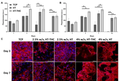
metabolic activity as well as cell morphology are shown in Figure 7. The metabolic 400
activity on both gelatin concentrations presented a similar behaviour being higher for the 401
2.5% w/v scaffolds without the presence of THC. The seeded chondrocytes presented 402
their metabolic activity peak on day 7, decreasing afterwards due to the high cell 403
confluence (as statistically significantly evident for the control represented by cells 404
seeded on Tissue Cultured Plate, TCP). It is important to note that the metabolic activity 405
on THC scaffolds was lower due to the THC presence; however, once released 406
(approximately 98 h as shown previously in section 3.3), cells achieved their maximum 407
w/v scaffolds, since their THC release was slower when compared to the 2.5% w/v 409
scaffolds. 410
Phalloidin and Dapi stainings was performed to study the cell morphology and 411
organisation within the different scaffolds. As shown in Figure 7, the cells seeded in 412
both, 2.5 and 4% gelatin scaffolds, showed a high metabolic activity and spreading at 413
day 3; particularly with visualisation of typical polygon shape of young chondrocytes at 414
4% gelatin [60, 61]. Subsequently, as the cells proliferated, it was possible to observe 415
the formation of cellular clusters in the 4% w/v scaffolds, either in presence and 416
absence of THC, upon 7 days of culture. Furthermore, cultured cells were gradually 417
aggregating, retaining typical polygons shapes and creating large interspaces; 418
suggesting a potential formation of lacuna-like cartilage [62]. In contrast, the cells in the 419
TCP control and 2.5% gelatin groups showed lower expression of F-actin (phalloidin 420
staining). This could be induced by the different level of gelatin and/or its biomechanical 421
effects closer to natural cartilage tissues, by providing an environment with more 422
favourable chondrogenic properties [63]. It is also verified that both gelatin 423
concentrations, with or without THC, showed healthy and proliferating cells in the 424
developed scaffolds. Interestingly, the stability and metabolic activity of the scaffolds in 425
growth media improved up to 2 weeks when compared to TCP control group, and it 426
could be that the specific biological properties of gelatin to promote cell adhesion and 427
cell-maritx interactions enhanced the biological stability of scaffolds [64]. 428
3.5. Antibacterial characterisation 429
In order to test the potential antimicrobial activity of THC infiltrated disks as an 430
shaped scaffolds (8 mm diameter) in the presence of the common nosocomial and joint-432
replacement/ wound infecting organisms S. aureus and P. aeruginosa. As shown in 433
Figure 8, there was a slight antimicrobial action exhibited by the THC containing 434
scaffolds with S. aureus in the area where the disks were placed (i.e. with the THC 435
there was a less dense growth), seen by individual colonies being visible, whereas 436
without the THC the bacteria grew all the way up to the scaffold. The same did not apply 437
for P. aeruginosa. We therefore have preliminary evidence that these materials have 438
possible antimicrobial capability that could augment their potential for clinical use in 439
orthopaedic surgery, because S. aureus strains represent a significant proportion of all 440
pathogens causing infections associated with orthopaedic implants, and the binding of 441
S. aureus to host tissues and plasma proteins is of critical importance in the 442
development of infections at the implant site [65]. 443
4. Conclusions 444
This study demonstrated the utility of lactose cross-linking and THC to modify and 445
improve the physico-chemical properties of gelatin-based porous scaffolds for tissue 446
engineering. The cross-linking reaction resulted in a more water stable scaffold with 447
enhanced mechanical properties and high water uptake capacity (> 800%). Although 448
porosity decreased with the increase of the initial gelatin concentration, the observed 449
increase of pore size from 118 ± 40 µm to 254 ± 57 µm, along with the cross-linking 450
reaction, reinforced the scaffolds, enhancing their mechanical properties. Results of in 451
vitro THC release and antibacterial experiments demonstrated that the THC-loaded 452
scaffolds were capable of effectively releasing THC in a controlled way, with 80% THC 453
method for the postoperative inflammation and pain management. Looking forward to 455
the intended clinical use, the main advantage of the system reported here was the THC 456
biocompatibility and antimicrobial potential with a tailored concentrations for relatively 457
rapid release to achieve a therapeutic effect minimising the risk of systemic side effects. 458
This study has therefore demonstrated that THC may be applied to the manufacture of 459
medical devices, particularly embedded in gelatin porous scaffolds, and it possesses 460
potential antimicrobial properties to augment itsÕ musculoskeletal applications. 461
Acknowledgments 462
Authors thank the University of the Basque Country (research group PPG17/18), the 463
Provincial Council of Gipuzkoa (Department of Economic Development, the Rural 464
Environment and Territorial Balance), the School of Engineering at Newcastle University 465
(UK), and the EPSRC for their financial support. Also thanks both the National EPSRC 466
XPS Users' Service (NEXUS) and Northern Institute for Cancer Research at Newcastle 467
Figure captions 469
Figure 1 ATR-FTIR spectra of non-heated (NH), heat-treated (HT) and HT-THC 470
scaffolds with (A) 2.5 and (B) 4% w/v gelatin; (C) measurement of the areas of the 471
bands at 1624 cm-1, 1650 cm-1, and 1680 cm-1 as a function of protein concentration 472
and heat treatment. 473
Figure 2 (A) Stress-strain curves of non-heated (NH), heat-treated (HT) and HT-THC 474
scaffolds with 2.5 and 4% w/v gelatin. (B) YoungÕs modulus and stress values 475
(calculated at 40% strain). Two means followed by the same letter in the same 476
parameter are not significantly (p > 0.05) different through the TukeyÕs multiple range 477
test. n = 3 is the minimum number of replications. 478
Figure 3 SEM micrographs of (A) non-heated (NH), (B) heat-treated (HT) and (C) HT-479
THC scaffolds with 2.5% w/v gelatin and (D) NH, (E) HT and (F) HT-THC scaffolds with 480
4% w/v gelatin. Bar = 200 µm. 481
Figure 4 (A) Average pore size (µm) and (B) pore size distribution (%) of non-heated 482
(NH) heat-treated (HT) and HT-THC scaffolds with 2.5 and 4% w/v gelatin. 483
Figure 5 Water uptake (WU) values for heat-treated (HT) and HT-THC scaffolds with 484
(A) 2.5 and (B) 4% w/v gelatin. (C)Scaffold degradation degree (DD) values calculated 485
after immersion in PBS for different interval times. Two means followed by the same 486
letter are not significantly (p > 0.05) different through the TukeyÕs multiple range test. n 487
= 3 is the minimum number of replications. 488
Figure 6 THC release from heat-treated (HT) scaffolds with 2.5 and 4% w/v gelatin. 489
Figure 7 Metabolic activity and morphological study of cells seeded on the porous 490
scaffolds. PrestoBlue assay of cells cultured on (A) 2.5% w/v and (B) 4% w/v gelatin 491
porous scaffolds after 1, 3, 7 and 14 days. (C) Confocal microscopy images of 492
chondrocytes cells after 3 and 7 days of culture. Scale bars: 75 µm. 493
Figure 8 Representative macro-photograph showing growth inhibition of (A) 494
Pseudomonas aeuroginosa and (B) Staphylococcus aureus at 24 h by 2.5% w/v gelatin 495
without and with THC addition (a and c), and 4% w/w gelatin without and with THC 496
addition (b and d). 497
A. Etxabide, R.D.C. Ribeiro, P. Guerrero, A.M. Ferreira, G.P. Stafford, K. Dalgarno, K. 500
de la Caba, P. Gentile. International Journal of Biological Macromolecules 501
Figure 1 502
503
504
C
Gelatin
concentration Scaffold 1624 cm
-1 1650 cm-1 1680 cm-1
NH 35.7 60.1 4.2
2.5% w/v HT 42.1 52.3 5.6
HT-THC 40.9 54.4 4.7
NH 35.5 59.4 5.1
4.0% w/v HT 48.2 44.3 7.5
HT-THC 46.9 46.3 6.8
[image:24.612.53.524.133.443.2]A. Etxabide, R.D.C. Ribeiro, P. Guerrero, A.M. Ferreira, G.P. Stafford, K. Dalgarno, K. 505
de la Caba, P. Gentile. International Journal of Biological Macromolecules 506
Figure 2 507
508
509
510
Scaffold Stress (MPa) YoungÕs modulus (MPa)
2.5% w/v 4.0% w/v 2.5% w/v 4.0% w/v
NH 0.173 ± 0.006a 0.282 ± 0.04c 1.22 ± 0.18a 1.51 ± 0.04ab
HT 0.232 ± 0.011bc 0.377 ± 0.050d 1.42 ± 0.18ab 2.43 ± 0.48c
HT-THC 0.155 ± 0.003a 0.289 ± 0.014c 0.92 ± 0.22a 2.04 ± 0.18bc A
[image:25.612.95.500.137.455.2]A. Etxabide, R.D.C. Ribeiro, P. Guerrero, A.M. Ferreira, G.P. Stafford, K. Dalgarno, K. 511
de la Caba, P. Gentile. International Journal of Biological Macromolecules 512
Figure 3 513
514
[image:26.612.67.534.123.363.2]A. Etxabide, R.D.C. Ribeiro, P. Guerrero, A.M. Ferreira, G.P. Stafford, K. Dalgarno, K. 516
de la Caba, P. Gentile. International Journal of Biological Macromolecules 517
Figure 4 518
519
520
A. Etxabide, R.D.C. Ribeiro, P. Guerrero, A.M. Ferreira, G.P. Stafford, K. Dalgarno, K. 521
[image:28.612.69.517.121.432.2]de la Caba, P. Gentile. International Journal of Biological Macromolecules 522
Figure 5
523
524
A. Etxabide, R.D.C. Ribeiro, P. Guerrero, A.M. Ferreira, G.P. Stafford, K. Dalgarno, K. 526
de la Caba, P. Gentile. International Journal of Biological Macromolecules 527
Figure 6 528
529
[image:29.612.200.396.146.284.2]A. Etxabide, R.D.C. Ribeiro, P. Guerrero, A.M. Ferreira, G.P. Stafford, K. Dalgarno, K. 531
de la Caba, P. Gentile. International Journal of Biological Macromolecules 532
Figure 7 533
534
[image:30.612.74.524.133.441.2]A. Etxabide, R.D.C. Ribeiro, P. Guerrero, A.M. Ferreira, G.P. Stafford, K. Dalgarno, K. 536
de la Caba, P. Gentile. International Journal of Biological Macromolecules 537
Figure 8 538
A. Etxabide, R.D.C. Ribeiro, P. Guerrero, A.M. Ferreira, G.P. Stafford, K. Dalgarno, K. 541
de la Caba, P. Gentile. International Journal of Biological Macromolecules 542
Scheme 1 543
544
545
Schiff base
C
C=O C C OH
O C OH CH2OH
gal
H2 C
O
NH (CH2)4CH
NH Amadori product
C
C OH
C C OH
O C OH CH2OH
gal
H C
O
(CH2)4CH
NH
N
-H2O
Lactose Lysineresidue
in gelatin +
C
C OH C C OH
O C OH CH2OH
gal
H O NH CH C
O
(CH2)4
NH2
References 546
[1] Q. Zhang, H. Lu, N. Kawazoe, G. Chen, Pore size effect of collagen scaffolds on cartilage 547
regeneration, Acta biomaterialia 10(5) (2014) 2005-2013. 548
[2] S.J. Hollister, Porous scaffold design for tissue engineering, Nature materials 4(7) (2005) 549
518-524. 550
[3] H. Lu, N. Kawazoe, T. Kitajima, Y. Myoken, M. Tomita, A. Umezawa, G. Chen, Y. Ito, 551
Spatial immobilization of bone morphogenetic protein-4 in a collagen-PLGA hybrid scaffold for 552
enhanced osteoinductivity, Biomaterials 33(26) (2012) 6140-6146. 553
[4] M.C. Echave, P. S‡nchez, J.L. Pedraz, G. Orive, Progress of gelatin-based 3D approaches for 554
bone regeneration, Journal of Drug Delivery Science and Technology (2017). 555
[5] A.I. Raafat, Gelatin based pH!sensitive hydrogels for colon!specific oral drug delivery: 556
Synthesis, characterization, and in vitro release study, Journal of applied polymer science 118(5) 557
(2010) 2642-2649. 558
[6] N.M. Siqueira, B. Paiva, M. Camassola, E.Q. Rosenthal-Kim, K.C. Garcia, F.P. dos Santos, 559
R.M.D. Soares, Gelatin and galactomannan-based scaffolds: Characterization and potential for 560
tissue engineering applications, Carbohydrate polymers 133 (2015) 8-18. 561
[7] T. Agarwal, R. Narayan, S. Maji, S. Behera, S. Kulanthaivel, T.K. Maiti, I. Banerjee, K. Pal, 562
S. Giri, Gelatin/Carboxymethyl chitosan based scaffolds for dermal tissue engineering 563
applications, International journal of biological macromolecules 93 (2016) 1499-1506. 564
[8] S. Kuttappan, D. Mathew, M.B. Nair, Biomimetic composite scaffolds containing 565
bioceramics and collagen/gelatin for bone tissue engineering-A mini review, International 566
journal of biological macromolecules 93 (2016) 1390-1401. 567
[9] A.A. Aldana, G.A. Abraham, Current advances in electrospun gelatin-based scaffolds for 568
tissue engineering applications, International journal of pharmaceutics 523(2) (2017) 441-453. 569
[10] K.G. Shankar, N. Gostynska, M. Montesi, S. Panseri, S. Sprio, E. Kon, M. Marcacci, A. 570
Tampieri, M. Sandri, Investigation of different cross-linking approaches on 3D gelatin scaffolds 571
for tissue engineering application: A comparative analysis, International journal of biological 572
macromolecules 95 (2017) 1199-1209. 573
[11] S. Chen, Q. Zhang, T. Nakamoto, N. Kawazoe, G. Chen, Gelatin scaffolds with controlled 574
pore structure and mechanical property for cartilage tissue engineering, Tissue Engineering Part 575
C: Methods 22(3) (2016) 189-198. 576
[12] C. Chung, J.A. Burdick, Engineering cartilage tissue, Advanced drug delivery reviews 60(2) 577
(2008) 243-262. 578
[13] C. Tang, C. Jin, X. Du, C. Yan, B.-H. Min, Y. Xu, L. Wang, An Autologous Bone Marrow 579
Mesenchymal Stem CellÐDerived Extracellular Matrix Scaffold Applied with Bone Marrow 580
Stimulation for Cartilage Repair, Tissue Engineering Part A 20(17-18) (2014) 2455-2462. 581
[14] B.P. Chan, K.W. Leong, Scaffolding in tissue engineering: general approaches and tissue-582
specific considerations, European spine journal 17(4) (2008) 467-479. 583
[15] M. Nikkhah, M. Akbari, A. Paul, A. Memic, A. Dolatshahi!Pirouz, A. Khademhosseini, 584
Gelatin!Based Biomaterials For Tissue Engineering And Stem Cell Bioengineering, Biomaterials 585
from Nature for Advanced Devices and Therapies (2016) 37-62. 586
[16] P. Hern‡ndez-Mu–oz, R. Villalobos, A. Chiralt, Effect of cross-linking using aldehydes on 587
properties of glutenin-rich films, Food Hydrocolloids 18(3) (2004) 403-411. 588
[17] M. Lacroix, K.D. Vu, Edible coating and film materials: proteins, Innovations in Food 589
Packaging (Second Edition), Elsevier2014, pp. 277-304. 590
[18] A. Ghosh, M.A. Ali, G.J. Dias, Effect of cross-linking on microstructure and physical 591
performance of casein protein, Biomacromolecules 10(7) (2009) 1681-1688. 592
[19] J. Liu, Q. Ru, Y. Ding, Glycation a promising method for food protein modification: 593
physicochemical properties and structure, a review, Food Research International 49(1) (2012) 594
[20] G.B. Naranjo, A.S.P. Gonzales, G.E. Leiva, L.S. Malec, The kinetics of Maillard reaction in 596
lactose-hydrolysed milk powder and related systems containing carbohydrate mixtures, Food 597
chemistry 141(4) (2013) 3790-3795. 598
[21] A. Gaowa, T. Horibe, M. Kohno, K. Sato, H. Harada, M. Hiraoka, Y. Tabata, K. Kawakami, 599
Combination of hybrid peptide with biodegradable gelatin hydrogel for controlled release and 600
enhancement of anti-tumor activity in vivo, Journal of Controlled Release 176 (2014) 1-7. 601
[22] A.H. Nguyen, J. McKinney, T. Miller, T. Bongiorno, T.C. McDevitt, Gelatin methacrylate 602
microspheres for controlled growth factor release, Acta biomaterialia 13 (2015) 101-110. 603
[23] A. Berardi, L. Bisharat, M. Cespi, I.A. Basheti, G. Bonacucina, L. Pavoni, H.S. AlKhatib, 604
Controlled release properties of zein powder filled into hard gelatin capsules, Powder 605
Technology 320 (2017) 703-713. 606
[24] S. Patel, S. Srivastava, M.R. Singh, D. Singh, Preparation and optimization of chitosan-607
gelatin films for sustained delivery of lupeol for wound healing, International journal of 608
biological macromolecules (2017). 609
[25] P.H. Long, Medical devices in orthopedic applications, Toxicologic pathology 36(1) (2008) 610
85-91. 611
[26] M. Zilberman, J.J. Elsner, Antibiotic-eluting medical devices for various applications, 612
Journal of Controlled Release 130(3) (2008) 202-215. 613
[27] A. Etxabide, V. Coma, P. Guerrero, C. Gardrat, K. De La Caba, Effect of cross-linking in 614
surface properties and antioxidant activity of gelatin films incorporated with a curcumin 615
derivative, Food hydrocolloids 66 (2017) 168-175. 616
[28] A. Etxabide, J. Uranga, P. Guerrero, K. de la Caba, Development of active gelatin films by 617
means of valorisation of food processing waste: A review, Food Hydrocolloids 68 (2017) 192-618
198. 619
[29] M. Dai, X. Zheng, X. Xu, X. Kong, X. Li, G. Guo, F. Luo, X. Zhao, Y.Q. Wei, Z. Qian, 620
Chitosan-alginate sponge: preparation and application in curcumin delivery for dermal wound 621
healing in rat, BioMed Research International 2009 (2009). 622
[30] A.B. Hegge, T. Andersen, J.E. Melvik, E. Bruzell, S. Kristensen, H.H. T¿nnesen, 623
Formulation and bacterial phototoxicity of curcumin loaded alginate foams for wound treatment 624
applications: studies on curcumin and curcuminoides XLII, Journal of pharmaceutical sciences 625
100(1) (2011) 174-185. 626
[31] C.N. Gajendragadkar, P.R. Gogate, Intensified recovery of valuable products from whey by 627
use of ultrasound in processing stepsÐA review, Ultrasonics sonochemistry 32 (2016) 102-118. 628
[32] A. Etxabide, J. Uranga, P. Guerrero, K. de la Caba, Improvement of barrier properties of 629
fish gelatin films promoted by gelatin glycation with lactose at high temperatures, LWT-Food 630
Science and Technology 63(1) (2015) 315-321. 631
[33] A. Etxabide, M. Urdanpilleta, P. Guerrero, K. de la Caba, Effects of cross-linking in 632
nanostructure and physicochemical properties of fish gelatins for bio-applications, Reactive and 633
Functional Polymers 94 (2015) 55-62. 634
[34] A. Etxabide, C. Vairo, E. Santos-Vizcaino, P. Guerrero, J.L. Pedraz, M. Igartua, K. de la 635
Caba, R.M. Hernandez, Ultra thin hydro-films based on lactose-crosslinked fish gelatin for 636
wound healing applications, International journal of pharmaceutics 530(1-2) (2017) 455-467. 637
[35] B.P. Kanungo, E. Silva, K. Van Vliet, L.J. Gibson, Characterization of mineralized 638
collagenÐglycosaminoglycan scaffolds for bone regeneration, Acta Biomaterialia 4(3) (2008) 639
490-503. 640
[36] D. ASTM, 570-98, Standard test method for water absorption of plastics (2005). 641
[37] A. Mart’nez-Ruvalcaba, F. Becerra-Bracamontes, J.C. S‡nchez-D’az, A. Gonz‡lez-çlvarez, 642
Polyacrylamide-gelatin polymeric networks: effect of pH and gelatin concentration on the 643
swelling kinetics and mechanical properties, Polymer bulletin 62(4) (2009) 539-548. 644
[38] S. James, J. Fox, F. Afsari, J. Lee, S. Clough, C. Knight, J. Ashmore, P. Ashton, O. Preham, 645
M. Hoogduijn, Multiparameter analysis of human bone marrow stromal cells identifies distinct 646
[39] N. BenBetta•eb, T. Karbowiak, S. Bornaz, F. Debeaufort, Spectroscopic analyses of the 649
influence of electron beam irradiation doses on mechanical, transport properties and 650
microstructure of chitosan-fish gelatin blend films, Food Hydrocolloids 46 (2015) 37-51. 651
[40] S. Basu, U.S. Shivhare, T.V. Singh, V.S. Beniwal, Rheological, textural and spectral 652
characteristics of sorbitol substituted mango jam, Journal of Food Engineering 105(3) (2011) 653
503-512. 654
[41] W.-q. Wang, Y.-h. Bao, Y. Chen, Characteristics and antioxidant activity of water-soluble 655
Maillard reaction products from interactions in a whey protein isolate and sugars system, Food 656
chemistry 139(1) (2013) 355-361. 657
[42] H. Souguir, F. SalaŸn, P. Douillet, I. Vroman, S. Chatterjee, Nanoencapsulation of curcumin 658
in polyurethane and polyurea shells by an emulsion diffusion method, Chemical engineering 659
journal 221 (2013) 133-145. 660
[43] K. Siimon, H. Siimon, M. JŠrvekŸlg, Mechanical characterization of electrospun gelatin 661
scaffolds cross-linked by glucose, Journal of Materials Science: Materials in Medicine 26(1) 662
(2015) 37. 663
[44] A. Bigi, G. Cojazzi, S. Panzavolta, N. Roveri, K. Rubini, Stabilization of gelatin films by 664
crosslinking with genipin, Biomaterials 23(24) (2002) 4827-4832. 665
[45] K.M. Rao, K.S.V.K. Rao, G. Ramanjaneyulu, C.-S. Ha, Curcumin encapsulated pH 666
sensitive gelatin based interpenetrating polymeric network nanogels for anti cancer drug 667
delivery, International journal of pharmaceutics 478(2) (2015) 788-795. 668
[46] N. Dave, A. Troullier, I. Mus-Veteau, M. Du–ach, G. Leblanc, E. Padr—s, Secondary 669
structure components and properties of the melibiose permease from Escherichia coli: a Fourier 670
transform infrared spectroscopy analysis, Biophysical Journal 79(2) (2000) 747-755. 671
[47] X.C. Tang, M.J. Pikal, Design of freeze-drying processes for pharmaceuticals: practical 672
advice, Pharmaceutical research 21(2) (2004) 191-200. 673
[48] C.W. Wong, H.B. Wijayanti, B.R. Bhandari, Maillard reaction in limited moisture and low 674
water activity environment, Water Stress in Biological, Chemical, Pharmaceutical and Food 675
Systems, Springer2015, pp. 41-63. 676
[49] C. Tonda-Turo, P. Gentile, S. Saracino, V. Chiono, V.K. Nandagiri, G. Muzio, R.A. Canuto, 677
G. Ciardelli, Comparative analysis of gelatin scaffolds crosslinked by genipin and silane 678
coupling agent, International journal of biological macromolecules 49(4) (2011) 700-706. 679
[50] J.-Y. Lai, D.H.-K. Ma, M.-H. Lai, Y.-T. Li, R.-J. Chang, L.-M. Chen, Characterization of 680
cross-linked porous gelatin carriers and their interaction with corneal endothelium: biopolymer 681
concentration effect, PLoS One 8(1) (2013) e54058. 682
[51] W. Chen, B. Sun, T. Zhu, Q. Gao, Y. Morsi, H. El-Hamshary, M. El-Newehy, X. Mo, 683
Groove fibers based porous scaffold for cartilage tissue engineering application, Materials 684
Letters 192 (2017) 44-47. 685
[52] R. Levato, W.R. Webb, I.A. Otto, A. Mensinga, Y. Zhang, M. van Rijen, R. van Weeren, 686
I.M. Khan, J. Malda, The bio in the ink: cartilage regeneration with bioprintable hydrogels and 687
articular cartilage-derived progenitor cells, Acta biomaterialia 61 (2017) 41-53. 688
[53] S. Wu, H. Dong, Q. Li, G. Wang, X. Cao, High strength, biocompatible hydrogels with 689
designable shapes and special hollow-formed character using chitosan and gelatin, Carbohydrate 690
Polymers 168 (2017) 147-152. 691
[54] M. Jafari, Z. Paknejad, M.R. Rad, S.R. Motamedian, M.J. Eghbal, N. Nadjmi, A. Khojasteh, 692
Polymeric scaffolds in tissue engineering: a literature review, Journal of Biomedical Materials 693
Research Part B: Applied Biomaterials 105(2) (2017) 431-459. 694
[55] A.G. Mikos, J.S. Temenoff, Formation of highly porous biodegradable scaffolds for tissue 695
engineering, Electronic Journal of Biotechnology 3(2) (2000) 23-24. 696
[56] X. Wu, Y. Liu, X. Li, P. Wen, Y. Zhang, Y. Long, X. Wang, Y. Guo, F. Xing, J. Gao, 697
Preparation of aligned porous gelatin scaffolds by unidirectional freeze-drying method, Acta 698
biomaterialia 6(3) (2010) 1167-1177. 699
[58] L.P. Yan, Y.J. Wang, L. Ren, G. Wu, S.G. Caridade, J.B. Fan, L.Y. Wang, P.H. Ji, J.M. 702
Oliveira, J.T. Oliveira, Genipin!cross!linked collagen/chitosan biomimetic scaffolds for articular 703
cartilage tissue engineering applications, Journal of Biomedical Materials Research Part A 95(2) 704
(2010) 465-475. 705
[59] W.N. Scott, Insall & Scott Surgery of the Knee E-Book, Elsevier Health Sciences2011. 706
[60] J. Liu, X. Liu, G. Zhou, R. Xiao, Y. Cao, Conditioned medium from chondrocyte/scaffold 707
constructs induced chondrogenic differentiation of bone marrow stromal cells, The Anatomical 708
Record 295(7) (2012) 1109-1116. 709
[61] X. Li, X. Ren, S. Li, J. Liang, X. Zhao, T. Wang, Z. Wang, Morphological, 710
Immunocytochemical, and Biochemical Studies of Rat Costal Chondrocytes Exposed to IL-1β
711
and TGF-β1, Journal of healthcare engineering 2017 (2017). 712
[62] K. Yang, J. Sun, D. Wei, L. Yuan, J. Yang, L. Guo, H. Fan, X. Zhang, Photo-crosslinked 713
mono-component type II collagen hydrogel as a matrix to induce chondrogenic differentiation of 714
bone marrow mesenchymal stem cells, Journal of Materials Chemistry B 5(44) (2017) 8707-715
8718. 716
[63] K. Brodkin, A. Garcõa, M. Levenston, Chondrocyte phenotypes on different extracellular 717
matrix monolayers, Biomaterials 25(28) (2004) 5929-5938. 718
[64] M. Santoro, A.M. Tatara, A.G. Mikos, Gelatin carriers for drug and cell delivery in tissue 719
engineering, Journal of controlled release 190 (2014) 210-218. 720
[65] M.C. Hudson, W.K. Ramp, K.P. Frankenburg, Staphylococcus aureus adhesion to bone 721
matrix and bone!associated biomaterials, FEMS microbiology letters 173(2) (1999) 279-284. 722