A STUDY OF ISOLATED ROOT CULTURE IN WHITE CLOVER.
Madeleine Jennifer Alice Ahearn
A Thesis submitted for the Degree of i\fasrer of Science at the Australian National University
This thesis contains no material which has been accepted for the award of any other degree or diploma in any University. To the best of my knowledge, it contaim no material previously published, or the result of any vork by another person, except where due reference is made in the text.
TABLE OF CONTE1 YTS ABSTRACT
ACKNOWLEDGEtvlEl"'ITS
GLOSSARY AND ABBREVIATIONS CHAPTER 1
1.1 1.2 1.3 1.4 1.5 1.6
INTRODUCTION
Nitrogen fixation: its relevance The nodulation process
Early excised root studies
Further development of excised root techniques
The aims of this Study Outline of thesis
CHAPTER TWO: MATERIALS Al'ID METHODS 1 2 3 4 5 6 7 8 9 10 11 12 13 14 15 16 17 18 19 20 21 22 23 24
Plant material Bacterial material
Incubation of bacterial cultures Gases
Chemicals
Media preparation Culture/selection media Rhizobium culture medium
Seed sterilization and germination Variations of the standard germination procedures
Inoculation of seedlings Incubation of plates Nodulation tests
Root excision techniques Growth of excised roots
Variations of growth of excised roots Sand culture
Vermiculite culture
Modified Fahraeus slide technique 1 Modified Fahraeus slide technique 2 Microscopy
Cytology
Lactophenol clearing techniques Preparation of root tip material for microscopic examination
CHAPTER TWO (continued) 25 26 27 28 Buffer
Enzyme assays
Variations of standard assay method Statistical analyses
33 33 35 36 CHAPTER THREE: ESTABLISHMENT OF EXCISED ROOT CULTURE
1 2 3 4 5 6 7 8
Isolated root culture of beans and soybean using the Raggio and Raggio technique
37 Effect of Rhizobium preculture on the
efficiency of the establishment of the symbiosis
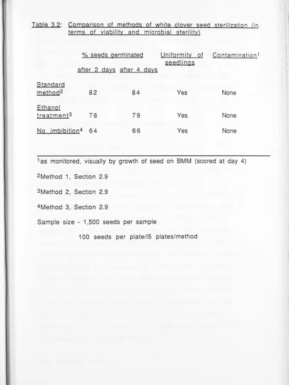
41 Comparison of methods of seed sterilization
in Trif olium rep ens 42
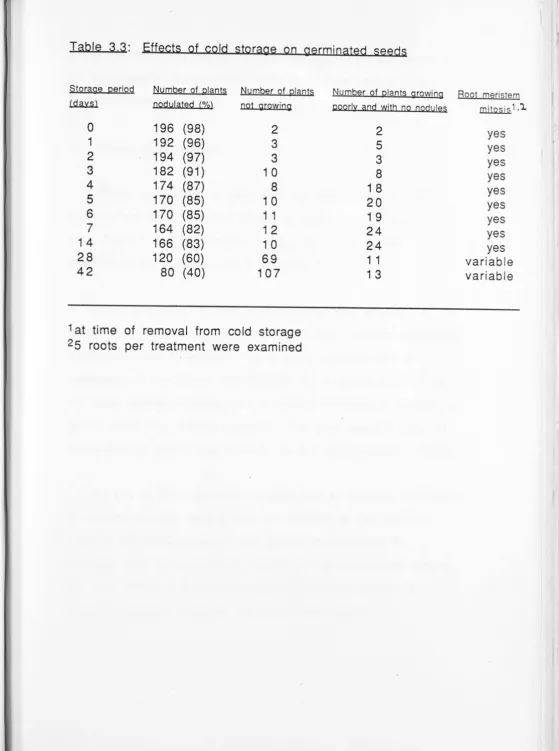
Effects of cold storage on germinated seeds 43
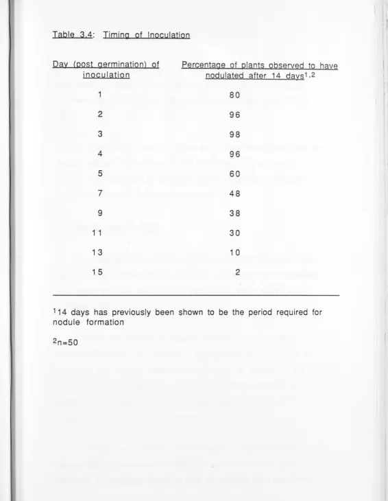
Timing of inoculation 44
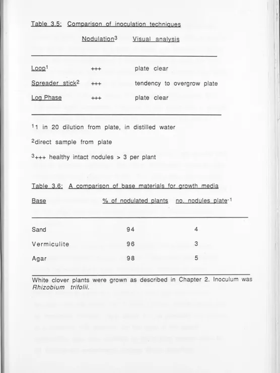
Inoculation techniques 45
Growth medium base 45
A comparison of media for establishment of growth of excised white clover roots 47 CHAPTER FOUR: EXPERI IENT AL MAL"\TJPULA TIONS
1
2
3
4 5
Growth of excised roots in varying light regimes
Age and timing of excision
Excision of white clover roots under N-BS versus air
Modified Fahraeus glass slide technique Maintenance of growth and symbiotic characteristics of white clover on medium 17 /12
62
63
64
65
CHAPTER 4 (continued)
6
7
8
Analysis of medium effects on nodulation in isolated root cultures
Root temperatures
Effect of explant age on establishment of nodulation in excised roots
CHAPTER FIVE: DISCUSSION
5. I
5.2
Discussion
Medium components and their relative effects
71
75
76
79
80
A. Components of the inorganic medium 8 0
5.3
5 .4
5.5
APPENDICES
REFERENCES
B. Components of the organic medium 8 2
P~ysical and environmental effects
Future directions
Conclusions
87
95
98
99
ABSTRACT
Root culture techniques were developed in order to facilitate the autonomous study of the leg ume-Rhizobium interation. This experimental system separates shoot from root in an in vitro culture system, thus controlling the level of input of exogenous substances such as phytohormones and photosynthate. This simplifies the analysis of the complex process of nodulation and nitrogen fixation.
In this study the excised roots of white clover (Trifolium repens) were successfully grown and nodulated on an agar medium. An essential aspect of the culture system was the use of sectored ( or Y) Petri plates whir;h allowed the physical separation of the
organic (OM) and inorganic (JOY!) media components. Once growth and nodulation were established, it was possible to investigate the effect of various phytohormones, nutrients, light regimes a.nd a variety of other environmental factors on the nodulation-nitrogen fixation process (i.e. temperature, excision conditions, physical substrate and culture methods).
The early work of Raggio and Raggio on excised roots of other legumes (i.e. Glycine max (L) ivlerr and Phaseolus vulgaris) was repeated, extended and modified. Roots were excised in the
hypocotyl region and ·grown in the dark at 23°C in 'Y-plates' on an agar-based organic and inorganic medium (the optimum medium was designated 17112). Maintenance of growth (0.8 - 1.2
Roots inoculated after excision were successfully nodulated and nitrogen fixation .( as measured by acetylene reduction) was
achieved. The effect of carbon and nitrogen sources and
phytohormone levels in the medium was investigated. The lack of a particular 'shoot factor' was postulated.
The techniques developed here have simplified the
investigation and use of isolated root cultures in legumes. They
have led to increased nodulation frequency and increased
acetylene reduction in the experimental materials. These
techniques are applicable to all legume root cultures and form the
basis of a wide range of experimental areas.
Acknowledgements
I am grateful to my supervisor, Dr. P.M. Gresshoff, for his enthusiastic encouragement, advice and criticism.
I would like to thank Dr. B. Carroll for his helpful suggestions and discussions, Mr M. Commons for his technical assistance, Evelyn for her continued support and last, but not least, my husband, Robert, for putting up with the gestation of this thesis!
The thesis is dedicated to my father, the late Jerome Morawski, who both in life and in death showed me the meaning of courage and dignity.
GLOSSARY A.t\TD ABBREVTATIO S
Acetylene reduction: the enzyme nitrogenase catalyses the conversion of the gas acetylene to ethylene. This is used as a measure of the activity of the nitrogenase enzyme. Bacteroid: BM: BMM: BS: FM: Il: I2: IAA: LB: LBG: NAA: OD: Sp: 2,4-D: asparagine
the symbiotic nitrogen fixing form of Rhizobium
basic medium
Bergersen's modified media nutrient medium as defined by Gamborg and Everleigh (1968) Fahraeus medium - a nitrogen-free growth medium
glutamine
inorganic medium 1 inorganic medium 2 indole acetic acid Luria broth
Luria broth with glucose Ci<. napthalene acetic acid optical density
spectinomycin
CHAPTER 1
INTRODUCTIO_
1.1 Nitrogen fixation: its relevance
Nitrogen is an essential component of the macromolecular building blo'cks of all biological systems. The dinitrogen molecule is the predominant gas component in the atmosphere (79 .08% by volume), however it is chemically inert and generally unavailable to the vast majority of organisms. It can enter biological systems only when it has been 'fixed' or combined with certain other elements such as oxygen or hydrogen (Bergersen, 1979).
Nitrogen is considered to be the most limiting element in
primary production due to the absolute requirement of most living organisms to obtain nitrogen in a combined form. This process can be accomplished industrially through the
'Haber-Bosch' process, via the manufacture of ammonia from hydrogen and atmospheric nitrogen and although the production of
ammonia and other related chemical fertilizers is now a major
industry, the bulk of all fixed nitrogen is of biological origin (Gresshoff and Rao, 1987).
Only a few specialized prokaryotic species pos ess the
in the case of Sesbania) on leguminous plants. Other bacteria fix
nitrogen in the free-living state or in associations.
1.2 The nodulation process
The legume-Rhizobium symbiosis can be seen as a controlled infection which requires the formation of an appropriate
environment in which to operate. This is found in the root nodule,
a specialized organ which is initiated and developed by a complex
interaction of bacterial, plant and environmental factors (Vincent,
1980). The bacteria, present in the rhizosphere, attach to an
emerging root hair and invade the plant root through an infection
thread. Proliferation of the root cortical tissues occur, resulting in
the formation of the nodule characteristic of the symbiosis (see
Fig. 1.1).
Once inside the nodule, bacteria differentiate into the
symbiotically active bacteroid form. Nitrogen is fixed via
nitrogenase in the bacteroids to produce ammonia, which rn turn
is translocated into the nodule cytoplasm. Once in the cytoplasm
ammonia is assimilated into other nitrogenous compounds via the
plant assimilatory enzymes. Hence fixed nitrogen is contributed to
the plant, and the Rhizobium receives carbon for metabolic processes in return (Gresshoff and Delves, 1986). This is
accompanied by the expre sion of genes required for the
functioning of the nitrogen-fixing enzyme complex nitrogenase
which reduces the dinitrogen molecule by the following reaction:
SH++ 2 + Se-+ nATP +nH20 -> 2 H3+ nADP + nPi + H2
Nitrogenase also catalyses the conversion of acetylene to ethylene, which is used as the basis of the acetylene reduction assay by gas chromatography. Legume symbioses are further characterised by the presence of high levels of leghaemoglobin within the nodules. Evidence suggests that it is located within the plant cytoplasm (Robertson et al., 1975) and that it facilitates the
diffusion of oxygen to the bacteroids (Bergersen et al., 1973) particularly at low ambient oxygen concentrations (Wittenberg,
1980). Plant genes code for the globin apoprotein, whilst the site
of the heme synthesis appears to be within the bacteroid ( adler
and Avissar, 1977). The actual assembly of leghaemoglobin is
thought to occur outside the bacteroid as no leghaemoglobin is found inside it.
Overall, the plant provides the energy for the process of nitrogen fixation while the bacteroid supplies the plant with
ammonia. To achieve this relationship requires a complex developmental process involving multiple signals between the bacterium and the plant in order to express their symbiotic
phenotype. Hence, the development of an effective symbiosis rn Rhizobium is dependent on the host-specificity of the Rhizobium
strain. (Most Rhizobium strains that nodulate the agriculturally important temperate legumes are restricted in their nodulation
capacity to a single legume genus, e.g. the bacteria that nodulate clover species Trifolium are Rhi:obium trifolii).
Symbiotic fixation of atmospheric nitrogen by leguminous plants is the principal process for maintaining nitrogen fertility in
agriculture and the interaction of plants of the family
Leguminoseae and the specific bacteria of the genus Rhizobium m this symbiotic relationship is not yet well understood. Research on
the symbiotic relationship has focussed either on isolated
bacteroids or whole plant studies. The latter at times are difficult
to interpret as all factors that affect the plant's fitness also affect
the symbiosis. Thus a reductionist approach, finding an
intermediate situation, would be favourable. Although the
symbiosis is a cellular process it has been impossible to
repeatedly establish infections of aseptically-cultured plant cell
cultures (see Gresshoff, 1982). However, some success has been
achieved with isolated organ culture. The excised root technique
has been developed as such an experimental system (Raggio and
Raggio, 1956; and this study). Used in conjunction with other
experimental observations it provides a method of further
expanding our knowledge of the complex legume-Rhizobium symbiosis.
1.3 Earlv exci ed root tudie
To inve tigate the establishment and development of
nodulation and nitrogen fixation m legumes a model system which
eliminates as many variables as po sible is neces ary. Excised root
studies may provide such a system. By excising the root in the
hypocotyl region one is able to control the photosynthate and
phytohormone upply to the roots. Thi allows investigation of the
physiological parameters of nodulation and nitrogen fixation as
well as the genetic components and allows discrimination between
the two. (Haberlandt { 1902} was the first to suggest that the
through severance of these relationships, that is, isolated cells and organs in vitro).
Kotte (1922) initiated studies on the growth of excised root tips in sterile culture solutions. Kotte examined growth of apical and subapical fragments of Pisum and Zea roots, Robbins (1922) reported growth of root tips of Pisum, Zea and Gossypium. Both Kotte and Robbins added extracts to the culture medium and reported increased growth and that the excised root meristem would, if it grew, differentiate into nJrmal root tissue. Robbins and Maneval (1923, 1924) tested a wider range of species and noted the beneficial effect on growth of moderate illumination of the cultures. White (1932, 1933) investigated optimum conditions for excised root growth and in 1934 began a four year growth period of excised tomato roots (1937). Other workers in this area included Chambers (1923), Dauphine (1929, 1930), Loo and Loo (1935), Gautheret (1935), and Geiger Huber and Burlet (1936).
the second week, then amino acids and vitamin B (regarded as a
phytohormone) replaced the yeast. No nodulation was observed.
Wipf and Cooper (1940) grew excised roots of pea on agar and
observed no nodulation on the solid medium.
Seppilli et al (1941) and McGonagle (1944) reported complete
failure in their attempts to nodulate excised legume roots.
McGonagle investigated cultures of excised roots of Pisum (maple
pea). She excis_ed radicles at 1-2 cm and placed 0.5 cm excised tips
in flasks. Growth was observed (10-12 cm in the first 14 days,
then up to 40 cm), and laterals were observed, however no
secondary thickening of roots or nodules were observed and only
sparse and short root hairs. The associated rhizobia multiplied yet
no infection was ohserved. McGonagle used liquid medium,
dropping the excised roots into flasks. The implications of the
techniques mentioned to this point will be further discussed in
Chapter 5.
1.4 Further development of excised root techniques
Technically, the success of excised root cultures requires a
healthy root culture system. The shoot must be replaced by 1n
vitro components, especially a carbon source and the root
environment must be free of carbon and nitrogen sources as a
carbon source would permit the excess growth of rhizobia and
most nitrogen sources interfere with nodulation (Carroll and
Gresshoff, 1983) (Note, these concerns are further discussed rn
Chapter 5). Hence, a two-chambered apparatus is required for the
physical separation of organic and inorganic moieties of the medium.
ln 1956 Raggio and Raggio developed a new method for the cultivation of isolated roots involving a separation of inorganic and organic media. The solid (organic) medium was poured into a glass vial and placed in a Petri dish containing the liquid
(inorganic) medium. Less browning of roots was observed using this method. ln 1957 Raggio et al., investigated the relationship between nodule formation and media components in excised root cultures of Glycine max and P haseolus vulgaris. These roots, inoculated with homologous Rhizobium strains developed some nodules. The advantages of their system were that nutrition was controlled, the shoot contribution was excluded and substances whose effects were to be tested could be added in known
amounts. For example, Raggio et al. (1959) monitored the
enhancement of nodulation of P haseolus vulgaris excised roots by inositol (see Discussion, Chapter 5).
McAlister and Krober (1951) observed that critical substances for growth and development of the primary root migrate from the cotyledons to the root in the first few days of germination.
secretions of nodule bacteria (Thornton, 1936) and the study of the
production of growth-substances by clover nodule bacteria (Chen,
1938) led to the inferences drawn by Raggio et al. (1957) that is (i) that the primary roots should nodulate if maintenance requirements
are met, (ii) that as nitrate and sucrose inhibit whole plant
nodulation they should do so in excised root, and (iii) that sucrose
around the root will lead to a prohibitory proliferation of Rhizobium.
Raggio et al. (1957) performed one of the first trials to determine
which physical substrate would be best for the inorganic phase of the
excised root apparatus. They also determined that light was not
necessary to achieving successful nodulation.
Following the initial successful work several investigations
were performed on the nature of media components. Raggio et al. ( 1964) investigated the interaction of nitrate and carbohydrates in
root nodule formation; Valera and Alexander (1965) determined
that nodulation of excised roots of M edicago sativa was enhanced by an extract of alfalfa seeds and that coconut water exerted a
similar influence on excised roots of Glycine max and Phaseolus
vulgaris. Cartwright (1967) reported that gibberellic acid reduced nodule number but not growth in excised roots of Phaseolus
vulgaris.
Higashi et al. (1971) investigated the influence of plant
hormones (indole acetic acid, kinetin, gibberellin, 2,4-D and NAA)
on infection thread formation in white clover root hairs. They
used excised roots in order to elucidate the primary stage of
used capillary tubes inserted into liquid media and their
nodulation results were relatively low (see Chapter 5 for further discussion). Abe and Higashi (1979) used excised root techniques in white clover to investigate the degree of root cell mass
necessary for infection thread production and nodulation. ln this study they used an extrinsic substance isolated from Rhizobium
trifolii 4S cells (ES6000) and found that initially root segments had apical meristems, infection threads and some nodules, that the root apical meristem was needed for infection (no threads observed when it was excised) and that the age of the root hairs may affect infection thread formation. They found no interaction between root elongation and promotion of infection thread
formation. Very low nodule numbers were reported in this study and no evidence of nitrogen fixation was presented.
Gautheret (1980) worked with isolated root cultures of Glycine max to study the physiology of nodule formation. He investigated three different physical placements of the root material (see
Chapter 5 for discussion of his apparatus and results) however, he reported that roots grew but did not nodulate.
Several researchers have reported Rhizobium infections of leguminous cells (Holsten et al., 1971; Hermina and Reporter, 1977) and Verma et al. (1978) reported them as an abnormal (parasitic penetration) of the leguminous callus and suspension cells by comparing features of bacterial morphology and nitrogen-fixing ability with normal tissue.
al. (1971) who used acetylene reduction techniques in the
investigation of symbiosis between Rhizobium and plant cells in
vitro, little work appears to have been done on a quantitative
measurement of the nitrogen-fixing ability of the nodules formed
on excised roots.
1.5 The aims of this study
From the previous information it can be seen that in order to
simplify the excised root system the nutrition of excised roots
grown in vitro must be controlled by the addition of substances
normally contributed by the shoot (such as phytohormones and photosynthate). The organic and inorganic moieties must be
physically separated, the system must provide an
easily-reproducible, fast-screening, consistent method, which allows both
the investigation of nodulation procedures and the establishment
and maintenance of nitrogen fixation. This study is concerned with
the development of such a system and investigates a wide range
of parameters in order to optimize the system. The system is
applicable to a wide range of legume hosts and whilst white clover
(Trifolium repens) has been the primary host investigated here,
other legumes (Glycine max, Phaseolus vulgaris) have been
1.6 Outline of thesis
Chapter Two describes the materials and methods used m this study.
Chapter Three outlines the 'background' work and
investigations leading up to the successful establishment of an excised root culture in Trifolium repens.
Chapter Four investigates the experimental manipulations performed on the system once it was established.
Figure 1.1: Nodule morphogenesis in the Rhizobium-legume
symbiosis. A schematic representation of the development of a nodule following infection by Rhizobium, divided into a series of phenotypically distinguishable steps as proposed by Vincent
Nodule morphogenesis rn the Rhizobium-Iegume symbiosis.
~~-~
~
~
'!)
Root colonization
Root ~dhesion
Hair branching
Hair curling
Infection
Nodule initiation
Inf ecti on-lh read branching
Nodule development
Bacterial release
Bacteriod development
Nitrogen fixation
Complementary functions
CHAPTER 2
1. Plant Material
Three plant species were studied, Phaseolus vulgaris, Glycine max and Trifolium repens. Table 2.1 shows the common name and seed type for each of these species. Figure 2.1 illustrates the experimental materials.
2. Bacterial Material
Rhizobium trifolii strain ANU 843 derived from strain SU
794 Sp was used as the white clover inoculant, Bradyrhizobium strain CB 1809 (= USDA 136 = TAL 102) was used as the Glycine
_max inoculant in this study and Rhizobium trifolii sp. was used as the Phaseolus vulgaris inoculant. Rhizobium colonies were
maintained on plates of modified Bergersen's medium
(Bergersen, 1961) containing 1 OOmg/1 spectinomycin (BMM spec)
(see Section 2.8). These inoculant stocks were stored at 40c and were routinely subcultured each month.
3. Incubation of bacterial cultures
Where possible bacteria were maintained on antibiotic
selection media. Liquid cultures were incubated in 250 ml Klett
flasks plugged with sterile cotton wool. These were shaken at
150 rpm in a gyratory shaker (New Brunswick Scientific Co. Inc. USA). Plates were sealed with a strip of Nescofilm (Nippon Shoji Kaishi, Osaka, Japan) and incubated inverted in a 280C incubator. Slopes were sealed with the associated sterile screw top and incubated on their sides in a 280C incubator.
Table 2.1: Plant species used for root culture experiments
Botanical Name Common Name Seed type/source
Glycine max soybean variety Bragg
(from Dr D Herridge,
Tamworth)
Phaseolus vulgaris wax bean Dr. Adrian Gibbs
(VERG, ANU)
Trifolium repens white clover New Zealand 5826
Fig 2.1:
The experimental materials
A. Phaseolus vulqaris
B. Glycine max
[image:27.612.7.570.18.734.2]4. Gases
High purity grade gases from CIG (Australia) were always
used to ensure that levels of contaminating 02, CO, CO2 were kept to a minimum.
5. Chemicals
All chemicals were of analytical reagent grade. Where
commercial preparations have been used, the product and the
manufacturer are cited.
6. Media Preparation
Difeo Bacto-agar (1 .5%) (Difeo Laboratories, USA) was used
for all solid media. With the exception of heat labile
components, all components were sterilized by autoclaving at
15 psi for 15-30 minutes, time being proportional to volume.
Heat labile components were freshly prepared and filter
sterilized (0.22 µm) before being added to autoclaved media
(cooled to 45-5QOC). For plates, the media were poured into
99mm diameter sterile disposable plastic Petri dishes, holding
25-30 ml (Disposable Products, Australia) or pipetted into
disposable plastic Petri 'Y-plates' holding 25-30 ml (Medical
Plastics, Australia) and divided into three compartments (see
Fig. 2.2 and Section 2.14 for specific method). All
post-sterilization manipulations were made in a laminar flow-hood
(Standardised Protection, Brookvale, NSW, 2100 Australia).
[image:29.615.18.611.16.755.2]Figure 2.2: Schematic representation of isolated root culture technique of white clover using 'Y-plates' (see Section 2.14 for further details).
II
[image:30.614.6.566.17.724.2]·.·.·.·.·.·.·.·.·.·.·.·.· ·.·.·.·.·.·.·.·.·.·.·.
·.· .. ·
.·.·.·.·.·.·.
·.·. ·
11
·
.
...
.
:;::: ::::·
16M
::::::: ::::
:
:::"
16M
:
:
::::
.. ·.·.·.·.·.·.·.·.·.·.· . . ·.·.·.·.·.·.·.·.·.·.·
·
.
:
::::::::::::::::::::
·.·.·.·.·.·.·.·.·
I
<
:::
1
.
C?~(33)JJ
::::::
OM\:/
/:
I
Plates scale(, incubated upright at 23°C in the dark
Excision of agar block
. . .
:J
:
i
>
... .. ....... . . . . . . . . . . . . . . . . . . . . . . .
. . . . . . . . . . . . . . . ... . . . . . . . .
.
• ... .
:::::::::::::::::::::-. :::::::::::::::::::::-. . . . . . . . .
. . . . . . . . . . . . . . . . .
. . . . . . . .
!:
:::::::::::::::::::::
::
::J
ffl
j±gJrj
Rhizobium inoculation
•' • • • • • •' • • • • ,-• •••••••-I I' • • • • • • • ' • •
·.·.·.·.·.·.·.·.·.·.·.·.
·.·.·.·.·.·.·.·.·.·.·.·., t.·.·.·.·.·.·.
-::::::::::::::::::::11:::::::::::::·.
1
1
::>:··
·
f~3
·
1
17. Culture/Selection Media
Stock solutions for media were prepared and added to
distilled water as required.
Fahraeus Medium (FM) White clover, soybean and french bean
plants were cultured on Fahraeus medium (FM) (Vincent, 1970).
The composition of the basic nitrogen-free Fahraeus medium is
given in Table 2.1 a
Iron Chelate (Gresshoff and Doy, 1974). Iron chelate was
prepared by dissolving 557 mg of FeS04 .7H20 and 745 mg of Na 2EDTA in 100 ml of distilled H20.
Trace Elements (Gresshoff and Doy, 1974). Trace elements were
prepared by dissolving 100 mg MnS04.H20, 30 mg H3803, 30 mg
ZnS04.7H20, 2.5 mg Na2Mo04.2H20, 2.5 mg CuS04.5H20, 2.5 mg CoCl2.6H 20 in 100 ml of distilled water.
Biotin For 1000 x stock: 10mg in 100ml H20 (final concentration 0.001 mg/I)
Thiamine-HCI For 1000 x stock: 100 mg in 100 m1 H2
o
(final concentration 0.001 mg/I)Fahraeus Medium with Nitrate (FMN03
l.
FMN03 composition is the same as Fahraeus medium (Table 2.1 a) with the addition of 0.5gm of potassium nitrate (KN03), i.e. 5 mM nitrate.Table 2.1
a
Fahraeus MediumComponent*
.Ein.a!
concentrationma
11Potassium dihy'drogen phosphate KH 2P04 1 0 O
Disodium hydrogen phosphate Na2H P04.12H2
o
1 5 OMagnesium sulphate MgS04.7H20 120
Calcium chloride CaCl 2 1 O O
Iron che1ate ** FeS04 .7H 2
o
2. 785Trace elements** MnS04 .H 20 0.300
'Difeo' agar
H3B03 0.090
ZnS04)H20 0.090
Na2Mo04_2H20
CuS04.SH20
CoCl2.6H20
0.0075
0.0075 0.0075
12000
735 419 487 901
10.0
1.8 1 .5 C.6
3.1x10⁻²
3.0 x10⁻² 3.2 x10⁻²
• Stock solutions of all the compounds were made and appropriate volumes were added to distilled water.
Medium for Raggio Repeat Experiment (Black waxbean and
soybean) see Table 2.1 b
N-BS Medium. This was prepared by dissolving 1.5 g of
NaH2P04.2H20, 7.5 mg of Kl, 5.0 g of KCI, 1.5 g Na2S04, 2.5 g Mg S04. 7H20 ·and 1.5 g CaCl 2.2H20 (dissolved separately) in 1000
ml of H20. This is an adaptation of the major salt stock of the
plant tissue culture medium BS (Gamborg and Eveleigh, 1968).
lnoroanic Media (General composition for inorganic component of
isolated root culture) see Table 2.1 c
Oroanic Media (General composition for organic component of
isolated root culture) see Table 2.2. The basic medium (Table
2.2) (plus sucrose and. nitrogen source where applicable) was
autoclaved at 10 psi for 20 mins. Hormones were filter
sterilized and added after autoclaving.
8. Rhizobium Culture Medium
Bergersen's Modified Medium (BMM) (see Table 2.1 d). All the
components of BMM were mixed before autoclaving at 10 psi for
20 minutes. BMM plates were supplemented with spectinomycin
(100 mg/I) (added after autoclaving) for strain ANU 843.
Luria Broth. see Table 2.1 e. Luria broth with glucose (LBG)
(Miller, 1972) was used as a test for contamination of inoculant
by other bacteria. Most Rhizobium strains cannot form colonies
Table 2, 1 b Medjum for Raggio Reoeat Experiment (Black wax bean and soybean)
Inorganic Medium
CaC03 CaCl2.2H20 CaS04.2H20 KCI K2HP04 MgS04.7H20 Na2S04.1 OH20 Kl FeCl3
Organic Medium
Glycine
Sucrose
Vitamins
Nicotinic acid Pyridoxine Thiamine HCI
+ H20 and agar (12g/l) to pH 6.8
a/litre
3
0.3 0.2 0.065 0.2 0.7 0.45 0.75mg 1.5mg
15 mg/I
10% (1 OOg/1)
2.Smg/1 0.5mg/l 0.Smg/1
Table
2
1 c Inorganic Media (General composition for inorganic component of isolated root culture)EM.*
Thiamine (mg/I) Biotin <mg/I)I II
+ +
*Fahraeus Medium 0.1 0.1
I - inorganic medium 1 11 - inorganic medium 2
0.1 0.1
Kinetio (mg/I)
0 .1
Table 2.1 d: Modified Bergersen's Medium (Bergersen, 1969)
Chemical* Final concentration (mg/I)
Na2HP04.12H20 360
MgS04.?H20 80
Thiaminea 0.001
Biotin a 0.001
FeCl3.6H20 3.0
CaCl2.2H20 150
MnS04.H20b 0.1
H3803 0.03
Zn SO 4. 7H2ob 0 03
NaMo04.2H 20b 0.0025
CuS_0 4.5H2ob 0.0025
CoCl 2.6H20b 0.0025
Sodium glutamate 500
Mannitol 10000
Yeast extract 500
Table 2.1 e: Luria Broth with glucose (LBG)
Compound
Peptone
NaCl
Yeast extract
Glucose
Agar
Final concentration q/1
1 0
5
5
5
1 0
All ingredients were mixed in one litre distilled water and autoclaved for 20 minutes at IOpsi.
Table 2.1f: Vitamins (Gresshoff and Doy, 1974)
Compound Concentration (mg)
Myo-inositol 1000
Thiamine-HCL 100
Nicotinic acid 1 0
Pyridoxine-HCL 1 0
Distilled water 100ml
Table 2.2: General Media Composition for Organic Component of Isolated Root Culture
Nitrogen sources 1 (m M)
Basic Medium Sucrose ~
™
~ 2 .9.l.l.!t:!l::!2 .LN.t:4)2~ IAA/um).2 kinetin/uM\2(BM)3 (w/v%)
1 + 1 0
2 + 1 0 1
3 + 1 0 0.3
4 + 1 0 1
5 + 1 0 0.3
6 + 1 0 1
7 + 1 0 0.3
8 + 1 0 1
9 +
1 0 + 1 1 + 1 2 +
1 3 +
1 4 +
1 5 +
1 6 +
1 7 +
1 8 +
1 9 +
20 +
21
1 0 0.3
1 0 1
1 0 0.3
1 0 0.25 5.0
1 0 0.25 0.5
1 0 0.25 5.0
1 0 0.25 5.0
1 0 0.25 5.0
1 0 0.25 5.0
1 0 0.25 5.0
20 0.25 5.0
1 0 2.5 0.25 5.0
22 +
23
24 +
25 + 20
1 as supplied to the final medium at concentrations indicated !AA
=
indole acetic acid, aspNH2=
asparagine, gluNH2=
glutamine2 hormones were filter sterilised and added after autoclaving
3 BM <Basic Medium)
Gresshoff and Doy vitamin stock solution Trace elements stock solution
Iron chelate stock solution
N minus B5 mineral salts stock solution
m..W
on LBG because of the high NaCl content, however most common
bacterial and fungal contaminants can grow on LBG.
P reRaratio n of antibiotics
Spectinomycin is water soluble so that the required amount
was dissolved in a nominal volume of water, filter sterilized
and added to the sterilized medium when cooled to 450c. Unless
otherwise stated amino acids and vitamins (other than those
normally added to bacterial media) were added to media after
dissolving in distilled water and filter sterilizing. Vitamins
were prepared as described in Table 2.1 f.
9. Seed Sterilization and Germination
Seeds of Trifolium repens were sterilized by one of the
three methods outlined below:
Method 1
(i) place one scoop (1 gm) of calcium hypochlorite powder in
about 50 ml H20, stir to dissolve, then leave to saturate at room
temperature.
(ii) at the same time, imbibe about 200 seeds in a plastic Petri
dish in sterile H20 (about 20-30 ml) with one small drop of the
nonionic detergent Sarkosyl NL30 (Ciba-Geigy Corp.). Ensure all
seeds are submerged. The detergent cleans the seeds and lowers
surface tension, thus facilitating complete submersion.
(iii) let seeds imbibe for 4-6 hours at room temperature. Using a 10ml pipette remove the detergent mix, add fresh sterile H
20
(15 ml) and 5 ml saturated calcium hypochlorite solution (do not pick up scum). Mix thoroughly. Stir gently. Leave for 10-12 mins.
(iv) withdraw sterilant solution by pipette, add new sterile H
2
o
as a rinse, withdraw wash liquid and add new sterile H20 to continue imbibing.
(v) take 10 seeds and test for sterility on LBG and BM M plates (see Section 2.8). Incubate overnight at 210c then examine for microbial contaminants.
(vi) after 20 hrs imbibing, in the dark, place seeds (about 40 per plate) on FM or FMN03 plates, seal with Nescofilm (Nippon Shoji Kaishi, Osaka, Japan) and incubate upright under lights (see Section 2.12).
(vii) seedlings are ready for inoculation after 48 hrs.
Method
2.
1. Seeds were rinsed in ethanol for five minutes then rinsed three times in sterile distilled water. The ethanol rinse
facilitates the breaking of seed surface tension.
2. Seeds were imbibed for 2 hrs in 10 ml of distilled water placed in a 9.9 cm Petri dish.
3. A saturated solution of calcium hypochlorite was made (see
Method 1 ).
4. 1 Om1 of the saturated calcium hypochlorite solution was
added to the distilled water in 2. Final concentration was
therefore 50%.
5. Seeds were 'swirled' in sterilent and left for ten minutes and then steps iv) to vii) of Method 1 were followed.
Method
3
.
Method 3 was similar to Method 1 with the omission of the
imbibing phase (steps (ii) and (iii)). Instead seeds were simply washed in sterile H20 (about 20-30 ml) with one small drop of
the nonionic detergent Sarkosyl NL30 (Ciba-Geigy Corp.) and allowed to soak for 15 minutes. Seeds were then washed in distilled water and sterilized, incubated, germinated as in
Method 1.
Seeds of Phaseolus vulgaris and Glycine max were
sterilized by the method outlined in Appendix 1.
1
o
.
Variations of the standard germination proceduresIn order to investigate the possibility of storage of germinated seeds (see Chapter 3.3) seeds were stored in the refrigerator from 1 day-14 days, 28 days, 35 days.
Following Method 1 for sterilization and germination, seedlings
on FM plates sealed with Nescofilm (Nippon Shoji Kaishi, Osaka,
Japan) were placed upright in baskets and stored in the
refrigerator for the required period.
11. Inoculation of seedlings
Three methods of inoculation of white clover seedlings were
investigated.
Method 1: Three days before seed inoculation a Rhizobium liquid
culture was prepared by transferring two loopsful of a bacterial
colony from a stock plate to a Klett flask containing 20 ml
liquid BMM (see Section 2.8). The flask was then incubated in a
gyratory shaker at 150rpm at 300c until stationary growth phase
was reached, approximately 350 Kletts). Growth of the culture
was estimated by its increase in density, as monitored by a
Klett-Summerson photo-electric colorimeter (Klett
Manufacturing Co. Inc. New York) using a 540nm green filter (No.
42). The colorimeter scale is graduated in units proportional to
optical density (OD). Klett values are converted to OD by the
formula:
Klett reading
=
OD
x1000
2
1ml of culture was diluted to 20 ml with sterile distilled water.
This mixture was used as the inoculant.
To inoculate seedlings, three drops of inoculant were spread
over the surface of the FM (Section 2.7) plate. Three clover
seedlings germinated as above (Section 2.9) were placed on the inoculated plate. During transfer it was important to avoid damage to seedlings through desiccation of the root tip or
I
through handling seedlings.
The plate was sealed with a Nescofilm strip (Nippon Shoji Kaisha Ltd. Osaka, Japan), 2.0 to 2.5 cm wide by 10 cm long. A small slit (approximately 2 cm) was cut in the seal at the top of the plate to allow for gas exchange (Rolfe et al., 1980).
Method 2:
BMM plates (see Section 2.8) were streaked withRhizobium .from stock plates 2-3 days before use. At the same time LBG (~ee Section 2.8) plates were streaked to test for contamination.
The following procedures for inoculation were used: i) in the laminar flow hood two loopsful of a bacterial colony from
prepared BMM plates were transferred to a sterile test tube containing 20 ml of sterile, distilled H2
o.
The mixture wasvortexed for approximately 4 seconds. This mixture was used as the inoculant and the procedure outlined in Method 1 for
inoculation was followed; ii) plates were inoculated by direct spreading from prepared colonies on BMM plates. The procedure outlined in Method 1 was then followed for transfer of seedlings and sealing of plates.
Method 3:
BMM slopes were prepared by pouring 25 ml of BMMLBG plates were also streaked to check for contamination.
Method 2 procedures were then followed.
Bradyrhizobium inoculant was prepared using Method 1.
Stationary growth phase (350 Kletts) occurred after
approximately one week Rhizobium phaseoli inoculant was
prepared by a modification of Raggio et al., (1957) method. Both
strains were cultured in the dark at room temperature on 8 ml
slants of BMM (see Section 2.8). Bacterial inoculations were
made with 4 day old cultures of R. phaseo!i. The average surface
area of each slant was 35 cm2 and the entire colony formed on
the slant was transferred by loop to a sterile test tube
containing 5 ml sterile distilled H20 per slant. This was mixed
and 2 ml of the suspension poured into each plate or Petri dish.
12. Incubation of Plates
Plates containing inoculated plants were placed in a growth
cabinet at 250c, with an 18 hour day and 6 hour night. The
cabinet was illuminated by a combination of incandescent and
fluorescent tubes, giving an average photon flux density of
approximately 365 ,uEm2s-1. Plates were placed in rows and
incubated vertically. A plate containing FM only was placed at
each end of a row to ensure uniform light conditions.
13. Nodulation tests
Nodulation was tested using the plate method (Rolfe et al.,
1980), an axenic plant culture technique. Approximately 30 ml
21
' I
Fahraeus medium was poured into a plastic Petri dish (diameter
99 mm). The solid agar provided a surface for inoculated
waxbean, soybean or clover seedling growth, allowing for
vertical incubation of the sealed plate. FM plates were
inoculated with 2 drops of a 1 :2 dilution in sterile distilled H2
o
of the appropriate inoculant strain and spread with a glass
spreader. Two germinated seedlings were washed in distilled
H20, as wetting facilitates adherence to the agar medium, and
placed approximately two-thirds of the way up the plate. The
plates were left in a horizontal position for about 60 minutes
then sealed with Nescofilm, ensuring that the seal was
complete to prevent leakage. A 1-2 cm slit was made along the
top section to allow gas exchange. The plates were incubated in
a growth cabinet ar 2soc for an 18 hour day, 6 hour night
photoperiod, with photon flux of approximately 365 µEm 2s-1.
The root system was screened from excessive light exposure by
stacking plates vertically and placing an FM plate at each end of
the rows.
The plate method provides an excellent opportunity to
accurately examine whether a plant has formed nodules, and to
accurately examine the rate of nodulation of large numbers of
plants. The method is more appropriate to clover plants as it
was found that soybean and waxbean plants were too large to
examine over long periods of time. Due to the larger size of
soybean and waxbean plants the method was modified and one
seedling only was grown per plate.
A modification of the above method was used to screen for
nodulation of excised roots. Roots were prepared as in Sections
2.9, 2.11, 2.12 and 2.14, and stored in incubators at 230c in the dark. Plants were then examined on the plates for nodulation. In
nodulation tests sterile distilled water was injected into the
plates through the slit if they appeared dry.
This basic nodulation technique was used in a variety of experimental situations. Precise descriptions of media
additions, environmental conditions and timing of excision or
inoculation are included with the results of the relevant
experiments in Chapters 3 and 4.
14. Root Excision ·Techniques
I. Preparation of 'Y-plates'
i) 'Y-plate' Petri dishes (Biomedical Plastics, Adelaide,
Australia) (see Figure 2.2) have a Y-shaped barrier to allow separation of media within the one plate. Media were pipetted
into each of the sections. Sections comprising inorganic medium
contained 18-20 ml of medium whereas those comprising
organic medium contained 9-10 ml of medium. Each medium was
allowed to cool before addition of other media. Once plates were
cooled they were sealed with Nescofilm and stored at 4°C until
needed.
ii) Immediately prior to root excision the organic medium was cut with a scalpel as indicated in Figure 2.2. The slices of agar
23
I
1,
- - -
-medium were then placed on the remaining organic -medium to
provide a platform of medium raised above the 'Y plate' division,
but not touching the lid of the Petri dish. It was crucial that the
medium did not come into contact with the lid of the Petri dish,
as mixing of. media can occur when liquid runs from the lid to
other compartments.
11. Root excision
i) Under sterile conditions whole plants were removed from FM
plates and cut just below the hypocotyl region. The root was
then gently pushed into the raised section of organic medium
and the remainder gently placed on the inorganic medium (see
Figure 2.3). It was -crucial to perform this action quickly to
minimize 'trauma' to the excised root. Hence, individual plants
were excised and placed in the medium.
ii) The procedure as outlined in i) was modified by performing
excision and transfer to plate under liquid N-85 (see Section
2. 7) to avoid xylem blockage by air bubbles and to lessen damage
to the cut end. In all cases, 2 roots per plate, i.e. on each side of
the barrier, were grown (see Figure 2.2).
15. Growth of excised roots
'Y plate' Petri dishes containing excised roots were wrapped
in aluminium foil (Comalco Australia) to prevent any light
entering the plate, and incubated at 230c. Plates were stored in
an upright position (i.e. the root was in its normal orientation).
24
Figure 2.3: A schematic representation of the root excision
[image:48.614.8.566.17.743.2]This orientation was indicated by arrows marked on the foil.
Light was permitted to enter during short term visual
examinations. Increase in root length was determined by placing a series of dots at the tip of the root (see Figure 2.4). The dots
were colour-coded to indicate date of measurement. The
distance between dots was then measured and recorded to give
an estimate of growth of roots. It was crucial to be aware of
the potentially large parallax error which could be introduced
here, so plates were measured in exactly the same position each time (see Figure 2.4).
16. Variations of Qrowth of excised roots
1. Temperature
An investigation into the optimum temperature for root growth was undertaken. This involved preparing excised roots as previously mentioned (see Section 2.14, 2.15). Roots were
then placed in incubators covering the range of temperatures
from 1 soc to 320c with 20c increments.
11. LiQht reQime
Excised roots were prepared as in I, and grown in i) total darkness ( except for viewing and recording); ii) 18hr light/6hr dark; iii) 24hr light. Plates were placed in growth cabinets at
230c. Cabinets were illuminated by a combination of fluorescent
and incandescent tubes, giving an average photon flux density of approximately 365 µ E m-2s-1, as measured with a Li-Car
f
ot placed hereII
:
:
:::
:
::::::::::::::
7W
Z f
::::::::::::\::::::\::::::::::::
.
jl
Fixed brace
Figure 2.4: Measurement of root growth using the 'dot' method (see
Section 2.15 for details).
Quantum/ Radiometer Photometer Model L 1185A. Plates were
placed in rows and incubated vertically. A plate containing
inorganic/organic media combinations used in the individual
experiment was placed at each end of a row to ensure uniform
light conditions in ii) and iii).
17.
Sand Culture
Method 1. Initially, the method of Raggio and Raggio (1956) was
followed. Acid washed river sand was sterilized by autoclaving
for three hours at 15 psi. The cut section of the excised root was
placed in a sterile micropipette tube filled with liquid organic
medium (no agar).This was then placed in a sterile sand-filled
Petri dish (also containing inorganic medium), so that the base
of the root tip was in the sand (see Figure 2.SA). This method
was then modified using 'Y-plates' to facilitate the separation
of the organic and inorganic media.
Method 2. Acid washed river. sand was sterilized by autoclaving
as above. The sand was placed in sterile 'Y plate' Petri dishes.
One section of the 'Y plate' was filled with liquid organic
medium (no agar) and the other two sections filled with liquid
inorganic medium. The media were replenished daily. Sections
containing inorganic medium were inoculated with
Bradyrhizobium where appropriate. Excised roots were carefully
placed on sand culture with the cut section in the organic
medium and the base of root tip in the inorganic medium . Plates
were sealed with Nescofilm and stored in the dark horizontally,
at 26cc.
Figure 2.5A: Sand culture of excised roots of Trifolium repens
18. Vermiculite Culture
Method 1. Initially, the method of Raggio and Raggio (1956) was
followed. Vermiculite was sterilized by autoclaving for three
hours at 15 psi. The cut section of the excised root was placed in
a sterile micropipette tube filled with liquid organic medium
(no agar).This was then placed in a sterile vermiculite-filled
Petri dish (also containing inorganic medium), so that the base
of the root tip was in the vermiculite.(see Figure 2.58) This
method was then modified using 'Y-plates'.
Method
2.
Vermiculite was sterilized by autoclaving for threehours at 15 psi. The vermiculite was placed in sterile 'Y plate'
Petri dishes. One section of the 'Y plate' was filled with liquid
organic medium and the other two sections filled with liquid
inorganic medium (no agar). The procedure outlined for sand
culture was then followed It is important that no pieces of
vermiculite are allowed to touch the lid as this may result in
contamination of media due to the medium running across the lid
and into another section.
19. Modified Fahraeus Slide Technique 1
This technique was a further modification of that proposed
by Nutman (1959). Each experimental plant was grown in a small
open-sided glass cell which stood in a plugged tube containing
nutrient solution.
Figure 2.58: Vermiculite culture of excised roots of Trifofium
repens excised at 14 days after germination.
II
a) To make the glass cell: four high drops of Araldite were
placed on a 76 x 26 mm glass slide. A 22 x 38 mm cover slip
was gently pressed onto the slide. It was important to keep as
great a spacing as possible between the cover slip and glass
slide. Slides were dried then placed in an oven for 1 hour at
1
oo
0c
to harden the Araldite. Slides were surface-sterilizedbefore use.
b) To assemble the apparatus: the sterilized seedling was placed
(see Section 2.9) under a coverslip to minimise 'trauma' to
roots. The root was placed under the slide so that the epicotyl,
cotyledon and shoot portion were above the coverslip (see Figure
2.6). The slide was placed into the glass tube and nutrient
medium (FM, see Section 2.7) was added so that it covered the
coverslip but not the leaves. The tube was closed with a sterile
plug and placed into a growth cabinet at 25°C with an 18 hour
day and 6 hour night. The cabinet was illuminated with a
combination of incandescent and fluorescent tubes, giving an
average photon flux density of approximately 365 µ E m-2s-1.
20. Modified Fahraeus Slide Technique 2
This technique was developed in order to view
microscopically the progress of the excised legume root. The
glass cell was prepared as in technique 1. A small glass vial
was prepared using the pointed end of a Pasteur pipette, which
was sealed in a flame then cut to form a vial approximately 1.5
cm long. The glass vials were autoclaved at 10 psi for 20
minutes. The appropriate organic medium (see Section 2.7) was
-
,,• •
•
Autoclave
Glass slide
Covers lip
+
•
•
Araldite
::::::::::::::::::::::::::::::::::::
-::::::: , '
Tubes placed in growth cabinet
h'1 FM
pipetted into the vial, filling it to the top. The vials were
placed in sterilized sand to facilitate ease of filling. Filled
vials were stored in a sealed container in a coldroom at 40c
until needed.
Assembly of the apparatus:
Place excised root (see Section 2.15) under the coverslip of
the glass cell. A drop of Araldite was then applied to the glass
slide above the coverslip. The top of the root was placed
carefully in the solid medium in the glass vial and the glass vial
attached to the slide with the Araldite. Inorganic nutrient
solution was added (see Section 2.7). The remainder of the
procedure was as stated for Technique 1 (above).
21. Microscopy
Microscope observations and photographs were made using
either a Zeiss camera system (Model M35476072/9901) mounted
on a microscope (Model 4650143) with a light source or an
Olympus camera system (Model PM-10-M mounted on an Olympus
microscope [Model BH]). For phase contrast work a green filter
was used. Agfa Pan Black and White SOL film was used to take
photographs.
22. Cytology
1. Observation of infection threads:
Inoculated white clover seedlings, soybean and waxbean
seedlings were removed from the appropriate culture medium
and examined for infection thread formation. Age of material
examined varied according to experiment and has been indicated
in the Results section (Chapters 3 and 4). The following
procedure was used: Roots were cut into 2 cm pieces and
stained for 5 to 7 minutes with 10% Loeffler's alkaline
methylene blue (Purchase, 1958). Root hairs stained light blue
and infection threads stained dark blue. Root segments were
then washed in distilled water, mounted on a microscope slide
and covered with a coverslip. Numbers of infection ihreads
were determined using a 40x objective with a 1 Ox ocular. It is
important to orient the root segment in the same manner as it
has grown on the culture medium as in agar cultures few root
hairs form on the lower surface of the root. Infection threads
are counted on upper and side surfaces only.
11. Observation of excised roots:
All excised roots were examined to determine whether 'bumps'
observed with the naked eye were lateral roots forming or intact
nodules. Roots were initially examined under a stereo
microscope where insufficient information could be obtained.
Roots were further examined under a light microscope (see
Section 2.21) using a lOx ocular and lOx and 40x objectives.
30
I
Ill. Fixation and Embedding of Nodules
Formaldehyde-glutaraldehyde fixative was used in potassium
phosphate buffer (see Table 2.3) as a fixative. Spurrs resin
(Spurr, 1969)_ (Polysciences, Inc.) was used to embed the tissue.
The routine fixation and embedding procedure of Hughes (1982)
was used. This is outlined in Appendix 2.
23. Lactophenol clearing techniques
Two methods of lactophenol clearing were used to prepare
root material for microscopic examination to determine
whether 'bumps' were nodules or lateral roots.
Method
1
(O'Brien and Mccully, 1981 ). Tissue was mounteddirectly in 75-80% lactic acid at 50-6ooc until clear. A
permanent preparation was made by sealing the edge of the
coverslip with clear nail polish. Slides were cleaned with
alcohol to remove lactic acid that had oozed from beneath the
coverslip (Simpson, 1929).
Method
2
(this is a modification of Wittman, 1965)The clearing solution was prepared in the following manner:
Lactophenol crystals were melted in a flask by immersing the
flask in very hot water under the fume hood. The other
components (see Table 2.4) were added and the solution
thoroughly mixed. The solution was then stored in a dark glass
bottle. Cleared roots were either stained directly with
haemotoxylin (see below) or stored for further use in 50%
glycerol.
Table 2.3: Formaldehyde-glutaraldehyde fixative
Component
i) paraformaldehyde
ii) distilled H20
iii) glutaraldehyde (8%)
Quantity
0.64 g
6.4 ml
10.0 ml
Combine i), ii) and iii) and heat to 65 C
IN NaOH 0.5 ml
pot_assium phosphate buffer (0.05M) 12.8 ml
distilled water make up above solution to 32 ml
pH 7.0 (above components added sequentially).
Buffer (0.025M)
Component
potassium di hydrogen phosphate
pH7.0
Quantity
3.402 g
1 litre
I
I.
Table 2.4: Components for Lactophenol clearing <Method 2)
Component
detached phenol crystals
lactic acid
glycerin
Quantity
20 g (20 ml melted)
20 ml
20 ml
40 g (32 ml)
The above components were added to melted lactophenol crystals in a flask.
Table 2.5: Haemotoxylin stain
Component
haemotoxylin
Quantity
2 gm
iron alum (ferric ammonium sulphate) 0.5 gm
Haemotoxylin Stain
Haemotoxylin stain differentiated meristematic cells of
nodules and lateral root primordia from cortical, epidermal,
endodermal and vascular cells. Haemotoxylin was prepared as
follows: the haemotoxylin stain components (see Table 2.5)
were stirred until powder dissolved (30 - 60 minutes) and
stored in a dark glass bottle. Roots were then washed with
distilled water several times and 5 roots immersed in 20ml of
distilled water containing 12 drops of haemotoxylin stain and
left overnight in the dark. Note that the solution was made just
prior to use, as it deteriorated if kept for a longer period of
time.
24. Preparation of root tip material for microscopic examination
Material obtained from both whole plant and excised roots
was treated as follows:.
2.0 cm was removed from the end of the root. This was then
placed in 0.02% colchicine (20mg in IOOmls) dissolved in buffer
(see Section 2.25) and left for 3 hours. The roots were then
fixed in a 3:1 solution of ethyl alcohol:acetic acid, for one hour.
The roots were then removed from the fixing solution and placed
in INHCI for one hour at room temperature or 5 minutes at 60°C.
After hydrolysis root tips were stored in 3:1 ethyl
alcohol/acetic acid solution in a refrigerator until needed for
staining. 10-15 cm was removed from the end of the root, placed
in a watch glass with several drops of lactopropionic orcein