RESEARCH
Upstream AUGs and upstream ORFs can
regulate the downstream ORF in
Plasmodium
falciparum
Mayank Kumar, Vivek Srinivas and Swati Patankar
*Abstract
Background: Upstream open reading frames (uORFs) and upstream AUGs (uAUGs) can regulate the translation of downstream ORFs. The AT rich genome of Plasmodium falciparum, due to the higher AT content of start and stop codons, has the potential to give rise to a large number of uORFs and uAUGs that may affect expression of their flank-ing ORFs.
Methods: A bioinformatics approach was used to detect uATGs associated with different genes in the parasite. To study the effect of some of these uAUGs on the expression of the downstream ORF, promoters and 5′ leaders con-taining uAUGs and uORFs were cloned upstream of a luciferase reporter gene. Luciferase assays were carried out in transient transfection experiments to assess the effects of uAUGs and mutations on reporter expression.
Results: The average number of uATGs and uORFs seen in P. falciparum coding sequences (CDS) is expectedly high compared to other less biased genomes. Certain genes, including the var gene family contain the maximum number of uATGs and uORFs in the parasite. They possess ~5 times more uORFs and ~4.5 times more uAUGs within 100 bases upstream of the start codons than other CDS of the parasite. A 60 bp upstream region containing three ORFs and five ATGs from var gene PF3D7_0400100 and a gene of unknown function (PF3D7_0517100) when cloned upstream of the luciferase start codon, driven by the hsp86 promoter, resulted in loss of luciferase activity. This was restored when all the ATGs present in the −60 bp were mutated to TTGs. Point mutations in the ATGs showed that even one AUG was sufficient to repress the luciferase gene.
Conclusions: Overall, this work indicates that the P. falciparum genome has a large number of uATGs and uORFs that can repress the expression of flanking ORFs. The role of AUGs in translation initiation suggests that this repression is mediated by preventing the translation initiation complex from reaching the main AUG of the downstream ORF. How the P. falciparum ribosome is able to bypass these uAUGs and uORFs for highly expressed genes remains a question for future research.
Keywords: Upstream ORFs, Upstream AUGs, Plasmodium falciparum, var genes, Malaria, Gene regulation, 5′ leader, Kozak sequence
© 2015 Kumar et al. This article is distributed under the terms of the Creative Commons Attribution 4.0 International License (http://creativecommons.org/licenses/by/4.0/), which permits unrestricted use, distribution, and reproduction in any medium, provided you give appropriate credit to the original author(s) and the source, provide a link to the Creative Commons license, and indicate if changes were made. The Creative Commons Public Domain Dedication waiver (http://creativecommons.org/ publicdomain/zero/1.0/) applies to the data made available in this article, unless otherwise stated.
Background
Plasmodium falciparum causes malaria in humans and results in around half a million deaths every year [1]. The parasite completes its life cycle in two hosts (humans and mosquitoes), which involves distinct morphological
stages mediated by differentially expressed genes [2–4]. Several studies have compared mRNA and protein abun-dance data of asexual stages of the parasite life cycle and mRNA profiling of steady state and polysome associated mRNA. These studies found a lag between maximum mRNA expression and expression of the correspond-ing protein for many genes, indicatcorrespond-ing the presence of post-transcriptional gene regulation (PTGR) in the para-site. This is proposed to be an effective and rapid way of
Open Access
*Correspondence: patankar@iitb.ac.in
controlling gene expression [5–7]. Polysome profiling of parasite mRNA has also shown that ribosomal coverage in 5′ leaders of mRNAs is a common feature in the para-site. Also, it was found that a larger proportion of genes that show PTGR have a higher 5′ leader read coverage than genes that do not show PTGR [5, 6]. Upstream open reading frames (uORFs) have been proposed to play an important role in this phenomenon [5].
Upstream ORFs and upstream AUGs (uAUGs) have emerged as important players in PTGR in different organ-isms where they repress translation. Upstream ORFs are open reading frames containing a start and an in-frame stop codon present within the 5′ leader of mRNA while uAUGs are AUGs without any in-frame stop codon within this region. Bioinformatics studies have shown the pres-ence of one or more uAUG/uORF in eukaryotic mRNAs [8,
9]. Indeed, 49 % of human transcripts and 44 % of mouse transcripts were shown to contain at least one uORF in their 5′ leaders. Also, for mammalian cells, mRNAs con-taining uORFs were found to have lower protein to mRNA ratio than mRNAs containing no uORF [9, 10].
Upstream AUGs and uORFs mediate their effects at the translation initiation step. In eukaryotes, translation ini-tiation begins with loading of the small ribosomal subu-nit onto the 5′ cap of mRNA, followed by scanning in the 3′ direction. When the scanning ribosome encounters an AUG, the translation machinery assembles and transla-tion begins [11]. However, the nucleotides surrounding the start codon, termed the Kozak sequence, determine the ability of AUGs to engage the scanning ribosome and begin translation [12, 13]. Because of the scanning mode of translation initiation in eukaryotes, the presence of uORFs and uAUGs can pose a challenge to the start codon if they engage the ribosome [14].
The strength of down-regulation of the main ORF by uAUGs and uORFs depends on several factors, one being the Kozak sequence. Upstream AUGs and uORFs with stronger Kozak sequences have a higher chance of engaging the ribosome and initiating translation at the expense of the downstream ORF [14]. The role of Kozak sequences in mediating the effect of uORFs was recently studied by introducing a 9 bases uORF upstream of the GFP start codon. By changing the Kozak sequences of the uORF and GFP, the authors were able to achieve relative GFP expression from 0.05 to 0.6 units [15]. In another study, effect of uAUGs on translation of the main ORF was tested by introducing synthetic 5′ leaders that con-tained varying numbers of uAUGs with different Kozak sequences [16].
This ability of uORFs to regulate translation has bio-logical relevance as shown in yeast [17], Neurospora [18], plant cells [19], Drosophila [20], human cells [21] and viruses [22]. Mutations that alter uORFs have also been
linked to diseases in humans [9]. A uAUG out of frame with the main AUG can also down-regulate the main ORF of embryonic proinsulin mRNA [23].
In P. falciparum, uORF mediated PTGR is seen for the var2csa gene, a member of the var gene family. The var genes code for erythrocyte membrane proteins called PfEMP1 (Plasmodium falciparum erythrocyte membrane protein 1) [24]. PfEMP1 proteins mediate sequestration and immune evasion of parasite infected RBCs [25, 26]. The parasite genome contains ~60 var genes [27] which show monoallelic expression [28]. This ensures that only one var gene is expressed at a time and to achieve this they are under different layers of regulation [29–33].
The VAR2CSA protein is thought to be responsible for binding of parasites to the placenta in pregnancy-associated malaria [34]. The var2csa transcripts are pre-sent in both pregnant and non-pregnant individuals [35,
36]. However, sera from non-pregnant individuals do not show reactivity against a laboratory strain of P. fal-ciparum selected for binding with chondroitin sulfate A (CSA) while sera from malaria-infected pregnant women do [34, 37, 38]. These data indicate that while transcripts are expressed, the protein is present only during preg-nancy suggesting translational regulation of var2csa.
Interestingly, a 364 bases upstream ORF (uORF) located in the 5′ leader of the var2csa mRNA has been shown to down-regulate the translation of a reporter gene in laboratory strains [39, 40]. Polysome profiling of in vitro grown parasites found a high ribosome density at two uORFs of var2csa mRNA, one of which is the 364 bases uORF [6]. This indicated that the uORFs were able to engage the scanning ribosome and initiate translation at the expense of the VAR2CSA protein.
reporter activity. This study shows that the presence of uAUGs is a common feature of parasite mRNAs and these uAUGs can down-regulate the main ORF. This work opens up future avenues of research regarding the mechanisms used by the parasite translation machinery to bypass these uAUGs and express abundant proteins.
Methods
Culturing of parasites
Plasmodium falciparum 3D7 strain was cultured in vitro in RPMI 1640 (Life Technologies) supplemented with 10 % human plasma or 0.5 % albumax (Life Technolo-gies), 48 mg L−1of hypoxanthine (Sigma-Aldrich),
2 mg ml−1of sodium bicarbonate (Sigma-Aldrich)
and 2 mg ml−1 glucose (Sigma-Aldrich) containing
50 µg ml−1 of gentamycin (Abbott). A haematocrit of 3 %
was maintained using human RBCs. Parasites were syn-chronized with 5 % sorbitol whenever necessary. Fresh RBCs were collected from volunteers after approval from the Institute Ethics Committee of IIT Bombay.
Extraction of uATGs from DNA sequences
A Python script was written for extracting uATGs from 3D7 genome sequence downloaded from [42] and randomized sequences. The script can be found online [43].
Cloning the −60 bp regions of var gene PF3D7_0400100 and gene of unknown function PF3D7_0517100
Single stranded oligonucleotides were annealed to generate the −60 bp upstream region of the var gene Pf3D7_0400100 and the gene PF3D7_0517100. Primers used for generating the mutant constructs are given below.
var gene Pf3D7_0400100
5′CATGCCAAACCATGTATGCCACGATATAAAC
C AC GTATG C ATGAC ATC ATGTAGTC GTG AACAA3′
5′CATGTTGTTCACGACTACATGATGTCATGCA
TAC GTG GT T TATATC GTG G C ATAC ATG G TTTGG3′
Gene PF3D7_0517100
5′CATGTAATGGTTAAGCATCAGGTTAATTTTC
CTATGTCATGTTCTTTATATGATATGCTTTAA A3ʹ
5′CATGTTTAAAGCATATCATATAAAGAACAT GACATAGGAAAATTAACCTGATGCTTAACCATT A3ʹ
The annealed oligonucleotides were ligated to plasmid Pf86 digested (kind gift from Kevin Militello and Dyann Wirth, Harvard School of Public Health, Boston) with
NcoI (Thermo Scientific). The cloning strategy involved disruption of the NcoI sites after successful ligation for ease of screening. A similar protocol was followed to make mutations in the −60 bp region cloned in the plasmid Pf86 (60var) to generate constructs Pf86-60var(mut2) and Pf86-60var(mut 2 to 6). Primers used for generating the mutant constructs are given below.
Pf86-60var(mut2)
5′CATGCCAAACCTAATATGCCACGATATAAAC
CACGTATGCATGACATCATGTAGTCGTGAACA A3ʹ
5′CATGTTGTTCACGACTACATGATGTCATGCA
TACGTGGTTTATATCGTGGCATATTAGGTTTG G3ʹ
Pf86-60var(mut 2 to 6)
5′CATGCCAAACCTTGTTTGCCACGATATAAAC
CACGTTTGCTTGACATCTTGTAGTCGTGAACA A3ʹ
5′CATGTTGTTCACGACTACATGATGTCAAGC AAACGTGGTTTATATCGTGGCAAACAAGGTTT GG3ʹ
All clones were confirmed by sequencing.
Site directed mutagenesis (SDM)
SDM was performed by PCR using non-overlapping forward and reverse primers, where one of the primers contained the desired mutation(s). Primers were phosphorylated by poly-nucleotide kinase (New England Biolabs) as per the manu-facturer’s protocol. PCR was carried out by KAPA HiFi™ PCR kit (Kapa Biosystems) as per the manufacturer’s proto-col. The PCR product was treated with DpnI (Thermo Sci-entific) to cleave the parental plasmid, after which the PCR products were ligated and transformed. SDMs were per-formed to generate constructs Pf86-60var(mut 1 to 6) using plasmid 60var(mut 2 to 6) as PCR template and Pf86-60var(mut1, 3, 4, 5, 6) and Pf86-60var(mut 1 to 5) using plas-mid Pf86-60var(mut 1 to 6) as PCR template. Primers used for generating the mutant constructs are given below.
Pf86-60var(mut 1 to 6)
5′CTCAACGGCCTTGCCAAACC3ʹ 5′ATTTTATTCGAAATGTGGGAAG3ʹ Pf86-60var(mut1, 3, 4, 5, 6)
5′GCCTTGCCAAACCATGTTTGCCAC3ʹ 5′CGTTGAGATTTTATTCGAAATGTGGG3ʹ Pf86-60var(mut 1 to 5)
5′GCTTGACATCATGTAGTCGTGAAC3ʹ
SDM was also used to mutate the ATGs present in the 60 bp upstream sequence of the gene PF3D7_0517100 cloned in the plasmid Pf86 [Pf86-60cntr mut(1 to 6)]. Primers used for this SDM were:
5′GTTAATTTTCCTTTGTCTTGTTCTTTATTTGA TTTGCTTTAAAC3ʹ
5′CTGATGCTTAACCAATACAAGGCCGTTGAG3ʹ
All the clones were confirmed by sequencing.
Cloning of the luciferase start codon with different Kozak sequences
Plasmid Pf86 was digested with BstBI (Thermo Scien-tific). One of the BstBI sites was present in the hsp86 5′ leader (at −16 bp upstream of the start codon) while the
other site was present in the luciferase coding sequence (at +165 bp downstream of the start codon). The
diges-tion of the plasmid resulted in the release of the lucif-erase start codon along with the Kozak sequence. To introduce different Kozak sequences, SDMs were carried out by PCR using plasmid Pf86 as template with a spe-cific set of forward primers intended to introduce desired Kozak sequences and a common reverse primer comple-mentary to +150 bp region described above. Names and
sequences of the primers used are provided in Additional file 1: Table S1. The PCR products were then digested with BstBI and ligated to BstBI digested plasmid Pf86. Clones were confirmed by sequencing.
Transfection and luciferase assay
Plasmids were isolated with the QIAGEN Maxi-prep kit as per the manufacturer’s protocol. Transfections were carried out using the pre-loaded RBC protocol [44]. For each transfection, 100 µg of test firefly luciferase plas-mid were mixed with 100 µg of Renilla plasplas-mid (pPfrluc) which acts as control to normalize the firefly luciferase readings. After transfection of un-infected RBCs, the RBC pellet was washed and re-suspended in 5 ml com-plete medium, and infected RBCs (iRBCs) at the late trophozoite stages were added to give a final parasitemia of 0.2–0.35 %. The medium was changed every 24 h and luciferase assays were performed 80–85 h post infection when most of the parasites were in late trophozoites. The parasite pellets for luciferase assays was obtained by saponin lysis of the iRBCs. The parasites were lysed by subjecting isolated parasites to three freeze–thaw cycles alternating between liquid nitrogen and 37 °C in 60 µl of 1 X Passive Lysis buffer (Promega). The cell debris was removed by centrifuging at 10,000 rpm for 1 min. The luciferase assays were carried out using the Promega Dual-luciferase kit as per the manufacturer’s protocol. The relative light units (RLUs) were measured
by a luminometer (Berthold Junior LB 9509) cumula-tively over 30 s or a scintillation counter (PerkinElmer, Tri-CarbR 2810TR) in the single photon counting (SPC)
default mode where it behaves as a luminometer. The scintillation counter gave counts per minute (CPM) after capturing the photons for 30 s. Both firefly and Renilla luciferase activities were measured for equal amount of time.
Results
Intergenic regions of the P. falciparum genome contain a large number of uATGs and uORFs
AT-rich genomes have a high frequency of uATGs and uORFs as start and stop codons are themselves AT-rich [41]. The genome of P. falciparum is ~90 % AT-rich in the inter-genic region [27]. As a result, the genome is expected to have a large number of uATGs. In agreement with this, a recent report shows that the parasite genome contains a large number of uORFs [45]. A Python script was written [43] to identify uATGs within 350 bases upstream of annotated start codons since the average 5′ leader length of parasite mRNAs is predicted to be ~346 bases [46]. The program identified 27,760 uATGs which were associated with 5283 annotated CDS. This indicates that 97.8 % of the parasite CDS contain at least one uATG (Fig. 1, Additional file 2: Table S2).
Among the genes which contained a stagger-ing number of 31 uATGs were three of the var genes (PF3D7_0412700, PF3D7_0412900, and PF3D7_1240600). Other genes with large numbers of uATGs were var gene (PF3D7_0808600), genes that
[image:4.595.304.540.482.657.2]code for phospholipase (PF3D7_0814400), transla-tion initiatransla-tion factor (PF3D7_1456000), plasmepsin (PF3D7_1430200), dynein (PF3D7_0927500) and some conserved proteins with unknown functions.
var genes contain higher numbers of uATGs and uORFs than other genes
Consistent with an earlier report [47], large numbers of uATGs were observed in var upstream regions. The var genes and their associated uATGs and uORFs [45] were further analysed. Each var gene has an average of ~7 uORFs and ~9 uATGs while other CDS contain ~4 uORFs and ~5 uATGs within 350 bases upstream of their start codons. Some var genes contained 29 uORFs and 31 uATGs within this length of upstream region (Fig. 1). Additionally, the average GC content of the 350 bases upstream sequences of the var genes was ~20 %, while that of other genes was ~13.3 %. This is in contrast with the expectation that GC rich sequences should have smaller number of uATGs and uORFs. This indicates that the large number of uATGs and uORFs found flanking var genes may not be entirely due to sequence composi-tion and might be biologically relevant.
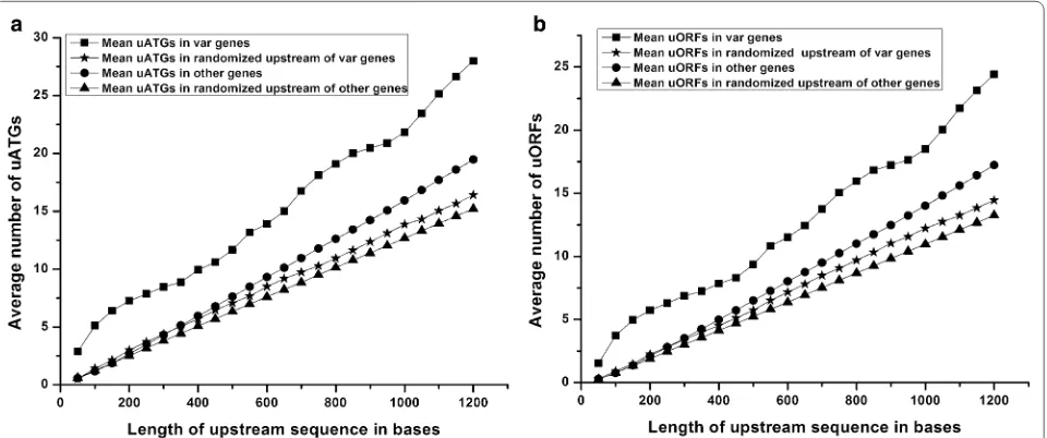
Further, relative frequencies of uATGs and uORFs were compared among the upstream regions of var and other genes and randomized sequences of the same. The average number of uATGs and uORFs was calculated in windows of 50 bases, up to 1200 bases upstream of the start codon. 1.2 kb was selected since the transcription start sites of three of the var genes have been predicted to be at around −1200 bases [31, 48, 49]. Interestingly,
the average number of uATGs and uORFs proximal to var genes was consistently higher than those found in other genes and their randomized upstream sequences throughout the length of the upstream regions under study (Fig. 2). Higher frequencies of uATGs and uORFs in var 5′ upstream regions in comparison to randomized sequence also suggest that it is not a random phenome-non and may have some functional significance.
Also of note is that the fold differences in the average number of uATGs and uORFs associated with var and other genes were not uniform (Fig. 3). Within 50 bases upstream of the start codon, there were ~5 times more uATGs and uORFs in var genes versus other genes. This is largely due to the fact that other genes have virtually no uATGs and uORFs in these regions while var genes have an average number of ~1.5 uORFs and ~3 uATGs in the same region. Up to 300 bases upstream of the start codon, the fold difference between var gene-associated uATGs and uORFs and other genes-associated uATGs and uORFs decreases until it reaches ~2; after 300 bases the fold difference in average uATGs and uORFs lev-els off at ~1.5. These data indicate a high frequency of uATGs and uORFs within 100 bases of the start codon of var genes. Should the ribosome initiate at one of these numerous uATGs and uORFs, profound effects on trans-lation of the downstream var gene could be envisaged.
Insertion of a var upstream sequence represses expression of a downstream luciferase reporter gene
To study the effect of var gene-associated uATGs/uORFs on the translation of the downstream ORF, the −60 bp region
[image:5.595.60.540.488.689.2](−60 to −1) from one of the var genes was cloned upstream of a luciferase reporter gene. The region was chosen because the 50 bp var upstream regions contain ~5 times more uORFs and uATGs than other CDS of the parasite (Fig. 3). The 60 bases var upstream sequence was expected to be part of the var mRNA since the average length of the 5′ leader sequences of the parasite mRNA is predicted to be 346 bases and the length of 5′ leader sequences of a few var genes is more than 1 kb [31, 48, 49]. Nevertheless, RT-PCR of var upstream regions carried out from parasite RNA revealed that the 60 bases var upstream sequence was indeed part of var mRNA (Additional file 3: Figure S1). Therefore, this 60 bp var upstream sequence was tested for its effect on the expression of a downstream ORF.
The effects of uORFs on var gene expression have been tested for the var2csa gene where a var2csa-uORF down-regulates the translation of a downstream ORF [39, 40]. These studies have used the approach of stable transfection of a plasmid carrying a drug resistance gene under a con-stitutive promoter and a reporter gene (luciferase coding sequence or drug resistance marker) driven by the var pro-moter and var 5′ leader. Effect of the mutations in the var uORF are tested by quantifying luciferase expression or by measuring the time taken for the stable line to be selected under drug pressure, when resistance marker is used.
In this report, a transient transfection approach was used to study the role of uAUGs and uORFs in regula-tion of the luciferase gene expression. In these experi-ments, the time taken for testing constructs is fast (80–85 h), however due to low transfection efficiencies, it
is not possible to test whether mutations in the 5ʹ leader have any effect on mRNA levels. Therefore, care has been taken to state that the results reflect the effects of uAUGs and uORFs on “gene expression” of the luciferase reporter. Gene expression is here defined as the sum of mRNA levels and translation.
To assess the effect of var sequences proximal to the start codon on the expression of the downstream ORF, the
−60 bp region of one of the var genes with five uATGs and three uORFs (PF3D7_0400100) was cloned upstream of the luciferase start codon in vector Pf86 (Pf86-60var). The upstream region inserted into Pf86 contained five ATGs (ATG2 to ATG5), but during cloning, an extra ATG
(ATG1) was introduced in the vector since the upstream
sequence was cloned at an NcoI site. Pf86 vector was used as positive control and the Pf86 vector that had a G to T mutation at the +4 position (resulting in generation of a stop codon immediately after the start codon of luciferase; Pf86-stop) was used as a negative control.
The construct Pf86-60var was transiently transfected and luciferase assays performed. The introduction of 60 bases of var upstream sequences resulted in a loss of luciferase activity to the level of the negative control (Fig. 4). To check the role of uAUGs in this phenomenon, a construct where all the six uATGs were mutated to
Fig. 3 Ratio of uATGs and uORFs. Ratio of average number of uATGs and uORFs associated with var and other genes of the parasite with increasing length of the regions upstream of the start codons. Ratios were calculated in windows of 50 bases, up to 1200 base pairs upstream of the start codons
[image:6.595.57.290.89.286.2] [image:6.595.306.540.419.589.2]TTG [Pf86-60var(mut 1 to 6)] was generated. Luciferase readings of the construct Pf86-60var(mut 1 to 6) were found to be restored to the level of the positive control Pf86 (Fig. 4). This result indicated that the uAUGs pre-sent in the 60 bp region are capable of repressing the expression of the luciferase gene.
A single uAUG is sufficient to repress expression of the luciferase gene
Further, to test the strength of individual uAUGs in repressing expression of the luciferase gene, constructs were generated to contain only one uATG at a time. Con-structs Pf86-60var(mut 2 to 6), Pf86-60var(mut 1, 3, 4, 5, 6) and Pf86-60var(mut 1 to 5) had all the ATGs in the 60 bp region mutated to TTG except ATG1, ATG2 and
ATG6 respectively. Construct Pf86-60var(mut 2) was also
generated in which only ATG2 was mutated.
Luciferase activities of the constructs were measured after transient transfections. The expression levels of the constructs were similar to the negative control indicating that even one uAUG is sufficient in repressing expres-sion of the downstream ORF (Fig. 5). However, individ-ual uAUGs showed varied levels of repression: construct Pf86-60var(mut 2 to 6) showed ~5 % of Pf86 luciferase
activity, while constructs Pf86-60var(mut 1 to 5) and Pf86-60var(mut 1, 3, 4, 5, 6) showed ~10 and ~20 % of Pf86 luciferase activity respectively (Fig. 5).
The loss of luciferase activities of all these con-structs appears to be due to the uATG having an in frame stop codon and leading to the formation of an ORF. The uATG1 initiates an ORF of 147 bases that overlaps with the luciferase ORF, while uATG2 initi-ates a uORF of 18 bases. The uATG6 is immediately followed by a stop codon. Thus, the absence of lucif-erase activity of the constructs appears to be due to the failure of the scanning ribosome to reach the luciferase start codon.
Upstream sequence from a gene of unknown function also represses luciferase gene expression
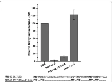
In order to test if the ability of the uAUGs to repress the downstream ORF is specific to var gene upstream sequence, a −60 bp sequence was cloned from a gene of unknown function, PF3D7_0517100. The gene was selected since the 60 bp upstream sequence of this gene also contained five uATGs and three uORFs and its sequence was different from that of the var gene upstream sequence cloned (Pf86-60var). During cloning, an extra ATG (ATG1) was introduced resulting in a total
of six uATGs. The clone (Pf86-60_0517100) was tran-siently transfected and luciferase assays were carried out. The construct resulted in only ~15 % of the luciferase activity in comparison to Pf86 (Fig. 6).
To check if the repression of the luciferase activity was mediated by uAUGs, all the six uATGs were mutated [Pf86-60_0517100(mut 1 to 6)]. The mutations restored luciferase activity indicating that the repression was mediated by uAUGs (Fig. 6). Therefore, it appears that the ability of uAUGs to repress the downstream ORF is a general phenomenon observed in P. falciparum.
The ability of a Kozak sequence to repress reporter expression does not correlate with the frequency in the P.
falciparum genome
As discussed earlier, the Kozak sequence is the most important feature governing the strength of uAUGs [15]. However, little is known about the strength of Kozak sequences in P. falciparum. In this report, individual uAUGs were capable of repressing the expression of lucif-erase gene effectively (Fig. 5). When the Kozak sequences of these uAUGs were analysed, it was observed that they were different, some of which (Kozak sequences of Pf86 and uATG1) were not found associated with any annotated parasite CDS while Kozak sequences of Pf86-60var and uATG2 were found at low frequencies. The observation that uAUGs with differing Kozak sequences could repress luciferase led us to check whether Kozak
[image:7.595.57.289.399.573.2]sequences frequently found in the parasite genome are able to drive higher reporter expression.
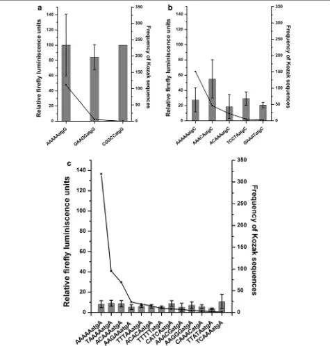
For this, the frequencies of Kozak sequences associ-ated with 5401 annotassoci-ated CDS of P. falciparum were calculated. The −5 to +4 positions of annotated start
codons were considered since a bioinformatics study [45] indicated these positions to be important in decid-ing the translational ability of a start codon of mRNA. Out of 4096 possible Kozak sequences, the parasite uses 1086 Kozak sequences (Additional file 4: Table S3). As expected due to the AT biased genome, AAAAAatgA was the most frequent Kozak sequence, (318) followed by TAAAAatgA (151).
In mammals, frequent Kozak sequences are found to be stronger in driving translation [50]. Therefore, the rela-tionship between the frequency and the ability of Kozak sequences to drive reporter expression in P. falciparum was tested. Due to lack of mRNA data, only the quanti-tative effect of a particular Kozak sequence on reporter gene expression has been tested. For this, 21 of the Kozak sequences of different frequencies from the most fre-quent (318) to least frefre-quent (0) sequence were selected.
The plasmid Pf86 was modified such that the lucif-erase start codon was surrounded by different Kozak sequences. Thus, 20 constructs were generated. Wild
type Pf86 was taken as a control. No correlation was observed between the reporter activity and the frequency of the Kozak sequences in P. falciparum, unlike in other eukaryotes. In fact, the nucleotides (−5 to −1) preced-ing the start codon showed scant effect on expression of the luciferase gene (Fig. 7). To reach this conclusion, the constructs were classified based on the nucleotide pre-sent at the +4 position as this was within the luciferase coding region and would alter the second amino acid of the luciferase protein. Comparisons were made between constructs having the same nucleotide at the +4 position with different nucleotides at the −5 to −1 position.
Constructs containing ‘G’ at +4 positions showed high firefly luciferase activities (~100 %) despite contain-ing very different bases at positions −1 to −5 (Fig. 7a). Similarly, for constructs containing ‘C’ at +4 posi-tions, luciferase activities remained around 20–30 % except one even after altering the preceding bases com-pletely (Fig. 7b). Finally, low firefly activities (~10 %) were obtained from all the constructs containing differ-ent Kozak sequences at −1 to −5 positions but ‘A’ at +4 positions (Fig. 7c). Therefore, it appears that nucleotides preceding the start codon (−5 to −1) do not play a sig-nificant role in determining the strength of the Kozak sequences. As a result the Kozak sequence which was found to be one of the strongest (CGGCCatgG) was not present in the parasite genome at all (Fig. 7).
Discussion
This report adds to the accumulating data on the effect of uAUGs and uORFs on the expression of the downstream ORF in P. falciparum.
Upstream AUGs are likely to regulate downstream ORFs via translation
In this report, the mRNA levels of different constructs in the transfection experiments have not been verified. Therefore, all statements have been made with regard to the effects of uAUGs and uORFs on the “expression” of the luciferase reporter, where expression is defined as the sum of mRNA and protein levels. However, the possibility of mRNA instability by introducing various point muta-tions in different constructs (Fig. 5) cannot be ruled out in the current study. Nevertheless, translational repres-sion might be the mechanism of uAUG action because of following reasons. It is unlikely that changing single A to U at different positions in the 5′ leader plasmid Pf86-60var (Fig. 5) would affect the stability of mRNAs. There are several reports where point mutations in uAUGs have been shown to de-repress the translation of downstream ORF without altering the mRNA levels [9, 20, 40, 51–56]. As indirect evidence, the stability of the −60 bp region was predicted by the Mfold algorithm [57] and mutating
[image:8.595.57.291.88.260.2]the uAUGs does not alter the stability of the −60 bases sequences significantly (ΔG of the most stable folding ranged between −14 and −9.00 kcal/mol). Finally, evi-dence supportive of the contention that mutations may not affect mRNA stability comes from our experiments where another set of mutations, altering uATGs to TTGs completely restores luciferase activity (Fig. 6), strongly indicating that the underlying phenomenon is translation regulation.
The effect of uAUGs on translation is mediated either in a peptide-dependent or peptide-independent manner. In the peptide-dependent mechanism, a peptide formed after the ribosome begins translation at the uAUG binds the ribosome and stalls it [58]. There are only few reports of uAUGs acting via a peptide-dependent mechanism [58, 59]. In the peptide-independent mechanism, when a scanning ribosome encounters a uAUG, it can begin translation resulting in down-regulation of the main ORF or can simply continue scanning and initiate translation from the next AUG in a favorable context [60]. In such cases, down-regulation of the main ORF takes place because of the reduced probability of the scanning ribo-some reaching the main AUG [61] or due to the presence of rare codons in the region between uAUG and main AUG [52] or due to the reduced concentration of transla-tion factors [61].
P.falciparum parasites might employ uAUGs to regulate the translation of mRNAs
hsp86 is a house-keeping gene and codes for a heat shock protein. The 5′ leader of hsp86 is 686 bases long and contains a single uAUG 495 bases upstream of the start codon. Since uAUG close to the mRNA cap is less efficient in repressing translation [62], this hsp86 uAUG may not affect translation of the downstream ORF. On the other hand, var genes contain on an average of ~5 uATGs within 50 bases upstream of the start codon. When 60 bases upstream region of a var gene and a gene with unknown function containing five uATGs was introduced within the 5′ leader of hsp86, luciferase activ-ity was completely lost (Figs. 5, 6), hinting at the role of uAUGs in translational efficiency and protein abundance.
For mammalian cells, mRNAs containing uORFs were found to have lower protein to mRNA ratio than mRNAs containing no uORF [9, 10]. This indicates that cells might use different numbers of uATGs/uORFs to fine tune the translation of a mRNA with genes encod-ing low abundance proteins havencod-ing higher numbers of uAUGs/uORFs. Hints that P. falciparum might use this strategy come from our data showing that parasite CDS have different numbers of uATGs/uORFs (Fig. 1) and that insertion of six uAUGs into a heterologous pro-moter drastically reduces luciferase activity (Figs. 5, 6).
To further explore this, a preliminary analysis of poly-some profiling data [5] was conducted by linking genes having exon reads of polysome-bound mRNAs with their number of uATGs (data from this report). This analysis revealed a weak negative correlation between the number of uAUGs and the raw exon reads of the CDS; mRNAs with higher read counts in their CDS are likely to contain lesser number of uAUGs/uORFs. Assuming that mRNAs with higher reads in the CDS are likely to have higher translational efficiency, this pre-liminary analysis hints at a possible correlation between the number of uAUGs and protein levels. However, the hypothesis needs to be tested further by quantifying par-asite proteins whose genes contain different numbers of uAUGs/uORFs.
Large numbers of uAUGs in parasite mRNAs will pose challenge for the translation initiation machinery scanning the leader
It appears that the nucleotides preceding the start codon do not play a large role in determining the strength of a Kozak sequence (Fig. 7). This is consistent with our data where uAUGs having different Kozak sequences were all able to repress the luciferase gene very strongly (Fig. 5). This leads to the next intriguing question: how is the translation of var and other mRNAs containing large number of uAUGs achieved? One interesting way to achieve this is by ribosome shunting, where a scanning ribosome skips a part of the 5′ leader of mRNA. Recently, ribosome shunting has been shown to skip a uAUG with a favourable Kozak sequence, and also a potential hair-pin loop in the 5′ leader of the mRNA [63]. Interestingly, differences in the secondary structure of the region sur-rounding uAUGs and main AUG were also observed (Fig. 8). uAUGs were found to be present in a more sta-ble secondary structure than main AUGs. Whether this difference is relevant for the translation machinery to skip uAUGs and recognize the main AUG remains to be investigated.
Conclusions
transcriptional gene regulation and how the parasite translation machinery skips uAUGs and starts transla-tion from the main AUGs.
Abbreviations
uAUG/ATG: upstream AUG/ATG; uORF: upstream open reading frame; CDS: coding sequences.
Authors’ contributions
MK and SP conceived the study and designed the experimental set up. VS car-ried out the bioinformatics work. MK carcar-ried out the experiments. MK and SP wrote the manuscript. All authors read and approved the final manuscript.
Acknowledgements
This work was funded in part by the Industrial Research and Consultancy Centre (IRCC), IIT Bombay. MK and VS were supported by Research Fellowships from the University Grants Commission (UGC). We thank IIT Bombay hospital and staff for assisting in blood collection from the volunteers for parasite cul-turing. We are thankful to Kevin Militello and Dyann Wirth (Harvard School of Public Health) for sharing the plasmids Pf86 and Pfrluc. We thank Kirk Deitsch (Cornell University), Rahul Chaudhari and Prasad Babar (IIT Bombay) for their critical comments on the manuscript.
Competing interests
The authors declare that they have no competing interests. Additional files
Additional file 1: Table S1. List of primers used to generate Pf86 con-taining luciferase start codon under different Kozak sequences. Additional file 2: Table S2. Numbers of uATGs associated with each annotated CDS in the parasite genome.
Additional file 3: Figure S1. RT-PCR of the mixed stage parasite RNA. Additional file 4: Table S3. Frequency of different Kozak sequences in annotated CDS in the parasite genome.
Received: 30 August 2015 Accepted: 8 December 2015
References
1. WHO. World Malaria Report 2014. Geneva: World Health Organization; 2014.
2. Florens L, Washburn MP, Raine JD, Anthony RM, Grainger M, Haynes JD, et al. A proteomic view of the Plasmodium falciparum life cycle. Nature. 2002;419:520–6.
3. Hayward RE, Derisi JL, Alfadhli S, Kaslow DC, Brown PO, Rathod PK. Shot-gun DNA microarrays and stage-specific gene expression in Plasmodium
falciparum malaria. Mol Microbiol. 2000;35:6–14.
4. Llinas M, Bozdech Z, Wong ED, Adai AT, DeRisi JL. Comparative whole genome transcriptome analysis of three Plasmodium falciparum strains. Nucleic Acids Res. 2006;34:1166–73.
5. Bunnik EM, Chung DW, Hamilton M, Ponts N, Saraf A, Prudhomme J, et al. Polysome profiling reveals translational control of gene expression in the human malaria parasite Plasmodium falciparum. Genome Biol. 2013;14:R128.
6. Caro F, Ahyong V, Betegon M, DeRisi JL. Genome-wide regulatory dynam-ics of translation in the asexual blood stages. Elife. 2014;3:e04106. 7. Le Roch KG, Johnson JR, Florens L, Zhou Y, Santrosyan A, Grainger M, et al.
Global analysis of transcript and protein levels across the Plasmodium
falciparum life cycle. Genome Res. 2004;14:2308–18.
8. Iacono M, Mignone F, Pesole G. uAUG and uORFs in human and rodent 5′untranslated mRNAs. Gene. 2005;349:97–105.
9. Calvo SE, Pagliarini DJ, Mootha VK. Upstream open reading frames cause widespread reduction of protein expression and are polymorphic among humans. Proc Natl Acad Sci USA. 2009;106:7507–12.
10. Matsui M, Yachie N, Okada Y, Saito R, Tomita M. Bioinformatic analysis of post-transcriptional regulation by uORF in human and mouse. FEBS Lett. 2007;581:4184–8.
11. Hunter AR, Jackson RJ, Hunt T. The role of complexes between the 40-S ribosomal subunit and Met-tRNA-Met-f in the initiation of protein synthe-sis in the wheat-germ system. Eur J Biochem. 1977;75:159–70.
12. Kozak M. Possible role of flanking nucleotides in recognition of the AUG initiator codon by eukaryotic ribosomes. Nucleic Acids Res. 1981;9:5233–52.
13. Kozak M. The scanning model for translation: an update. J Cell Biol. 1989;108:229–41.
14. Kozak M. Selection of initiation sites by eucaryotic ribosomes: effect of inserting AUG triplets upstream from the coding sequence for preproin-sulin. Nucleic Acids Res. 1984;12:3873–93.
15. Ferreira JP, Overton KW, Wang CL. Tuning gene expression with synthetic upstream open reading frames. Proc Natl Acad Sci USA. 2013;110:11284–9.
16. Wang XQ, Rothnagel JA. 5′-untranslated regions with multiple upstream AUG codons can support low-level translation via leaky scanning and reinitiation. Nucleic Acids Res. 2004;32:1382–91.
17. Hinnebusch AG. Translational regulation of yeast GCN4. A window on factors that control initiator-trna binding to the ribosome. J Biol Chem. 1997;272:21661–4.
18. Luo Z, Sachs MS. Role of an upstream open reading frame in mediating arginine-specific translational control in Neurospora crassa. J Bacteriol. 1996;178:2172–7.
19. Jorgensen RA, Dorantes-Acosta AE. Conserved peptide upstream open reading frames are associated with regulatory genes in angiosperms. Front Plant Sci. 2012;3:191.
20. Medenbach J, Seiler M, Hentze MW. Translational control via protein-regulated upstream open reading frames. Cell. 2011;145:902–13. 21. Morris DR, Geballe AP. Upstream open reading frames as regulators of
mRNA translation. Mol Cell Biol. 2000;20:8635–42.
22. Alderete JP, Child SJ, Geballe AP. Abundant early expression of gpUL4 from a human cytomegalovirus mutant lacking a repressive upstream open reading frame. J Virol. 2001;75:7188–92.
23. Hernandez-Sanchez C, Mansilla A, de la Rosa EJ, Pollerberg GE, Martinez-Salas E, de Pablo F. Upstream AUGs in embryonic proinsulin mRNA control its low translation level. EMBO J. 2003;22:5582–92.
[image:11.595.57.290.87.269.2]24. Su XZ, Heatwole VM, Wertheimer SP, Guinet F, Herrfeldt JA, Peterson DS, et al. The large diverse gene family var encodes proteins involved in cytoadherence and antigenic variation of Plasmodium falciparum -infected erythrocytes. Cell. 1995;82:89–100.
25. Biggs BA, Anders RF, Dillon HE, Davern KM, Martin M, Petersen C, et al. Adherence of infected erythrocytes to venular endothelium selects for antigenic variants of Plasmodium falciparum. J Immunol. 1992;149:2047–54.
26. Leech JH, Barnwell JW, Miller LH, Howard RJ. Identification of a strain-spe-cific malarial antigen exposed on the surface of Plasmodium falciparum -infected erythrocytes. J Exp Med. 1984;159:1567–75.
27. Gardner MJ, Hall N, Fung E, White O, Berriman M, Hyman RW, et al. Genome sequence of the human malaria parasite Plasmodium falcipa-rum. Nature. 2002;419:498–511.
28. Kyes S, Pinches R, Newbold C. A simple RNA analysis method shows var and rif multigene family expression patterns in Plasmodium falciparum. Mol Biochem Parasitol. 2000;105:311–5.
29. Freitas-Junior LH, Hernandez-Rivas R, Ralph SA, Montiel-Condado D, Ruvalcaba-Salazar OK, Rojas-Meza AP, et al. Telomeric heterochroma-tin propagation and histone acetylation control mutually exclusive expression of antigenic variation genes in malaria parasites. Cell. 2005;121:25–36.
30. Guizetti J, Martins RM, Guadagnini S, Claes A, Scherf A. Nuclear pores and perinuclear expression sites of var and ribosomal DNA genes correspond to physically distinct regions in Plasmodium falciparum. Eukaryot Cell. 2013;12:697–702.
31. Lopez-Rubio JJ, Gontijo AM, Nunes MC, Issar N, Hernandez Rivas R, Scherf A. 5′ flanking region of var genes nucleate histone modification patterns linked to phenotypic inheritance of virulence traits in malaria parasites. Mol Microbiol. 2007;66:1296–305.
32. Merrick CJ, Dzikowski R, Imamura H, Chuang J, Deitsch K, Duraisingh MT. The effect of Plasmodium falciparum Sir2a histone deacetylase on clonal and longitudinal variation in expression of the var family of virulence genes. Int J Parasitol. 2010;40:35–43.
33. Ralph SA, Scheidig-Benatar C, Scherf A. Antigenic variation in Plasmodium
falciparum is associated with movement of var loci between subnuclear
locations. Proc Natl Acad Sci USA. 2005;102:5414–9.
34. Salanti A, Dahlback M, Turner L, Nielsen MA, Barfod L, Magistrado P, et al. Evidence for the involvement of VAR2CSA in pregnancy-associated malaria. J Exp Med. 2004;200:1197–203.
35. Duffy MF, Caragounis A, Noviyanti R, Kyriacou HM, Choong EK, Boysen K, et al. Transcribed var genes associated with placental malaria in Malawian women. Infect Immun. 2006;74:4875–83.
36. Rottmann M, Lavstsen T, Mugasa JP, Kaestli M, Jensen AT, Muller D, et al. Differential expression of var gene groups is associated with morbidity caused by Plasmodium falciparum infection in Tanzanian children. Infect Immun. 2006;74:3904–11.
37. Salanti A, Staalsoe T, Lavstsen T, Jensen AT, Sowa MP, Arnot DE, et al. Selec-tive upregulation of a single distinctly structured var gene in chondroitin sulphate A-adhering Plasmodium falciparum involved in pregnancy-associated malaria. Mol Microbiol. 2003;49:179–91.
38. Kyes SA, Christodoulou Z, Raza A, Horrocks P, Pinches R, Rowe JA, et al. A well-conserved Plasmodium falciparum var gene shows an unusual stage-specific transcript pattern. Mol Microbiol. 2003;48:1339–48. 39. Amulic B, Salanti A, Lavstsen T, Nielsen MA, Deitsch KW. An upstream
open reading frame controls translation of var2csa, a gene implicated in placental malaria. PLoS Pathog. 2009;5:e1000256.
40. Bancells C, Deitsch KW. A molecular switch in the efficiency of transla-tion reinitiatransla-tion controls expression of var2csa, a gene implicated in pregnancy-associated malaria. Mol Microbiol. 2013;90:472–88. 41. Patakottu BR, Mamidipally C, Patankar S, Noronha S. In Silico analysis
of translation initiation sites from P. falciparum. Online J Bioinform. 2009;10:259–79.
42. Aurrecoechea C, Brestelli J, Brunk BP, Dommer J, Fischer S, Gajria B, Gao X, Gingle A, Grant G, Harb OS, et al. PlasmoDB: a functional genomic database for malaria parasites. Nucleic Acids Res. 2009;37:D539–43. 43. http://www.bio.iitb.ac.in/~patankar/software/.
44. Deitsch K, Driskill C, Wellems T. Transformation of malaria parasites by the spontaneous uptake and expression of DNA from human erythrocytes. Nucleic Acids Res. 2001;29:850–3.
45. Srinivas V, Kumar M, Noronha S, Patankar S. ORFpred: a machine learning program to identify translatable small open reading frames in intergenic regions of the Plasmodium falciparum genome. Curr Bioinform. (In Press).
46. Watanabe J, Sasaki M, Suzuki Y, Sugano S. Analysis of transcriptomes of human malaria parasite Plasmodium falciparum using full-length enriched library: identification of novel genes and diverse transcription start sites of messenger RNAs. Gene. 2002;291:105–13.
47. Brancucci NM, Witmer K, Schmid C, Voss TS. A var gene upstream element controls protein synthesis at the level of translation initiation in
Plasmo-dium falciparum. PLoS One. 2014;9:e100183.
48. Deitsch KW, del Pinal A, Wellems TE. Intra-cluster recombination and var transcription switches in the antigenic variation of Plasmodium falcipa-rum. Mol Biochem Parasitol. 1999;101:107–16.
49. Voss TS, Kaestli M, Vogel D, Bopp S, Beck HP. Identification of nuclear proteins that interact differentially with Plasmodium falciparum var gene promoters. Mol Microbiol. 2003;48:1593–607.
50. Kozak M. An analysis of 5′-noncoding sequences from 699 vertebrate messenger RNAs. Nucleic Acids Res. 1987;15:8125–48.
51. Child SJ, Miller MK, Geballe AP. Translational control by an upstream open reading frame in the HER-2/neu transcript. J Biol Chem. 1999;274:24335–41.
52. Col B, Oltean S, Banerjee R. Translational regulation of human methio-nine synthase by upstream open reading frames. Biochim Biophys Acta. 2007;1769:532–40.
53. Jeon S, Kim J. Upstream open reading frames regulate the cell cycle-dependent expression of the RNA helicase Rok1 in saccharomyces cerevi-siae. FEBS Lett. 2010;584:4593–8.
54. Jousse C, Bruhat A, Carraro V, Urano F, Ferrara M, Ron D, Fafournoux P. Inhibition of CHOP translation by a peptide encoded by an open reading frame localized in the chop 5′UTR. Nucleic Acids Res. 2001;29:4341–51. 55. Kos M, Denger S, Reid G, Gannon F. Upstream open reading frames
regulate the translation of the multiple mRNA variants of the estrogen receptor alpha. J Biol Chem. 2002;277:37131–8.
56. Mueller PP, Hinnebusch AG. Multiple upstream AUG codons mediate translational control of GCN4. Cell. 1986;45:201–7.
57. Zuker M. Mfold web server for nucleic acid folding and hybridization prediction. Nucleic Acids Res. 2003;31:3406–15.
58. Fang P, Wang Z, Sachs MS. Evolutionarily conserved features of the arginine attenuator peptide provide the necessary requirements for its function in translational regulation. J Biol Chem. 2000;275:26710–9. 59. Law GL, Raney A, Heusner C, Morris DR. Polyamine regulation of
ribo-some pausing at the upstream open reading frame of S-adenosylmethio-nine decarboxylase. J Biol Chem. 2001;276:38036–43.
60. Kozak M. Point mutations define a sequence flanking the AUG initia-tor codon that modulates translation by eukaryotic ribosomes. Cell. 1986;44:283–92.
61. Hood HM, Neafsey DE, Galagan J, Sachs MS. Evolutionary roles of upstream open reading frames in mediating gene regulation in fungi. Annu Rev Microbiol. 2009;63:385–409.
62. Sedman SA, Gelembiuk GW, Mertz JE. Translation initiation at a down-stream AUG occurs with increased efficiency when the updown-stream AUG is located very close to the 5′ cap. J Virol. 1990;64:453–7.