RESEARCH
YKL-40 is highly expressed
in the epicardial adipose tissue of patients
with atrial fibrillation and associated with atrial
fibrosis
Qing Wang
†, Hua Shen
†, Jie Min
†, Yang Gao, Kai Liu, Wang Xi, Jie Yang, Liang Yin, Jibin Xu, Jian Xiao
*and Zhinong Wang
*Abstract
Background: YKL-40 (CHI3L1) is a novel biomarker for inflammation, tissue remodeling, and fibrosis, as well as cardio-vascular diseases. We investigated the association between YKL-40 expression in epicardial adipose tissue (EAT) and atrial fibrosis in patients with atrial fibrillation (AF).
Methods: Blood samples, subcutaneous adipose tissue (SAT), paracardial adipose tissue (PAT), EAT, and adjacent atrial myocardium were acquired from patients receiving coronary artery bypass grafts. The patients were divided into the AF group (n = 28) and the sinus rhythm (SR) group (n = 36).
Results: We did not detect a significant difference in the serum YKL-40 levels in the SR and AF groups (P = 0.145). Quantitative real-time PCR showed that YKL-40 (CHI3L1) mRNA levels in the EAT were significantly higher than in the SAT or PAT of AF patients, or the EAT of SR patients (All P < 0.001). We found similar results for YKL-40 protein levels by immunohistochemistry. Masson staining showed significantly more fibrosis in AF patients than in SR patients (P < 0.001). Western blotting indicated that AF patients had significantly higher expression of collagen I (P = 0.039). We found a linear relationship between YKL-40 mRNA expression and the collagen volume fraction of the atrial myo-cardium (y = 3.576x + 26.205, P < 0.001). Multivariate linear regression analysis revealed that body mass index is an independent risk factor for YKL-40 expression in EAT (β = 0.328, P = 0.011).
Conclusions: YKL-40, which is highly expressed in the EAT of patients with AF, is affected by body mass index and associated with atrial fibrosis, which may contribute to the development of AF.
Keywords: YKL-40, Epicardial adipose tissue, Atrial fibrosis, Atrial fibrillation, Body mass index
© The Author(s) 2018. This article is distributed under the terms of the Creative Commons Attribution 4.0 International License (http://creat iveco mmons .org/licen ses/by/4.0/), which permits unrestricted use, distribution, and reproduction in any medium, provided you give appropriate credit to the original author(s) and the source, provide a link to the Creative Commons license, and indicate if changes were made. The Creative Commons Public Domain Dedication waiver (http://creat iveco mmons .org/ publi cdoma in/zero/1.0/) applies to the data made available in this article, unless otherwise stated.
Background
Atrial fibrillation (AF), the commonest sustained arrhyth-mia in clinics, burdens millions of people worldwide, with increasing prevalence, mortality, and morbidity [1]. As a result, it has become a hotspot in cardiovascu-lar research, both in clinical and basic research settings.
However, its underlying mechanism is still elusive. Atrial remodeling plays a key role in the pathogenesis of AF [2]. Atrial fibrosis, the fundamental process of atrial struc-tural remodeling, greatly contributes to the incidence and maintenance of AF [3].
Epicardial adipose tissue (EAT), once merely thought to be a protective and metabolic tissue for the heart, has recently been found to be related to several cardiovascu-lar diseases, especially AF [4]. Nevertheless, the specific relationship between EAT and AF remains elusive. YKL-40 (CHI3L1), an emerging biomarker in cardiovascu-lar disease, is named for its first 3 terminal amino acids
Open Access
*Correspondence: 18916066266@189.cn; wangzn007@163.com
†Qing Wang, Hua Shen and Jie Min contributed equally to this work
[tyrosine (Y), lysine (K), and leucine (L)] and its appar-ent molecular weight [5]. YKL-40 is expressed by the gene chitinase-3-like protein 1 (CHI3L1) in several cell types, such as vascular smooth muscle cells, monocytes, and macrophages [6]. YKL-40 was first described as an inflammatory glycoprotein, which involved in asthma and lung function [7]. Recent research showed that it can be highly expressed in visceral adipose tissue [8] and par-ticipate in cardiovascular disease and diabetes [5]. In this study, we investigated YKL-40 expression in EAT and its relationship with atrial fibrosis in AF patients.
Methods
Study population
From March 2016 to Dec 2017, a total of 64 patients who received coronary artery bypass grafts in Changzheng Hospital affiliated with the Second Military Medical University, were enrolled in this study: 36 patients with sinus rhythm (SR) and 28 patients with persistent or per-manent AF. Exclusion criteria included structural heart disease, severe hepatic or renal dysfunction, metabolic disease, infectious disease, cancer, or age over 80 years. AF was determined by 12-lead electrocardiogram. The baseline characteristics of all patients were collected for analysis.
Sample acquisition
Peripheral venous blood samples were acquired in the first morning after the admission. The serum was centri-fuged and stored at − 80 °C. Adipose tissue was obtained during surgery, before the cardiopulmonary bypass. The different types of adipose tissue (average 0.5 g each) were collected from each patient, including subcutane-ous adipose tissue (SAT) at the chest incision, paracar-dial adipose tissue (PAT) from the pericardium, and EAT from the atrioventricular groove next to the right atrial appendage. Each biopsy was divided into two portions; one was frozen immediately at − 80 °C for RNA isolation, and the other was immersed in neutralized formalin for immunohistochemistry. The adjacent atrial myocardium (average 0.1 g) from the right atrial appendage tissue was also collected and likewise divided into two portions.
Enzyme‑linked immunosorbent assay
A commercial YKL-40 two-site, sandwich-type enzyme-linked immunosorbent assay kit (Quidel Corporation, San Diego, CA, USA) was used to measure serum YKL-40 levels.
RNA isolation and quantitative real‑time PCR
TRIzol® reagent (Invitrogen, Carlsbad, CA, USA) was used to extract total RNA; the purity of the isolated RNA was assessed by measurement of the optical density at
260 nm and 280 nm. Reverse transcription was per-formed using a High-Capacity cDNA Reverse Transcrip-tion Kit (Applied Biosystems, Foster City, CA, USA), according to the manufacturer’s instructions. SYBR® Premix Ex Taq™ (TakaRA Bio, Tokyo, Japan) was used to prepare the quantitative real-time PCR samples. An ABI Prism 7900 Detector System (Applied Biosystems) was used to perform the quantitative real-time PCR. The primers were designed using Primer Premier 6.0 software (Premier Biosoft, Palo Alto, CA, USA). The primer sequences used in this study were as follows: YKL-40 (CHI3L1), forward 5′-GTG AAG GCG TCT CAA ACA GG-3′, reverse 5′-GAA GCG GTC AAG GGC ATC T-3′; collagen I (COL1A1), forward 5′-CCA AGA CGA AGA CAT CCC ACC-3′, reverse 5′-CAG TTG TCG CAG ACG CAG AT-3′; collagen III (COL3A1), forward 5′-TCG CTC TGC TTC ATC CCA CTAT-3′, reverse 5′-CTT CCA GAC ATC TCT ATC CGCAT-3′; β-actin (ACTB), forward 5′-CAT GTA CGT TGC TAT CCA GGC-3′, reverse 5′-CTC CTT AAT GTC ACG CAC GAT-3′. Relative gene expres-sion was calculated using the threshold cycle value (CT)
and the formula 2−ΔΔCT.
Immunohistochemistry
The paraffin-embedded sections were placed at 60 °C for 1 h, then dewaxed, rehydrated, and rinsed 2–3 times with PBS, each time for 3 min. Next, the sections were subjected to 3% hydrogen peroxide solution at room temperature for 8 min, then rinsed 3 times with PBS (pH 7.2–7.6), each time for 5 min. Sections were incubated with primary antibodies (CHI3L, Abcam, Cambridge, UK) overnight at 4 °C in a moist chamber, then rinsed 3 times with PBS (pH 7.2–7.6). Then, the sections were incubated with secondary antibodies for 1 h at 4 °C, then rinsed 3 times with PBS; 50–100 μl DAB reagent was added. The slices were observed under a microscope. The integrated optical density (IOD) of positively stained tis-sue was calculated to quantify the expression of YKL-40. The IOD of each tissue section was calculated from eight different 400× magnified fields.
Masson staining
1–2 min. The samples were sliced, dehydrated, rendered transparent with xylene, and sealed with optical rubber. The sections were observed under a light microscope, and the image data were collected to calculate the volume fraction of collagen (CVF% = average collagen area/area of total field × 100).
Western blotting
The atrial myocardium samples were weighed and treated with cold Radio-Immunoprecipitation Assay (RIPA) lysis buffer (1 ml/100 mg). The homogenized samples were centrifuged (12,000 rpm, 10 min, 4 °C) and a BCA pro-tein assay kit (Yeason Biotech, Shanghai, China) was used to determine their protein concentrations. Loading buffer was added to diluted supernatants to adjust concentra-tions and volumes. Afterward, samples with equal protein content were subjected to sodium dodecyl sulfate–poly-acrylamide gel electrophoresis on a 10% polysulfate–poly-acrylamide gel, then transferred to a polyvinylidene difluoride mem-brane (Millipore, Boston, MA, USA). Next, the samples were blocked with blocking buffer containing 5% bovine serum albumin for 1 h at room temperature, followed by incubation with antibodies including anti-collagen I (Boster Biological Technology, Wuhan, China) and anti-collagen III (Boster Biological Technology, Wuhan, China), overnight at 4 °C, with anti-GAPDH used as an internal control. The membranes were rinsed 3 times for 10 min with TBST, followed by incubation with the sec-ondary antibody for 1 h. The membranes were washed 3 times with TBST for 15 min. Then, the membranes were developed with chemiluminescence solution A and B mixed at a 1:1 ratio, along with the developing substrate. Image J software was used to calculate the relative optical density of the bands.
Statistical analysis
Continuous data, when distributed normally, were expressed as mean ± standard deviation, while the cat-egorical data were expressed as percentages. Abnormally distributed variables were described as median and inter-quartile range (IQR). The baseline characteristics of the two groups were compared with Student’s t-test and the Chi square test. Immunohistochemistry IOD and mRNA levels between groups were compared using Mann– Whitney test, while the differences among the three types of adipose tissue were detected by Kruskal–Wallis test. The CVF% calculated from the Masson staining and the relative optical density of western blotting between two groups were compared with Student’s t-test. A cor-relation analysis between mRNA expression and CVF% was conducted by univariate linear regression. A multi-variate linear regression model was used to analyze the risk factors affecting the expression of YKL-40 in EAT,
and a enter pattern was adopted for building the model. All data were analyzed with SPSS 22.0 software (IBM, Almonte, NY, USA). Differences were considered signifi-cant when P < 0.05.
Results
Patient characteristics
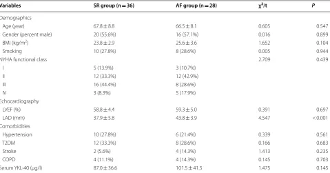
Patient baseline characteristics are shown in Table 1. The demographic data were similar in the two groups includ-ing age, gender, body mass index (BMI), and smokinclud-ing status (P > 0.05). In addition, there were no cardiac func-tion disparities between the two groups, as manifested in NYHA class or left ventricular ejection fraction (LVEF) (P > 0.05). The patients in both groups had similar pro-portions of comorbidities, including hypertension, type 2 diabetes mellitus, stroke, and chronic obstructive pul-monary disease (COPD). However, the average left atrial diameter (LAD) of the AF group was significantly larger than that of the SR group (t = 4.547, P < 0.001). Showed in Fig. 1b, We also tested serum YKL-40, which was higher in the AF group, but the difference was not statistically significant (t = 1.475, P = 0.145).
YKL‑40 expression
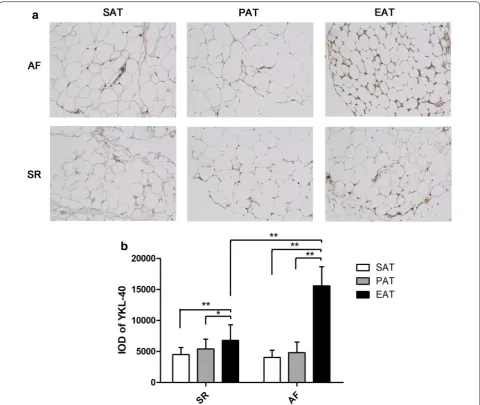
YKL-40 mRNA expression in the adipose tissue of the two groups is shown in Fig. 1a. In the SR group, we detected no significant differences among the three types of adipose tissue (χ2= 5.686, P = 0.058). In the AF group,
YKL-40 mRNA expression differed significantly among the types of adipose tissue (χ2= 52.531, P < 0.001); it was
significantly more highly expressed in EAT than in SAT or PAT (EAT vs SAT: 3.25 (2.09) vs 1.30 (1.70), P < 0.001; EAT vs PAT: 3.25 (2.09) vs 1.45 (0.80), P < 0.001). YKL-40 mRNA expression in SAT and PAT did not differ between the groups (SAT: P = 0.256; PAT: P = 0.735). However, it was significantly more highly expressed in the EAT of the AF group than the SR group (P < 0.001).
sections (EAT vs SAT: 15,582 ± 3080 vs 4808 ± 1693, P < 0.001; EAT vs PAT: 15,582 ± 3080 vs 4032 ± 1166, P < 0.001). Between groups, the IOD of the EAT sec-tions was higher in the AF group than in the SR group (t = 12.528, P < 0.001), although it did not differ in the SAT (t = 1.569, P = 0.122) and PAT sections (t = 1.466, P = 0.148).
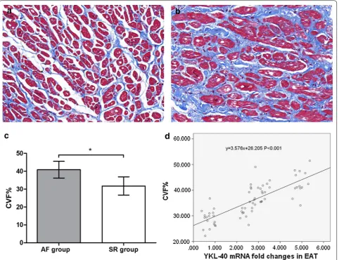
Atrial fibrosis
The Masson-stained sections of atrial myocardium are shown in Fig. 3a and b. The representative sections indi-cated more fibrotic and disordered myocardia in the AF group than in the SR group. As shown in Fig. 3c, the quantitative CVF% of the AF group was signifi-cantly higher than that of the SR group (40.87 ± 4.67 vs Table 1 Baseline characteristics of patients in the atrial fibrillation (AF) and sinus rhythm (SR) groups
BMI body mass index, LVEF left ventricular ejection fraction, LAD left atrial diameter, T2DM type 2 diabetes mellitus, COPD chronic obstructive pulmonary disease,
NYHA New York Heart Association
Variables SR group (n = 36) AF group (n = 28) χ2/t P
Demographics
Age (year) 67.8 ± 8.8 66.5 ± 8.1 0.605 0.547
Gender (percent male) 20 (55.6%) 16 (57.1%) 0.016 0.899
BMI (kg/m2) 23.8 ± 2.9 25.6 ± 3.6 1.652 0.104
Smoking 10 (27.8%) 8 (28.6%) 0.005 0.944
NYHA functional class 2.709 0.439
I 5 (13.9%) 3 (10.7%)
II 12 (33.3%) 12 (42.9%)
III 16 (44.4%) 8 (28.6%)
IV 3 (8.3%) 5 (17.9%)
Echocardiography
LVEF (%) 58.8 ± 4.4 59.3 ± 5.0 0.391 0.697
LAD (mm) 37.9 ± 5.8 43.8 ± 3.9 4.547 < 0.001
Comorbidities
Hypertension 10 (27.8%) 6 (21.4%) 0.339 0.561
T2DM 12 (33.3%) 8 (28.6%) 0.166 0.683
Stroke 2 (5.6%) 4 (14.3%) 1.413 0.235
COPD 4 (11.1%) 4 (14.3%) 0.145 0.703
Serum YKL-40 (μg/l) 87.0 ± 36.6 101.5 ± 41.5 1.475 0.145
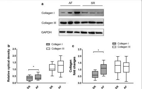
[image:4.595.58.538.101.352.2] [image:4.595.66.538.415.557.2]31.74 ± 5.11, t = 7.357, P < 0.001). The western blotting and quantitative real-time PCR results conformed with the Masson staining, which indicated significantly higher collagen I mRNA and protein expression in the AF group (mRNA: 1.06 ± 0.39 vs 0.62 ± 0.24, t = 5.528, P < 0.001; protein: 0.44 ± 0.19 vs 0.34 ± 0.16, t = 2.109, P = 0.039), whereas there were no significant differences in collagen III expression between the 2 groups (mRNA: 1.11 ± 0.54 vs 1.24 ± 0.47, t = 1.048, P = 0.299; protein: 1.27 ± 0.47 vs 1.08 ± 0.30, t = 1.915, P = 0.060), as shown in Fig. 4. Univariate linear regression revealed a significant cor-relation between YKL-40 mRNA expression and CVF% (y = 3.576x + 26.205, P < 0.001), as shown in Fig. 3d.
Multivariate linear regression analysis
Table 2 shows the results of the multivariate linear regres-sion analysis for YKL-40 expresregres-sion in EAT. Of the base-line characteristic enrolled in this model, only BMI was significantly associated with IOD of EAT in all patients (β = 0.328, P = 0.011).
Discussion
[image:5.595.58.539.87.492.2]Atrial fibrosis is related to the structural remodeling of the atrium, which lays the basis for persistent and per-manent AF [10]. EAT, or pericardial adipose tissue, previ-ously thought to play a protective and metabolic role for the cardiovascular system, has been found to be highly correlated with the incidence and recurrence of AF in recent epidemiologic studies [11, 12]. The association between EAT and AF remains poorly understood; how-ever, current evidence indicates that the arrhythmogenic mechanisms of EAT may include adipocyte infiltration, pro-fibrotic and pro-inflammatory paracrine effects, and oxidative stress [13]. We found that YKL-40 is more highly expressed in EAT than in other types of adipose tissue in AF patients, which may contribute to the devel-opment of AF.
YKL-40 plays a role in the activation of the innate immune system, extracellular matrix remodeling, and the differentiation and maturation of macrophages [14]. Several epidemiologic studies focused on the relation-ship between serum YKL-40 and AF, and showed that AF patients have higher levels of serum YKL-40, which is related to disease severity [15, 16]. However, it cannot be used as a biomarker for predicting the results of electrical cardioversion [16]. We found that YKL-40 mRNA levels were higher in the EAT of AF patients, while there was no significant difference in serum levels between the AF and SR groups, which suggested that EAT may be a depot for YKL-40 and affect the atrium by paracrine secretion. The difference between our results and previous stud-ies in serum YKL-40 level can be explained in two major Fig. 3 Masson staining and quantitative results of atrial myocardium from patients with atrial fibrillation (AF, n = 28) and sinus rhythm (SR, n = 36).
[image:6.595.59.542.88.458.2]reasons. On one hand, Marott [15] led a cohort study focusing on the serum YKL-40 and the risk of develop-ing AF, while we tested it on patients with existed AF. Also, our patients were all complicated by CAD, which
was reported to be associated with YKL-40 level [17]. On the other hand, there was indeed a higher level of YKL-40 for AF patients, but the difference is not statistical signifi-cant, and the reason might be attributed to the relatively smaller sample size than previous study [16]. Although adipocytes may not express YKL-40, the macrophages and other inflammatory cells in the EAT can express it, as confirmed by the study of Catalan et al., who revealed higher expression of YKL-40 in the visceral adipose tissue of obese patients.
YKL-40, acting as a key factor in fibroblast prolifera-tion and matrix deposiprolifera-tion, is related to organic fibrosis, including lung [18] and hepatic fibrosis [19]. Based on our previous findings and the role played by atrial fibro-sis in AF, we hypothesized that YKL-40 may induce AF by promoting atrial fibrosis. Hence, we determined the fibrosis level in all patients and analyzed its relationship with YKL-40 expression. We found that atrial fibrosis is prominent in AF patients compared with SR patients, and the quantitative index of CVF% has a linear relation-ship with YKL-40 mRNA levels. We also assessed the Fig. 4 Western blotting and quantitative real-time PCR results of collagen I (COL1A1) and collagen III (COL3A1) expression in the atrial myocardium of patients with atrial fibrillation (AF, n = 28) and sinus rhythm (SR, n = 36). a Western blotting results of AF patients and SR patients (representative samples). b Relative optical density of western blotting results in the AF and SR groups. Relative optical density of Collagen I in AF group was significantly higher than SR group. c Quantitative real-time PCR results from the AF and SR groups. Collagen I mRNA expression of AF group was significantly higher than SR group. *P < 0.001
Table 2 Multivariate linear regression analysis of the IOD of YKL-40 and baseline characteristics
BMI body mass index, T2DM type 2 diabetes mellitus
Variables B SE β T P
Constant 625.609 8256.052 – 0.076 0.940
Gender 2082.884 1565.724 0.200 1.330 0.189
Age − 44.122 99.980 − 0.072 − 0.441 0.661
Smoking − 844.645 1588.415 − 0.074 − 0.532 0.597
BMI 519.585 196.793 0.328 2.640 0.011
NYHA − 543.476 914.067 − 0.091 − 0.595 0.555
T2DM 1257.607 1471.844 0.113 0.854 0.397
Hypertension − 1607.303 1718.569 − 0.135 − 0.935 0.354
COPD 2694.578 1988.461 0.173 1.355 0.181
[image:7.595.61.539.89.389.2] [image:7.595.56.290.499.642.2]expression of representative collagen I and collagen III in all atrium samples, and noted a marked reduction in collagen I expression in the atrium, with no difference in collagen III. Similarly, Iwata et al. [20] found that YKL-40 secreted from macrophages in adipose tissue inhibits type I collagen degradation.
Furthermore, we tried to find possible risk factors affecting the expression of YKL-40 in EAT, and found that BMI is an independent risk factor. Epidemiologic studies and clinical data both indicate that obesity is commonly associated with AF, and stable weight loss decreases AF burden and AF recurrence following treat-ment [21]. Mild inflammation, insulin resistance, and structural remodeling in obese patients are believed to be the major reasons for the development of AF [22]. Another study found that serum YKL-40 is elevated in morbidly obese patients and declines after weight loss [23]. However, Nielsen et al. demonstrated that YKL-40 is independent of BMI as a biomarker for type 2 diabe-tes mellitus. Our study also found that the serum YKL-40 level did not differ between the AF and SR groups. The similarity between the two groups may be attributable to the limited sample size, but the expression in the EAT varies greatly between the groups.
Four aspects of the limitations of this study should be noted. Firstly, although significant differences have been identified, the relatively inadequate sample size is a dis-advantage and the findings require evidence from fur-ther clinical and basic science studies. Secondly, all of the patients enrolled have coronary artery disease, which may affect the results. Thirdly, the study lacks healthy controls because healthy samples of EAT cannot be obtained for ethical reasons. Lastly, despite similar base-line characteristics, the results can still be influenced by mixed clinical factors. Notably, AF develops in multifac-eted ways and is affected by numerous factors; the results in this study only provide some evidences that YKL-40 might be a booster to atrial fibrosis and AF, which require more experimental studies to validate.
Conclusions
In summary, our study demonstrates that YKL-40 is highly expressed in EAT, especially in AF patients. YKL-40 expression in EAT is closely associated with atrial fibrosis, which is more marked in AF patients. We also found that BMI is the only risk factor for the expression of YKL-40 in EAT. Future studies should focus on the cause–effect relationship of YKL-40 and AF, and basic research should address how YKL-40 activates fibrotic processes in the atrium, which may provide novel targets for the prevention and treatment of AF.
Abbreviations
AF: atrial fibrillation; SR: sinus rhythm; EAT: epicardial adipose tissue; SAT: subcutaneous adipose tissue; PAT: paracardial adipose tissue; CVF%: volume fraction of collagen; IOD: integrated optical density; BMI: body mass index; LVEF: left ventricular ejection fraction; LAD: left atrial diameter; T2DM: type 2 diabetes mellitus; COPD: chronic obstructive pulmonary disease; NYHA: New York Heart Association.
Authors’ contributions
(1) Study conception and design: QW, JM and HS; (2) Sample acquisition and collection of clinical data: YG, JBX and KL; (3) Experiments conduct: HS and WX; (4) Data analysis: JY and LY; (5) Manuscript writing: JM and Q W; (6) Editing and reviewing: JX and ZNW. All authors read and approved the final manuscript.
Acknowledgements Not applicable.
Competing interests
The authors declare that they have no competing interests.
Availability of data and materials
The datasets used and/or analyzed during the current study are available from the corresponding author on reasonable request.
Consent for publication Not applicable.
Ethics approval and consent to participate
This study was approved by the Committee on Ethics of Biomedicine of the Second Military Medical University. This study also complied with the Declara-tion of Helsinki, and signed, written informed consent was obtained from all subjects included in this study.
Funding
This study was funded by the National Nature Science Foundation of China (No. 81670299), Shanghai Shenkang Medicine Developing Project (SHDC12014107), Shanghai Science and Technology Committee Medicine Leading Project (No. 15411960100), Shanghai Leading Talent Program (LJRC-WZN), and PLA Medical Scientist Cultivation Program (17QNP013).
Publisher’s Note
Springer Nature remains neutral with regard to jurisdictional claims in pub-lished maps and institutional affiliations.
Received: 17 April 2018 Accepted: 3 August 2018
References
1. Chugh SS, Havmoeller R, Narayanan K, Singh D, Rienstra M, Benjamin EJ, Gillum RF, Kim YH, McAnulty JH, Zheng ZJ, et al. Worldwide epidemiology of atrial fibrillation a global burden of disease 2010 study. Circulation. 2014;129:837–47.
2. Nattel S, Harada M. Atrial remodeling and atrial fibrillation. J Am Coll Cardiol. 2014;63:2335–45.
3. Jalife J, Kaur K. Atrial remodeling, fibrosis, and atrial fibrillation. Trends Cardiovasc Med. 2015;25:475–84.
4. Hatem SN, Sanders P. Epicardial adipose tissue and atrial fibrillation. Cardiovasc Res. 2014;102:205–13.
5. Rathcke CN, Vestergaard H. YKL-40-an emerging biomarker in cardiovas-cular disease and diabetes. Cardiovasc Diabetol. 2009;8:61.
6. Wynn TA, Barron L. Macrophages: master regulators of inflammation and fibrosis. Semin Liver Dis. 2010;30:245–57.
•fast, convenient online submission
•
thorough peer review by experienced researchers in your field
• rapid publication on acceptance
• support for research data, including large and complex data types
•
gold Open Access which fosters wider collaboration and increased citations maximum visibility for your research: over 100M website views per year
•
At BMC, research is always in progress.
Learn more biomedcentral.com/submissions
Ready to submit your research? Choose BMC and benefit from: 8. Catalan V, Gomez-Ambrosi J, Rodriguez A, Ramirez B, Rotellar F, Valenti V,
Silva C, Gil MJ, Salvador J, Fruhbeck G. Increased circulating and visceral adipose tissue expression levels of YKL-40 in obesity-associated type 2 diabetes are related to inflammation: impact of conventional weight loss and gastric bypass. J Clin Endocrinol Metab. 2011;96:200–9.
9. Lip GY, Tse HF, Lane DA. Atrial fibrillation. Lancet. 2012;379:648–61. 10. Burstein B, Nattel S. Atrial fibrosis: mechanisms and clinical relevance in
atrial fibrillation. J Am Coll Cardiol. 2008;51:802–9.
11. Al Chekakie MO, Welles CC, Metoyer R, Ibrahim A, Shapira AR, Cytron J, Santucci P, Wilber DJ, Akar JG. Pericardial fat is independently associated with human atrial fibrillation. J Am Coll Cardiol. 2010;56:784–8. 12. Chao T-F, Hung C-L, Tsao H-M, Lin Y-J, Yun C-H, Lai Y-H, Chang S-L, Lo L-W,
Hu Y-F, Tuan T-C, et al. Epicardial adipose tissue thickness and ablation outcome of atrial fibrillation. PLoS ONE. 2013;8:e74926.
13. Wong CX, Ganesan AN, Selvanayagam JB. Epicardial fat and atrial fibrilla-tion: current evidence, potential mechanisms, clinical implications, and future directions. Eur Heart J. 2016;38:1294–302.
14. Johansen JS. Studies on serum YKL-40 as a biomarker in diseases with inflammation, tissue remodelling, fibroses and cancer. Dan Med Bull. 2006;53:172–209.
15. Marott SCW, Benn M, Johansen JS, Jensen GB, Tybjaerg-Hansen A, Nord-estgaard BG. YKL-40 levels and atrial fibrillation in the general population. Int J Cardiol. 2013;167:1354–9.
16. Henningsen KM, Therkelsen SK, Johansen JS, Bruunsgaard H, Svendsen JH. Plasma YKL-40, a new biomarker for atrial fibrillation? Europace. 2009;11:1032–6.
17. Kastrup J, Johansen JS, Winkel P, Hansen JF, Hildebrandt P, Jensen GB, Jespersen CM, Kjoller E, Kolmos HJ, Lind I, et al. High serum YKL-40 concentration is associated with cardiovascular and all-cause mortality in patients with stable coronary artery disease. Eur Heart J. 2009;30:1066–72. 18. Zhou Y, Peng H, Sun H, Peng X, Tang C, Gan Y, Chen X, Mathur A, Hu
B, Slade MD, et al. Chitinase 3-like 1 suppresses injury and promotes fibroproliferative responses in mammalian lung fibrosis. Sci Transl Med. 2014;6:240ra76.
19. Berres M-L, Papen S, Pauels K, Schmitz P, Zaldivar MM, Hellerbrand C, Mueller T, Berg T, Weiskirchen R, Trautwein C, Wasmuth HE. A functional variation in CHI3L1 is associated with severity of liver fibrosis and YKL-40 serum levels in chronic hepatitis C infection. J Hepatol. 2009;50:370–6. 20. Iwata T, Kuwajima M, Sukeno A, Ishimaru N, Hayashi Y, Wabitsch M,
Mizusawa N, Itakura M, Yoshimoto K. YKL-40 secreted from adipose tissue inhibits degradation of type I collagen. Biochem Biophys Res Commun. 2009;388:511–6.
21. Nalliah CJ, Sanders P, Kottkamp H, Kalman JM. The role of obesity in atrial fibrillation. Eur Heart J. 2016;37:1565–72.
22. Ergun G, Basaran O, Dogan V, Dogan MM, Biteker M. Obesity and atrial fibrillation. Int J Cardiol. 2016;223:159–60.