A DNA-binding Gd chelate for the
detection of cell death by MRI
The Harvard community has made this
article openly available.
Please share
how
this access benefits you. Your story matters
Citation
Garanger, Elisabeth, Scott A. Hilderbrand, Joseph T. Blois, David
E. Sosnovik, Ralph Weissleder, and Lee Josephson. 2009. “A
DNA-Binding Gd Chelate for the Detection of Cell Death by MRI.”
Chemical Communications, no. 29: 4444. https://doi.org/10.1039/
b907375b.
Citable link
http://nrs.harvard.edu/urn-3:HUL.InstRepos:41384353
Terms of Use
This article was downloaded from Harvard University’s DASH
repository, and is made available under the terms and conditions
applicable to Other Posted Material, as set forth at http://
A DNA-binding Gd chelate for the detection of cell death by MRI
†Elisabeth Garangera, Scott A. Hilderbranda, Joseph T. Bloisa, David E. Sosnovika,b, Ralph Weissledera,c, and Lee Josephsona
a Center for Molecular Imaging Research, Massachusetts General Hospital, Harvard Medical
School, Boston MA, USA. E-mail: ljosephson@mgh.harvard.edu; Fax: 1 617 726 5708; Tel: 1 617 726 6478
b Cardiology Division, Massachusetts General Hospital, Harvard Medical School, Boston MA, USA
c Center for Systems Biology, Massachusetts General Hospital and the Department of Systems
Biology, Harvard Medical School, Boston MA, USA
Abstract
GadoTO, a MR contrast agent for the detection of cell death, consists of a nucleic acid-binding fluorophore attached to a gadolinium chelate.
Techniques for assessing cell death can be useful in analyzing diverse biological processes,1,
2 or to provide guidance for the response to cancer chemotherapy,3 the success of organ
transplantation,4 and the assessment of ischemia-damaged myocardium.5 Magnetic resonance-based techniques for assessing cell death can utilize the properties of hydrogen or carbon nuclei intrinsic to biological systems or take advantage of synthetic, water relaxation-enhancing magnetic probes. Among the former are techniques such as lipid proton MR spectroscopy,6 and diffusion weighted MRI,7 while the latter include annexinV-magnetic nanoparticles (MNP).8 We reasoned that a gadolinium probe binding dead (necrotic) cells might add substantially to MR methods for studying cell death, especially since it might be used along with our previously described annexinV-MNP which can image phosphatidylserine expression on apoptotic and dead cells in tissues by MRI.9
We here describe GadoTO (Scheme 1, 4), which for the first time uses a vital dye (TO-PRO-1) to target a paramagnetic metal (Gd) to dead cells.10 The binding of TO-PRO-1 to nucleic acids occurs through an intercalation of the thiazole orange (TO) moiety and electrostatic interaction between the negative phosphate backbone and cationic side chain in a non-sequence specific manner.11 TO-PRO-1 was selected as a nucleic acid-targeting dye in the design of our Gd-based probe for several reasons. First, we hypothesized that GadoTO, like TO-PRO-1, would be impermeable to healthy cells and selectively bind dead cells. Some DNA-binding, nucleus-staining dyes (e.g. Hoechst 33342) are permeable to healthy, apoptotic, and necrotic cells, and were deemed unsuitable for this application. Second, TO-PRO-1’s fluorescence (exc. 515 nm, em. 531 nm) would allow the facile tracking of GadoTO by fluorescence microscopy or flow cytometry, two widely used techniques to evaluate probe specificity. Third, unlike some vital dyes, a TO-PRO-1-like derivative could be readily synthesized affording a free carboxylate for the attachment of a Gd chelator while maintaining the two positive charges needed for DNA binding. The synthesis and characterization of GadoTO are detailed, and its accumulation in dead cells was measured by fluorescence and MR-based techniques. Conversely, a non-fluorescent and non-DNA binding Gd chelate, Gd-DTPA, had no effect on T1.
†Electronic supplementary information (ESI) available: Experimental procedures. See DOI: 10.1039/b907375b
NIH Public Access
Author Manuscript
Chem Commun (Camb). Author manuscript; available in PMC 2009 September 29.
Published in final edited form as:
Chem Commun (Camb). 2009 August 7; (29): 4444–4446. doi:10.1039/b907375b.
NIH-PA Author Manuscript
NIH-PA Author Manuscript
The synthesis of GadoTO 4 was performed in three steps from 1 (Scheme 1). A
6-aminohexanoate linker was installed on the iodopropyl chain by nucleophilic substitution (step a). Saponification of the ethyl ester, followed by reverse phase HPLC purification, provided the intermediate 2, featuring the two quaternary amines of TO-PRO-1 and an additional carboxylate handle (b). The acid was converted into an activated N-hydroxysuccinimide ester (c) and coupled with p-aminobenzyldi-ethylenetriaminepentaacetic acid (p-NH2-Bn-DTPA)
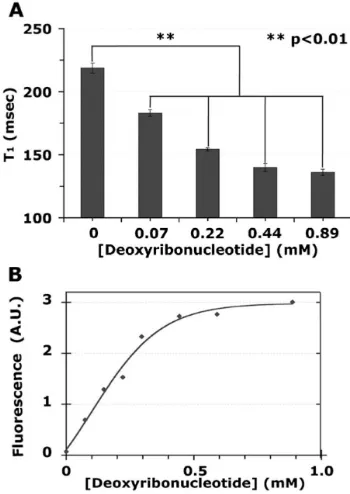
to provide the intermediate 3 (d). The reaction required heating at 60 °C and the use of a large excess of base due to the low nucleophilicity and reactivity of the p-toluidine moiety. The paramagnetic Gd(III) was complexed with 3 to obtain GadoTO 4 as confirmed by MS-ESI (e). The interaction of GadoTO with DNA was examined by relaxometry and fluorescence. Increasing concentrations of DNA decreased the T1 (GadoTO 1 mM) from 220 to 135 msec
(Fig. 1A) and produced a 50-fold increase in fluorescence (Fig. 1B). Samples from Fig. 1A were diluted for the more sensitive fluorescence measurements. To further characterize the interaction between GadoTO and DNA, we measured relaxation times as the concentrations of GadoTO were varied. With the addition of DNA, R1 increased from 4.23 ± 0.06 to 6.5 ± 0.2
(mM.sec)−1, while R2 increased from 4.80 ± 0.08 to 8.2 ± 0.1 (mM.sec)−1 (ESI) †. The R1 and
R2 of GadoTO in the absence of DNA were similar to those of Gd-DTPA. Our data indicate
that GadoTO binds to DNA and exhibits an increased fluorescence and relaxivity. The increased R1 likely reflects a slower tumbling time (τR) when GadoTO binds to DNA, since
similar effects have been noted with many Gd chelates binding proteins.12 The half-maximal point for the T1 change was 0.089 mM(95%confidence interval 0.078–0.101) and was lower
than the half-maximal concentration for fluorescence change (0.179 mM, 0.149–0.214). This may reflect differences in amplification of relaxivity (less than two fold) and fluorescence (fifty fold) upon DNA binding.
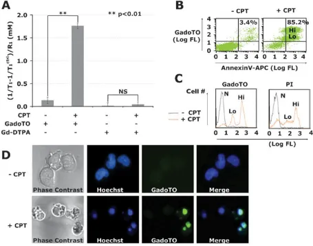
The interaction of GadoTO with camptothecin-treated (+CPT) and untreated (−CPT) Jurkat cells was studied by relaxometry, flow cytometry, and fluorescence microscopy. In all experiments, Jurkat T cells were treated with 10 μM CPT for 24 h, a common procedure for studying chemotherapy-induced cell death,13 and incubated with GadoTO (or the control chelate, Gd-DTPA). For relaxometry, cells were pelleted and lysed, and the T1 of cell lysates
determined. Gd concentrations were obtained by the relation Δ(1/T1)/R1 (Fig. 2A).
Accumulation of GadoTO was observed in CPT-treated cells while Gd-DTPA failed to accumulate. Dual-wavelength flow cytometry with suspended untreated cells indicated 3.4% cells bound both GadoTO and annexinV (upper right quadrant) (Fig. 2B). In our hands, healthy populations of various cell lines, like the Jurkat T cells employed here, typically have 1 to 10% of cells in the annexinV positive, PI positive quadrant. The binding of GadoTO to untreated cells was therefore limited and typical. Two populations of annexinV positive, GadoTO positive cells were noticeable with CPT treatment denoted as “Lo” or “Hi”. We compared the uptake of GadoTO with a “gold standard” vital dye, PI, using single channel flow cytometry (Fig. 2C). Three distinct populations of cells were apparent: non-binding/normal cells (N), low PI and low GadoTO binding cells (Lo), and high PI and high GadoTO binding cells (Hi). Binding of GadoTO and PI to CPT-treated cells were similar. The intracellular distribution of GadoTO was visualized by microscopy (Fig. 2D). Phase contrast (PC) images showed membrane blebbing with CPT-treated cells, absent in healthy cells. Hoechst 33342 uptake showed a diffuse, nuclear staining with healthy cells, while CPT-treated cells showed highly condensed nuclei. Healthy cells failed to take GadoTO, while most CPT-treated cells were positive for GadoTO confirming the presence of GadoTO in condensed nuclei of those cells.
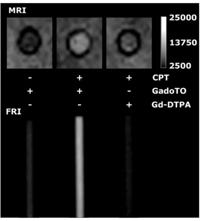
To demonstrate the ability of GadoTO to image cell death, cells were suspended in matrigel in 3 mm (ID) capillary tubes (Fig. 3). Tubes were imaged using a T1-weighted pulse sequence
(MRI) or subjected to fluorescence reflectance imaging (FRI). CPT-treated cells took up GadoTO and were hyperintense (bright), consistent with the binding of GadoTO to DNA in
Garanger et al. Page 2
Chem Commun (Camb). Author manuscript; available in PMC 2009 September 29.
NIH-PA Author Manuscript
NIH-PA Author Manuscript
dead/necrotic cells. FRI confirmed the uptake of GadoTO. The MRI of CPT-treated cells was notably brighter when cells were exposed to GadoTO instead of Gd-DTPA, indicating the important role played by the DNA-binding TO moiety of GadoTO in image brightness of necrotic cells.
Hence, GadoTO is a multimodal, fluorescent/water relaxation rate enhancing compound that enables the detection of cell death by MR or fluorescence. Future studies with GadoTO in animal models appear justified.
Supplementary Material
Refer to Web version on PubMed Central for supplementary material.
Notes and references
1. Majno G, Joris I. Am J Pathol 1995;146:3. [PubMed: 7856735] 2. Blankenberg FG. Cancer Biol Ther 2008;7:1525. [PubMed: 18836289] 3. Sen S. Biol Rev Cambridge Philos Soc 1992;67:287. [PubMed: 1420728]
4. Narula J, Acio ER, Narula N, Samuels LE, Fyfe B, Wood D, Fitzpatrick JM, Raghunath PN, Tomaszewski JE, Kelly C, Steinmetz N, Green A, Tait JF, Leppo J, Blankenberg FG, Jain D, Strauss HW. Nat Med 2001;7:1347. [PubMed: 11726976]
5. Korngold EC, Jaffer FA, Weissleder R, Sosnovik DE. Heart Failure Rev 2008;13:163.
6. Blankenberg FG, Katsikis PD, Storrs RW, Beaulieu C, Spielman D, Chen JY, Naumovski L, Tait JF. Blood 1997;89:3778. [PubMed: 9160684]
7. Valonen PK, Lehtimaki KK, Vaisanen TH, Kettunen MI, Grohn OH, Yla-Herttuala S, Kauppinen RA. J Magn Reson Imaging 2004;19:389. [PubMed: 15065161]
8. Schellenberger EA, Sosnovik D, Weissleder R, Josephson L. Bioconjugate Chem 2004;15:1062. 9. Sosnovik DE, Schellenberger EA, Nahrendorf M, Novikov MS, Matsui T, Dai G, Reynolds F, Grazette
L, Rosenzweig A, Weissleder R, Josephson L. Magn Reson Med 2005;54:718. [PubMed: 16086367] 10. Lee LG, Chen CH, Chiu LA. Cytometry 1986;7:508. [PubMed: 2430763]
11. Fechter EJ, Olenyuk B, Dervan PB. J Am Chem Soc 2005;127:16685. [PubMed: 16305259] 12. Lauffer RB. Magn Reson Q 1990;6:65. [PubMed: 2202424]
13. Poot M, Gibson LL, Singer VL. Cytometry 1997;27:358. [PubMed: 9098628]
NIH-PA Author Manuscript
NIH-PA Author Manuscript
Fig. 1.
Binding of GadoTO to DNA by relaxometry and fluorescence.
Garanger et al. Page 4
Chem Commun (Camb). Author manuscript; available in PMC 2009 September 29.
NIH-PA Author Manuscript
NIH-PA Author Manuscript
Fig. 2.
Accumulation of GadoTO in CPT-treated cells by relaxometry and cytometry.
NIH-PA Author Manuscript
NIH-PA Author Manuscript
Fig. 3.
Imaging necrotic cells by MRI and FRI in capillary tubes.
Garanger et al. Page 6
Chem Commun (Camb). Author manuscript; available in PMC 2009 September 29.
NIH-PA Author Manuscript
NIH-PA Author Manuscript
Scheme 1.
Synthesis and structure of GadoTO 4