MOLECULAR GENETIC AND IMMUNOLOGICAL STUDIES IN SYSTEMIC LUPUS ERYTHEMATOSUS.
A thesis submitted for the degree of Doctor of Philosophy
in the
Australian National University
by
HEATHER DUNCKLEY
John Curtin School of Medical Research The Australian National University
Canberra
STATEMENT
This thesis describes the results of a research project carried out under the supervision of Dr. Sue Serjeantson, at the John Curtin School of Medical Research, Australian National University, from January 1985 to April 1988, during which time I recieved an Australian National University Ph.D scholarship.
The experimental work and data analysis presented in this thesis are my own original work, except when otherwise acknowledged in the text or acknowledgements.
-III-ACKNOWLEDGEMENTS
I would like to thank my supervisor Dr. Sue Serjeantson, to whom I am very grateful for her help, encouragement and invaluable advice during the course of the work and writing of this thesis.
I would also like to thank Dr. Paul Gatenby for providing samples from the SLE and CTD patients, and for their clinical review.
I am grateful to Dr. Sue Wilson for assistance in the logistic regression and discriminant analysis and for critical reading of the relevant parts of this thesis, and also to Dr. Elizabeth Jazwinska for critically reading the manuscript.
I would like to thank Mrs. Michelle Reid and Mr. Brett White for occassional technical assistance, Mrs. Pam Ranford for help in the C4 and C2 work, Ms. Marj Coggan for teaching me the method of C3
phenotyping, and Dr. Maija Kohonen-Corish for assistance in interpreting some DR types.
I appreciate the help given to me by Mrs. Rosalyn Morrison and Mrs. Julie Keeffe in typing parts of this thesis, and thank Mr. Stewart
Butterworth and his staff for excellent photography.
ABSTRACT
This thesis examines genetic factors in susceptibility to the autoimmune disease systemic lupus erythematosus (SLE). Candidate genes examined for a possible role in the predisposition to SLE included those encoding the HLA class I and class II antigens, the complement
components C4A, C4B and C2, and the T-cell antigen receptor a, ß and y chains. Immunoglobulin allotypes, C3 phenotypes, and lymphocytotoxic activity of SLE sera has also been examined. Genetic profiles were established for approximately 30 Caucasoid SLE patients, for additional patients with other connective tissue disorders and for appropriate controls.
Several earlier studies have shown associations between ehe HLA class II DR antigens and SLE, but these have often been conflicting. This may be due to the well known difficulty in cellular and serological
typing of HLA-D and -DR antigens in SLE patients, and due to possible clinical heterogeneity between SLE patients from different centres. In this study, the technique of DNA-DR typing, which gives unequivocal results, was carried out in parallel with serological HLA-DR typing-in SLE and showed a potentially high error rate in HLA-DR serology in lupus. This can be due to incorrect antigen assignment as well as a failure to detect some DR antigens. These errors have the potential to introduce bias into serological studies of HLA-DR in lupus as some DR antigens, such as DR2, are more easily detected than others.
-V-DP genes have also been made, but no role for the DQ or -V-DP antigens in SLE was apparent from the RFLP analyses.
Deficiencies of the MHC class III complement components C2 and C4 have also been implicated in the pathogenesis of lupus, but due to linkage disequilibrium with the HLA-B and -DR genes, it has been difficult to distinguish the primary susceptibility locus. In this study, the role of C4 deficiency has been examined in SLE patients from three populations with different MHC gene linkage arrangements.
Caucasoid, Japanese and Chinese SLE patients and controls were C4 allotyped and the maximum likelihood method used to calculate gene frequencies. This study showed a significant increase in the C4A null allele (C4A*Q0) frequency in each of the three patient populations when compared with their respective controls, indicating a major role for complete and partial C4A deficiency in the pathogenesis of lupus.
Inherited deficiencies of C4B and C2 were not implicated in SLE in this study.
Logistic regression analysis was used to compare the relative contributions of the C4A null gene and DR3 to lupus in Caucasoids, as these two alleles are in significant linkage disequilibrium on the haplotype A1.B8.DR3. This statistical analysis strongly supported the notion that C4A*Q0 is the primary MHC disease promoting locus, with no significant contributions made by HLA-DR3, or C4B*Q0 genes.
fragments detected with the Tcr-ot, -ß or - y chain probes, nor any interaction between TCR RFLPs and HLA class II RFLPs.
Because of the diversity of clinical features in lupus,
discriminant analysis has been carried out in an attempt to correlate clinical features of lupus with genetic variables. This revealed a significant decrease in frequency of a DPß/EcoRV RFLP in patients with pleuritis, suggesting a possible role for HLA-DP in some lupus subtypes. Discriminant analysis between SLE and various connective tissue diseases was also performed, and together with inherited partial C4A deficiency, lymphocytotoxic activity against B-cells was an important discriminating variable, suggesting that lymphocytotoxins may be more important
pathogenically than previously thought.
This study has shown that SLE is a genetically heterogeneous disease, but one important susceptibility factor, in Chinese and
PUBLICATIONS Original papers:
Dunckley H, Gatenby PA, Serjeantson SW. 1986. DNA typing of HLA-DR
antigens in systemic lupus erythematosus. Immunogenetics 24:158-162. Dunckley H, Hawkins B. 1986. An unusual C4B variant in Cantonese. In: HLA
in Asia-Oceania 1986. Proceedings of the Third Asia-Oceania
Histocompatibility Workshop. Eds: Aizawa M, Natori T, Wakisaka A, Konoeda Y. Sapporo, Hokkaido University Press, p. 571-573.
Dunckley H, Naito S, Serjeantson SW. 1986. DNA-DR typing of Japanese
systemic lupus erythematosus patients. In: HLA in Asia-Oceania 1986. Proceedings of the Third Asia-Oceania Histocompatibility Workshop. Eds: Aizawa M, Natori T, Wakisaka A, Konoeda Y. Sapporo, Hokkaido University Press, p. 419-422.
Dunckley H, Gatenby PA, Hawkins B, Naito S, Serjeantson SW. 1987. Deficiency of C4A is a genetic determinant of systemic lupus
erythematosus in three ethnic groups. J .Immunogenetics. In press. Dunckley H, Gatenby PA, Serjeantson SW. 1988. T-cell receptor and HLA
class II RFLPs in systemic lupus erythematosus. Immunogenetics 27: 392-395.
Serjeantson SW, Kohonen-Corish MRJ, Dunckley H, Reid MA. 1986. HLA class II RFLPs are haplotype specific. Cold Spring Harbor Symposium on
Quantitative Biology. Vol LI (1): 83-89.
Hawkins BR, Wong KL, Wong RWS, Chan KH, Dunckley H, Serjeantson SW. 1987. Strong association between the major histocompatibility complex and systemic lupus erythematosus in Southern Chinese. J. Rheumatol. 14: 1128-1131.
Ranford P, Serjeantson SW, Hay J, Dunckley H. 1987. A high frequency of inherited deficiency of complement component C4 in Darwin Aborigines. Aust. NZ. J. Med. 17: 420-423.
Jazwinska EC, Dunckley H, Propert D, Gatenby PA, Serjeantson SW. 1988. Gm allotyping by immunoglobulin heavy chain RFLP analysis. A m . J . H u m . Genet. In press.
lupus erythematosus. (Human Genetics Society of Australasia, 10th Annual Scientific Meeting, 1986, Canberra). Aust. Paediatr. J. 23: 72.
Dunckley H, Hawkins BR. 1987. An unusual C4B variant is increased in
frequency in Cantonese. (Human Genetics Society of Australasia, 10th Annual Scientific Meeting, 1986, Canberra). Aust. Paediatr. J. 23: 77.
Dunckley H, Gatenby PA, Hawkins B, Naito S, Serjeantson SW. 1987. Partial C4A deficiency increases predisposition to systemic lupus
-IX-TABLE OF CONTENTS
THESIS TITLE I
STATEMENT II
ACKNOWLEDGEMENTS III
ABSTRACT IV
PUBLICATIONS VII
TABLE OF CONTENTS IX
CHAPTER 1
GENERAL INTRODUCTION
1.1 INTRODUCTION. 1
1.2 EPIDEMIOLOGY. 1
1.3 DIAGNOSIS OF SLE. 2
1.4 CLINICAL AND LABORATORY FEATURES OF SLE. 4
1.5 ENVIRONMENTAL OR GENETIC? 8
1.6 ANIMAL MODELS OF SLE. 9
1.7 CANDIDATE GENES FOR SLE IN HUMANS. 10
1.8 THE MAJOR HISTOCOMPATIBILITY COMPLEX. 11 1.8.1 THE HLA-A, -B, AND -C ANTIGENS. 11
1.8.2 THE HLA-D ANTIGENS. 12
1.5.3 GENETIC ORGANIZATION OF THE
MHC CLASS II GENES. 13
1.9 ASSOCIATION OF THE CLASS I AND II GENES
WITH DISEASE ' 16
1.9.1 ASSOCIATION OF THE CLASS I AND II MHC
GENES WITH SLE. 17
1.10 THE CLASS III GENES IN THE MHC. 18
1.10.1 THE COMPLEMENT SYSTEM. 18
1.10.2 COMPLEMENT DEFICIENCIES. 21
1.10.3 COMPLEMENT COMPONENT C4. 24
1.10.4 COMPLEMENT COMPONENT C 2 . 26
1.11 STRUCTURE AND FUNCTION OF THE T-CELL RECEPTOR. 27
1.11.1 THE aß RECEPTOR. 28
1.11.3 T-CELL RECEPTOR GENES. 29 1.11.3.1 TCR-ß GENE ORGANIZATION. 30 1.11.3.2 TCR-Ot GENE ORGANIZATION. 32 1.11.3.3 TCR-Y GENE ORGANIZATION. 33 1.11.4 DIVERSITY OF THE T-CELL RECEPTOR. 34
1.12 AIMS OF THESIS. 34
CHAPTER 2 METHODS
2.1 INTRODUCTION. 37
2.1.1 PATIENTS AND CONTROLS. 37
2.2 HLA-A, -B, -C, AND -DR SEROLOGICAL TYPING. 38
2.2.1 T-CELLS. 39
2.2.2 NON-ROSETTING CELLS. 40
2.2.3 TISSUE TYPING. 40
2.2.4 AET TREATMENT OF SHEEP RED BLOOD CELLS. 40 2.3 DNA EXTRACTION, DIGESTION, AND SOUTHERN BLOTS. 41
2.3.1 DNA EXTRACTION. 41
2.3.2 DNA DIGESTION. 41
2.3.3 SOUTHERN BLOTS. 42
2.4 PREPARATION AND NICK TRANSLATION OF THE PROBES. 43
2.5 HYBRIDIZATIONS. 44
2.5.1 REHYBRIDIZATIONS. 45
2.6 C4 ALLOTYPING. 45
2.6.1 MATERIALS AND METHOD. 46
2.6.2 HAEMOLYTIC OVERLAY. 48
2.7 C2 ALLOTYPING. 48
2.7.1 MATERIALS AND METHOD. 49
2.8 C2 DEFICIENCY ASSAY. 50
2.9 C3 TYPING. 51
2.9.1 MATERIALS AND METHOD. 52
2.10 TESTING FOR LYMPHOCYTOTOXINS. 53
-XI-CHAPTER 3
DR GENOTYPING IN SLE AND OTHER MHC CLASS II RFLPS
3.1 INTRODUCTION. 54
3.1.1 HLA-DR TYPING IN SLE. 54
3.1.2 HLA-DR AND HLA-DQ IN CONNECTIVE TISSUE
DISEASES. 56
3.1.3 HLA-DQ AND HLA-DP GENES IN SLE. 56
3.1.4 AIMS. 57
3.2 MATERIALS AND METHODS. 58
3.2.1 PATIENTS AND CONTROLS. 58
3.2.2 SEROLOGICAL HLA CLASS I AND II TYPING. 58
3.2.3 DNA-DR TYPING. 59
3.2.4 OTHER HLA CLASS II HYBRIDIZATIONS. 59
3.2.5 PROBES. 64
3.3 RESULTS. 64
3.3.1 DNA-DR TYPING. 64
3.3.2 RELIABILITY OF SEROLOGICAL DR TYPING IN
SLE. 66
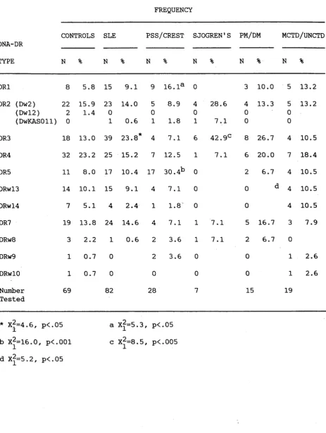
3.3.3 HLA-DR IN THE CAUCASOID SLE PATIENTS. 69 3.3.4 HLA-DR IN CONNECTIVE TISSUE DISEASES. 69 3.3.5 HLA-DQ IN THE CAUCASOID SLE
AND CTD PATIENTS. 71
3.3.6 DQß/BamHI RFLPs IN THE SLE
AND CTD PATIENTS. - 71
3.3.7 HLA-DRa/Bglll RFLPs IN SLE AND THE CTD
PATIENTS. 72
3.3.8 HLA-DP IN SLE AND THE CONNECTIVE TISSUE
DISEASES. 72
3.3.9 HLA-DR AND HLA-DQ IN JAPANESE PATIENTS
WITH SLE. 75
3.4 DISCUSSION. 77
3.4.1 DNA-DR TYPING IN SLE. 77
3.4.2 HLA-DQ IN SLE. 79
3.4.3 HLA-DR AND HLA-DQ IN THE CONNECTIVE TISSUE
DISEASES. 80
3.4.4 HLA-DP RFLPs IN THE SLE AND CTD PATIENTS 81 3.4.5 DR TYPING IN JAPANESE SLE PATIENTS. 81
3.5 CONCLUSION. 81
CHAPTER 4
COMPLEMENT AND SLE
4.1 INTRODUCTION. 83
4.2 MATERIALS AND METHODS. 87
4.2.1 PATIENTS AND CONTROLS. 87
4.2.2 C4 TYPING. 87
4.2.3 STATISTICAL ANALYSIS. 88
4.2.4 ESTIMATION OF NULL ALLELE FREQUENCIES. 88
4.2.5 LOGISTIC REGRESSION. 89
4.2.6 HYBRIDIZATIONS. 89
4.2.7 C2 TYPING. 90
4.2.8 C2 FUNCTIONAL ASSAYS. 90
4.3 RESULTS. 90
4.3.1 C4 IN CAUCASOID, JAPANESE, AND CHINESE SLE PATIENTS AND IN CAUCASOID CTD PATIENTS. 90 4.3.2 C4A DEFICIENCY VERSUS HLA-DR3 IN SLE. 103 4.3.3 C4B DEFICIENCY VERSUS DR3 IN SLE. 105 4.3.4 C2 DEFICIENCY IN CAUCASOID, JAPANESE AND
CHINESE SLE PATIENTS AND CAUCASOID CTD
PATIENTS. 109
4.4 DISCUSSION. 114
4.4.1 SIGNIFICANCE OF C4A*Q0 IN SLE PATIENTS. 114
4.4.2 C2 IN SLE. 117
4.4.3 C4 AND C2 IN CONNECTIVE TISSUE DISEASES. 118
4.5 CONCLUSION. 119
CHAPTER 5
THE T-CELL ANTIGEN RECEPTOR IN SLE
5.1 INTRODUCTION. 121
5.1.1 THE T-CELL RECEPTOR IN AUTOIMMUNITY. 121
5.1.2 AIMS. 122
5.2 METHODS. 122
5.2.1 PATIENTS AND CONTROLS. 122
5.2.2 PROBES. 123
5.2.3 RESTRICTION ENZYMES. 123
5.3 RESULTS. 125
5.3.1 TCR-a, -ß, and -y RFLPS. 125
5.3.2 TCR-ß IN THE SLE AND CTD PATIENTS. 127 5.3.3 TCR-y IN THE SLE AND CTD PATIENTS. 130 5.3.4 TCR-a IN THE SLE AND CTD PATIENTS. 130 5.3.5 TCR RFLPS AND HLA-DR PHENOTYPES. 133
5.4 DISCUSSION. 135
5.4.1 THE T-CELL RECEPTOR IN SLE. 135 5.4.2 THE T-CELL RECEPTOR IN CONNECTIVE TISSUE
DISEASES. 137
-XIII-CHAPTER 6
DISCRIMINANT ANALYSIS IN SLE AND OTHER IMMUNOLOGICAL VARIABLES.
6.1 INTRODUCTION. 139
6.1.1 THE IMMUNOGLOBULINS. 140
6.1.2 COMPLEMENT COMPONENT C 3 . 141
6.1.3 LYMPHOCYOTOXIC ANTIBODIES. 142
6.1.4 RHEUMATOID FACTOR. 142
6.1.5 AIMS. 142
6.2 METHODS. 143
6.2.1 PATIENTS AND CONTROLS. 143
6.2.2 Gm TYPING. 143
6.2.3 C3 TYPING. 143
6.2.4 LYMPHOCYTOTOXIC ANTIBODY SCREENING. 144 6.2.5 RHEUMATOID FACTOR SCREENING. 144
6.2.6 STATISTICS. 144
6.3 RESULTS. 144
6.3.1 GM AND KM ALLOTYPING. 144
6.3.2 COMPLEMENT COMPONENT C 3 . 147
6.3.3 LYMPHOCYTOTOXIC ANTIBODIES. 147
6.3.4 RHEUMATOID FACTOR. 149
6.3.5 DISCRIMINANT ANALYSIS BETWEEN SLE
AND THE CTD s . 149
6.3.6 ANALYSIS OF CLINICAL SUBSETS OF SLE. 153
6.3.6.1 PHOTOSENSITIVITY. 153
6.3.6.2 PLEURITIS. 155
6.3.6.3 ANTI-DNA ANTIBODIES. 157
6.4 DISCUSSION. 158
6.4.1 DISCRIMINATION OF SLE FROM CTDs. 158
6.4.2 CLINICAL SUBSETS OF SLE. 160
6.4.3 GM AND C3 IN THE CTD PATIENTS. 161
6.5 CONCLUSION. 162
CHAPTER 7
GENERAL DISCUSSION: DISEASE MECHANISMS IN SLE 163
-1
-1.1 INTRODUCTION.
Systemic lupus erythematosus (SLE or lupus) is an autoimmune connective tissue disease, the aetiology of which is unknown.
Originally, SLE was thought to be a cutaneous disease but in 1872 Kaposi recognized that other organs could be involved and that the disease was therefore systemic in nature. Clearer definition of the systemic
features was later provided by Osler, Libman and Sacks, and Klemperer. In 1948, Hargraves discovered the "LE cell” (lupus erythematosus cell) phenomonen which was later shown to represent an antibody to DNP
(deoxyribose nucleoprotein). Since then, many other autoantibodies, particularly against nuclear antigens, have been found in the sera of patients with SLE, with the fluorescent antinuclear antibody (ANA) test being used as a routine diagnostic clinical test by the late 1950's. A wide variety of more specific ANAs and other autoantibodies have now been recognized and described, and have contributed to the understanding of the disease (reviewed by Reichlin 1981). The history of lupus has been reviewed in more detail by Graninger et al. (1987).
1.2 EPIDEMIOLOGY.
SLE is a disease primarily affecting females from 15 to 35 years of age, but can occur in all age groups including the newborn and the
Grennan 1980). The differing levels of incidence in Caucasoids may be partly due to ethnic heterogeneity in the Caucasiods studied, or
differences in the way that the cases are ascertained. There is no large scale survey of the prevalence of SLE in the Australian community.
1.3 DIAGNOSIS OF SLE.
SLE can vary from a mild cutaneous problem with appropriate serology to a severe, life threatening, multi-organ disease, and can also go through stages of active disease or remission in individuals, with a wide variety of clinical symptoms. In addition, lupus is a disease that can develop very quickly or can take many years to evolve.
The extreme diversity of symptoms seen in lupus, many of which are non-specific, make the diagnosis difficult, particularly when several other connective tissue disases (CTDs) have the same or similar
features. The issue is further complicated by the fact that SLE may well be collection of several diseases rather than one specific disease. This complexity has lead to a number of attempts to develop criteria for the diagnosis of SLE, for use in both epidemiological surveys and in scientific studies, to ensure some uniformity with regard to what is described as a case of lupus.
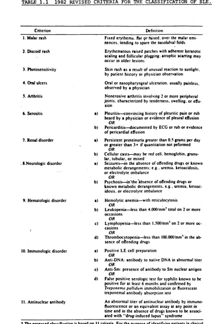
In 1971, the "Preliminary Criteria for the Classification of
Systemic Lupus Erythematosus" (Cohen et al. 1971) were published by the American Rheumatism Association (ARA) which did establish some
- 3
-TABLE 1 . 1 1982 REVISED CRITERIA FOR THE CLASSIFICATION OF SLE.
Criterion Definition
1. Malar rash Fixed erythema, flat prfaised. over the malar emi nences. tending to spare the nasolabial folds 2 Discoid rash Erythematous raised patches with adherent keratotic
scaling and follicular plugging, atrophic scarring may occur in older lesions
3. Photosensitivity Skin rash as a result of unusual reaction to sunlight, by patient history or physician observation
4. Oral ulcers Oral or nasopharyngeal ulceration, usually painless, observed by a physician
5. Arthritis Nonerosive arthritis involving 2 or more peripheral joints, characterized by tenderness, swelling, or effu
sion
6. Serositis a) Pleuritis—convincing history of pleuritic pain or rub heard by a physician or evidence of pleural effusion
O R
b) Pericarditis—documented by ECG or rub or evidence of pericardial effusion
7. Renal disorder a) Persistent proteinuria greater than 0.5 grams per day or greater than 3+ if quantitation not peformed
O R
b) Cellular casts—may be red cell, hemoglobin, granu lar, tubular, or mixed
. 8.Neurologic disorder a) Seizures—in the absence of offending drugs or known metabolic derangements; e g . uremia, ketoacidosis, or electrolyte imbalance
O R
b) Psychosis—in^the absence of offending drugs or known metabolic derangements, e g . uremia, ketoac idosis. or electrolyte imbalance
9. Hematologic disorder a) Hemolytic anemia—with reticulocytosis O R
b) Leukopenia—less than 4.000'mm' total on 2 or more occasions
O R
c) Lymphopenia—less than 1.300/mm’ on 2 or more oc casions
O R
d) Thrombocytopenia—less than lOO.OOO'mm’ in the ab sence of offending drugs
10. Immunologic disorder a) Positive LE cell preparation b)
O R
Anti-DNA: antibody to native DNA in abnormal titer O R
c) Anti-Sm: presence of antibody to Sm nuclear antigen O R
d) False positive serologic test for syphilis known to be positive for at least 6 months and confirmed by T rep o n em a p a llid u m immobilization or fluorescent treponema! antibody absorption test
II. Antinuclear antibody An abnormal titer of antinuclear antibody by immuno fluorescence or an equivalent assay at any point in time and in the absence of drugs known to be associ ated with “ drug-induced lupus" syndrome
• The proposed classification is based on 11 criteria. For the purpose of identifying patients in clinical studies, a person shall be said to have systemic lupus erythematosus if any 4 or more of the 11 criteria are present, serially or simultaneously, during any interval of observation.
[image:17.549.48.476.53.705.2]activity (Tan et al. 1982) and these have subsequently been shown to be highly specific for the diagnosis of SLE (Passas et a l . 1985). The classification criteria are shown in Table 1.1 (taken from Tan et al.
1982). For a patient to be diagnosed with SLE, he or she must have any four of the criteria, either serially or simultaneously during the course of observation.
It should be mentioned however that these criteria are exclusive, rather than inclusive. The criteria are designed to exclude other autoimmune CTDs, arthritides and certain systemic illnesses (Tan et al. 1982), and therefore may exclude individuals whose diagnosis is almost certainly SLE.
1.4 CLINICAL AND LABORATORY FEATURES OF SLE.
The clinical features of lupus have been described in depth by several authors (Dubois 1976; Ropes 1976; Rothfield 1985) and will only be reviewed briefly here. Table 1.2, (taken from Schur 1983), gives the incidence with which some of clinical features occur in lupus. These frequencies can vary in different studies and may be due to the way in which patients are ascertained.
Most patients with lupus suffer from a number of generalized
symptoms seen in many diseases including fatigue, weight loss, fever and a general lack of well being.
-5
-TABLE 1.2 FREQUENCY OF CLINCAL FEATURES IN SLE.
FEATURE PERCENT
Fever 83
Weight loss 62
Arthritis, arthralgia 90
Skin 74
Butterfly rash 42
Photosensitivity 30
Mucous membrane lesions 12
Alopecia 27
Raynaud1s phenomonen 17
Purpura 15
Urticaria 8
Renal 53
Nephrosis 18
Gastrointestinal 38
Pulmonary 47
Pleurisy 45
Effusion 24
Pneumonia 29
Cardiac 46
Pericarditis 27
Murmurs 23
ECG changes 39
Lymphadenopathy 46
Splenomegaly 15
Hepatomegaly 25
Central nervous system 32
Psychosis 15
Convulsions 15
and nose, and sensitivity to sunlight which can exacerbate the malar rash and erythematous maculopapula eruptions. Alopecia is also fairly common in SLE but is not specific enough to be part of the diagnostic criteria.
Systemic features in SLE involve the joints, kidneys, lungs, heart, central nervous system, gastrointestinal tract, liver and spleen. Joint disease is very common in lupus, ranging from arthralgias and minor tendonitis to joint swelling and arthritis. The arthritis is rarely erosive and deformity is seldom seen in contrast to rheumatoid arthritis (Labowitz and Schumacher 1971).
Renal disease is seen clinically in about 50% of patients, although pathology usually indicates a higher incidence. Renal disorders can vary in severity from mild focal lupus nephritis to acute and ultimately chronic renal failure.
About 50% of SLE patients are affected with lung problems, with pleuritic pain the most common feature. Interstitial lung involvement with acute lupus pneumonitis and diffuse chronic interstitial lung disease can also occur, and lung function abnormalities may be found in asymptomatic patients.
Pericarditis is the most common cardiac disorder, in lupus, and is usually acute. Myocardial involvement is uncommon, while vascular disease may be seen at autopsy, but is rarely a problem during life.
Central nervous system manifestations include seizures and
psychoses, both of which are diagnostic criteria of SLE. Patients also suffer from headaches (migraine or a more diffuse type), while a small number of patients develop cerebrovascular disease. Peripheral
neuropathy has also been described.
-7
-the diagnostic criteria for lupus, and includes anorexia, nausea and vomiting (seen in about 20% of patients), while abdominal pain occurs in about 10% of patients. Hepatomegaly is sometimes seen, but clinical hepatitis is very rare.
Slight to moderate splenomegaly is seen in about 20% of SLE
patients, especially those with haemolytic anaemia. Approximately 50% of patients have enlarged lymph nodes at some stage during the course of the disease.
Vascular disease is also seen in lupus, varying from Raynaud’s phenomonen (mild to severe), to venous and arterial thrombosis. The thrombosis has been associated with anti-phospholipid antibodies (Harris et al. 1983).
Several haematological abnormalities have been observed in lupus including lymphopenia, leukopenia, thrombocytopenia, and haemolytic anaemia (all part of the diagnostic criteria). Leukopenia occurs in nearly 50% of lupus patients while haemolytic anaemia and
thrombocytopenia are seen in about one fifth of patients (Tan et al. 1982). Thrombocytopenia may be due to the presence of anti-platelet antibodies (Maini 1977).
Serology has revealed a number of autoantibodies in SLE including antinuclear antibodies (ANAs, seen in more than 95% of patients), anti-double standed DNA (anti-dsDNA) antibodies (found in 70% of
patients) and anti-Sm antibodies (seen in about 30% of patients), where Sm is an acidic nucleoprotein. Each of these antibodies are diagnostic criteria for SLE, as are the LE cell and a consistently false positive standard test for syphilis.
but not specific), anti-Ro (SS-A) and anti-La (SS-B) antibodies which are also seen in patients with Sjogren's syndrome, and anti-nRNP antibodies which are seen in a high proportion of mixed connective tissue disease (MCTD) patients.
The levels of complement components C3 and C4, and the total haemolytic complement activity are usually decreased during active disease. However, results of complement assays in SLE patients,
particularly C 4 , should be interpretated carefully as inherited C4 null alleles may predispose an individual to SLE.
Cold reactive lympnocytotoxins specific for B- and T-cells have also been observed in SLE sera, which are frequently specific for a patients own lymphocytes (Terasaki et al. 1970). These are of the IgM sub class (Winfield et al. 1975a), and are detectable with the
complement dependant microcytotoxicity test. Rheumatoid factor (RF) is another IgM antibody found in the sera of about 20% of SLE patients and
is reactive against IgG antibodies (Fong et al. 1985). RF was one of the original criteria for the diagnosis of SLE but has since been omitted as it is not highly specific for SLE.
1.5 ENVIRONMENTAL OR GENETIC?
Both environmental and genetic factors have been implicated in the pathogenesis of SLE. Studies have shown that the age of onset of SLE in sibling pairs occurs closer in the calendar year than in the actual age of the siblings (Arnett and Schulman 1976; Kaplan et al. 1984) which would indicate an environmental agent. Further support comes from
-9
-frequency in the sera of close household contacts of patients with SLE. Laboratory workers who handle SLE blood also have a higher incidence of lymphocytotoxic antibodies (LCAs) compared with the general population (De Horatius et al. 1979).
On the other hand, close blood relatives of SLE patients, with little patient contact, have an increased incidence of LCAs (De Horatius and Messner 1975) and a study by Elkon et al. (1983) has shown an
increase in circulating immune complex levels in first degree relatives of SLE patients, both with and without close household contact, which would tend to support a genetic basis. In addition, there is a higher concordance rate of disease in identical twins (57%) compared to
dizygotic twin pairs and sibs (Block et al. 1975) with disease
expression very similar between identical twins. It has been observed that approximately 10% of SLE patients have a first or second degree relative with SLE (Arnett et al. 1984) which is suggestive of genetic involvement.
The most convincing evidence for the role of genetic factors in the pathogenesis of SLE is provided by the association of major
histocompatibility complex (MHC) genes with the disease, as reviewed later in this chapter and Chapter 3. Further evidence for genetic involvement is provided by animal models of SLE.
1.6 ANIMAL MODELS OF SLE.
disease very similar to that seen in humans. The parental NZW mouse is phenotypically normal, and any autoimmune symptoms are rarely seen before 18 months of age. The NZB mouse does have some autoimmune manifestations such as haemolytic anaemia and occassionally anti-ssDNA or anti-dsDNA or anti-histone antibodies. Renal disease rarely occurs when the NZB mice are less than one year old and is very mild. However,
in the (NZB x NZW)F1 hybrids, anti-dsDNA and anti-histone antibodies are present, and the mice suffer from fatal immune complex
glomerulonephritis. The female FI mice have an average life span of only eight months. Some new strains of mice with lupus like diseases are now available for study such as the MRL mouse and 3XSB mouse. However, none of the mice have the same H-2 type (the mouse equivalent of the human MHC genes), nor the same immunoglobulin heavy chain type. Work on the T cell receptor (TCR) in these mice strains indicates that the NZW mice do have differences in their TCR genes, but whether these have an effect on the pathogenesis of the lupus like disease in the mice
is not clear. Different accelerating factors are also seen in the mice. For example, hormones play a role in the (NZB x NZW)F1 mice with female mice dying much sooner than male mice and the lymphoproliferative (lpr) gene in MRL mice reduces life expectancy three-fold. How these
accelerating factors work is unknown. The murine models suggest that there are several mechanisms or abnormalities which can lead to lupus; the mice lupus models are reviewed by Theofilopoulos et al. (1986).
1.7 CANDIDATE GENES FOR SLE IN HUMANS.
-11
-histocompatibility complex and the T-cell receptor (TCR) genes. The molecular organization, structure and function of the MHC and T-cell receptor genes have been the subject of intense investigation and are reviewed here in some detail.
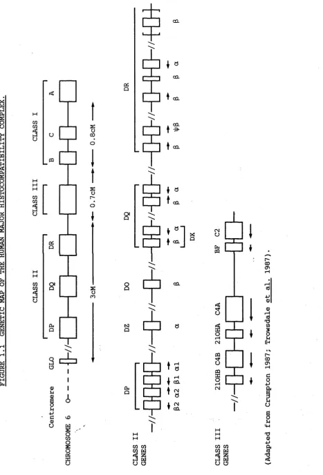
1.8 THE MAJOR HISTOCOMPATIBILITY COMPLEX.
The MHC is found on the short arm of chromosome 6, at the 6p21.3 band, and encodes some of the most polymorphic human genes. The MHC is comprised of three classes of genes, (Figure 1.1), which encode the class I HLA-A, -3, and -C antigens found on the surface of the majority of nucleated cells, the class II HLA-D antigens found primarily on the surface of 3-lymphocytes, and the class III serum complement components C2, C4 and 3F and also the 21-hydroxylase enzyme. Recently, the tumor necrosis factor (TNF) a and 0 chain genes have been localized between the class I and III genes.
1.8.1 THE HLA-A, -3, AND -C ANTIGENS.
The class I antigens, (the classic transplantation antigens), consist of a polymorphic transmembrane glycoprotein about 4£.kDa in size encoded in the MHC, and a noncovalently associated 12kDa 82
microglobulin (Strominger et al. 1977) which is encoded by a gene on chromosome 15 (Goodfellow et al. 1975). The heavy chain has three external domains, a transmembrane domain and a cytoplasmic domain. The 02 microglobulin is associated with the highly conserved third external domain next to the cell membrane, while the first two extracellular domains are the most polymorphic (Yokoyama and Nathenson 1983).
Polymorphism in the class I antigens is evident by serological, cellular and biochemical techniques with the Ninth International
Histocompatibility Workshop recognizing 23 HLA-A, 47 HLA-B and eight HLA-C antigens, which were detected serologically (Bodmer and Bodmer
1984). The class I antigens are important in the discrimination between self and nonself during an immune response, as they are found on the majority of cells, and therefore play an important role in
transplantation. In addition, T-cytotoxic cells recognize antigen in association with the MHC class I molecules during an immune response.
1.8.2 THE HLA-D ANTIGENS.
The class II HLA-D antigens are polymorphic heterodimers comprised of a heavy a chain about 33 kDa in size and a light ß chain (28 kDa)
(Snary et al. 1977; Springer et al. 1977). Each polypeptide chain has two extracellular domains, the first showing the most polymorphism, a transmembrane domain and a cytoplasmic domain (Kaufman and Strominger 1982). The first external domains of the a and ß chains probably
combine together to bind antigen. Three HLA-D antigens have so far been detected, the DR, DQ and DP antigens, with the possibility of a fourth indicated from molecular genetic (Trowsdale et al. 1985) and biochemical studies (Carra and Acoila 1987). The Ninth International
Histocompatibility Workshop recognized 14 DR, three DQ and six DP serologically distinct antigens (Bodmer and Bodmer 1984). During an immune response, 3-cells, macrophages and other antigen presenting cells (which carry the class II antigens on their cell surface), process
- 13
-recognizes foreign antigen in association with the class II protein, are then able to interact and become activated and function as T-helper cells, stimulating B-cell involvement and antibody production.
1.3.3 GENETIC ORGANIZATION OF THE MHC CLASS II GENES.
The organization of the genes in the MHC is shown in Figure 1.1. The MHC covers an area of about 2.5cM and the order of the genes was established initially with family studies of people with HLA recombinant haplotypes, both serologically and by recombinant DNA techniques. More recently, the new method of pulsed field gel electrophoresis (PFGE) and the use of overlapping cosmid clones {Dunham et al. 1987; Carroll et al.
1987) has proved very useful as larger areas of DNA can be covered. The class II region contains four sets of genes, the DR, DQ, DP, and DO/DZ genes. By linkage of ccsmid clones (Korman et al. 1985) and
southern blotting {Wake et al. 1982a; Bohme et al. 1983), the DR locus has been shown to consist of one a gene and a variable number of ß chain genes. The number of ß chain genes correlates with serological DR types
(Bohme et al. 1985). For example, DRw8 has one ß gene while DR4 has at least 3 ß genes (Bohme et al. 1985). The ß2 gene appears to be a
- 15
-There are two pairs of genes in the DQ region, known as DQa and D Q ß , and DXa and DXß , which have not been linked with cosmid cloning
(Okada et al. 1985a; Korman et al. 1985). The DQ RFLPs show strong linkage disequilibrium with DR types unlike those at the DX locus
(Auffray et al. 1984; Trowsdale et al. 1983; Spielman et al. 1984), and with supporting evidence from deletion mutant cell lines it has been
suggested that the DX genes are centromeric to the DR and DQ genes, (Auffray et al. 1983a), and that there is a hot spot for recombination between the DX and DR/DQ genes (Trowsdale et al. 1985). The DX gene pair do not appear to be expressed as no mRNA or protein has yet been detected, but from sequencing, the genes do not appear to be pseudogenes
(Auffray et al. 1984; Okada et a l . 1985a). The DQa and DQß genes show variability in the splicing points at their 3' ends which can result in deletion of part of the untranslated region (Schenning et al. 1984). The DX gene pair show limited polymorphism but both the DQa and DQß genes have extensive polymorphism when examined for RFLPs (Auffray et a l . 1983b; Spielman et al. 1984; Cohen et al. 1984). The variability in the DQa gene has been shown to be clustered to amino acid residues in the first external domain (Chang et al. 1983; Trowsdale et al. 1985).
The DP region covers about lOQkb of DNA (Okada et al. 1985b) and has been extensively characterized (Roux-Dosseto et al. 1983; Gorski et a l . 1984; Trowsdale et al. 1985). Two pairs of genes have been found in this region, namely DPal and DPßl, and DPa2 and DPß2 and these have been linked by cosmid cloning. Sequencing suggests that the a2 and ß2 gene pair are pseudogenes due to frame shift mutations and defective slicing
patterns have been observed as well as variation in the site for the poly A tail (Kelly and Trowsdale 1985). The DP genes were originally
thought to show little polymorphism (Spielman et al. 1984), but it is now evident that they are highly polymorphic, with genetic variation clustered in regions of the second exon (Bugawan et al. 1987).
The fourth set of genes in the HLA-D region are the newly identified DOß and DOa/DZa genes. The DZa gene was originally
identified by Spielman. et al. (1984) and was later shown to be identical to the DOa gene which was isolated by Inoko et al. (1985). DO appears to lie between DQ and DP from PFGE and cosmid cloning studies (Hardy et a l . 1986; Erlich et al. 1986; Amar et a l . 1987). DOß and DOa/DZa may not be a gene pair as PFGE indicates that they lie about lOOkb apart in the MHC and DOa/DZa is inducible by y interferon while DOß is not. From their sequences, both could be functional genes. Northern blots have shown a low level of expression of the DO genes in some B-cell lines, but a very large mRNA was detected (Trowsdale et al. 1985). No RFLPs have yet been found (Trowsdale et al. 1985).
1.9 ASSOCIATION OF THE CLASS I AND II GENES WITH DISEASE.
The class I and II genes have shown associations with several diseases which are characterized by abnormalities in the immune system such as insulin dependent diabetes mellitus, rheumatoid arthritis, and multiple sclerosis. These genetic associations may be directly
-17
-susceptibility gene (a non HLA gene) which could be carried on the same haplotype. A high level of linkage disequilibrium is seen in the MHC and may be a consequence of selective pressure for certain benefical genes, for example during a disease epidemic. However, this linkage disequilibrium may have resulted in some less beneficial genes (with regard to autoimmunity) being carried on the haplotype, a feature which has been called "hitch hiking" (Bodmer and Bodmer 1978). There is still an area of the MHC, between the class I and III genes, where new genes could be located, or a disease susceptibility gene could lie outside the MHC. A third explanation for disease associations with genes in the MHC
is that certain MHC types are necessary, but not sufficient, for the development of disease, with other non-MHC genes and environmental factors contributing to disease development in autoimmune disorders.
1.9.1 ASSOCIATION OF THE CLASS I AND II MHC GENES WITH SLE.
SLE was originally reported to be associated with HLA-B5 and HLA-B8 in several populations, but it was later shown that the class II
antigens DR2 and DR3 were also increased in frequency in SLE (reviewed by Tiwari and Terasaki 1985). However, because of the linkage
diseqilibrium seen in the MHC, as observed between B8 and DR3 for
1.10 THE CLASS III GENES IN THE MHC.
The early acting complement components C2, C4, and BF are encoded in the class MHC III region along with the genes for 21-hydroxylase (Figure 1.1). Mapped with overlapping cosmid clones (Carroll et al. 1984) the class III genes lie between the class I and HLA-D genes with C2 and BF only 421bp apart, while the C4 genes are about 30kb away from BF. The C4A and C4B genes lie about lQkb apart from each other while the 21-OH genes are located close to the 3' ends of the C4 genes
(Carroll et al. 1985; White et al. 1985). PFGE has established the gene order as HLA-B, C 2 , BF, C4A, 21-OHA, C4B, 21-OHB, HLA-DR (Dunham et al.
1987; Carroll et al. 1987).
1.10.1 THE COMPLEMENT SYSTEM.
The complement system is comprised of about 18 plasma proteins which are involved in a series of sequential reactions and which show a wide variety of immunological activities including solubilization of
immune complexes, viral neutralization, lysis of foreign cells,
opsonization, and stimulation of the inflammatory response. There are two pathways in the complement system - the classical, and the
alternative which join together in the terminal attack cascade (Figure 1.2).
F IG URE 1 . 2 T H E COMPLE ME NT CASCA DE
-19
-Ss 0 o <0 c 3 §s ■H CO 0) 03 •H 5-1
03 -C u o cd CO >
rH a) o I-(
au 12
3
X fH
a) o
r-l CO
a, a £ -H
0
ü d) Jd ^ O' <2
iJ ••
01 it) Di
c{ hU <d o> M u 01 M 2 C3i M
ä
03 0 12 •I- ( +J C ■=c 01 c <D O' •H 4-> c ii o u u id i4 0 4J o id b 12CQ 3d M
Ü S-l
to the Ag:Ab:Cl complex. In the presence of magnesium ions, C2 binds to C4b and is cleaved by Cls to C2Ä (released) and C2b. C4b2b is a C3 convertase enzyme, cleaving C3 to C3a and C3b through a proteolytic site on C2b. C3a, an anaphylatoxin, is released into the circulation and C3b binds to the Ag:Ab:C14b2b complex and cleaves complement component C5 to
its active form, resulting in the activation of the terminal attack cascade and the formation of C5b-9b, (the terminal attack complex), which is extremely hydrophobic and inserts into the lipid bilayer of cells and causes lysis.
In the alternative pathway, factor B (BF) is activated by antigens particularly those carrying complex polysaccharides on their surface such as bacteria, as well as large insoluble immune complexes and IgA carrying immune complexes. Factor D cleaves factor B forming Ba
(released) and Bb which binds to C3b. Properdin stabilizes this C3 convertase and the terminal attack pathway is activated. A positive feedback loop, controlled by Factors I and H, is set up as C3 is part of its own C3 convertase. In the classical pathway, Cl is controlled by Cl esterase inhibitor and C4 is inactivated by C4 binding protein and
factor I which cleaves C4b to C4c and C4d (for general references on the complement system see Dias da Silva 1986; Schur 1985).
Both pathways act together to clear immune complexes from the body, the alternative pathway being essential for the solubilization of immune complexes while the classical pathway is essential for the prevention of precipitation of immune complexes, (Webb and Whaley 1986; Schifferli
1986a). To facilitate the clearance of immune complexes from the circulation, C3b and C4b bind to a receptor, CR1, which is found in large numbers on erythrocytes so that immune complexes can be
-21
-This prevents interaction of immune complexes with endothelium and reduces local inflammation and tissue damage from the activation of complement. C3b also plays a role in opsonization, coating immune complexes which facilitates binding to macrophages which carry a C3b receptor (Bianco et a l . 1975).
1.10.2 COMPLEMENT DEFICIENCIES.
Deficiencies of most of the complement proteins are seen in the general population with approximately equal numbers of males and females affected. C2 deficiency is the most common with about 1% of Caucasoids heterozygous for C2 deficiency (Glass et al. 1976). Individuals with complete deficiencies of one of the early complement components Cl, C2, or C4 often present with a lupus-like disease but usually lack the characteristic LE cell, antinuclear antibodies or Ig and C3 deposits in the skin, which are seen in lupus. Deficiencies of components C5 to C8 result in an increased susceptibility to bacterial infections, in
The early complement components C 2 , C 4 , and 3F are encoded by genes in the class III region of the MHC, and are therefore important in
disease studies because of the association of the MHC class I and II genes with diseases such as IDDM (Svejgard 1980) and because of the
linkage disequilibrium between genes in the MHC. Due to the linkage disequilibrium it is often difficult to pinpoint the locus conferring the greatest risk in diseases such as SLE where associations are sometimes weak. However, in the case of SLE where deficiencies of
complement components Clq, Clr, and Cls are associated with a lupus-like disease (Schur 1986; Agnello 1986) and are not encoded by genes in the MHC and where acquired deficiencies of C4 and C2 (eg. from Cl esterase inhibitor deficiency) (Frank et al. 1976) also result in a lupus-like disease, it would appear that low levels of complement could predispose an individual to SLE. This could happen in several ways. For example, a complement deficient individual may not be able to eliminate a
pathogen from the body fast enough so that it causes a chronic illness or else the subject may not be able to process immune complexes and these could be deposited causing the tissue damage seen in lupus.
Figure 1.3 taken from Webb and Whaley (1986) shows possible
FI GURE 1 . 3 P A TH W A Y S T O I M M U N E COMP LE X D I S E A S E .
-23
-ä >i TO O rQ • H -p
s
< c a) O' •H ■p a) c >4 ft 0 •H o -p •p cc 0 to0
a) rH > •H •rH rH •H-p
o tO -p <0 •H u rH iw rH (0 C4
a) (0 a) TO c 4-4 TO
0 0 •p •H -p
c -p u c
a) o 0 a)
e c -p e
cd d •H 0)
rH 4-4 rH ft 1 •H C4
5 C R
0 0 c 0
U z H u
1.10.3 COMPLEMENT COMPONENT C 4 .
Complement component C4 is encoded by two genes in the MHC, C4A and C4B (O'Neill et al. 1978), which arose through gene duplication (White et al. 1985) and which show considerable polymorphism with at least 13 C4A and 21 C4B alleles detectable by agarose gel electrophoresis of desialized C4 followed by immunofixation (Mauff et al. 1983), making C4 one of the most polymorphic serum proteins in Caucasoids. More
polymorphism is evident at the DNA level with a few reported RFLPs already subdividing some of the known C4 alleles (Whitehead et al. 1984). Only one RFLP specific for a C4 allele, (C4A*6), has been reported (Palsdottir et al. 1983). In addition to the variation in C4 generated by the large number of alleles at each locus, there can be duplication of C4A or C4B loci on some haplotypes (Bruun-Peterson et al. 1982; Raum et al. 1984; Uring-Lambert et al. 1984), and null alleles of both C4A and C4B have been observed.
C4 is synthesized in a proenzyme form, about 200 kDa in size and 1722 amino acids in length, which is cleaved to produce a protein
comprised of three polypeptide chains a (95 kDa), ß (75 kDa), and y (30 kDa) linked by disulphide bonds (Schreiber and Mulier-Eberhard 1974; Reid and Porter 1981). The a chain of C4A is about 96 kDa in size while that of C4B is 94 kDa, however they have been shown to be identical in length suggesting that conformational changes are responsible for the observed size differences (Roos et al. 1982). Sequencing has shown that there are only 15 nucleotides which are different between the C4A and C4B proteins (Belt et al. 1985), twelve of which are located close to the internal thiolester bond and are responsible for the different
-25
-FIGURE 1.4 A POSSIBLE MECHANISM FOR DELETION OF THE C4A GENE.
IX
[image:39.549.64.530.64.745.2]result in amino acid substitutions, six are believed to be isotypic and four allotypic. C4A gene products show less haemolytic activity than those of the C4B gene, with C4A*6 on certain haplotypes showing the
least haemolytic activity of all (O'Neill et al. 1980; Teisberg et al. 1980). Dodds et al. (1985) have shown that the C5 convertase formed with C4A*6 is less active which explains the lower haemolytic activity of C4A*6.
Homozygous C4A deficiency is not common in the general population, estimated to be present in about 1% of individuals while homozygous C4B deficiency is more prevalent at the 5% level (O'Neill et al. 1978). Complete C4 deficiency is rare and is usually associated with SLE
(Agnello 1986). In about 50% of cases, C4 null alleles are believed to have arisen through deletions (Schneider et al. 1986), which are
probably due to unequal crossing over, resulting in one chromosome
having a deletion in a C4 gene (Raum et al. 1984; Schneider et al. 1986) (Figure 1.4), but in other cases transcriptional or translational
defects are believed responsible. It has also been suggested that a C4 gene could be converted to code for the product at the alternate locus, therefore only C4A or C4B alleles would be observed (Palsdottir et al. 1987). In one family, C4 deficiency has been associated with a gene which is not linked to the MHC (Muir et al. 1984). The deficiency was
incomplete and appeared to be autosomal dominant.
1.10.4 COMPLEMENT COMPONENT C 2 .
- 2 7
-alternative pathway), indicating they probably arose through gene
duplication. The C2 gene encodes a 102 kDa glycoprotein (Kerr and Porter 1978), which is 732 amino acids in length. C2 is a serine protease but shares an uncommon feature with BF in being much longer than the
majority of serine proteases. C2 does not show such extensive
polymorphism as C4 with 4 alleles detectable with isoelectric focussing followed by a haemolytic overlay, but like C4, greater variation is seen at the DNA level (Bentley et al. 1985; Cross et al. 1985).
Southern blot analysis of the C2 gene has shown no major
rearrangments or deletions in individuals who are C2 deficient (Cole et a l . 1985). The lack of detectable mRNA in these people led Cole and his colleagues to propose that a transcriptional or pretranslational defect leads to C2 deficiency. C2 deficiency is stongly associated with the MHC haplotype A25.B18.BF*S.C4A*4.C4B*2.DR2(Dw2) with about 67% of C2 deficient individuals carring this extended haplotype (Hauptmann et al.
1982). This makes it difficult to say whether the complement deficiency or another gene (or genes) on the haplotype are important in the
pathogenesis of SLE.
1.11 STRUCTURE AND FUNCTION OF THE T-CELL RECEPTOR.
1.11.1 THE aß RECEPTOR.
The T-cell antigen receptor (TCR) is a polymorphic heterodimer responsible in part for discrimination between self and nonself (Yague st al. 1985), conferring clonal variability in T-cell recognition of antigen/MHC complexes (Dembic et al. 1986). The TCR has a protein structure similar to other members of the immunoglobulin superfamily with the characteristic immunoglobulin homology unit and both constant and variable domains (Hunkarpillar and Hood, 1986). There are two types of T-cell receptor, the first is comprised of an acidic a and a basic ß polypeptide chain linked with a disulphide bond. The a chain is about 45 kDa in size while the ß chain is about 40 kDa (Allison et al. 1982; Haskins et al. 1983; Meuer et al. 1983a; Meuer et al. 1983b). The aß receptor is found on the surface of the majority of T-cells and is MHC restricted in activity - that is, it requires compatible or self MHC protein to react. The aß receptor on the T-helper subset of lymphocytes
recognizes antigen in association with an MHC class II protein presented on the surface of antigen presenting ceils such as macrophages (Epplen
1987). Another T-helper cell surface protein, CD4, is also involved in this recognition process, possibly increasing the affinity between the T-cell and the antigen presenting cell by binding to the MHC class II molecule (Epplen et al. 1987). A third T-cell protein, CD3, comprised of three polypeptide chains y, 5 and e, (Borst et al. 1983), is
noncovalently associated with the T-cell receptor and is thought to transmit a signal into the cell which triggers T-cell activation (Weiss and Stobo 1984; Oettgen et al. 1985; Samelson et al. 1985). T-cytotoxic
Usually
-29
-protein, CD8, is thought to be involved in the recognition of the MHC protein and CD3 is believed to transmit an activation signal across the cell membrane.
1.11.2 THE y6 RECEPTOR.
The second type of T-cell receptor is comprised of a y and a
proposed 6 polypeptide chain (Brenner et al. 1986; Moingeon et a l . 1986) with the y polypeptide chain about 55kDa in size and the putative 5 chain about 40kDa (Brenner et al. 1986; Littman et al. 1987). This receptor is expressed early in T-cell maturation which takes place in the thymus. In adults, the y 6 receptor is found mainly on the least mature T-cells in the thymus, (Fowlkes et al. 1985), and on the CD3+CD4“CD8~ subset of peripheral T-cells, (about 2% of T-cells),
(Brenner et al. 1986; Borst et a l . 1987), with both a disulphide and non-disulphide linked form of the y 6 receptor having been observed
(Brenner et a l . 1987; Borst et al. 1987). Unlike the aß receptor carrying cells, T-cells with the y & receptor are not MHC restricted in their cytotoxic activity (Borst et al. 1987; Brenner et al. 1987). This y6 receptor is associated with the CD3 protein complex which has been shown to be involved in signal transduction and activation of Tcr-Y<$ lymphocytes (Weiss et al. 1986; Krangel et al. 1987).
1.11.3 T-CELL RECEPTOR GENES.
The genes for the TCR-a, TCR-ß and TCR-y polypeptide chains have been localized and characterized , the gene maps to chromosome 7
1985), while the Y. gene is believed to be at 7pl5 (Murre et a l . 1985). The TCR-a gene is found on chromosome 14 at 14qll-12 (Caccia et al. 1985; Collins et al. 1985). cDNA clones believed to encode the TCR-<5 protein have recently been isolated (Hata et al. 1987; Band et al.
1987), and the gene is believed to lie just upstream of the TCR-a gene cluster (Takihara et al. 1987).
1.11.3.1 TCR-8 GENE ORGANIZATION.
There is a high degree of similarity between the immunoglobulin genes and those encoding the T-cell receptor. Like the immunoglobulin genes, the TCR genes are encoded by variable (V), joining (J), and constant (C) gene segments, and in the case of TCR-3 there is also a diversity (D) gene region (Figure 1.5). The TCR-3 gene was the first isolated and therefore is the best characterized TCR gene. Of all the TCR genes it is the one most like that of the immunoglobulins. There are between 60 and 100 TCR-3 variable genes (Concannon et al. 1986a; Kimura et al. 1986) with each variable region gene comprised of two exons, one for the leader sequence and one for the protein variable domain (Siu et al. 1984). The 3' ends of the V region genes are very diverse (Concannon et al. 1986b) and it has been shown that some V region genes are utilized more often than others (Behlke et al. 1985: Sim and Augustin, 1985). Between 25-30% of the variable genes are
-covers about 24kb of DNA with six J region genes in each cluster and the two C regions about lOkb apart (Toyonaga et al. 1985; Mak et al. 1986). The two C regions are composed of four exons each, and shew similar sequence and genomic organization, the first two exons encode the
extracellular domains, the third the transmembrane domain and the fourth the cytoplasmic carboxy terminal domain and the 3' untranslated region. The 95 bp 3' to the first exon have been shown to be highly conserved among C region genes, but the other intron sequences are not (Toyonaga et al. 1985). Rearrangements between the V, D, and J gene segments occur with the use of recombination signals similar to those seen in the immunoglobulin genes (Early et al. 1980; Tonegawa 1983). These are immediately adjacent to the V, D, and J coding sequences and comprise a highly conserved heptamer followed by a variable spacer and then an AT rich nanomer (Toyonaga et al. 1985; Clark et al. 1984; Siu et al. 1984). The spacer in between the heptamer and the nanomer is either 12 or 23 bp in length, the 12 bp spacer is found 5 ’ to the D-ß and J-ß genes and the 23 bp spacers are found 3' to the V-ß and D-ß genes. Early and his colleagues showed that a short spacer is always involved in
recombination with a long spacer in the immunoglobulin genes (Early et a l . 1980) . Therefore, the arrangement of spacers seen in the TCR genes allows optional use of the D and J gene segments.
1.11.3.2 TCR-a GENE ORGANIZATION.
Unlike the TCR-ß gene, the TCR-a gene does not appear to contain diversity gene segments but does have a large number of V genes,
- 3 3
-cross-hybridizing families (Yoshikai et al. 1986). Like the TCR-3 V genes, the TCR-a V genes are composed of two exons, the first a signal peptide and the second encoding the variable protein domain (Yoshikai et a l . 1985). It has been observed that the coding sequences of the
joining regions are several codons longer than those of TCR-8 and the immunoglobulins and there are many more of them, (Yoshikai et al. 1985) covering an area of about 70kb of DNA and separated by about lkb. The
first J region gene is found about 4kb upstream of the single constant TCR-q gene. Recombination signals like those seen in the TCR-8 gene are located next to the variable and joining gene segments (Yoshikai et al. 1985; Baer et a l . 1986). The C region genes are comprised of four exons, the first two encoding the extracellular domains, the third the transmembrane and cytoplasmic tail domains and the fourth the 3'
untranslated region. The unusual feature of the poly A tail being almost entirely encoded by one exon is also seen in the MHC genes (Baer et al. 1986; Yoshikai et al. 1985).
1.11.3.3 TCR-Y GENE ORGANIZATION.
The TCR-y gene locus contains at least 9 variable regions covering an area of about 54 kb, which can be divided into two groups on their DNA homology, one group with 8 members, the other with one (LeFranc et a l . 1986a). Of the former group, 4 of the 8 have been shown to be
pseudogenes (LeFranc et al. 1986a). Recombination signals like those in the immunoglobulin and other TCR genes have been observed 3' to the variable gene segments and 5' to joining segments (LeFranc et al.
ha s three exons but Cy 2 has 4 exons as the second exon has been
duplicated and both copies have lost the codon for the cysteine residue which is thought to be involved in forming the disulphide bond between the y and 6 polypeptide chains (LeFranc et al. 1986b; Pelicci et al.
1987). This explains the two forms of the y<5 TCR which have been seen, with use of the Cy 1 gene resulting in the disulphide linked heterodimer and use of the Cy2 gene in the non-disulphide linked form (Krangel et a l . 1987; Littman et al. 1987).
1.11.4 DIVERSITY OF THE T-CELL RECEPTOR.
Diversity in the T-cell receptor is generated by flexibility in the joining position of the V, (D), J and C regions when they are rearranged as well as the many possible combinations of gene segments. In
addition, random nucleotides can be added during joining of the V, J, (and D) gene segments, (N-region diversity), a phenomenon also seen in the rearrangements of the heavy chain immunoglobulin genes (Alt and Baltimore 1982). However, unlike the immunoglobulin genes which can also generate variablility by somatic hypermutation, TCR genes do not use somatic mutation, probably because T cells are first selected for nonreactivity against self antigens before leaving the thymus, and further somatic mutation after this could result in self reactive clones.
1.12 AIMS OF THESIS.