City, University of London Institutional Repository
Citation
: Hadar, A. A., Makris, S. and Yarrow, K. (2012). The truth-telling motor cortex:
Response competition in M1 discloses deceptive behaviour. Biological Psychology, 89(2),
pp. 495-502. doi: 10.1016/j.biopsycho.2011.12.019
This is the unspecified version of the paper.
This version of the publication may differ from the final published
version.
Permanent repository link:
http://openaccess.city.ac.uk/894/
Link to published version
: http://dx.doi.org/10.1016/j.biopsycho.2011.12.019
Copyright and reuse:
City Research Online aims to make research
outputs of City, University of London available to a wider audience.
Copyright and Moral Rights remain with the author(s) and/or copyright
holders. URLs from City Research Online may be freely distributed and
linked to.
City Research Online:
http://openaccess.city.ac.uk/
[email protected]
The Truth-telling Motor Cortex: Response Competition in
M1 Discloses Deceptive Behaviour
Aviad Hadar
1*, Stergios Makris
1& Kielan Yarrow
11. Department of Psychology,
City University London
Running head: Response Competition Reveals Deception
* Author for correspondence:
Aviad Hadar,
Social Science Building,
City University,
Northampton Square,
London EC1V 0HB
Tel: +44 (0)20 7040 4597
Fax: +44 (0)20 7040 8580
Abstract
Neural circuits associated with response conflict are active during deception. Here we use transcranial magnetic stimulation to examine for the first time whether competing responses in primary motor cortex can be used to detect lies. Participants used their little finger or thumb to respond either truthfully or deceitfully regarding facial familiarity. Motor-evoked-potentials (MEPs) from muscles associated with both digits tracked the development of each motor plan. When preparing to deceive, the MEP of the non-responding digit (i.e. the plan corresponding to the truth) exceeds the MEP of the responding digit (i.e. the lie), whereas a mirror-reversed pattern occurs when telling the truth. This give away response conflict interacts with the time of stimulation during a speeded reaction period. Lies can even activate digit-specific cortical representations when only verbal responses are made. Our findings support neurobiological models which blend cognitive decision-making with motor programming, and suggest a novel index for discriminating between honest and intentionally false facial recognition.
Highlights
Keywords
• Response competition; • Response selection; • Deception;
• Lie detection;
• Motor evoked potential (MEP);
1. Introduction
Deception is commonplace in human communication, engaging multiple cognitive processes
( [Ekman, 2009] and [Spence, 2004]). Social interactions often involve deceptive behaviours, used to
maximize personal gain or avoid punishment ( [Nardini, 1987] and [DePaulo et al., 2003]). At present the most widely used tool for lie-detection is the polygraph (Pollina et al., 2004) which is based on indirect peripheral physiological measures. Recent imaging studies have demonstrated that neural circuits associated with response conflict and response inhibition are strongly implicated in deception
( [Abe et al., 2006], [Abe et al., 2007], [Bhatt et al., 2009], [Kozel et al., 2004], [Lee et al.,
2010], [Langleben et al., 2005], [Nunez et al., 2005] and [Schumacher et al., 2010]). Indeed, cognitive
models of deception posit the activation of the truth as one of the early processes underlying deception, which could lead to conflict. However, to date the involuntary activation of a population of neurons representing the truth in lying has not been demonstrated.
Several researchers have noted the consistent involvement of the anterior cingulate cortex (ACC) and dorsolateral prefrontal cortex (DLPFC), areas associated with cognitive control and conflict monitoring, in deceptive behaviour ( [Abe et al., 2006], [Bhatt et al., 2009], [Lee et al., 2010], [Nunez
et al., 2005] and [Schumacher et al., 2010]). Abe et al. (2006) used positron emission tomography
(PET) to show that the activation of conflict monitoring areas such as the ACC is specific for denying knowledge of an event rather than confabulating knowledge of an event. In a study by Nunez et al.
(2005), increased activation within the ACC and DLPFC was found particularly when participants
falsified autobiographical facts. They also found parallel behavioural effects expressed in increased reaction time (RT) for deceptive responses, which they attributed to interference from a potent true response, similar to the effects found in traditional conflict paradigms such as the Stroop and the Flanker tasks ( [Eriksen and Eriksen, 1974] and [Stroop, 1935]). More recently Bhatt and colleagues
(2009) asked participants to deliberately misidentify familiar faces in a format similar to police
‘line-ups’ used with crime suspects. Triads of faces, with one face being shown prior to the task, were streamed, and participants were instructed to deny recognition of the familiar face and instead indicate familiarity of an alternative novel face. As in previous work with verbal questioning, imaging data showed that increased activation in a network of areas, including the ACC and DLPFC, was associated with deceptive behaviour.
such an attempt, by using electromyography (EMG) to detect conflict at the motor output level. In their task, which is similar to intentional false responding in facial recognition, participants had to falsely indicate that some previously presented words were new, whilst responding truthfully to a second group of previously presented words. Additionally a third ‘filler’ group of unseen words was also shown, requiring a truthful (‘new’) response. In this experiment, responses were given with either the right or the left hand and EMG was recorded from two corresponding muscles. Analysis of correct trials revealed a greater number of partial errors (partial activation of the incorrect response) in the first condition, in which false responding was required. The authors interpreted this pattern as evidence for a conflict in the response preparation stage within a serial model incorporating competition between (1) automatic familiarity and (2) a slow level-headed recollection process. Thus, falsifying information reliably resulted in conflict between responding muscles.
The transformation of an abstract cognitive conflict (induced by deceptive behaviour) into a tangible motor conflict opens new avenues for the development of lie detection techniques, and can also shed light on the mechanisms underlying motor decision making. Verbal yes/no responses, which serve as the basis for most lie detection research, can be easily substituted with manual responses, requiring consistent activation of two distinct muscles. [Cisek, 2006] and [Cisek, 2007] constructed a model, based upon single-cell recording experiments in the macaque brain, simulating how plans for moving a digit in two opposing directions compete in pre-motor areas. According to the model the selection of a single motor output is achieved via mutual inhibitory competition between neural populations representing different directions for movement. He speculated that motor decision-making processes are constantly biased by projections from other brain areas. Importantly Cisek's computational simulations and the confirmatory neural data suggest that response selection entails parallel response preparation for multiple candidate responses. Therefore, the process of selecting a response does not necessary entail a discrete selection mechanism. Indeed, in the past decade evidence has accumulated in favour of such parallel activation views of response conflict which cast doubt on the traditional idea (Pashler, 1991) of a serial selection process with discrete stages of selection and motor preparation (e.g. [Desoto et al., 2001], [Fleming et al., 2009], [Taylor et al., 2007] and [Verleger
et al., 2009]).
compared with the non-responding hand. However, this effect was modulated by the irrelevant flankers, e.g. MEPs in the responding hand were larger in compatible conditions compared with incompatible or neutral conditions. Crucially, MEPs of the non-responding hand decreased closer to response execution and the reverse pattern was evident for MEPs of the responding hand. Thus, the relationship between the MEP amplitude of two responses can in principal be used to reveal how premature or even concealed response tendencies evolve during motor preparation.
2. Experiment 1
2.1. Method
2.1.1. Participants
Eight right-handed participants (5 male; mean age = 26, SD = 3.4) were tested. Participants were screened for contraindications for TMS, and also their ability to relax their muscles fully between manual responses. They were compensated financially for their time. The study was approved by the City University Psychology Department Ethics Committee.
2.1.2. Stimuli
Sixteen faces served as stimuli. Four were of famous politicians and four were of famous film actors/actresses (in both cases half male, half female; pictures found online). For each famous person, the face of a non-famous person from the Karolinskaor NimStimstimulus sets ( [Lundqvist et
al., 1998] and [Tottenham et al., 2009]) was matched on sex, skin colour, gaze direction and facial
expression. All faces were presented as greyscale 100 × 130 pixels portraits (∼4.94 × 5.81° visual angle).
2.1.3. Apparatus
signal. Note that the verbal responses had previously been associated with manual responses during a training phase.
EMG recording. Two 22 mm × 28 mm surface Ag/AgCl EMG electrodes recorded from the Abductor DigitiMinimi (ADM) of the right hand. Two others recorded from the first dorsal interosseous (FDI) of the same hand. EMG (bandpass filtered 20–450 Hz) was collected at 1000 Hz via a 13 bit A/D Biometrics Datalink system (version 7.5, Biometrics Ltd., Ladysmith, VA, U.S.A., 2008) and stored on a dedicated PC. EMG was also passed to a speaker to provide a warning when muscles were not fully relaxed. Participants were prompted to monitor their motor activity by relaxing their muscle whenever loud muscle noise persisted between responses.
TMS protocol. Pulses were applied using a 70 mm figure-of-eight coil (external casing diameter ∼90 mm for each loop) connected to a MagstimRapid2 biphasic stimulator (The Magstim Co. Ltd.,
Whitland, Carmarthenshire, U.K.). The coil was held tangentially to the skull, over the optimal spot at the left M1 to elicit MEPs in both the ADM and FDI, with the handle pointing backwards/laterally approximately midway between the saggital and coronal planes. Intensity of pulses was set around 110–117% of resting motor threshold (RMT) in order to elicit MEPs of around 1 mV amplitude in both the ADM and the FDI. Individual RMTs were determined prior to the experiment as the minimal intensity required to elicit an MEP ∼50 μV in amplitude (peak to peak) in at least 3 out of 6 single pulses when the hand was fully relaxed. A post-report form was used to document any adverse effects of TMS (suspected seizures, syncope, headaches, muscular discomfort and anxiety) which are reported elsewhere (Hadar et al., in press).
2.2. Statistical analysis
Data was aligned to the time of TMS and analysed offline. Each MEP was visually inspected for EMG activity in the 300 ms preceding TMS. The decision to reject trials was based on the detection of EMG activity greater than 50 μV peak to peak within this time window. Such trials were discarded. In all experiments reported here this filtering procedure included the exclusion of trials in which a response was given prior to pulse delivery. The EMG screening was conducted using a display that showed digital signals aligned to TMS pulses, but provided no indication of experimental condition. Peak-to-peak MEP amplitude was then calculated for correct trials. For each participant, amplitudes were z-transformed (by separately combining all data for the FDI and ADM) in order to give an equivalent measure for the two responses. Medians were then taken for the different conditions. Inferential statistics were two-tailed, with Greenhouse–Geisser corrections where appropriate.
2.3. Design and procedure
participant's digit-response mapping (little finger/thumb presses mapped to “yes”/“no” responses), which was reversed for half of the sample.
The allocation of faces to lie and truth conditions was counterbalanced across subjects in the following manner. The sixteen faces were divided into four sets, each containing two famous and two matched non-famous faces. Each participant was required to lie about just one set and tell the truth about the remaining three sets of faces. However, just one of the three truth sets was selected for TMS. Four distinct combinations of one lie and one truth set were therefore prepared for TMS trials, with a quarter of participants receiving each combination. The 16 faces were each presented six times during an experimental block. Single-pulse TMS was administered in one half (i.e. three) of the presentations of (1) the lie set and (2) the selected truth set (so once at each stimulation interval). The remaining two sets of truth faces served only as fillers, in order to dilute the TMS trials. Hence, each block consisted of 96 trials in pseudo-randomised order except that TMS could not occur on two consecutive trials. Seven such blocks generated 672 trials, containing 14 TMS trials at each combination of interval, honesty, and face-type.
3. Results and discussion
Accuracy data was averaged across the sample revealing an overall error rate of 6.8% (SD = 5.7%). No significant differences in accuracy were found between the lie and truth conditions. Incorrect trials were excluded when deriving MEP measures.
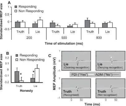
Fig. 2. Results from Experiment 1, showing activity of the neural population associated with the non-responding digit in lying but not in truth telling. Error bars denote standard errors. Asterisks (*) denote statistical significance for the key pairwise comparisons reported in the main text. (A) Mean of all participants’ median peak-to-peak MEP z-scores in Experiment 1, presenting the full set of conditions. (B) The same data are shown for responding and non-responding digits in lie and truth trials, having been collapsed across stimulation intervals in line with the results of the ANOVA (see main text). (C) Average MEPs for the Abductor DigitiMinimi (ADM) and First Dorsal Interosseous (FDI) of a single subject from Experiment 1 at stimulation interval 200 ms. This participant used the thumb (FDI) to indicate ‘yes’ and the little finger (ADM) for ‘no’. In both ‘lie’ conditions (i.e. lies made in response to either famous or non-famous faces) but not in the corresponding ‘true’ conditions, the MEP of the non-responding digit (ADM when falsely claiming recognition, FDI when falsely denying recognition) appears more active than that of the responding digit.
[image:13.595.74.481.70.411.2]4. Experiment 2
4.1. Method
Experiment 2 used the same practice session, stimuli, apparatus and statistical analyses as Experiment 1. Thirteen right-handed participants were tested. One participant was rejected following data analysis, because they had < 5 MEPs per condition after screening for muscular pre-activation leaving a final sample of 12 (7 females, mean age = 25.6, SD = 6.1).
4.2. Design and procedure
5. Results and discussion
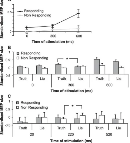
Fig. 3. (A) Digit by interval interaction in Experiment 2, illustrating the progressive selection of one response over the other in M1 during the decision-making process. (B) Further modulation of this interaction depending upon honesty of response, emphasising a significant digit by honesty interaction, but only in the middle stimulation interval. (C) Significant three-way (interval by digit by honesty) interaction in Experiment 3, where verbal responses were given without the need for hand motor planning. The digit by honesty interaction was significant only in the middle stimulation interval, showing a transient increase in the MEP of the non-responding digit in lying but not in truth telling.
[image:16.595.71.501.79.554.2]6. Experiment 3
6.1. Method
We employed identical stimuli, apparatus and statistical analyses to those used in Experiment 1. Four right-handed participants (4 females, mean age = 25.3, SD = 2.9) were tested.
6.2. Procedure
One hundred simple yes/no type questions served as training material, establishing the association between verbal and motor responses. This set of questions was randomised and presented 17.5 times during the experiment. Within these phases, on each trial one question was presented for 2500 ms followed by a tone, after which participants depressed one of the two response keys indicating yes/no whilst simultaneously saying “yes”/“no”. There were 1600 such trials at the start of the experiment. A shorter top-up phase (50 trials) followed each of the first three deception blocks.
7. Results and discussion
8. General discussion
Using single-pulse TMS, we examined whether conflict between competing responses in M1 can discriminate between intentionally false and true facial recognition. In Experiments 1 and 2 naïve participants used their little finger or thumb to indicate familiarity with famous and unknown faces. In Experiment 3 participants provided verbal responses which had been associated in advance with the same manual responses. By stimulating M1 prior to response execution, we could compare the MEPs linked to the two competing responses. We compared MEPs in correct trials from the digit that was used (the “responding digit”) and the passive digit (the “non-responding digit”). In all three experiments we identified critical times when the MEPs of the non-responding digit were greater than the MEPs of the responding digit in trials where the former coded the truth, i.e. when subjects lied. By contrast, MEPs of the non-responding digit were smaller than the MEPs of the responding digit when the former response coded a lie, i.e. when subjects told the truth.
Our data are suggestive of intra-hemispheric mutual inhibition during response preparation. In the second experiment, as RT unfolded the population of neurons representing the to-be-executed response became increasingly active whilst the alternative response appeared inhibited. Notably, this progressive process was biased or delayed in deception trials: the truth was initially prepared, then some neural signal prompted the alternative response (i.e. the lie) to become preeminent. Biasing influences could originate from prefrontal and cingulate areas ( [Frith, 2000], [Passingham,
1993] and [Lau et al., 2006]). Both areas have been implicated previously in the resolution of
response conflicts and the act of deception ( [Abe et al., 2006], [Abe et al., 2007], [Bhatt et al.,
2009], [Kozel et al., 2004], [Lee et al., 2010], [Langleben et al., 2005], [Nunez et al.,
2005] and [Schumacher et al., 2010]). Excitability of specific populations in M1 might for example be
biased via cortico-cortical pathways to supplementary motor area (SMA), or striatal projections to M1 and the SMA.
and reward system, like that which accompanies real-life deception. Such studies should also investigate the degree to which the partial activation of the truth is determined by the frequency of lying within the task (e.g. Verschuere et al., 2011).
Neurobiological principles of response selection can also be tested against our data. The parallel process of excitation of one response and the suppression of the alternative one argues against diffusion models of decision making that stipulate independent activation in both of two response alternatives in a race, particularly in regard to intra-hemispheric competition (Smith and Ratcliff, 2004). Put differently, our data support the notion of reciprocal inhibition between two motor response alternatives (although this inhibition may not be direct). Inhibitory links which still allow activation of multiple response alternatives are central to the leaky competing accumulator model of simple perceptual choices (Usher and McClelland, 2001) and the affordance competition model of motor decision making (Cisek, 2007).
The results of Experiment 3 suggest that verbal responses can be associated with digit-specific motor plans strongly enough to trigger them involuntarily. A verbal lie will then briefly activate a manual motor plan associated with the truth, presumably via the activation of the verbal truth plan. This is consistent with the theory of event coding (Hommel et al., 2001). The finding may remove one barrier to developing a lie detector based on TMS. However, in many techniques, lie-detection accuracy has been shown to be weakened by participants’ countermeasures (e.g. when using the ERP P300; [Mertens and Allen, 2008] and [Rosenfeld et al., 2001]). These would also hamper MEP-based detection, which requires voluntary muscle relaxation.
References
Abe, N., Fujii, T., Hirayama, K., Takeda, A., Hosokai, Y., Ishioka, T., . . . Takahashi, S. (2009). Do
parkinsonian patients have trouble telling lies? The neurobiological basis of deceptive
behaviour. Brain132, 1386.
Abe, N., Suzuki, M., Mori, E., Itoh, M., & Fujii, T. (2007). Deceiving others: Distinct neural responses of
the prefrontal cortex and amygdala in simple fabrication and deception with social
interactions. Journal of Cognitive Neuroscience,19, 287-295.
Abe, N., Suzuki, M., Tsukiura, T., Mori, E., Yamaguchi, K., Itoh, M., & Fujii, T. (2006). Dissociable roles
of prefrontal and anterior cingulate cortices in deception. Cerebral Cortex, 16, 192.
Bestmann, S., Harrison L. M., Blankenburg, F., Mars, R. B., Haggard, P., Friston, K. J., & Rothwell, J. C.
(2008). Influence of uncertainty and surprise on human corticospinal excitability during preparation
for action. Current Biology.18, 775–780.
Bhatt, S., Mbwana, J., Adeyemo, A., Sawyer, A., Hailu, A., & Vanmeter, J. (2009). Lying about facial
recognition: An fMRI study. Brain andCognition, 69, 382-390.
Botvinick, M., Nystrom, L. E., Fissell, K., Carter, C. S., & Cohen, J. D. (1999). Conflict monitoring versus
selection-for-action in anterior cingulate cortex. Nature, 402, 179-180.
Braver, T. S., Reynolds, J. R., & Donaldson, D. I. (2003). Neural mechanisms of transient and sustained
cognitive control during task switching. Neuron, 39, 713-726.
Carter, C. S., Braver, T. S., Barch, D. M., Botvinick, M. M., Noll, D., & Cohen, J. D. (1998). Anterior
Cisek, P., & Kalaska, J. F. (2005). Neural correlates of reaching decisions in dorsal premotor cortex:
Specification of multiple direction choices and final selection of action. Neuron,45, 801-814.
Cisek, P. (2006). Integrated neural processes for defining potential actions and deciding between
them: A computational model. Journal of Neuroscience, 26(38), 9761.
Cisek, P. (2007). Cortical mechanisms of action selection: The affordance competition hypothesis.
Philosophical Transactions of the Royal Society B: Biological Sciences, 362(1485), 1585.
DePaulo, B. M., Lindsay, J. J., Malone, B. E., Muhlenbruck, L., Charlton, K., & Cooper, H. (2003). Cues to
deception. Psychological Bulletin, .129, 74-118.
Desoto, M. C., Fabiani, M., Geary, D. C., & Gratton, G. (2001). When in doubt, do it both ways: Brain
evidence of the simultaneous activation of conflicting motor responses in a spatial stroop task.
Journal of Cognitive Neuroscience, 13, 523-536.
Ekman, P. (2009). Telling lies: Clues to deceit in the marketplace, politics, and marriage. WW Norton &
Company.
Eriksen, B. A., & Eriksen, C. W. (1974). Effects of noise letters upon the identification of a target letter
in a nonsearch task. Perception & Psychophysics, 16, 143-149.
Fleming, S. M., Mars, R. B., Gladwin, T. E., & Haggard, P. (2009). When the brain changes its mind:
Flexibility of action selection in instructed and free choices. Cerebral Cortex,
Frith, C.D. The role of the dorsolateral prefrontal cortex in the selection of action (2000). In Attention
and performance XVIII: Control of cognitive processes, Monsell, S. & Driver, J. Cambridge MA: MIT
Press, p. 549–64.
Gandevia, S., & Rothwell, J. (1987). Knowledge of motor commands and the recruitment of human
Hadar, A. A., Makris, S., & Yarrow, K. (in press). Single-pulse TMS related syncopal spell in a healthy
subject. Brain Stimulation.
Hommel, B., Müsseler, J., Aschersleben, G., & Prinz, W. (2001). The theory of event coding (TEC): A
framework for perception and action planning. Behavioral and Brain Sciences, 24, 849-878.
Kiers, L., Fernando, B., & Tomkins, D. (1997). Facilitatory effect of thinking about movement on
magnetic motor-evoked potentials. Electroencephalography and Clinical
Neurophysiology/Electromyography and Motor Control, 105, 262-268.
Kozel, F. A., Johnson, K. A., Mu, Q., Grenesko, E. L., Laken, S. J., & George, M. S. (2005). Detecting
deception using functional magnetic resonance imaging. Biological Psychiatry, 58, 605-613.
Kozel, F. A., Padgett, T. M., & George, M. S. (2004). A replication study of the neural correlates of
deception. Behaviour Neuroscience, 118, 852-856.
Langleben, D. D., Loughead, J. W., Bilker. W.B., Ruparel, K., Childress, A. R., Busch, S. I., & Gur, R. C.
(2005). Telling truth from lie in individual subjects with fast event-related fMRI. Human Brain
Mapping, 26, 262-272.
Lau, H, Rogers, R.D., Passingham, R.E. (2006) Dissociating response selection and conflict in the
medial frontal surface. Neuroimage,29, 446–451.
Lee, T. M. C., Lee, T. M. Y., Raine, A., Chan, C. C. H., & Manzoni, O. J. (2010). Lying about the valence of
affective pictures: An fMRI study. PloS One5, 244-245.
Lundqvist, D., Flykt, A., & Öhman, A. (1998). The Karolinska Directed Emotional Faces - KDEF, CD ROM
from the Department of Clinical Neuroscience, Psychology section, Karolinska Institutet, Stockholm,
Makris, S., Hadar, A.A., Yarrow, K. (2011). Viewing objects and planning actions: On the
potentiation of grasping behaviours by visual objects.
Brain and Cognition
,
77
,
257-264
Mazurek, M. E., Roitman, J. D., Ditterich, J., & Shadlen, M. N. (2003). A role for neural integratorsin perceptual decision making. Cerebral Cortex, 13, 1257.
Mertens, R., & Allen, J. J. B. (2008). The role of psychophysiology in forensic assessments: Deception
detection, ERPs, and virtual reality mock crime scenarios. Psychophysiology,45, 286-298.
Nardini, W. (1987). The polygraph technique: An overview. Journal of Police Science Administration,
15, 239-249.
Nunez, J. M., Casey, B., Egner, T., Hare, T., & Hirsch, J. (2005). Intentional false responding shares
neural substrates with response conflict and cognitive control. NeuroImage, 25, 267-277.
Pashler, H. (1991). Shifting visual attention and selecting motor responses: Distinct attentional
mechanisms. Journal of Experimental Psychology, 17, 1023-1040.
Passingham, R. 1993. The Frontal Lobes and Voluntary Action. (Oxford, UK: Oxford University Press)
Pollina, D., Dollins, A., Senter, S., Krapohl, D., & Ryan, A. (2004). Comparison of polygraph data
obtained from individuals involved in mock crimes and actual criminal investigations. Journal of
Applied Psychology, 89, 1099-1105.
Rosenfeld, J. P., Miller, A. R., Rao, A., & Soskins, M. (2001). Event-related potentials in detection of
deception. Handbook of Polygraphy. (New York, Academic Press).
Schumacher, E. H., Seymour, T. L., & Schwarb, H. (2010). Brain activation evidence for response
conflict in the exclude recognition task. Brain Research, 1329, 113-123.
Seymour and Schumacher, 2009 T.L. Seymour, E.H. Schumacher Electromyographic evidence for
response conflict in the exclude recognition task Cognitive, Affective, & Behavioral Neuroscience, 9,
Smith, P. L., & Ratcliff, R. (2004). Psychology and neurobiology of simple decisions. Trends in
Neuroscience, 27, 161-168.
Spence, S. A. (2004). The deceptive brain. Journal of the Royal Society of Medicine, 97, 6.
Stroop, J. R. (1935). Studies of interference in serial verbal reactions. Journal of Experimental
Psychology. 18(6):643-662
Taylor, P. C. J., Nobre, A. C., & Rushworth, M. F. S. (2007). FEF TMS affects visual cortical activity.
Cerebral Cortex, 17, 391.
Tottenham, N., Tanaka, J., Leon, A.C., McCarry, T., Nurse, M., Hare, T.A., Marcus, D.J., Westerlund, A.,
Casey, B.J., & Nelson, C.A. (2009). The NimStim set of facial expressions: judgments from untrained
research participants. Psychiatry Research,168, 242-249.
Usher, M., & McClelland, J. L. (2001). The time course of perceptual choice: The leaky, competing
accumulator model. Psychological Review, 108, 550-592.
Verleger, R., Kuniecki, M., Möller, F., Fritzmannova, M., & Siebner, H. R. (2009). On how the motor
cortices resolve an inter-hemispheric response conflict: An event-related EEG potential-guided TMS
study of the flankers task. European Journal of Neuroscience, 30, 318-326.
Verschuere, B., Spruyt, A., Meijer, E. H., & Otgaar, H. (2011). The ease of lying. Consciousness and