R E S E A R C H A R T I C L E
Open Access
Analysis of the gut microbiota of walking sticks
(Phasmatodea)
Matan Shelomi
1, Wen-Sui Lo
2,3,4, Lynn S Kimsey
1and Chih-Horng Kuo
2,3,5*Abstract
Background:Little is known about the Phasmatodea gut microbial community, including whether phasmids have symbiotic bacteria aiding in their digestion. While symbionts are near ubiquitous in herbivorous insects, the Phasmatodea’s distinctively thin body shape precludes the gut enlargements needed for microbial fermentation. High-throughput sequencing was used to characterize the entire microbiota of the fat bodies, salivary glands, and anterior and posterior midguts of two species of walking stick.
Results:Most bacterial sequences belonged to a strain ofSpiroplasma(Tenericutes) found primarily in the posterior midgut of the parthenogenetic speciesRamulus artemis(Phasmatidae). Beyond this, no significant differences were found between theR.artemismidgut sections or between that species andPeruphasma schultei(Pseudophasmatidae). Histological analysis further indicated a lack of bacteriocytes.
Conclusions:Phasmids are unlikely to depend on bacteria for digestion, suggesting they produce enzymes endogenously that most other herbivorous insects obtain from symbionts. This conclusion matches predictions based on phasmid anatomy. The role of Spiroplasmain insects warrants further study.
Keywords:Phasmatodea, Microbiota, 16S rDNA, Symbionts, Digestive system
Background
Research on insect endosymbionts has historically focused on insects with limited diets, such as wood-feeders or the nitrogen-limited phloem feeders [1,2]. This body of work has revealed several obligate symbioses, such as the aphid-Buchnerasystem where the insect cannot survive without its bacterial mutualist [3], as well as the many termite symbioses with gut bacteria and/or flagellates that assist in lignocellulose digestion [4]. Many such insects have spe-cialized cells called bacteriocytes in which the microbes are housed [5], while in others the symbionts persist within the midgut lining [2]. As most microbes, including the majority of endocellular symbionts, cannot be cultured [6], high-throughput sequencing has been used to char-acterize insect microbiota and identify possible symbionts [7], with successes in groups such as honey bees [8] and moths [9]. Gut microbes and their enzymes have received
much recent attention by the biofuel industry’s search for novel cellulases and xylanases, which has biased the gut mi-crobe literature towards xylophages like termites, roaches, and beetles [10,11].
Less understood are the digestive mechanisms and gut microbes of leaf eaters such as Lepidoptera larvae and orthopteroids. While leaves are not as difficult a diet as dry wood, they still contain abundant cellulase as plant cell walls [12], as well as toxic secondary chemicals, waxes, trichomes, and other obstacles to consumption and digestion by insects [13]. Symbiotic microbes would be beneficial to leaf eaters by assisting in cellulose break-down [14], nitrogen fixation or amino-acid metabolism [15], detoxifying or neutralizing plant defensive com-pounds [16], and recycling nitrogenous wastes [17].
While some microbial work has been performed on
Orthoptera sensu stricto[18-20], and a few papers have
looked at specific microbes within phasmids [21,22], no complete microbial inventory of the Phasmatodea gut has ever been performed. As obligate leaf eaters (as op-posed to certain periodically cannibalistic orthopteroids), the phasmids would certainly stand to benefit from having microbial symbionts. However, several factors suggest that * Correspondence:[email protected]
2
Institute of Plant and Microbial Biology, Academia Sinica, Taipei, Taiwan 3Molecular and Biological Agricultural Sciences Program, Taiwan International Graduate Program, National Chung Hsing University and Academia Sinica, Taipei, Taiwan
Full list of author information is available at the end of the article
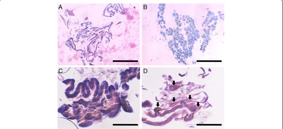
phasmids would not have digestive symbionts. Their char-acteristic body shape places restrictions on their gut morphology, which is straight and narrow tubes (Figure 1) with very short gastric caecae and no obvious diverticulae or fermentation chambers [23,24] of the kind that house microbes in other insects [1]. The only likely places for a phasmid symbiont to exist would be the midgut (the main site of phasmid digestion), the salivary glands, and the fat bodies. One paper studying insect fibre digestion sug-gested phasmids do not rely on microbes for digestion, but it based this on counts of culturable microbes, mean-ing up to 99% of the microbe diversity in the gut may have been ignored [21]. In our paper, microscopy and next gen-eration sequencing was used to catalog the entire micro-biota of the aforementioned phasmid organs from two species, with the goal of finding potential symbionts and possibly elucidating their functions, as well as increasing our knowledge of a poorly described alimentary canal.
Methods
The phasmids used were lab-reared specimens ofRamulus
artemis(Westwood) (Phasmatidae), fed rose (Rosa) leaves, andPeruphasma schultei(Conle & Hennemann) (Pseudo-phasmatidae), fed privet (Ligustrum). All insects were maintained and used as per the University of California,
Davis’ Institutional Animal Care and Use Committee
guidelines.
To check for the presence of bacteriocytes, histological analysis was performed [25]. Whole insects with longitu-dinal slits across the body wall were fixed in Bouin’s Fluid for three days and stored in 70% ethanol. Tissue samples were dissected out, dehydrated in an ethanol-butanol series, and embedded in paraffin. Sections were slide mounted using Meyer’s albumin and stained in Giemsa or Heidenhaim’s haematoxylin and eosin for light microscopy [26,27].
For molecular analysis, adult females were starved for two days to clear their guts and reduce chloroplast con-tamination of the samples. The digestive tract and its contents were dissected out, and the midguts divided into two sections reflecting the phasmid’s unique gut anatomy (Figure 1): the heavily pleated anterior midgut (AMG) and the unpleated posterior midgut (PMG) stud-ded with tubules of a currently unknown function [23,28]. The gut sections as well as the fat bodies and salivary glands were preserved in 100% ethanol prior to DNA extraction.
The total DNA from each sample was extracted using the Wizard® Genomic DNA Purification Kit (Promega; Fitchburg, Wisconsin, USA) following the manufac-turer’s protocol. To identify gut microbes, PCR was done to amplify the 16S ribosomal DNA using the universal primers 27F (AGAGTTTGATCMTGGCTCAG) and 511R (GCGGCTGCTGGCACRKAGT) with the appropriate 454 Life Sciences adaptor sequence. In addition, the forward primer used for each sample contained a unique 6-bp barcode for multiplexed sequencing. The barcodes used are described in NCBI BioSamples SAMN02318746-SAMN 02318757. To minimize biases that might occur in individ-ual PCR reactions, three independent reactions were per-formed for each sample and the products were pooled
before sequencing. Each of the 50 μL of PCR mixture
consisted of 1μL PfuUltra II Fusion HS DNA polymerase
(Stratagene; La Jolla, California, USA), 5μL of supplied
10× buffer, 2.5μL of 5 mM dNTP mix (MBI Fermentas;
Burlington, Ontario, Canada), 0.5μL of 10 mg/mL BSA
(New England Biolabs; Ipswich, Massachusetts, USA),
1μL of each 10μM primer, and 50 ng of template DNA.
[image:2.595.58.540.547.703.2]The PCR program included one denaturing step at 95°C for 3 min, 25 cycles of 95°C for 40 sec, 55°C for 40 sec, and 72°C for 40 sec, followed by a final extension at 72°C for 7 min. Gel electrophoresis was used to check the existence of a single band of expected size for each
PCR product. For the positive samples, PCR products were purified with the QIAquick PCR Purification Kit (Qiagen; Venlo, Netherlands). To further confirm the successful amplification of bacterial 16S rDNA in our broad range PCR, the purified PCR products were cloned using the CloneJet PCR Cloning Kit (Fermentas Life Sci-ence; Burlington, Ontario, Canada) and transformed into HIT-JM 109 competent cells (RBC Bioscience; Zhonghe City, Taipei County, Taiwan). A limited number of clones were sequenced using the BigDye Terminator v3.1 Cycle Sequencing Kit on an ABI Prism 3700 Genetic Analyzer (Applied Biosystems; Foster City, California, USA) to verify the presence of expected 16S rDNA fragment, multiplexing barcodes, and the adapters for 454 sequencing.
For high-throughput DNA sequencing, the positive samples were pooled in equal proportions and se-quenced on a 454 Jr. sequencer (454 Life Sciences; Branford, Connecticut, USA). The pyrosequencing flow-grams were converted to sequence reads with corre-sponding quality scores using the standard software provided by 454 Life Sciences. All raw reads were de-posited in NCBI Sequence Read Archive (accession num-ber SRR955712). The sequences were quality-trimmed using the default settings of LUCY [29]. Reads that were shorter than 400-bp after the quality trimming were re-moved from the data set. After the quality trimming, the sample-specific barcode and the primer sequence were identified and trimmed from each sequence; sequences that lacked a recognizable barcode and PCR primer were discarded. To identify the operational taxonomic units (OTUs) in these samples, the partial 16S rDNA sequences
were hierarchically clustered at 100%, 99%, and 97% se-quence identity using USEARCH version 5.2.32 [30]. The 97% sequence identity threshold was chosen be-cause it is commonly used to define bacterial species [31,32]. For taxonomic assignment, the representative sequence of each OTU was used as the query for the CLASSIFIER [33] program provided by the Ribosomal Database Project [34] with the 16S rRNA training set (version 2.5). The OTUs that were identified as originat-ing from plant chloroplasts or mitochondria were ex-cluded from downstream analyses. Furthermore, OTUs that could not be assigned to a particular genus with at least 70% confidence level were removed because these sequences are likely to represent chimeras or other ar-tifacts introduced during the PCR or pyrosequencing process [35]. For verification of the CLASSIFIER results and taxonomic assignment at species level, BLASTN [36,37] similarity search against the NCBI nt database [38] was performed for the representative sequence of each OTU. We limited the BLASTN search to the subject sequences from Bacteria (‘taxid2’). Additionally, subject sequences from environmental samples or metagenomes were excluded because these sequences often do not con-tain reliable taxonomic assignments. The e-value cutoff and other parameters of the BLASTN search were set to default; the top one hit was used as the representative for each query.
[image:3.595.58.539.475.694.2]To compare the microbiota composition among indi-vidual samples, we utilized the software package Fast UniFrac [39] to perform Principal Coordinate Analysis (PCoA) and hierarchical clustering. The OTUs were
weighted by abundance and the branch lengths were normalized. To generate a reference tree for the Fast UniFrac analyses, the representative sequences from all OTUs were aligned using the RDP ALIGNER [34]. The resulting multiple sequence alignment was examined to ensure that the 5’-end of each sequence was mapping to the expected location of 16S rDNA. The program FastTree [40] was then used to infer a maximum likeli-hood phylogeny of the OTUs. Additionally, the PVCLUST package [41] for R Statistical Software [42] was used to perform an alternative hierarchical clustering analysis with a phylogeny-independent approach.
Results
The broad range PCR amplification results for bacterial 16S rDNA were negative for the fat body and salivary gland samples examined, so they were excluded after the initial quality check steps. This negative result is consist-ent with phasmid histological studies, which did not show obvious mycetocytes in fat body or salivary gland tissue, nor any obvious endosymbionts in the midgut tis-sues (Figure 2). All sevenP.schulteiPMG samples tested negative for bacterial DNA as well. In total, bacterial 16S
rDNA was recovered from four P. schultei AMGs, four
R.artemisAMGs, and threeR.artemisPMGs, and these samples were included in the 454 sequencing and final analysis. In total, we obtained 26,006 high quality se-quence reads for these 11 midgut samples.
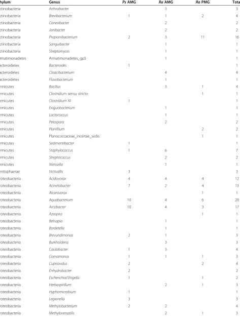
Among the midgut samples, an overwhelming majority of the sequencing reads originated from the 16S rDNA of plant chloroplasts or mitochondria (Table 1). After re-moving these contaminants, we obtained a total of 162 bacterial OTUs covering 64 genera (Table 2). Rarefaction curves (Figure 3) suggest that the sampling depths achieved in this study may not be sufficient to fully characterize the microbiota of Phasmatodea guts. With the exception of a large Tenericute population in the R. artemis PMGs, most of the microbes isolated were Proteobacteria, followed by Actinobacteria and Firmicutes (Table 1 and Figure 4). Nearly all of the Tenericute reads (4,902 out of 5,412 Tenericute sequences) belonged to a single OTU (OTU_ID: CXTIA; Additional file 1) found
mostly in theR. artemisposterior midgut. The BLASTN
search against the NCBI nucleotide database showed that this sequence is 99.0% identical toSpiroplasmasp.‘Gent’ (GenBank AY569829), an uncultured, male-killing
entomo-pathogen identified from the housefly Fannia manicata.
The next most common OTU had only 161 reads, and all other OTUs had less than 50 reads (Additional file 1). All but one of the 10 most abundant OTUs appeared to be Spiroplasma, which are known to be associated with a wide range of insects [43,44]. The most abundant non-Spiroplasma OTU was represented by 31 reads and
showed high similarity (99.3% sequence identity) to Sphingobium sp. KR5 (GenBank JQ433940).
Comparison of the microbiota composition based on the PCoA plots (Figure 5) indicated that the relative
abundance ofSpiroplasma reads is the major
determin-ant that differentiates different sample types. When all 5,679 bacterial reads are considered, the threeR.artemis PMG samples form a tight cluster and the PCO1 ex-plains 81.77% of the variance (Figure 5A). Although one P. schulteiAMG sample (from individual #5) appears to share a similar microbiota composition with the threeR. artemis PMG samples, this P. schultei AMG sample should be considered as an outlier because it contains only
one reads assigned to the most abundant Spiroplasma
OTU. When the putative Spiroplasma reads were
ex-cluded, no clear pattern exists to distinguish among sam-ple types (Figure 5B).
[image:4.595.305.538.102.462.2]The hierarchical clustering analyses based on either phylogeny-dependent approach (Figure 6 panels A and B) or phylogeny-independent approach (Figure 6 panels C and D) produced the same patterns inferred from the Table 1 Summary of reads received from phasmid samples
P. schultei
anterior midgut
R. artemis
anterior midgut
R. artemis
posterior midgut
Total
# of positive samples 4 4 3 All sequences:
# reads passed quality control
9,255 9,697 7,054 26,006
Average # reads per sample
2,313.75 2,424.25 2,351.33
# 100% id OTUs 8,090
# 99% id OTUs 2,702
# 97% id OTUs 844
After RDP CLASSIFIER:
# chloroplast 16S 14,541
# mitochondrial 16S and other contaminants
5,786
# bacterial 16S reads 112 171 5,396 5,679 Average # reads
per sample
28 43 1,799
# reads (# OTUs) by phylum
Actinobacteria 3(2) 13(9) 13(4) 29(13) Armatimonadetes 0 1(1) 0 1(1) Bacteroidetes 1(1) 5(3) 0 6(4) Firmicutes 3(3) 16(9) 5(4) 24(15) Lentisphaerae 3(1) 0 0 3(1) Proteobacteria 87(37) 65(39) 45(26) 197(75) Tenericutes 15(6) 65(10) 5,332(47) 5,412(47)
TM7 0 6(5) 1(1) 7(6)
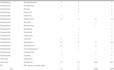
Table 2 The number of reads for each microbe genus per gut type (pooled samples)
Phylum Genus PsAMG RaAMG RaPMG Total
Actinobacteria Arthrobacter 3 3
Actinobacteria Brevibacterium 1 1 2 4
Actinobacteria Conexibacter 2 2
Actinobacteria Janibacter 2 2
Actinobacteria Propionibacterium 2 3 11 16
Actinobacteria Sanguibacter 1 1
Actinobacteria Streptomyces 1 1
Armatimonadetes Armatimonadetes_gp5 1 1
Bacteroidetes Bacteroides 1 1
Bacteroidetes Cloacibacterium 4 4
Bacteroidetes Flavobacterium 1 1
Firmicutes Bacillus 3 1 4
Firmicutes Clostridiumsensu stricto 1 1
Firmicutes ClostridiumXI 1 1
Firmicutes Exiguobacterium 1 1
Firmicutes Lactococcus 1 1
Firmicutes Pelospora 2 2
Firmicutes Planifilum 2 2
Firmicutes Planococcaceae_incertae_sedis 1 1
Firmicutes Sedimentibacter 1 1
Firmicutes Staphylococcus 1 6 7
Firmicutes Streptococcus 2 2
Firmicutes Weissella 1 1
Lentisphaerae Victivallis 3 3
Proteobacteria Acidovorax 4 4 4 12
Proteobacteria Acinetobacter 7 2 4 13
Proteobacteria Alcanivorax 1 1
Proteobacteria Aquabacterium 10 4 6 20
Proteobacteria Arcobacter 10 4 3 17
Proteobacteria Azospira 1 1
Proteobacteria Belnapia 1 1
Proteobacteria Bordetella 1 1
Proteobacteria Brevundimonas 2 1 3
Proteobacteria Burkholderia 3 3
Proteobacteria Caulobacter 1 3 4
Proteobacteria Comamonas 1 1 1 3
Proteobacteria Cupriavidus 2 2 4
Proteobacteria Enhydrobacter 2 2
Proteobacteria Escherichia/Shigella 1 1 2
Proteobacteria Herbaspirillum 2 1 3
Proteobacteria Hyphomicrobium 1 1
Proteobacteria Legionella 3 3
Proteobacteria Methylobacterium 2 2 4
PCoA plots. When all bacterial reads are considered
(Figure 6 panels A and C), the three R. artemis PMG
samples (together with the Ps5_AMG sample that
con-tains only one Spiroplasma read) form a single clade
with short distances among each other and high
diver-gences with other samples. When the Spiroplasmareads
were excluded (Figure 6 panels B and D), individual samples of the same type do not cluster together. Add-itionally, the branches that separate samples of the same type are similar in length compared to those separating samples of different types.
Discussion
The overabundance of chloroplast contamination greatly reduced the amount of bacterial reads available for ana-lysis. Starving the phasmids for two days was insufficient to clear their digestive tracts. For future study, a longer time period of starvation (e.g., five to ten days) would be preferable. While 16S primers that select against chloro-plasts do exist, they also bias the results of broad range
PCR and are not suitable replacements for “universal”
[image:6.595.59.539.100.395.2]primers when studying total microbial community. Des-pite the relatively low abundance of bacterial reads, the
Table 2 The number of reads for each microbe genus per gut type (pooled samples)(Continued)
Proteobacteria Novosphingobium 5 2 7
Proteobacteria Ochrobactrum 1 2 3
Proteobacteria Pantoea 3 3
Proteobacteria Paracoccus 2 2
Proteobacteria Pelomonas 1 1
Proteobacteria Pseudomonas 4 8 4 16
Proteobacteria Raoultella 1 1
Proteobacteria Rheinheimera 1 1
Proteobacteria Rhizobacter 4 4
Proteobacteria Shewanella 1 1
Proteobacteria Simplicispira 4 4
Proteobacteria Smithella 1 1 2
Proteobacteria Sphingobium 21 6 9 36
Proteobacteria Sphingomonas 4 1 2 7
Proteobacteria Stenotrophomonas 1 1
Proteobacteria Undibacterium 1 2 3
Proteobacteria Zoogloea 3 1 4
Tenericutes Haloplasma 1 1
Tenericutes Spiroplasma 15 65 5,331 5,411
TM7 TM7_genera_incertae_sedis 6 1 7
Sum 112 171 5,396 5,679
The taxomic assignment at genus level was based on RDP CLASSIFIER.Ps=P.schultei.Ra=R.artemis.
50 100 150 0
20 40 60
0
0 20 40 60 80 100
0 2,000 4,000 6,000
Number of OTUs
A Ps_AMG
20 40 60 80
0
0 50 100 150 200
Number of reads
B Ra_AMG
C Ra_PMG
[image:6.595.63.539.569.693.2]use of next generation sequencing techniques still pro-vided thousands of usable reads of mostly uncultivated mi-crobes. This data set provides at least one order of magnitude more reads than would be available using clone libraries or denaturing gradient gel electrophoresis.
The low number of bacterial reads compared to chlo-roplast reads suggests the abundance of bacterial cells in
the phasmid gut is relatively low. These results, together with the lack of bacterial DNA in the fat body and saliv-ary glands and the lack of bacteriocytes in the tissue slices, do not support the hypothesis that phasmids have obligate microbial symbioses. Had such mutualist endo-symbionts existed in these samples, they would have
been detected at similar levels to the Spiroplasma
0 20 40 60 80 100 Ps_AMG
Ra_AMG
Ra_PMG
Relative abundance (%)
A
All bacterial reads (5,679 reads/162 OTUs)B
Excluding Spiroplasma reads (268 reads/115 OTUs)0 20 40 60 80 100 Ps_AMG
Ra_AMG
Ra_PMG
Relative abundance (%)
Actinobacteria Armatimonadetes Bacteroidetes Firmicutes
Lentisphaerae Proteobacteria Tenericutes TM7
112 # reads
171
5,396
97 # reads
106
65 Group
[image:7.595.57.549.89.356.2]Group
Figure 4Relative abundances of reads of the different phyla. (A)All 5,679 bacterial 16S rDNA reads.(B)ExcludingSpiroplasmareads.
Ra_AMG Ra_PMG Ps_AMG
PCO1 (81.77%)
PCO2 (8.44%)
-0.4 -0.2 0.0 0.2 0.4
-0.4 -0.2 0.0 0.2
5 2
6
3
4
2
5 6
5 3 6
PCO1 (50.82%)
PCO2 (20.19%)
-0.4 0.0 0.4
-0.2 0.0 0.2 0.4 0.6
2
6 3
6 4
2 5
6 5
3
B
Excluding Spiroplasma readsA
All bacterial reads0.8
[image:7.595.59.539.481.685.2]infestation. While lab-reared phasmids will likely have different gut microbiota from wild specimens, they are expected to retain any symbiotic microbes essential for survival. Symbiont absence in the lab suggests absence in the field as well, but this remains to be demonstrated. The data suggests that most of the microbes isolated were environmental, picked up by the phasmids from their diet or housing [18,45].
The lack of beneficial symbiotic microbes in phasmids is unsurprising given the aforementioned restrictions on their digestive system due to their distinct body shape. The phasmid gut lacks the space to develop the signifi-cantly modified sections seen in symbiont-housing or-ganisms, such as the enlarged hindguts of termites. Given these restrictions, one would expect a phasmid to compensate for lack of symbionts either with modified feeding behavior (such as increased consumption and/or slower gut transit time) or through endogenous produc-tion of digestive enzymes such as cellulases, which re-search predicts the phasmids can produce [12,21,46]. No research on phasmid enzymes has been published yet,
however. If phasmids did depend on gut bacteria to breakdown their leafy diet, one would expect a gut microbiota consisting predominantly of Bacteroidetes and Firmicutes, as one sees in the roaches and termites [2]. The abundance of Proteobacteria resembles the mostly transient microbes found in Orthoptera [18], which supports the closer phylogenetic relationship be-tween that order and the Phasmatodea [47].
The nature of R. artemis’ Spiroplasma infestation
needs investigation. Of the 162 OTUs, 32 were closely
related to Spiroplasma sp. ‘Gent’ (GenBank AY569829),
and another eight were closely related to Spiroplasma
ixodetes (GenBank GU585671). The infection’s localiza-tion to the PMG suggests the bacteria may be colonizing the PMG’s enigmatic appendices, but histological data did not confirm this (Figure 2D).Spiroplasmainfestation
may also explain why R. artemiscan be parthenogenetic
in cultures, as some Spiroplasma species are known
male-killing parasites [48] similar to Wolbachia [49].
Several other phasmid species, most notably the laboratory stick insect,Carausius morosus, are also parthenogenetic
0.2 0.1
Ra4_AMG
Ps2_AMG Ps6_AMG
Ps3_AMG
Ra2_AMG
Ra6_AMG Ra5_AMG Ra3_PMG
Ps5_AMG
Ra5_PMG Ra6_PMG
0.05
Ra4_AMG
Ps2_AMG Ps6_AMG
Ps3_AMG
Ra2_AMG
Ra6_AMG
Ra5_AMG
Ra3_PMG Ra5_PMG Ra6_PMG
0.05
A
Ra5_AMG Ra6_AMG
Ra2_AMG
Ra3_PMG
Ra5_PMG
Ra6_PMG
Ra4_AMG
Ps2_AMG Ps6_AMG
Ps3_AMG
D
Ra4_AMG
Ps2_AMG Ps6_AMG Ps3_AMG
Ra2_AMG Ra6_AMG
Ra5_AMG Ra3_PMG
Ps5_AMG
Ra5_PMG Ra6_PMG
C
B
All bacterial reads Excluding Spiroplasma reads
PVCLUST
[image:8.595.57.539.90.416.2]Fast UniFrac
[50], so checking these cultures for Spiroplasma infesta-tions may be revealing. DiBlasi et al. [22] concluded that theSpiroplasmais transmitted in phasmids by mites, yet the cultures used here were mite-free, suggesting vector-less transmission.
The most common non-Spiroplasmamicrobe isolated
was identified as a member of Sphingobium, which may
be involved in the degradation of aromatic hydrocarbons [51]. It may perform this role for the phasmid, aiding in digestion by degrading plant defensive compounds, but whether it is a true symbiont transmitted between indi-viduals, an environmental microbe that can colonize the gut, or an allochthonous (transient) microbe found on the leaves and just passing through is unknown.
Conclusions
This paper marks the first attempt to catalog the micro-bial diversity of the Phasmatodea (Additional file 1: Table S1). While a clade of symbiont-dependent phas-mids may exist, the likelihood is low. All evidence sug-gests the only heritable symbionts, allegedly ubiquitous in the Insecta [6], in the Phasmatodea are
reproduction-manipulators like Spiroplasma. Still, the phasmid gut
microbial community is diverse and merits further inves-tigation. The possibility of fungal or other eukaryotic symbionts in the gut remains. The microbial community may function together to benefit the phasmid in ways akin to their functions on leaf surfaces, such as second-ary chemical detoxification [52]. Lastly, if phasmid diges-tion is truly microbe-independent, then the enzymology of the gut demands further analysis given the potential for finding novel lignocellulases and other compounds of possible human industrial applications.
Availability of supporting data
A supplementary table (Table S1) the lists the taxonomic assignment OTUs is included as Additional file 1.
Additional file
Additional file 1: Table S1.Taxonomic assignment of the OTUs based on RDP CLASSIFIER and BLASTN sequence similarity search against the NCBI nt database.
Competing interests
The authors declare that they have no competing interests.
Authors’contributions
MS and CHK conceived of the study. MS and WSL carried out the experiments. MS and CHK performed the data analysis. MS, LSK, and CHK wrote the manuscript. All authors read and approved the final manuscript.
Acknowledgements
We thank the curation staff at the Bohart Museum of Entomology insect culture care, Dr. Robert Kimsey for histology advising, and the staff at Academia Sinica for assistance in DNA sequencing (Ms. Mei-Jane Fang at the Institute of Plant and Microbial Biology for Sanger sequencing and Dr. Shu-Yun Tung at the
Institute of Molecular Biology for 454 pyrosequencing). MS was supported by the USA National Science Foundation’s East Asia and Pacific Summer Institutes (EAPSI) program (NSF proposal number 1209449) and the co-sponsorship by the Taiwanese National Science Council’s Summer Institutes in Taiwan (SIT) program. Funding for this work was provided by research grants from Academia Sinica to CHK, and the University of California Davis and Humanities Graduate Research Fellowship.
Author details
1
Department of Entomology, University of California, Davis, USA.2Institute of Plant and Microbial Biology, Academia Sinica, Taipei, Taiwan.3Molecular and Biological Agricultural Sciences Program, Taiwan International Graduate Program, National Chung Hsing University and Academia Sinica, Taipei, Taiwan.4Graduate Institute of Biotechnology, National Chung Hsing University, Taichung, Taiwan.5Biotechnology Center, National Chung Hsing University, Taichung, Taiwan.
Received: 20 February 2013 Accepted: 5 September 2013 Published: 11 September 2013
References
1. Buchner P:Endosymbiosis of Animals with Plant Microorganisms.New York: Interscience Publishers; 1965.
2. Ishikawa H:Insect symbiosis: an introduction.InInsect Symbiosis.Edited by Bourtzis K, Miller TA. Boca Raton: CRC Press; 2003:1–21.
3. Douglas AE:Buchnera bacteria and other symbionts of aphids.InInsect Symbiosis.Edited by Bourtzis K, Miller TA. Boca Raton: CRC Press; 2003:23–38. 4. Lo N, Eggleton P:Termite phylogenetics and co-cladogenesis with
symbionts.InBiology of Termites: A Modern Synthesis.Edited by Bignell DE, Roisin Y, Lo N. London: Springer; 2011:27–50.
5. Douglas AE:Lessons from studying insect symbioses.Cell Host Microbe 2011,10:359–367.
6. Moran NA, McCutcheon JP, Nakabachi A:Genomics and evolution of heritable bacterial symbionts.Annu Rev Genet2008,42:165–190. 7. Dharne M, Patole M, Shouche YS:Microbiology of the insect gut: tales
from mosquitoes and bees.J Biosci2006,31:293–295.
8. Moran NA, Hansen AK, Powell JE, Sabree ZL:Distinctive gut microbiota of honey bees assessed using deep sampling from individual worker bees.
PLoS One2012,7:e36393.
9. Belda E, Pedrola L, Pereto J, Martinez-Blanch JF, Montagud A, Navarro E, Urchueguia J, Ramon D, Moya A, Porcar M:Microbial diversity in the midguts of field and lab-reared populations of the European corn borer Ostrinia nubilalis.PLoS One2011,6:e21751.
10. Scharf ME, Tartar A:Termite digestomes as sources for novel lignocellulases.Biofuels, Bioprod Biorefin2008,2:540–552.
11. Warnecke F, Luginbuhl P, Ivanova N, Ghassemian M, Richardson TH, Stege JT, Cayouette M, McHardy AC, Djordjevic G, Aboushadi N, Sorek R, Tringe SG, Podar M, Martin HG, Kunin V, Dalevi D, Madejska J, Kirton E, Platt D, Szeto E, Salamov A, Barry K, Mikhailova N, Kyrpides NC, Matson EG, Ottesen EA, Zhang X, Hernández M, Murillo C, Acosta LG, Rigoutsos I, Tamayo G, Green BD, Chang C, Rubin EM, Mathur EJ, Robertson DE, Hugenholtz P, Leadbetter JR:Metagenomic and functional analysis of hindgut microbiota of a wood-feeding higher termite.Nature2007,450:560–565. 12. Watanabe H, Tokuda G:Cellulolytic systems in insects.Annu Rev Entomol
2010,55:609–632.
13. Shelomi M, Perkins LE, Cribb BW, Zalucki MP:Effects of leaf surfaces on first instar Helicoverpa armigera (Hübner)(Lepidoptera: Noctuidae) behaviour.Australian J Entomol2010,49:289–295.
14. Wilson DB:Aerobic microbial cellulase systems.InBiomass Recalcitrance: Deconstructing the Plant Cell Wall for Bioenergy.Edited by Himmel ME. Oxford: Blackwell Publishing Ltd; 2008:374–392.
15. Moran NA, Plague GR, Sandstrom JP, Wilcox JL:A genomic perspective on nutrient provisioning by bacterial symbionts of insects.Proc Natl Acad Sci U S A2003,100(Suppl 2):14543–14548.
[image:9.595.307.539.294.738.2]18. Idowua AB, Edemaa MO, Oyedepo MT:Extracellular enzyme production by microflora from the gut region of the variegated grasshopper Zonocerus variegatus (Orthoptera: Pyrgomorphidae).Int J Trop Insect Sci 2009,29:229–235.
19. Kaufman MG, Klug MJ:The contribution of hindgut bacteria to dietary carbohydrate utilization by crickets (Orthoptera: Gryllidae).
Comp Biochem Physiol1991,98A:117–123.
20. Mead LJ, Khachatourians GG, Jones GA:Microbial ecology of the gut in laboratory stocks of the migratory grasshopper, Melanoplus sanguinipes (Fab.) (Orthoptera: Acrididae).Appl Environ Microbiol1988,54:1174–1181. 21. Cazemier AE, den Camp HJMO, Hackstein JHP, Vogels GD:Fibre digestion
in arthropods.Comp Biochem Physiol1997,118A:101–109. 22. DiBlasi E, Morse S, Mayberry JR, Avila LJ, Morando M, Dittmar K:New
Spiroplasma in parasitic Leptus mites and their Agathemera walking stick hosts from Argentina.J Invertebr Pathol2011,107:225–228. 23. Cameron AE:Structure of the alimentary canal of the stick-insect, Bacillus
rossii Fabr.; with a note on the parthenogenesis of this species.Proc Zool Soc London1912,82:172–182.
24. Chopard L:Ordre des Chéleutoptères.InTraité de Zoologie: Anatmie, Systématique, Biologie.Edited by Grassé P-P. Paris: Masson; 1949:594–617. 25. Lasker R, Giese AC:Cellulose digestion by the silverfish Ctenolepisma
Lineata.J Exp Biol1940,33:542–553.
26. Coupland RE:Observations on the normal histology and histochemistry of the fat body of the locust (Schistocera gregaria).J Exp Biol1957,
34:290–296.
27. Gurr E:A Practical Manual of Medical and Biological Staining Techniques.New York: Interscience Publishers, Inc.; 1953.
28. Ramsay JA:The excretory system of the stick insect Dixippus morosus (Orthoptera, Phasmidae).J Exp Biol1955,32:183–199.
29. Chou HH, Holmes MH:DNA sequence quality trimming and vector removal.Bioinformatics2001,17:1093–1104.
30. Edgar RC, Haas BJ, Clemente JC, Quince C, Knight R:UCHIME improves sensitivity and speed of chimera detection.Bioinformatics2011,27:2194–2200. 31. Drancourt M, Raoult D:Sequence-based identification of new bacteria: a
proposition for creation of an orphan bacterium repository.J Clin Microbiol2005,43:4311–4315.
32. Janda JM, Abbott SL:16S rRNA gene sequencing for bacterial identification in the diagnostic laboratory: pluses, perils, and pitfalls.
J Clin Microbiol2007,45:2761–2764.
33. Wang Q, Garrity GM, Tiedje JM, Cole JR:Naive Bayesian classifier for rapid assignment of rRNA sequences into the new bacterial taxonomy.
Appl Environ Microbiol2007,73:5261–5267.
34. Cole JR, Wang Q, Cardenas E, Fish J, Chai B, Farris RJ, Kulam-Syed-Mohideen AS, McGarrell DM, Marsh T, Garrity GM, Tiedje JM:The Ribosomal Database Project: improved alignments and new tools for rRNA analysis.Nucl Acids Res2009,37:D141–D145.
35. Ochman H, Worobey M, Kuo C-H, Ndjango J-BN, Peeters M, Hahn BH, Hugenholtz P:Evolutionary relationships of wild hominids recapitulated by gut microbial communities.PLoS Biol2010,8:e1000546.
36. Altschul SF, Gish W, Miller W, Myers EW, Lipman DJ:Basic local alignment search tool.J Mol Biol1990,215:403–410.
37. Camacho C, Coulouris G, Avagyan V, Ma N, Papadopoulos J, Bealer K, Madden T:BLAST+: architecture and applications.BMC Bioinforma2009,
10:421.
38. Benson DA, Karsch-Mizrachi I, Clark K, Lipman DJ, Ostell J, Sayers EW:
GenBank.Nucl Acids Res2012,40:D48–D53.
39. Hamady M, Lozupone C, Knight R:Fast UniFrac: facilitating high-throughput phylogenetic analyses of microbial communities including analysis of pyrosequencing and PhyloChip data.ISME J2010,4:17–27. 40. Price MN, Dehal PS, Arkin AP:FastTree 2–Approximately
Maximum-Likelihood Trees for Large Alignments.PLoS ONE2010,5:e9490. 41. Suzuki R, Shimodaira H:Pvclust: an R package for assessing the
uncertainty in hierarchical clustering.Bioinformatics2006,22:1540–1542. 42. R Development Core Team. R:A language and environment for statistical
computing.Vienna, Austria: R Foundation for Statistical Computing; 2012. 43. Gasparich GE, Whitcomb RF, Dodge D, French FE, Glass J, Williamson DL:
The genus Spiroplasma and its non-helical descendants: phylogenetic classification, correlation with phenotype and roots of the Mycoplasma mycoides clade.Int J Syst Evol Microbiol2004,54:893–918.
44. Regassa LB, Gasparich GE:Spiroplasmas: evolutionary relationships and biodiversity.Front Biosci2006,11:2983–3002.
45. Thakuria D, Schmidt O, Finan D, Egan D, Doohan FM:Gut wall bacteria of earthworms: a natural selection process.ISME J2010,4:357–366. 46. Sun J-Z, Scharf ME:Exploring and integrating cellulolytic systems of
insects to advance biofuel technology.Insect Sci2010,17:163–165. 47. Flook PK, Rowell CHF:Inferences about orthopteroid phylogeny and
molecular evolution from small subunit nuclear ribosomal DNA sequences.Insect Mol Biol1998,7:163–178.
48. Jiggins FM, Hurst GD, Jiggins CD, Schulenburg JH VD, Majerus ME:The butterfly Danaus chrysippus is infected by a male-killing Spiroplasma bacterium.Parasitol2000,120:439–446.
49. Huigens ME, Stouthamer R:Parthenogenesis associated with Wolbachia.
InInsect Symbiosis.Edited by Bourtzis K, Miller TA. Boca Raton: CRC Press; 2003:247–266.
50. More E:Parthenogenesis explained.Phasmid Studies1996,52:62–69. 51. Singh A, Lal R:Sphingobium ummariense sp. nov., a
hexachlorocyclohexane (HCH)-degrading bacterium, isolated from HCH-contaminated soil.Int J Syst Evol Microbiol2009,59:162–166.
52. Ohmart CP, Thomas JR, Bubela B:Surfactant-producing microorganisms isolated from the gut of a Eucalyptus-feeding sawfly, Perga affinis affinis.
Oecologia1988,77:140–142.
doi:10.1186/1756-0500-6-368
Cite this article as:Shelomiet al.:Analysis of the gut microbiota of walking sticks (Phasmatodea).BMC Research Notes20136:368.
Submit your next manuscript to BioMed Central and take full advantage of:
• Convenient online submission
• Thorough peer review
• No space constraints or color figure charges
• Immediate publication on acceptance
• Inclusion in PubMed, CAS, Scopus and Google Scholar
• Research which is freely available for redistribution