R E S E A R C H A R T I C L E
Open Access
Genetic variation at
CYP3A
is associated with age
at menarche and breast cancer risk: a case-control
study
Nichola Johnson
1,2*, Frank Dudbridge
3, Nick Orr
1,2, Lorna Gibson
3, Michael E Jones
4, Minouk J Schoemaker
4,
Elizabeth J Folkerd
5, Ben P Haynes
5, John L Hopper
6, Melissa C Southey
7, Gillian S Dite
6, Carmel Apicella
6,
Marjanka K Schmidt
8, Annegien Broeks
8, Laura J Van
’
t Veer
8, Femke Atsma
9, Kenneth Muir
10, Artitaya Lophatananon
10,
Peter A Fasching
11,12, Matthias W Beckmann
11, Arif B Ekici
13, Stefan P Renner
11, Elinor Sawyer
14, Ian Tomlinson
15,16,
Michael Kerin
17, Nicola Miller
17, Barbara Burwinkel
18,19, Frederik Marme
18, Andreas Schneeweiss
18, Christof Sohn
18,20,
Pascal Guénel
21,22, Therese Truong
21,22, Emilie Cordina
21,22, Florence Menegaux
21,22, Stig E Bojesen
23,24,
Børge G Nordestgaard
23,24, Henrik Flyger
25, Roger Milne
26, M Pilar Zamora
27, Jose Ignacio Arias Perez
28,
Javier Benitez
29,30, Leslie Bernstein
31, Hoda Anton-Culver
32, Argyrios Ziogas
32, Christina Clarke Dur
33,
Hermann Brenner
34,35, Heiko Müller
34, Volker Arndt
34, Aida Karina Dieffenbach
34,35, Alfons Meindl
36, Joerg Heil
18,
Claus R Bartram
37, Rita K Schmutzler
38, Hiltrud Brauch
39,40, Christina Justenhoven
39,40, Yon-Dschun Ko
41, The GENICA
(Gene Environment Interaction and Breast Cancer in Germany) Network, Heli Nevanlinna
42, Taru A Muranen
42,
Kristiina Aittomäki
43, Carl Blomqvist
44, Keitaro Matsuo
45, Thilo Dörk
46, Natalia V Bogdanova
47, Natalia N Antonenkova
48,
Annika Lindblom
49, Arto Mannermaa
50,51,52, Vesa Kataja
50,51,53, Veli-Matti Kosma
50,51,52, Jaana M Hartikainen
50,51,52,
Georgia Chenevix-Trench
54, Jonathan Beesley
54, kConFab Investigators, Australian Ovarian Cancer Study Group,
Anna H Wu
55, David Van den Berg
55, Chiu-Chen Tseng
55, Diether Lambrechts
56,57, Dominiek Smeets
56,57,
Patrick Neven
58, Hans Wildiers
58, Jenny Chang-Claude
59, Anja Rudolph
59, Stefan Nickels
59, Dieter Flesch-Janys
60,61,
Paolo Radice
62, Paolo Peterlongo
62,63, Bernardo Bonanni
64, Valeria Pensotti
63,65, Fergus J Couch
66, Janet E Olson
67,
Xianshu Wang
66, Zachary Fredericksen
67, Vernon S Pankratz
67, Graham G Giles
6,68, Gianluca Severi
6,68,
Laura Baglietto
6,68, Chris Haiman
56, Jacques Simard
69, Mark S Goldberg
69, France Labrèche
70, Martine Dumont
71,
Penny Soucy
71, Soo Teo
72,73, Cheng Har Yip
72, Sze Yee Phuah
72,73, Belinda K Cornes
74, Vessela N Kristensen
75,76,
Grethe Grenaker Alnæs
76, Anne-Lise Børresen-Dale
75,76, Wei Zheng
77, Robert Winqvist
78, Katri Pylkäs
78,
Arja Jukkola-Vuorinen
79, Mervi Grip
80, Irene L Andrulis
81,82, Julia A Knight
81,83, Gord Glendon
81,84,
Anna Marie Mulligan
85,86, Peter Devillee
87, Jonine Figueroa
88, Stephen J Chanock
88, Jolanta Lissowska
89,
Mark E Sherman
88, Per Hall
90, Nils Schoof
90, Maartje Hooning
91, Antoinette Hollestelle
92, Rogier A Oldenburg
93,
Madeleine Tilanus-Linthorst
93, Jianjun Liu
94, Angie Cox
95, Ian W Brock
95, Malcolm WR Reed
96, Simon S Cross
97,
William Blot
77,98, Lisa B Signorello
99,100,101, Paul DP Pharoah
102, Alison M Dunning
102, Mitul Shah
103, Daehee Kang
103,
Dong-Young Noh
103, Sue K Park
104,105,106, Ji-Yeob Choi
103, Mikael Hartman
107,108,109, Hui Miao
99,100, Wei Yen Lim
108,109,
Anthony Tang
110, Ute Hamann
111, Asta Försti
112,113, Thomas Rüdiger
114, Hans Ulrich Ulmer
115, Anna Jakubowska
116,
Jan Lubinski
116, Katarzyna Jaworska-Bieniek
116,117, Katarzyna Durda
116, Suleeporn Sangrajrang
118, Valerie Gaborieau
119,
Paul Brennan
119, James McKay
119, Susan Slager
67, Amanda E Toland
120, Celine Vachon
67, Drakoulis Yannoukakos
121,
* Correspondence:nichola.johnson@icr.ac.uk †Equal contributors
1Breakthrough Breast Cancer Research Centre, The Institute of Cancer
Research, 237 Fulham Road, London SW3 6JB, UK
2Division of Breast Cancer Research, The Institute of Cancer Research, 237
Fulham Road, London SW3 6JB, UK
Full list of author information is available at the end of the article
Chen-Yang Shen
122,123, Jyh-Cherng Yu
124, Chiun-Sheng Huang
125, Ming-Feng Hou
126,127, Anna González-Neira
29,
Daniel C Tessier
128, Daniel Vincent
128, Francois Bacot
128, Craig Luccarini
102, Joe Dennis
129, Kyriaki Michailidou
129,
Manjeet K Bolla
129, Jean Wang
129, Douglas F Easton
102,129, Montserrat García-Closas
1,2,4, Mitch Dowsett
5,
Alan Ashworth
1,2, Anthony J Swerdlow
1,2,4, Julian Peto
3, Isabel dos Santos Silva
3†and Olivia Fletcher
1,2†Abstract
Introduction:
We have previously shown that a tag single nucleotide polymorphism (rs10235235), which maps to
the
CYP3A
locus (7q22.1), was associated with a reduction in premenopausal urinary estrone glucuronide levels and
a modest reduction in risk of breast cancer in women age
≤
50 years.
Methods:
We further investigated the association of rs10235235 with breast cancer risk in a large case control study of
47,346 cases and 47,570 controls from 52 studies participating in the Breast Cancer Association Consortium. Genotyping
of rs10235235 was conducted using a custom Illumina Infinium array. Stratified analyses were conducted to determine
whether this association was modified by age at diagnosis, ethnicity, age at menarche or tumor characteristics.
Results:
We confirmed the association of rs10235235 with breast cancer risk for women of European ancestry but found
no evidence that this association differed with age at diagnosis. Heterozygote and homozygote odds ratios (ORs) were
OR = 0.98 (95% CI 0.94, 1.01;
P
= 0.2) and OR = 0.80 (95% CI 0.69, 0.93;
P
= 0.004), respectively (
P
trend= 0.02). There was
no evidence of effect modification by tumor characteristics. rs10235235 was, however, associated with age at menarche
in controls (
P
trend= 0.005) but not cases (
P
trend= 0.97). Consequently the association between rs10235235 and breast
cancer risk differed according to age at menarche (
P
het= 0.02); the rare allele of rs10235235 was associated with a
reduction in breast cancer risk for women who had their menarche age
≥
15 years (OR
het= 0.84, 95% CI 0.75, 0.94;
OR
hom= 0.81, 95% CI 0.51, 1.30;
P
trend= 0.002) but not for those who had their menarche age
≤
11 years (OR
het= 1.06,
95% CI 0.95, 1.19, OR
hom= 1.07, 95% CI 0.67, 1.72;
P
trend= 0.29).
Conclusions:
To our knowledge rs10235235 is the first single nucleotide polymorphism to be associated with both
breast cancer risk and age at menarche consistent with the well-documented association between later age at menarche
and a reduction in breast cancer risk. These associations are likely mediated via an effect on circulating hormone levels.
Introduction
Family history is a well-established risk factor for breast
can-cer. First-degree relatives of women with breast cancer have
an approximately twofold increased risk of developing the
disease relative to the general population [1]. Twin studies
are consistent with this familial clustering having, at least in
part, a genetic origin [2,3]. Mutations in high-risk
suscepti-bility genes (mainly
BRCA1
and
BRCA2
) explain most large
multiple-case families, but account for only 15 to 20% of the
excess familial risk [4]. Genome-wide association studies
[5,6] have identified more than 70 common variants that are
associated with breast cancer susceptibility but they account
for only another approximately 15% of the excess familial
risk. The so-called
‘
missing heritability
’
may be explained by
common variants with very small effects and/or by rarer
variants with larger effects, neither of which can be
identi-fied by current genome-wide association studies. A
statisti-cally efficient alternative is to increase power by trying to
identify variants associated with known quantitative
pheno-typic markers of susceptibility to breast cancer [7], and then
to test them for association with breast cancer risk. This
approach might also improve our understanding of the
bio-logical mechanisms involved in breast cancer pathogenesis.
breast cancer risk but only in women aged 50 years or
younger at diagnosis (odds ratio (OR) = 0.91, 95% CI =
0.83, 0.99;
P
= 0.03) [19].
The aim of the present study was to further investigate
an association between rs10235235 and breast cancer risk
using a much larger set of subjects
–
the Breast Cancer
Association Consortium (BCAC)
–
comprising data from
49 additional studies, and to assess whether there was
evi-dence of effect modification by age at diagnosis, ethnicity,
age at menarche or tumour characteristics.
Materials and methods
Sample selection
Samples for the case
–
control analyses were drawn from
52 studies participating in the BCAC: 41 studies from
populations of predominantly European ancestry, nine
studies of Asian ancestry and two studies of
African-American ancestry. The majority were population-based
or hospital-based case
–
control studies, but some studies
were nested in cohorts, selected samples by age,
over-sampled for cases with a family history or selected
sam-ples on the basis of tumour characteristics (Table S1 in
Additional file 1). Studies provided ~2% of samples in
duplicate for quality control purposes (see below). Study
subjects were recruited on protocols approved by the
In-stitutional Review Boards at each participating
institu-tion, and all subjects provided written informed consent
(Additional file 2).
Genotyping and post-genotyping quality control
Genotyping for rs10235235 was carried out as part of a
collaboration between the BCAC and three other
con-sortia (the Collaborative Oncological Gene-environment
Study (COGS)). Full details of SNP selection, array
de-sign, genotyping and post-genotyping quality control
have been published [5]. Briefly, three categories of SNPs
were chosen for inclusion in the array: SNPs selected on
the basis of pooled genome-wide association study data;
SNPs selected for the fine-mapping of published risk
loci; and candidate SNPs selected on the basis of
previ-ous analyses or specific hypotheses. rs10235235 was a
candidate SNP selected on the basis of our previous
ana-lyses [19].
For the COGS project overall, genotyping of 211,155
SNPs in 114,225 samples was conducted using a custom
Illumina Infinium array (iCOGS; Illumina, San Diego,
CA, USA) in four centres. Genotypes were called using
Illumina
’
s proprietary GenCall algorithm. Standard quality
control measures were applied across all SNPs and all
samples genotyped as part of the COGS project. Samples
were excluded for any of the following reasons:
genotypi-cally not female XX (XY, XXY or XO,
n
= 298); overall
call rate <95% (
n
= 1,656); low or high heterozygosity
(
P
< 10
−6, separately for individuals of European, Asian
and African-American ancestry,
n
= 670); individuals not
concordant with previous genotyping within the BCAC
(
n
= 702); individuals where genotypes for the duplicate
sample appeared to be from a different individual (
n
= 42);
cryptic duplicates within studies where the phenotypic data
indicated that the individuals were different, or between
studies where genotype data indicated samples were
dupli-cates (
n
= 485); first-degree relatives (
n
= 1,981); phenotypic
exclusions (
n
= 527); or concordant replicates (
n
= 2,629).
Ethnic outliers were identified by multidimensional
scaling, combining the iCOGS array data with the three
Hapmap2 populations, based on a subset of 37,000
un-correlated markers that passed quality control
(includ-ing ~1,000 selected as ancestry informative markers).
Most studies were predominantly of a single ancestry
(European or Asian), and women with >15% minority
ancestry, based on the first two components, were
ex-cluded (
n
= 1,244). Two studies from Singapore (SGBCC)
and Malaysia (MYBRCA; see Table S1 in Additional file 1
for all full study names) contained a substantial fraction of
women of mixed European/Asian ancestry (probably of
South Asian ancestry). For these studies, no exclusions for
ethnic outliers were made, but principal components
ana-lysis (see below) was used to adjust for inflation in these
studies. Similarly, for the two African-American studies
(NBHS and SCCS), no exclusions for ethnic outliers were
made.
Principal component analyses were carried out
separ-ately for the European, Asian and African-American
subgroups, based on a subset of 37,000 uncorrelated
SNPs. For the analyses of European subjects, we
in-cluded the first six principal components as covariates,
together with a seventh component derived specific to
one study (LMBC) for which there was substantial
infla-tion not accounted for by the components derived from
the analysis of all studies. Addition of further principal
components did not reduce inflation further. Two
princi-pal components were included for the studies conducted
in Asian populations and two principal components were
included for the African-American studies.
Hardy
–
Weinberg equilibrium in any of the
contribut-ing studies (Table S2 in Additional file 1).
We did not test for an association between rs10235235
and age at menarche in our hypothesis-generating study
[19]. Therefore, to maximise our power to detect an
as-sociation, we included menarche data from BBCS cases
(
n
= 2,508) and controls (
n
= 1,650) and from UKBGS
cases (
n
= 3,388) and controls (
n
= 4,081) in this
ana-lysis. Age at menarche was not available for samples
from BIGGS. Full details of genotyping of rs10235235
in BBCS and UKBGS samples have been published
previously [19]. Briefly, genotyping was carried out
using competitive allele-specific polymerase chain
reac-tion KASPar chemistry (KBiosciences Ltd, Hoddesdon,
Hertfordshire, UK). Call rates were 98.0% (BBCS) and
96.6% (UKBGS); there was no evidence for deviation from
Hardy
–
Weinberg equilibrium (
P
= 0.29 (BBCS);
P
= 0.92
(UKBGS)), and the duplicate concordance based on a 1%
(BBCS) and 5% (UKBGS) random sample of duplicates
was 100% for both studies.
Statistical analysis
We estimated per-allele and genotypic log odds ratios
(ORs) for the European, Asian and African-American
subgroups separately using logistic regression, adjusted
for principal components and study [5]. To test for
de-parture from a multiplicative model we compared
multi-plicative and unconstrained models using a one degree
of freedom likelihood ratio test. Heterogeneity in ORs
between studies within each subgroup (European, Asian
and African-American), and between subgroups, was assessed
using the Cochrane
Q
statistic and quantified using the
I
2measure [20].
Analyses stratified by oestrogen receptor status (+/
–
),
progesterone receptor status (+/
–
), morphology (ductal
or lobular), grade (1,2,3), lymph node involvement (+/
–
)
or age at diagnosis (≤50 and >50 years) were restricted
to studies of European ancestry due to the small number
of studies of Asian and African-American ancestry. In
addition, studies were excluded if they had selected cases
on the basis of the stratifying variable, or had collected
data on that variable for less than 5% of cases or less
than 10 cases in total. Availability of data for each of the
stratifying variables in each study is shown in Table S3
in Additional file 1. To assess the relationship between
each of the stratifying variables and genotype,
stratum-specific ORs were calculated using logistic regression.
Cases in each stratum were compared with all control
subjects, adjusted for study and principal components.
Case-only logistic regression was used to test for
hetero-geneity between strata (binary stratifying variables) or
across strata (stratifying variables with three or more
strata).
P
values were estimated using likelihood ratio
tests with one degree of freedom.
We assessed whether rs10235235 was associated with
age at menarche in cases and controls separately. Studies
that had not collected data on age at menarche in both
cases and controls were excluded (Table S4 in Additional
file 1). We used linear regression, adjusted for principal
components and study, to estimate the relationship
be-tween age at menarche (years) and rs10235235 genotype
(0, 1, 2 rare alleles) and logistic regression adjusted for
principal components and study to estimate the
associ-ation between age at menarche and breast cancer risk.
To test for effect modification of an association between
rs10235235 and breast cancer risk by age at menarche,
we used logistic regression adjusted for principal
compo-nents, study and age at menarche (grouped as
≤11, 12,
13, 14 and
≥15 years) with and without an interaction
term(s). We considered four models: no interaction
(zero interaction terms); assuming a linear interaction
between genotype and menarche group (one interaction
term); assuming a linear interaction between genotype
and menarche group but allowing the linear term to
dif-fer between women who were heterozygous and those
who were homozygous for the rare allele (two
inter-action terms); and one interinter-action term for each possible
genotype/menarche group combination (eight interaction
terms). Nested models were compared using likelihood
ratio tests. All statistical analyses were performed using
STATA version 11.0 (StataCorp, College Station, TX,
USA). All
P
values reported are two-sided.
Results
The case
–
control analysis comprised genotype data for
47,346 invasive breast cancer cases and 47,569 controls
from 49 studies, including 80,518 (84.8%) subjects of
reported European ancestry, 12,419 (13.1%) of
self-reported Asian ancestry and 1,978 (2.1%) of self-self-reported
African-American ancestry. The mean (± standard
devi-ation) age at diagnosis was 56.1 (± 11.6) years for European
cases, 51.1 (± 10.5) years for Asian cases and 53.1 (± 10.7)
years for African-American cases. There were ethnic
differences in the estimated minor allele frequency
(MAF) of rs10235235 (
Q
= 7317.1, two degrees of
free-dom;
P
for heterogeneity (
P
het) = 0). The overall MAF
for European control women was 0.089 (95% CI = 0.087,
0.091), but with strong evidence of between-study
hetero-geneity (
P
het= 1 × 10
−22) that was accounted for by the
three Finnish studies (HEBCS, MAF = 0.15; KBCP, MAF =
0.21; and OBCS, MAF = 0.15;
P
het= 0.01); no evidence
of heterogeneity remained after taking account of these
studies (MAF = 0.087 (95% CI = 0.085, 0.089);
P
het= 0.23).
Relative to Europeans, the overall MAF was higher for
African-Americans (0.213, 95% CI = 0.195, 0.232;
P
het=
The case
–
control analysis was consistent with a modest
association between rs10235235 and breast cancer risk for
women of European ancestry, with an estimated per-allele
OR of 0.96 (95% CI = 0.93, 0.99;
P
for linear trend (
P
trend) =
0.02). Genotype-specific ORs were 0.98 (95% CI =
0.94, 1.01;
P
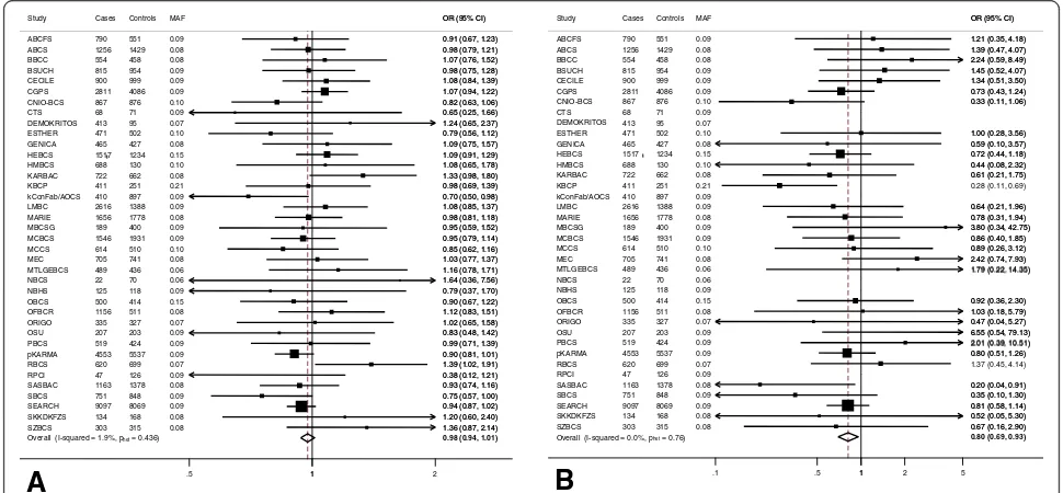
= 0.21) for AG versus AA (Figure 1A) and
0.80 (95% CI = 0.69, 0.93;
P
= 0.004) for GG versus AA
(Figure 1B), with no evidence of between-study
hetero-geneity for either OR estimate (
P
het= 0.44,
I
2= 1.9%
and
P
het= 0.76
, I
2= 0.0% for heterozygote and
homo-zygote OR estimates respectively). There was, however,
marginally significant evidence that the genotypic OR
esti-mates departed from those expected under a multiplicative
model with the inverse association of the GG genotype
be-ing more than the square of that of the AG genotype (test
for deviation from multiplicative model,
P
= 0.04).
Data for rs10235235 in women of Asian or
African-American ancestry were more limited, with just two
African-American studies (1,046 cases and 932 controls)
and nine Asian studies (5,795 cases and 6,624 controls).
In addition, this SNP was sufficiently rare in Asian
pop-ulations (MAF = 0.002) that we were unable to estimate
the heterozygote OR in two Asian studies (SEBCS, one
carrier among 1,114 cases and no carriers among 1,129
controls; TWBCS, one carrier among 236 controls and no
carriers among 774 cases; Table S2 in Additional file 1)
and we could not estimate a homozygote OR for any
Asian study (Table S2 in Additional file 1). There was no
clear evidence that this SNP was associated with breast
cancer risk for women of Asian ancestry (heterozygote
OR = 1.06, 95% CI = 0.76, 1.49) or African-American
ancestry (heterozygote and homozygote ORs were OR =
1.09, 95% CI = 0.90, 1.32 and OR = 0.94, 95% CI = 0.62,
1.42 respectively; Figure S1 in Additional file 1). This
ana-lysis, however, had low power to detect associations in
non-Europeans and these OR estimates were not
incon-sistent with the magnitude of the observed OR estimates
for European women (
P
het= 0.51).
Stratifying cases by oestrogen receptor (
P
het= 0.83) or
progesterone receptor (
P
het= 0.19) status, tumour grade
(
P
het= 0.63) or nodal involvement at diagnosis (
P
het= 0.51)
showed no evidence of effect modification (Table 1). There
was some evidence of effect modification by morphology
(
P
het= 0.03). For ductal cancers we estimated a very
modest reduction of risk for heterozygotes (OR
het=
0.98, 95% CI = 0.93, 1.02;
P
= 0.30) and a stronger,
sig-nificant reduction for homozygotes (OR
hom= 0.74, 95%
CI = 0.61, 0.90;
P
= 0.003). For lobular cancers there was
no such trend (OR
het= 1.07, 95% CI = 0.98, 1.17;
P
= 0.14
and OR
hom= 0.91, 95% CI = 0.64, 1.27;
P
= 0.57).
The SNP rs10235235 maps to a locus (
CYP3A
) that
has been considered an
a priori
candidate for
involve-ment in determining age at menopause and age at
me-narche [21,22]. Stratifying cases by age at diagnosis (≤50
Overall (I-squared = 1.9%, phet= 0.436) OBCS HEBCS MEC MCCS GENICA CGPS RBCS ABCFS SZBCS OSU MBCSG RPCI BBCC SEARCH SKKDKFZS MTLGEBCS Study BSUCH DEMOKRITOS SBCS MCBCS NBCS CNIO-BCS KBCP ORIGO CTS CECILE OFBCR ESTHER NBHS pKARMA ABCS PBCS kConFab/AOCS LMBC MARIE HMBCS KARBAC SASBAC 500 1517 705 614 465 2811 620 790 303 207 189 47 554 9097 134 489 Cases 815 413 751 1546 22 867 411 335 68 900 1156 471 125 4553 1256 519 410 2616 1656 688 722 1163 414 1234 741 510 427 4086 699 551 315 203 400 126 458 8069 168 436 Controls 954 95 848 1931 70 876 251 327 71 999 511 502 118 5537 1429 424 897 1388 1778 130 662 1378 0.15 0.15 0.08 0.10 0.08 0.09 0.07 0.09 0.08 0.09 0.09 0.09 0.08 0.09 0.08 0.06 MAF 0.09 0.07 0.09 0.09 0.06 0.10 0.21 0.07 0.09 0.09 0.08 0.10 0.09 0.09 0.08 0.09 0.09 0.09 0.08 0.10 0.08 0.08
0.98 (0.94, 1.01)
0.90 (0.67, 1.22) 1.09 (0.91, 1.29)
1.03 (0.77, 1.37) 0.85 (0.62, 1.16) 1.09 (0.75, 1.57) 1.07 (0.94, 1.22)
1.39 (1.02, 1.91) 0.91 (0.67, 1.23)
1.36 (0.87, 2.14) 0.83 (0.48, 1.42) 0.95 (0.59, 1.52)
0.38 (0.12, 1.21) 1.07 (0.76, 1.52)
0.94 (0.87, 1.02) 1.20 (0.60, 2.40) 1.16 (0.78, 1.71) OR (95% CI)
0.98 (0.75, 1.28)
1.24 (0.65, 2.37)
0.75 (0.57, 1.00) 0.95 (0.79, 1.14)
1.64 (0.36, 7.56) 0.82 (0.63, 1.06)
0.98 (0.69, 1.39)
1.02 (0.65, 1.58) 0.65 (0.25, 1.66) 1.08 (0.84, 1.39)
1.12 (0.83, 1.51) 0.79 (0.56, 1.12)
0.79 (0.37, 1.70)
0.90 (0.81, 1.01) 0.98 (0.79, 1.21)
0.99 (0.71, 1.39) 0.70 (0.50, 0.98) 1.08 (0.85, 1.37) 0.98 (0.81, 1.18) 1.08 (0.65, 1.78) 1.33 (0.98, 1.80)
0.93 (0.74, 1.16) 0.90 (0.67, 1.22) 1.09 (0.91, 1.29)
1.03 (0.77, 1.37) 0.85 (0.62, 1.16) 1.09 (0.75, 1.57) 1.07 (0.94, 1.22)
1.39 (1.02, 1.91) 0.91 (0.67, 1.23)
1.36 (0.87, 2.14) 0.83 (0.48, 1.42) 0.95 (0.59, 1.52)
0.38 (0.12, 1.21) 1.07 (0.76, 1.52)
0.94 (0.87, 1.02) 1.20 (0.60, 2.40) 1.16 (0.78, 1.71) OR (95% CI)
0.98 (0.75, 1.28)
1.24 (0.65, 2.37)
0.75 (0.57, 1.00) 0.95 (0.79, 1.14)
1.64 (0.36, 7.56) 0.82 (0.63, 1.06)
0.98 (0.69, 1.39)
1.02 (0.65, 1.58) 0.65 (0.25, 1.66) 1.08 (0.84, 1.39)
1.12 (0.83, 1.51) 0.79 (0.56, 1.12)
0.79 (0.37, 1.70)
0.90 (0.81, 1.01) 0.98 (0.79, 1.21)
0.99 (0.71, 1.39) 0.70 (0.50, 0.98) 1.08 (0.85, 1.37) 0.98 (0.81, 1.18) 1.08 (0.65, 1.78) 1.33 (0.98, 1.80)
0.93 (0.74, 1.16)
1
.5 1 2
A
Overall (I-squared = 0.0%, phet= 0.76) DEMOKRITOS KBCP SEARCH OSU pKARMA ESTHER RBCS CNIO-BCS ABCS NBHS GENICA KARBAC OBCS HEBCS NBCS MTLGEBCS SKKDKFZS ABCFS CTS MBCSG ORIGO PBCS kConFab/AOCS SBCS OFBCR SZBCS CGPS SASBAC BSUCH Study RPCI LMBC HMBCS MARIE MCBCS CECILE BBCC MCCS MEC 413 411 9097 207 4553 471 620 867 1256 125 465 722 500 1517 22 489 134 790 68 189 335 519 410 751 1156 303 2811 1163 815 Cases 47 2616 688 1656 1546 900 554 614 705 95 251 8069 203 5537 502 699 876 1429 118 427 662 414 1234 70 436 168 551 71 400 327 424 897 848 511 315 4086 1378 954 Controls 126 1388 130 1778 1931 999 458 510 741 0.07 0.21 0.09 0.09 0.09 0.10 0.07 0.10 0.08 0.09 0.08 0.08 0.15 0.15 0.06 0.06 0.08 0.09 0.09 0.09 0.07 0.09 0.09 0.09 0.08 0.08 0.09 0.08 0.09 MAF 0.09 0.09 0.10 0.08 0.09 0.09 0.08 0.10 0.08
0.80 (0.69, 0.93)
0.28 (0.11, 0.69)
0.81 (0.58, 1.14) 6.55 (0.54, 79.13)
0.80 (0.51, 1.26) 1.00 (0.28, 3.56)
1.37 (0.45, 4.14) 0.33 (0.11, 1.06) 1.39 (0.47, 4.07)
0.59 (0.10, 3.57)
0.61 (0.21, 1.75)
0.92 (0.36, 2.30) 0.72 (0.44, 1.18)
1.79 (0.22, 14.35)
0.52 (0.05, 5.30) 1.21 (0.35, 4.18)
3.80 (0.34, 42.75)
0.47 (0.04, 5.27)
2.01 (0.39, 10.51)
0.35 (0.10, 1.30) 1.03 (0.18, 5.79)
0.67 (0.16, 2.90) 0.73 (0.43, 1.24)
0.20 (0.04, 0.91) 1.45 (0.52, 4.07) OR (95% CI)
0.64 (0.21, 1.96) 0.44 (0.08, 2.32)
0.78 (0.31, 1.94)
0.86 (0.40, 1.85) 1.34 (0.51, 3.50) 2.24 (0.59, 8.49)
0.89 (0.26, 3.12) 2.42 (0.74, 7.93)
0.81 (0.58, 1.14) 6.55 (0.54, 79.13)
0.80 (0.51, 1.26) 1.00 (0.28, 3.56) 0.33 (0.11, 1.06) 1.39 (0.47, 4.07)
0.59 (0.10, 3.57)
0.61 (0.21, 1.75)
0.92 (0.36, 2.30) 0.72 (0.44, 1.18)
1.79 (0.22, 14.35)
0.52 (0.05, 5.30) 1.21 (0.35, 4.18)
3.80 (0.34, 42.75)
0.47 (0.04, 5.27)
2.01 (0.39, 10.51)
0.35 (0.10, 1.30) 1.03 (0.18, 5.79)
0.67 (0.16, 2.90) 0.73 (0.43, 1.24)
0.20 (0.04, 0.91) 1.45 (0.52, 4.07) OR (95% CI)
0.64 (0.21, 1.96) 0.44 (0.08, 2.32)
0.78 (0.31, 1.94)
0.86 (0.40, 1.85) 1.34 (0.51, 3.50) 2.24 (0.59, 8.49)
0.89 (0.26, 3.12) 2.42 (0.74, 7.93)
1
.1 .5 1 2 5
[image:5.595.55.540.437.662.2]B
or >50 years) as a proxy for menopausal status at
diagno-sis showed no evidence of effect modification (
P
het= 0.89;
Table 2), and excluding cases who were diagnosed between
age 46 and 55 as potentially perimenopausal did not alter
this result (
P
het= 0.28). Data on age at menarche were
available for 21,736 cases and 22,686 controls (Table S4 in
Additional file 1); to increase the power of the analysis
we included additional data from BBCS and UKBGS
(5,737 cases, 5,572 controls; Table S4 in Additional file 1)
[19]. There was a 1.5% (95% CI = 0.5%, 2.7%;
P
= 0.004)
reduction in breast cancer risk associated with each
additional year
’
s increase in age at menarche. Mean age
at menarche was positively associated with number of
copies of the minor allele of rs10235235 for controls
(
P
trend= 0.005; Table 3) but not for cases (
P
trend= 0.97;
Table 3). Consequently, there was an inverse trend in
the magnitude of the heterozygote and homozygote
breast cancer ORs with mean age at menarche (
P
het=
0.02; Table 4); being a carrier of one or two rare alleles
of rs10235235 was associated with an estimated 16%
(OR
het= 0.84, 95% CI = 0.75, 0.94;
P
= 0.003) or 19%
(OR
hom= 0.81, 95% CI = 0.51, 1.30;
P
= 0.39) (
P
trend=
0.002) reduction in breast cancer risk for women who
had their menarche at ages
≥15 years but there was no
evidence of reduction for those with a menarche at
age
≤11 years (OR
het= 1.06, 95% CI = 0.95, 1.19;
P
= 0.30
and OR
hom= 1.07, 95% CI = 0.67, 1.72;
P
= 0.78) (
P
trend=
[image:6.595.58.539.99.478.2]0.29). There was no evidence that the inverse trend in
the magnitude of ORs with mean age at menarche
dif-fered between heterozygous and homozygous carriers
(
P
= 0.97) and no evidence that the trend was nonlinear
(
P
= 0.70).
Table 1 Association of rs10235235 with risk of breast cancer for women of European ancestry: stratified analysis
Cases Controls ORhet 95% CI P1 ORhom 95% CI P1 Phet
ER status
ER-positive 24,780 38,739 0.99 0.95, 1.03 0.61 0.83 0.70, 0.99 0.04
ER-negative 5,851 38,739 1.02 0.95, 1.10 0.60 0.60 0.43, 0.86 0.005
NK 8,339
Total 38,970a 38,739 0.99 0.95, 1.03 0.74 0.79 0.67, 0.94 0.006 0.83
PR status
PR-positive 18,497 39,033 0.98 0.93, 1.02 0.32 0.82 0.67, 0.99 0.04
PR-negative 8,193 39,033 1.02 0.96, 1.09 0.53 0.74 0.56, 0.98 0.03
NK 12,111
Total 38,801b 39,033 0.99 0.94, 1.03 0.52 0.80 0.67, 0.95 0.01 0.19
Morphology
Ductal 22,123 31,803 0.98 0.93, 1.02 0.30 0.74 0.61, 0.90 0.003
Lobular 3,921 31,803 1.07 0.98, 1.17 0.14 0.91 0.64, 1.27 0.57
Other and NK 5,995
Total 32,039 31,803 0.99 0.95, 1.04 0.64 0.77 0.64, 0.92 0.004 0.03
Grade
Grade 1 5,944 37,285 0.97 0.90, 1.05 0.46 0.86 0.65, 1.15 0.31
Grade 2 13,427 37,285 1.00 0.95, 1.06 0.92 0.80 0.63, 0.98 0.04
Grade 3 8,638 37,285 0.98 0.92, 1.05 0.58 0.61 0.46, 0.82 0.001
NK 8,769
Total 36,778 37,285 0.99 0.95, 1.03 0.56 0.76 0.64, 0.90 0.001 0.63
Nodal status
Node-negative 17,463 37,836 0.98 0.93, 1.03 0.47 0.86 0.71, 1.04 0.12
Node-positive 10,746 37,836 0.98 0.92, 1.04 0.46 0.72 0.57, 0.93 0.01
NK 9,359
Total 37,568 37,836 0.98 0.94, 1.02 0.31 0.81 0.68, 0.96 0.02 0.51
Association of rs10235235 with risk of breast cancer for women of European ancestry stratified by oestrogen receptor (ER) status, progesterone receptor (PR) status, morphology, grade and nodal status. ORhet, odds ratio comparing rs10235235 AG genotype versus AA genotype; H0, null hypothesis; NK, not known;
ORhom, odds ratio comparing rs10235235 GG genotype versus AA genotype;P1, test of H0no association between rs10235235 and breast cancer risk;Phet, test of
H0no difference between stratum specific estimates for variables with two strata or test of H0no linear trend in stratum specific estimates for variables with three
strata.a
Excludes seven studies that selected all ER-negative cases (CTS, DEMOKRITOS, NBCS, NBHS, OSU, RPCI and SKKDKFZS) and one study (PBCS) that selected all ER-positive cases.b
Discussion
This study of more than 47,000 breast cancer cases and
47,000 controls has confirmed that rs10235235, mapping
to 7q22.1 (
CYP3A
), is associated with a reduction in breast
cancer risk for women of European ancestry. Previously,
our hypothesis-generating study of 10,000 breast cancer
cases and 17,000 controls found a per-allele OR estimate
of 0.96 (95% CI = 0.90, 1.02;
P
= 0.2), with marginally
sig-nificant evidence of an inverse association for breast cancer
diagnosed age 50 years or younger (OR = 0.91, 95% CI =
0.83, 0.99;
P
= 0.03) but no evidence of an association for
breast cancer at later ages (OR = 1.01, 95% CI = 0.93, 1.10;
P
= 0.82) [19]. In this considerably larger study, we found a
heterozygote OR estimate of 0.98 (95% CI = 0.94, 1.01;
P
= 0.21) and a homozygote OR estimate of 0.80 (95%
CI = 0.69, 0.93;
P
= 0.004) with marginally significant
evidence that the inverse association for homozygotes is
greater than predicted by a multiplicative model (
P
= 0.04).
To our knowledge, rs10235235 is the first SNP to be
associated with both breast cancer risk and age at
me-narche, consistent with the well-documented association
between later age at menarche and a reduction in breast
cancer risk [23]. Genome-wide association studies have
identified more than 70 breast cancer risk variants [5,6]
and more than 30 variants associated with age at
menar-che [22], none of which map to the
CYP3A
locus.
rs10235235 was originally identified on the basis of a
highly significant association with hormone levels,
ac-counting for 4.9% of the variation in premenopausal
urinary oestrone glucuronide levels [19]. In this current
analysis, rs10235235 accounted for only 0.01% of the
variation across controls in age at menarche and we
esti-mate that this SNP explains just 0.01% of the familial
excess breast cancer risk. Our data thus illustrate the
po-tential statistical efficiency of studies of intermediate
phenotypes in the identification of rarer (MAF < 10%)
risk alleles with modest associations. Our analysis shows
some inconsistency with a recent genome-wide study of
circulating oestradiol, testosterone and sex
hormone-binding globulin in postmenopausal women [24]. In that
study there was no genome-wide significant association
observed with plasma oestradiol levels in either the
pri-mary analysis of approximately 1,600 postmenopausal
women who were not taking postmenopausal hormones
at blood draw or the secondary analysis that included
approximately 900 current postmenopausal hormone
users. Further studies will be needed to determine whether
the lack of an association between
CYP3A
variants and
postmenopausal plasma oestradiol levels reflects a
differ-ence in the menopausal status of the study subjects, the
hormone/metabolite that was analysed or chance.
[image:7.595.56.539.100.169.2]One possible explanation for the apparent effect
modi-fication of the rs10235235
–
breast cancer risk association
by age at menarche is that this is a function of
genotyp-ing a marker SNP rather than the true causal variant.
For example, if rs10235235 was perfectly correlated with
a causal variant, SNP X, with a MAF substantially lower
than that of rs10235235 (
D′
~ 1.0,
r
2< 1.0), then there
would be three types of chromosome in the population:
type i, chromosomes carrying the common allele of
rs10235235 and the common allele of SNP X; type ii,
chromosomes carrying the rare allele of rs10235235 and
the common allele of SNP X; and type iii, chromosomes
carrying the rare allele of rs10235235 and the rare
(pro-tective) allele of SNP X. Only chromosomes carrying the
rare allele of rs10235235 and the rare (protective) allele of
Table 3 Association of rs10235235 with age at menarche for women of European ancestry by case-control status
rs10235235 genotype Cases Age at menarche (years) Ptrend Controls Age at menarche (years) Ptrend
AA 22,954 12.83 23,383 12.95
AG 4,312 12.83 4,627 13.02
GG 207 12.83 248 13.05
Total 27,473 12.83 0.97 28,258 12.96 0.005
[image:7.595.56.539.656.726.2]H0, null hypothesis;Ptrend, test of H0no linear trend in age at menarche according to rs10235235 genotype.
Table 2 rs10235235 and risk of breast cancer for women of European ancestry by age at diagnosis
Age at diagnosis Casesa Controlsa ORhet 95% CI P1 ORhom 95% CI P1 Phet
≤50 years 11,794 34,988 0.99 0.93, 1.05 0.69 0.68 0.53, 0.86 0.003
> 50 years 23,264 34,988 0.97 0.93, 1.02 0.24 0.84 0.70, 1.00 0.04
NK 554
Total 35,612 34,988 0.98 0.94, 1.02 0.23 0.79 0.67, 0.92 0.003 0.89
a
Five studies (ABCFS, MARIE, MEC, MTLGEBCS and SASBAC) that selected all cases on the basis of age at diagnosis (Table S3 in Additional file1) were excluded from this stratified analysis; two small studies (CTS and NBCS) that had no heterozygote or rare homozygote cases in one of the age stratum were also excluded. H0, null hypothesis; NK, not known; ORhet, odds ratio comparing rs10235235 AG genotype versus AA genotype; ORhom, odds ratio comparing rs10235235 GG
genotype versus AA genotype;P1, test of H0no association between rs10235235 and breast cancer risk;Phet, test of H0no difference between stratum
SNP X (type iii) would be enriched in controls.
Genotyp-ing the marker (rs10235235) rather than the causal variant
leads to misclassification. As the causal variant is
associ-ated with a protective effect on breast cancer risk, the
pro-portion of chromosomes carrying both the rare allele of
the causal variant and the marker (type iii) compared with
the common allele of the causal variant and the rare allele
of the marker (type ii) will be greater in controls than in
cases such that the extent of misclassification will be
greater for cases than controls. This will attenuate the
as-sociation between genotype and age at menarche to a
greater extent in cases than in controls creating an
appar-ent effect modification. Fine mapping and functional
stud-ies will be required to identify the causal variant and to
determine the true relationship between the causal
vari-ant, age at menarche and breast cancer risk.
Despite our original finding of a strong association
be-tween rs10235235 and hormone levels, we found no
evi-dence that the association between this SNP and breast
cancer risk differed by the hormone receptor status of the
tumour, and nor did we find any evidence that the
associ-ation differed by stage, grade or lymph node involvement.
There was marginally significant evidence that the
associ-ation between rs10235235 and breast cancer risk differed
between ductal and lobular cancers (
P
het= 0.03). Given
the number of stratified analyses that we carried out (six
stratifying variables) and given that there is no biological
basis to support an interaction between rs10235235 and
morphology, this is probably a chance observation.
In contrast to our earlier study [19], we found no
evi-dence of an interaction with age at diagnosis when we
stratified cases by age
≤/>50 years, either including or
excluding cases diagnosed between age 46 and 55 years
as potentially perimenopausal. We used age at diagnosis
as a proxy for menopausal status at diagnosis because
menopausal status at diagnosis is difficult to determine
by questionnaire, especially given the use of hormone
replacement therapies; while information on age at
diagnosis was available for all but 1.4% (
n
= 554) of
cases, information on age at natural menopause was
missing for 65.6% (
n
= 26,552) of cases of European
ancestry. Similarly, although rs10235235 is a plausible
candidate for association with age at menopause, we
did not test this due to the limited amount of data on
age at natural menopause for controls of European
an-cestry (
n
= 11,294, 28.2%) and the difficulty in ascertaining
whether treatment for breast cancer had influenced
re-ported age at menopause for cases.
The strengths of our study include the large size of
this combined analysis, and the availability of
informa-tion on tumour characteristics for the majority of cases
and on age at menarche for the majority of cases and
controls. Limitations include low power of the study to
examine an association between genotype and breast
cancer risk for non-Europeans.
Conclusions
In summary, we have confirmed that rs10235235 is
asso-ciated with breast cancer, have shown for the first time
that rs10235235 is associated with age at menarche in
controls and have suggested a potential mechanism for
these associations. rs10235235, which maps to the
CYP3A
locus, probably tags a causal variant that affects
expression of one or more
CYP3A
genes.
Additional files
Additional file 1:Contains Table S1 presenting details of participating BCAC studies; Table S2 presenting rs10235235 genotypes for breast cancer cases and controls from 49 BCAC studies; Table S3 presenting availability of data on age at diagnosis, hormone receptor status, morphology, grade and nodal status for breast cancer cases from 38 European BCAC studies; Table S4 presenting availability of data on age at menarche for breast cancer cases and controls from 40 European BCAC studies; and Figure S1 showing association of the rs10235235-AG genotype with breast cancer risk for women of Asian and African-American ancestry.
Additional file 2:Presents details of ethical committees that approved each study.
Abbreviations
[image:8.595.56.539.99.199.2]BCAC:Breast Cancer Association Consortium; CI: confidence interval; COGS: Collaborative Oncological Gene-environment Study; MAF: minor allele frequency; OR: odds ratio;Ptrend:Pvalue for linear trend; SNP: single nucleotide polymorphism.
Table 4 rs10235235 and risk of breast cancer for women of European ancestry by age at menarche
Age at menarche (years) Cases Controls ORhet 95% CI P1 ORhom 95% CI P1 Phet
≤11 4,818 4,749 1.06 0.95, 1.19 0.30 1.07 0.67, 1.72 0.78
12 5,655 5,720 0.92 0.83, 1.02 0.10 0.83 0.54, 1.28 0.41
13 7,308 7,379 0.93 0.85, 1.02 0.11 0.77 0.54, 1.09 0.14
14 5,307 5,743 0.96 0.86, 1.06 0.42 0.69 0.45, 1.06 0.09
≥15 4,385 4,667 0.84 0.75, 0.94 0.003 0.81 0.51, 1.30 0.39
Total 27,473 28,258 0.94 0.90, 0.98 0.007 0.81 0.67, 0.98 0.03 0.02
H0, null hypothesis; ORhet, odds ratio comparing rs10235235 AG genotype versus AA genotype; ORhom, odds ratio comparing rs10235235 GG genotype versus AA
Competing interests
The authors state that they have no competing interests.
Authors’contributions
OF, FD and NO performed the statistical analyses. OF, IdSS and NJ drafted the manuscript. NJ, FD, NO, LG, MEJ, MJS, EJF, BPH, MG-C, MDo, AA, AJS, JP, IdSS and OF comprised the writing group that was responsible for the interpretation of the results and for critically reviewing the manuscript. AC, AJ, AHW, AMa, BBu, C-YS, DL, ES, GC-T, HN, HBre, HBra, ILA, JC-C, J-YC, JLH, LBa, MKB, HMi, PAF, PR, RW, SEB, TD, MKS and UH also significantly contributed to the interpretation of the results. OF, IdSS, NJ, JP, LG, DFE, MKB and JW conceived of the original design of the study and participated in subject recruitment and in acquisition of data. JBen, AG-N, RM, DCT, DV, FB, CL, JD, JS and KMi carried out the genotyping and/or data analysis. FD, NO, MEJ, MJS, EJF, BPH, JLH, MCS, GSD, CA, MKS, AB, LJVV, FA, KMu, ALo, PAF, MWB, ABE, SPR, ES, IT, MK, NM, BBu, FMa, AS, CS, PG, TT, EC, FMe, SEB, BGN, HF, RMi, MPZ, JIAP, JBen, LBe, HA-C, AZ, CCD, HBre, HMü, VA, AKD, AMe, JH, CRB, RKS, HBra, CJ, Y-DK, The GENICA Network, HN, TAM, KA, CB, KMa, TD, NVB, NNA, ALi, AMa, VK, V-MK, JMH, GC-T, JBee, kConFab Investigators, Australian Ovarian Cancer Study Group, AHW, DVdB, C-CT, DL, DS, PN, HW, JC-C, AR, SN, DF-J, PR, PP, BBo, VP, FJC, JEO, XW, ZF, VSP, GGG, GS, LBa, CH, JS, MSG, FL, MDu, PS, ST, CHY, SYP, BKC, VNK, GGA, A-LB-D, WZ, RW, KP, AJ-V, MG, ILA, JAK, GG, AMM, PD, JF, SJC, JLis, MES, PH, NS, MHo, AH, RAO, MT-L, JLiu, AC, IWB, MWRR, SSC, WB, LBS, PDPP, AMD, MS, DK, D-YN, SKP, J-YC, MHa, HMi, WYL, AT, UH, AF, TR, HUU, AJ, JLu, KJ-B, KD, SSa, VG, PB, JM, SSl, AET, CV, DY, C-YS, J-CY, C-SH, M-FH, AG-N, DCT, DV, FB, CL, JD, KMi, MKB, JW, DFE, MG-C, MDo, AA and AJS made substantial contributions in recruiting subjects and acquiring data, and in critically reviewing the manuscript. All authors take responsibility for the work and read and approved the final version of the manuscript.
Acknowledgements
The authors thank all of the individuals who took part in these studies and all of the researchers, clinicians, technicians and administrative staff who have enabled this work to be carried out.
ABCFS would like to thank Maggie Angelakos, Judi Maskiell and Gillian Dite. ABCS would like to thank Ellen van der Schoot and Sanquin Amsterdam. The ACP study wishes to thank the participants in the Thai Breast Cancer study. Special thanks also go to the Thai Ministry of Public Health (MOPH) doctors and nurses who helped with the data collection process. The study would like to thank Dr Prat Boonyawongviroj, the former Permanent Secretary of MOPH and Dr Pornthep Siriwanarungsan, the Department Director-General of Disease Control who have supported the study throughout. BBCS would like to thank Eileen Williams, Elaine Ryder-Mills and Kara Sargus. BIGGS would like to thank Niall McInerney, Gabrielle Colleran, Andrew Rowan and Angela Jones. CNIO-BCS would like to thank Charo Alonso, Tais Moreno, Guillermo Pita, Primitiva Menendez and Anna González-Neira. The authors would like to acknowledge the contribution of the staff of the Génome Québec-genotyping unit under the supervision of Dr Sylvie LaBoissière, as well as Frédérick Robidoux from the McGill University and Génome Québec Innovation Centre. ESTHER would like to thank Hartwig Ziegler, Sonja Wolf and Volker Hermann. GC-HBOC would like to thank Bernd Frank. HEBCS would like to thank Dr Sofia Khan, Dr Kirsimari Aaltonen and Dr Karl von Smitten, and research nurses Irja Erkkilä and Virpi Palola. KBCP would like to thank Eija Myöhänen and Helena Kemiläinen. kConFab/AOCS would like to thank Heather Thorne, Eveline Niedermayr, the AOCS Management Group (D Bowtell, G Chenevix-Trench, A deFazio, D Gertig, A Green, P Webb) and the ACS Management Group (A Green, P Parsons, N Hayward, P Webb, D Whiteman). LAABC thanks all of the study participants and the entire data collection team, especially Annie Fung and June Yashiki. LMBC would like to thank Gilian Peuteman, Dominiek Smeets, Thomas Van Brussel and Kathleen Corthouts.
MARIE would like to thank Tracy Slanger, Elke Mutschelknauss, Ramona Salazar, S Behrens, R Birr, W Busch, U Eilber, B Kaspereit, N Knese and K Smit. MBCSG would like to thank Siranoush Manokian, Bernard Peissel and Daniela Zaffaroni of the Fondazione Istituto Nazionale dei Tumori, Milan, Monica Barile of the Istituto Europeo di Oncologia, Milan and Loris Bernard and personnel of the Cogentech Cancer Genetic Test Laboratory, Milan, Italy. MTLGEBCS would like to thank Martine Tranchant (Cancer Genomics Laboratory, CRCHUQ), Marie-France Valois, Annie Turgeon and Lea Heguy (McGill University Health Center, Royal Victoria Hospital; McGill University) for DNA extraction, sample management and skillful technical assistance. JS is Chairholder of the Canada Research Chair in Oncogenetics. MYBRCA would
like to thank Phuah Sze Yee, Peter Kang, Kang In Nee, Kavitta Sivanandan, Shivaani Mariapun, Yoon Sook-Yee, Daphne Lee, Teh Yew Ching and Nur Aishah Mohd Taib for DNA Extraction and patient recruitment. NBHS thanks study participants and research staff for their contributions and commitment to the study. OBCS would like to thank Meeri Otsukka and Kari Mononen. OFBCR would like to thank Teresa Selander and Nayana Weerasooriya. ORIGO thanks E Krol-Warmerdam and J Blom for patient accrual, administering questionnaires and managing clinical information. The LUMC survival data were retrieved from the Leiden hospital-based cancer registry system (ONCDOC) with the help of Dr J Molenaar. PBCS would like to thank Louise Brinton, Mark Sherman, Stephen Chanock, Neonila Szeszenia-Dabrowska, Beata Peplonska, Witold Zatonski, Pei Chao and Michael Stagner. pKARMA would like to thank The Swedish Medical Research Counsel. RBCS would like to thank Petra Bos, Jannet Blom, Ellen Crepin, Elisabeth Huijskens, Annette Heemskerk and the Erasmus MC Family Cancer Clinic. SASBAC would like to thank The Swedish Medical Research Counsel. SBCGS thanks study participants and research staff for their contributions and commitment to the study. SBCS would like to thank Sue Higham, Helen Cramp and Dan Connley. SEARCH would like to thank The SEARCH and EPIC teams. SGBCC would like to thank the participants and research coordinator Kimberley Chua. SKKDKFZS are grateful to all of the patients for their participation and thank the physicians and other hospital staff, scientists, research assistants and study staff who contributed to the patient recruitment, data collection and sample preparation. UKBGS thanks Breakthrough Breast Cancer and the Institute of Cancer Research for support and funding of the Breakthrough Generations Study, and the study participants, study staff, and the doctors, nurses and other healthcare providers and health information sources who have contributed to the study.
Consortia members
The GENICA network: Dr Margarete Fischer-Bosch-Institute of Clinical Pharmacology, Stuttgart, and University of Tübingen, Germany (Christina Justenhoven, Hiltrud Brauch); Department of Internal Medicine, Evangelische Kliniken Bonn gGmbH, Johanniter Krankenhaus, Bonn, Germany (Yon-Dschun Ko, Christian Baisch); Institute of Pathology, University of Bonn, Germany (Hans-Peter Fischer); Molecular Genetics of Breast Cancer, Deutsches Krebsforschungszentrum (DKFZ), Heidelberg, Germany (Ute Hamann); Institute for Prevention and Occupational Medicine of the German Social Accident Insurance (IPA), Bochum, Germany (Thomas Bruening, Beate Pesch, Sylvia Rabstein, Anne Spickenheuer); and Institute for Occupational Medicine and Maritime Medicine, University Medical Center Hamburg-Eppendorf, Germany (Volker Harth).
kConFab Investigators: David Amor, Lesley Andrews, Yoland Antill, Shane Armitage, Rosemary Balleine, Agnes Bankier, Patti Bastick, John Beilby, Barbara Bennett, Ian Bennett, Anneke Blackburn, Michael Bogwitz, Meagan Brennan, Melissa Brown, Michael Buckley, Matthew Burgess, Jo Burke, Phyllis Butow, Ian Campbell, Alice Christian, Georgia Chenevix-Trench, Christine Clarke, Alison Colley, Dick Cotton, Bronwyn Culling, Margaret Cummings, Sarah-Jane Dawson, Anna DeFazio, Martin Delatycki, Rebecca Dickson, Alexander Dobrovic, Tracy Dudding, Ted Edkins, Stacey Edwards, Gelareh Farshid, Susan Fawcett, Georgina Fenton, Michael Field, James Flanagan, Peter Fong, John Forbes, Stephen Fox, Juliet French, Clara Gaff, Mac Gardner, Mike Gattas, Graham Giles, Grantley Gill, Jack Goldblatt, Sian Greening, Scott Grist, Eric Haan, Marion Harris, Stewart Hart, Nick Hayward, Sue Healey, Louise Heiniger, John Hopper, Clare Hunt, Paul James, Mark Jenkins, Rick Kefford, Alexa Kidd, Belinda Kiely, Judy Kirk, James Kollias, Jessica Koehler, Serguei Kovalenko, Sunil Lakhani, Jennifer Leary, Geoff Lindeman, Lara Lipton, Liz Lobb, Graham Mann, Deborah Marsh, Bettina Meiser, Roger Milne, Gillian Mitchell, Shona O’Connell, Nick Pachter, Briony Patterson, Lester Peters, Kelly Phillips, Melanie Price, Lynne Purser, Tony Reeve, Edwina Rickard, Bridget Robinson, Barney Rudzki, Elizabeth Salisbury, Christobel Saunders, Joe Sambrook, Jodi Saunus, Robyn Sayer, Clare Scott, Elizabeth Scott, Rodney Scott, Adrienne Sexton, Raghwa Sharma, Andrew Shelling, Peter Simpson, Melissa Southey, Amanda Spurdle, Graeme Suthers, Pamela Sykes, Jessica Taylor, Ella Thompson, Heather Thorne, Sharron Townshend, Alison Trainer, Kathy Tucker, Janet Tyler, Jane Visvader, Logan Walker, Paul Waring, Robin Ward, Bev Warner, Rachael Williams, Ingrid Winship, Mary Ann Young (Peter MacCallum Cancer Center, Melbourne, Australia).
Financial support
Part of this work was supported by the European Community’s Seventh Framework Programme under grant agreement number 223175 (grant number HEALTH-F2-2009-223175) (COGS). This work was partly supported by the Canadian Institutes of Health Research for the‘CIHR Team in Familial Risks of Breast Cancer’program (JS, DFE), and the Ministry of Economic Development, Innovation and Export Trade of Quebec–grant number PSR-SIIRI-701 (JS, DFE, PH).
The ABCFS and OFBCR work was supported by the United States National Cancer Institute, National Institutes of Health (NIH) under RFA-CA-06-503 and through cooperative agreements with members of the Breast Cancer Family Registry (BCFR) and Principal Investigators, including Cancer Care Ontario (U01 CA69467), Northern California Cancer Center (U01 CA69417) and University of Melbourne (U01 CA69638). Samples from the NC-BCFR were processed and distributed by the Coriell Institute for Medical Research. The content of this manuscript does not necessarily reflect the views or policies of the National Cancer Institute or any of the collaborating centers in the BCFR, nor does mention of trade names, commercial products, or organizations imply endorsement by the US Government or the BCFR. ABCFS was also supported by the National Health and Medical Research Council of Australia, the New South Wales Cancer Council, the Victorian Health Promotion Foundation (Australia) and the Victorian Breast Cancer Research Consortium. JLH is a National Health and Medical Research Council (NHMRC) Australia Fellow and a Victorian Breast Cancer Research Consortium Group Leader. MCS is a NHMRC Senior Research Fellow and a Victorian Breast Cancer Research Consortium Group Leader. The ABCS study was supported by the Dutch Cancer Society (grants NKI 2001-2423 and 2007-3839) and the Dutch National Genomics Initiative. The ACP study is funded by the Breast Cancer Research Trust, UK. The work of the BBCC was partly funded by ELAN-Fond of the University Hospital of Erlangen. BBCS is funded by Cancer Research UK and Breakthrough Breast Cancer, and acknowledges NHS funding to the NIHR Biomedical Research Centre and the National Cancer Research Network. BCAC is funded by CR-UK (C1287/A10118 and C1287/A12014). Meetings of the BCAC have been funded by the European Union COST programme (BM0606). DFE is a Principal Research Fellow of CR-UK. ES (BIGGS) is supported by NIHR Comprehensive Biomedical Research Centre, Guy’s & St. Thomas’NHS Foundation Trust in partnership with King’s College London, UK. IT is supported by the Oxford Biomedical Research Centre. The BSUCH study was supported by the Dietmar-Hopp Foundation, the Helmholtz Society and the German Cancer Research Center (DKFZ). CGPS was supported by the Chief Physician Johan Boserup and Lise Boserup Fund, the Danish Medical Research Council and Herlev Hospital. CNIO-BCS was supported by the Genome Spain Foundation, the Red Temática de Investigación Cooperativa en Cáncer and grants from the Asociación Española Contra el Cáncer and the Fondo de Investigación Sanitario (PI081583 and PI081120). CTS was supported by the California Breast Cancer Act of 1993, the NIH (grants R01 CA77398 and the Lon V Smith Foundation (LVS39420)) and the California Breast Cancer Research Fund (contract 97-10500). Collection of cancer incidence data used in this study was supported by the California Department of Public Health as part of the statewide cancer reporting program mandated by California Health and Safety Code Section 103885. The ESTHER study was supported by a grant from the Baden Württemberg Ministry of Science, Research and Arts. Additional cases were recruited in the context of the VERDI study, which was supported by a grant from the German Cancer Aid (Deutsche Krebshilfe).
GC-HBOC was supported by Deutsche Krebshilfe (107054), the Dietmar-Hopp Foundation, the Helmholtz Society and the German Cancer Research Centre (DKFZ). GENICA was funded by the Federal Ministry of Education and Research (BMBF) Germany grants 01KW9975/5, 01KW9976/8, 01KW9977/0 and 01KW0114, the Robert Bosch Foundation, Stuttgart, Deutsches Krebsforschungszentrum (DKFZ), Heidelberg, Institute for Prevention and Occupational Medicine of the German Social Accident Insurance (IPA), Bochum, as well as the Department of Internal Medicine, Evangelische Kliniken Bonn gGmbH, Johanniter Krankenhaus, Bonn, Germany. HEBCS was supported by the Academy of Finland (132473), Helsinki University Central Hospital Research Fund, the Sigrid Juselius Foundation, the Finnish Cancer Society and the Nordic Cancer Union. HERPACC was supported by a Grant-in-Aid for Scientific Research on Priority Areas and on Innovative Area from the Ministry of Education, Science, Sports, Culture and Technology of Japan and by a Grant-in-Aid for the Third Term Comprehensive 10-Year Strategy
for Cancer Control from Ministry Health, Labour and Welfare of Japan. HMBCS was supported by short-term fellowships from the German Academic Exchange Program (NVB) and the Friends of Hannover Medical School (NVB). KBCP was financially supported by the special Government Funding (EVO) of Kuopio University Hospital grants, Cancer Fund of North Savo, the Finnish Cancer Organizations, the Academy of Finland and by the strategic funding of the University of Eastern Finland. kConFab is supported by grants from the National Breast Cancer Foundation, the NHMRC, the Queensland Cancer Fund, the Cancer Councils of New South Wales, Victoria, Tasmania and South Australia and the Cancer Foundation of Western Australia. The kConFab Clinical Follow Up Study was funded by the NHMRC (145684, 288704, 454508). Financial support for the AOCS was provided by the United States Army Medical Research and Materiel Command (DAMD17-01-1-0729), the Cancer Council of Tasmania and Cancer Foundation of Western Australia and the NHMRC (199600). GC-T and P Webb are supported by the NHMRC. LAABC is supported by grants (1RB-0287, 3PB-0102, 5PB-0018, 10PB-0098) from the California Breast Cancer Research Program. Incident breast cancer cases were collected by the USC Cancer Surveillance Program (CSP), which is supported under subcontract by the California Department of Health. CSP is also part of the National Cancer Institute’s Division of Cancer Prevention and Control Surveillance, Epidemiology, and End Results Program, under contract number N01CN25403. LMBC is supported by the‘Stichting tegen Kanker’(232-2008 and 196-2010). DL is supported by the KULPFV/10/016-SymBioSysII.
National Institutes of Health (R01 CA092447). Data on SCCS cancer cases used in this publication were provided by the Alabama Statewide Cancer Registry; Kentucky Cancer Registry, Lexington, KY; Tennessee Department of Health, Office of Cancer Surveillance; Florida Cancer Data System; North Carolina Central Cancer Registry, North Carolina Division of Public Health; Georgia Comprehensive Cancer Registry; Louisiana Tumor Registry; Mississippi Cancer Registry; South Carolina Central Cancer Registry; Virginia Department of Health, Virginia Cancer Registry; and Arkansas Department of Health, Cancer Registry, Little Rock. The Arkansas Central Cancer Registry is fully funded by a grant from National Program of Cancer Registries, Centers for Disease Control and Prevention (CDC). Data on SCCS cancer cases from Mississippi were collected by the Mississippi Cancer Registry, which participates in the National Program of Cancer Registries of the CDC. The contents of this publication are solely the responsibility of the authors and do not necessarily represent the official views of the CDC or the Mississippi Cancer Registry. SEARCH is funded by programme grants from Cancer Research UK (C490/A10124 and C8197/ A10123) and NIH grant 5U01CA098216-07. SEBCS was supported by the Korea Health 21 R&D Project (AO30001), Ministry of Health and Welfare, Republic of Korea. SGBCC is funded by the National Medical Research Council start-up Grant and Centre Grant (NMRC/CG/NCIS /2010). Additional controls were recruited by the Singapore Consortium of Cohort Studies-Multi-ethnic cohort (SCCS-MEC), which was funded by the Biomedical Research Council, grant number: 05/1/21/ 19/425. SKKDKFZS is supported by the DKFZ, Heidelberg, Germany. KJ-B (SZBCS) is a fellow of International PhD program, Postgraduate School of Molecular Medicine, Warsaw Medical University, supported by the Polish Foundation of Science. TBCS was funded by The National Cancer Institute, Thailand. TNBCC was supported by an NIH Specialized Program of Research Excellence (SPORE) in Breast Cancer (CA116201), the Breast Cancer Research Foundation, a generous gift from the David F and Margaret T Grohne Family Foundation and the Ting Tsung and Wei Fong Chao Foundation; The Stefanie Spielman Breast Cancer Fund and the OSU Comprehensive Cancer Center; the European Union (European Social Fund–ESF) and Greek national funds through the Operational Program‘Education and Lifelong Learning’of the National Strategic Reference Framework (NSRF)–Research Funding Program of the General Secretariat for Research & Technology: ARISTEIA. TWBCS is supported by the Taiwan Biobank project of the Institute of Biomedical Sciences, Academia Sinica, Taiwan. UKBGS is funded by Breakthrough Breast Cancer and the Institute of Cancer Research (ICR). ICR acknowledges NHS funding to the NIHR Biomedical Research Centre.
Author details 1
Breakthrough Breast Cancer Research Centre, The Institute of Cancer Research, 237 Fulham Road, London SW3 6JB, UK.2Division of Breast Cancer
Research, The Institute of Cancer Research, 237 Fulham Road, London SW3 6JB, UK.3Non-communicable Disease Epidemiology Department, London
School of Hygiene and Tropical Medicine, Keppel Street, London WC1E 7HT, UK.4Division of Genetics and Epidemiology, The Institute of Cancer Research,
15 Cotswold Road, Belmont, Sutton, Surrey SM2 5NG, UK.5The Academic Department of Biochemistry, The Royal Marsden Hospital, Fulham Road, London SW3 6JJ, UK.6Centre for Molecular, Environmental, Genetic and Analytic Epidemiology, University of Melbourne, 1-100 Gratton Street, Parkville, Melbourne, Victoria 3010, Australia.7Genetic Epidemiology Department, Department of Pathology, The University of Melbourne, 1-100 Gratton Street, Parkville, Melbourne, Victoria 3010, Australia.8Division of Molecular Pathology, Netherlands Cancer Institute, Antoni van Leeuwenhoek Hospital, Plesmanlaan 121, 1066CX Amsterdam, The Netherlands.9Sanquin, Radboud Universiteit Nijmegen, 6525 GA, Nijmegen, The Netherlands.
10
Warwick Medical School, University of Warwick, Coventry CV4 7AJ, UK.
11University Breast Center, Department of Gynecology and Obstetrics,
University Hospital Erlangen, Postfach 2306, D-91012 Erlangen, Germany.
12David Geffen School of Medicine, Department of Medicine, Division of
Hematology and Oncology, University of California, 10833 Le Conte Avenue, Los Angeles, CA 90095, USA.13Institute of Human Genetics, Friedrich
Alexander University Erlangen- Nuremberg, Schlossplatz 4, 91054 Erlangen, Germany.14Division of Cancer Studies, NIHR Comprehensive Biomedical
Research Centre, Guy’s & St. Thomas’NHS Foundation Trust in partnership with King’s College London, Guy’s Hospital, Great Maze Pond, London SE1 9RT, UK.15Welcome Trust Centre for Human Genetics, University of Oxford, Roosevelt Drive, Oxford OX3 7BN, UK.16Oxford Biomedical Research Centre,
University of Oxford, The Churchill Hospital, Old Road, Headington OX3 7LE Oxford UK.17Surgery, Clinical Science Institute, Galway University Hospital
and National University of Ireland, University Road, Galway, Ireland.
18
Department of Obstetrics and Gynecology, University of Heidelberg, Vosstrasse 9, 69115 Heidelberg, Germany.19Unit Molecular Epidemiology
C080, German Cancer Research Center, DKFZ, Im Neuenheimer Feld 280, 69120 Heidelberg, Germany.20National Center for Tumor Diseases, University
of Heidelberg, Im Neuenheimer Feld 400, 69120 Heidelberg, Germany.
21Inserm (National Institute of Health and Medical Research), CESP (Center for
Research in Epidemiology and Population Health), U1018, Environmental Epidemiology of Cancer, 101 rue de Tolbiac, Villejuif, 75654 Paris, France.
22
University Paris-Sud, UMRS 1018, 101 rue de Tolbiac, Villejuif, 75654 Paris, France.23Copenhagen General Population Study, Herlev Hospital,
Copenhagen University Hospital, Herlev Rinvej 75, 2730 Herlev, Copenhagen, Denmark.24Department of Clinical Biochemistry, Herlev Hospital,
Copenhagen University Hospital, Herlev Rinvej 75, 2730 Herlev, Copenhagen, Denmark.25Department of Breast Surgery, Herlev Hospital, Copenhagen
University Hospital, Herlev Rinvej 75, 2730 Herlev, Copenhagen, Denmark.
26Genetic and Molecular Epidemiology Group, Human Cancer Genetics
Program, Spanish National Cancer Research Centre (CNIO), Calle de Melchor Fernandez Almagro, 3, 28029 Madrid, Spain.27Servicio de Oncología Médica,
Hospital Universitario La Paz, Paseo de la Castellana, 261, 28046 Madrid, Spain.28Servicio de Cirugía General y Especialidades, Hospital Monte
Naranco, Avda. Dres. Fernández Vega, 107 Oviedo, Spain.29Human Genotyping-CEGEN Unit, Human Cancer Genetics Program, Spanish National Cancer Research Centre (CNIO), Calle de Melchor Fernandez Almagro, 3, 28029 Madrid, Spain.30Centro de Investigación en Red de Enfermedades
Raras (CIBERER), Calle de Melchor Fernandez Almagro, 3, 28029 Madrid, Spain.31Division of Cancer Etiology, Department of Population Sciences,
Beckman Research Institute of the City of Hope, Duarte, CA, USA.
32Department of Epidemiology, School of Medicine, 224 Irvine Hall,
University of California Irvine, Irvine, California 92697-7550, USA.33Cancer Prevention Institute of California, 2201 Walnut Avenue, Suite 300, Fremont, California 95438, USA.34Division of Clinical Epidemiology and Aging Research, German Cancer Research Center, Im Neuenheimer Feld 280, 69121 Heidelberg, Germany.35German Cancer Consortium (DKTK), Im Neuenheimer Feld 280, 69121 Heidelberg, Germany.36Clinic of Gynecology and Obstetrics,
Division of Tumor Genetics, Klinikum rechts der Isar, Technical University Munich, Ismaninger Strasse 22, D-81675 Munich, Germany.37Institute of
Human Genetics, University of Heidelberg, Im Neuenheimer Feld 366, 69121 Heidelberg, Germany.38Division of Molecular Gyneco-Oncology, Department
of Gynaecology and Obstetrics, Center of Molecular Medicine Cologne (CMMC), University Hospital of Cologne, ZMMK-Forschungsgebäude, Robert-Koch-Strasse 21, 50931 Cologne, Germany.39Dr. Margarete Fischer-Bosch-Institute of Clinical Pharmacology, Robert Bosch Stiftung GmbH, Heidehofstrasse 31, 70184 Stuttgart, Germany.40University of Tübingen, Geschwister-Scholl-Platz, 72074 Tübingen, Germany.41Department
of Internal Medicine, Evangelische Kliniken Bonn GGmbH, Johanniter Krankenhaus, 53113 Bonn, Germany.42Department of Obstetrics and
Gynecology, Helsinki University Central Hospital, University of Helsinki, P.O. Box 140Haartmaninkatu 2, FIN-00029 Helsinki, Finland.43Department of
Clinical Genetics, Helsinki University Central Hospital, P.O. Box 140Haartmaninkatu 2, FIN-00029 Helsinki, Finland.44Department of
Oncology, Helsinki University Central Hospital, P.O. Box 140Haartmaninkatu 2, FIN-00029 Helsinki, Finland.45Division of Epidemiology and Prevention, Aichi
Cancer Center Research Institute, 1-1Kanokoden, Chikusa-ku, Nagoya 464-8681, Japan.46Department of Obstetrics and Gynaecology, Hannover
Medical School, Carl-Neuberg-Str. 1, 30625 Hannover, Germany.
47Department of Radiation Oncology, Hannover Medical School,
Carl-Neuberg-Str. 1, 30625 Hannover, Germany.48N.N. Alexandrov Research Institute of Oncology and Medical Radiology, 223040, p. Lesnoy, Minsk, Belarus.49Department of Molecular Medicine and Surgery, Karolinska Institutet, Solnavägen 1, 171 77 Solna, Stockholm, Sweden.50School of
Medicine, Institute of Clinical Medicine, Pathology and Forensic Medicine, University of Eastern Finland, Yliopistonranta 1, P.O. Box 1627, FI-70211 Kuopio, Finland.51Biocenter Kuopio, Cancer Center of Eastern Finland, University of Eastern Finland, Yliopistonranta 1, P.O. Box 1627, FI-70211 Kuopio, Finland.52Imaging Center, Department of Clinical Pathology, Kuopio University Hospital, P.O. Box 100, FI-70029 Kuopio, Finland.53Imaging Center,
Department of Clinical Pathology, Kuopio University Hospital, P.O. Box 100, FI-70029 Kuopio, Finland.54Department of Genetics, Queensland Institute of