HISTOPATHOLOGICAL ANALYSIS AND STUDY
OF EXPRESSION OF PSMA IN THYROID
FOLLICULAR NEOPLASMS
DISSERTATION SUBMITTED FOR
M.D.PATHOLOGY
(BRANCH-III)
MAY 2019
THE TAMILNADU
CERTIFICATE FROM THE DEAN
This is to certify that the dissertation entitled
“HISTOPATHOLOGICAL ANALYSIS AND STUDY OF
EXPRESSION OF PSMA IN THYROID FOLLICULAR
NEOPLASMS” submitted by Dr.SYED ABDULLAH MOHAMED
AMEEN to the Faculty of Pathology, The Tamilnadu Dr.M.G.R. Medical University, Chennai in partial fulfilment of the requirement for the reward of M.D. Degree in Pathology is a bonafide work carried out by him during the period 2016-2018.
Place: Madurai Prof.Dr.D. MARUTHUPANDIAN
Date: M.S., F.I.C.S., F.A.I.S., FAC Dean,
CERTIFICATE FROM HEAD OF DEPARTMENT
This is to certify that the dissertation entitled
“HISTOPATHOLOGICAL ANALYSIS AND STUDY OF
EXPRESSION OF PSMA IN THYROID FOLLICULAR
NEOPLASMS” submitted by Dr.SYED ABDULLAH MOHAMED
AMEEN to the Faculty of Pathology, The Tamilnadu Dr.M.G.R. Medical University, Chennai in partial fulfilment of the requirement for the reward of M.D. Degree in Pathology is a bonafide work carried out by him during the period 2016-2018 under my direct supervision and guidance.
Place: Madurai Dr.T.GEETHA, M.D.,
Date: Professor and Head,
CERTIFICATE FROM THE GUIDE
This is to certify that the dissertation entitled
“HISTOPATHOLOGICAL ANALYSIS AND STUDY OF
EXPRESSION OF PSMA IN THYROID FOLLICULAR
NEOPLASMS” submitted by Dr.SYED ABDULLAH MOHAMED
AMEEN to the Faculty of Pathology, The Tamilnadu Dr.M.G.R. Medical University, Chennai in partial fulfilment of the requirement for the reward of M.D. Degree in Pathology is a bonafide work carried out by him during the period 2016-2018 under my direct supervision and guidance.
Place: Madurai Dr.G.MEENAKUMARI, M.D.,
Date: Professor of Pathology,
DECLARATION BY CANDIDATE
I, Dr.SYED ABDULLAH MOHAMED AMEEN, solemnly
declare that the dissertation titled “HISTOPATHOLOGICAL ANALYSIS AND STUDY OF EXPRESSION OF PSMA IN
THYROID FOLLICULAR NEOPLASMS” is a bonafide work done by me at Department of Pathology, Madurai Medical College & Government Rajaji Hospital, Madurai during the period from May 2016 to August 2018. I also declare that this bonafide work or a part of this work was not submitted by me or any other for any reward, degree and diploma to any university, board either in India or abroad. This dissertation is submitted to The Tamilnadu Dr.M.G.R. Medical University, towards partial fulfilment of requirement for the reward of M.D Degree in PATHOLOGY.
Place: Madurai. Dr.SYED ABDULLAH MOHAMED AMEEN
ACKNOWLEDGEMENT
My profound thanks and gratitude to The Dean, Madurai Medical College and Government Rajaji Hospital, Madurai and the Ethical committee for permitting me to carry out this study.
I wish to express my heartfelt thanks to the respected Professor
Dr.T.Geetha, M.D., Professor and head of the Department of Pathology, Madurai Medical College, Madurai for her valuable suggestions, constant encouragement and guidance throughout this work.
I express my gratitude to all the Professors
Dr.G.Meenakumari, M.D., Dr.N.SharmilaThilagavathy, M.D.,
Dr.M.Sivakami, M.D., and all the Assistant Professors and Tutors for their valuable suggestions and guidance in this work.
I am grateful to Professor and Head of the Department of Surgery and Professor and Head of the Department of Surgical Oncology, Government Rajaji Hospital, Madurai for permitting me to carry out this study.
CONTENTS
S.NO. TITLE PAGE
1 INTRODUCTION 1
2 AIMS AND OBJECTIVES 3
3 REVIEW OF LITERATURE 4
4 MATERIAL AND METHODS 46
5 OBSERVATION AND RESULTS 49
6 DISCUSSION 70
7 SUMMARY 75
8 CONCLUSION 77
9. ANNEXURES
ANNEXURE I –WHO CLASSIFICATION OF THYROID TUMOURS
ANNEXURE II –TNM STANGING SCHEME FOR TUMOURS OF THE THYROID GLAND ANNEXURE III –PROFORMA
ANNEXURE IV – HAEMATOXYLIN AND EOSIN STAINING METHOD
ANNEXURE V- KEY TO MASTER CHART ANNEXURE VI- MASTER CHARTS
ANNEXURE VII - LIST OF ABBREVIATIONS ANNEXURE VIII- BIBLIOGRAPHY
ANNEXURE IX- ETHICAL COMMITTEE APPROVAL
1
INTRODUCTION
Thyroid cancers are one of the common endocrine cancers. There is a notable increase in the incidence of thyroid cancers worldwide over the past two decades, probably due to the use of more sensitive detection techniques and environmental changes. Most thyroid neoplasms are primary tumours. Traditionally, they have been classified as adenomas and carcinomas.
Lesions exhibiting follicular cell differentiation comprise more than 95% of the total cases. (1)
Thyroid follicular neoplasms are composed of wide range of lesions which includes benign adenomatous nodule, follicular adenoma (FA), follicular carcinoma (FC) and follicular variant of papillary carcinoma (FVPC).
The Diagnosis of follicular carcinoma is based on the presence of capsular invasion or vascular invasion or nodal or distant metastasis, which is sometimes problematic for pathologists (2)
2
Prostate Specific Membrane Antigen (PSMA) is a type II integral membrane glycoprotein expressed in prostate cancer cells. Later it was discovered that it is also expressed in the neovasculature of the various other solid tumours. But neither normal endothelium nor endothelial cells of benign tissue are positive for PSMA (3).
AIMS
AND
3
AIMS AND OBJECTIVES
1. To study the frequency of occurrence of Thyroid Follicular neoplasm in specimens received at the Department of pathology, Madurai Medical College, Madurai.
2. To study the age and sex related incidence of thyroid follicular neoplasms.
REVIEW
OF
4
REVIEW OF LITERATURE
Historical Aspects
THOMAS WHARTON, an English physician and anatomist from London in 1656 named this endocrine gland as thyroid gland as it resembled the shield used in ancient Greece.
Embryology
The thyroid gland develops as an epithelial proliferation in the floor of pharynx between the tuberculum impar and copula at a point which is later represented as foramen cecum. After development, the thyroid descends in front of the pharyngeal gut as a bilobed diverticulum.
During the course of this migration the thyroid gland remains connected to the tongue by a narrow canal, called the thyroglossal duct which disappears later.
With further development, the thyroid gland descends in front of hyoid bone and laryngeal cartilages. By seventh week it reaches its final position.
5
coordinated action of specific transcription factors such as TTF-1, TTF-2, PAX8, and HHEX, and the altered expression of which likely plays an important role in thyroid dysgenesis. (4)
Anatomy
The thyroid is the largest of the endocrine glands, weighing between 20 and 25 gm. The thyroid gland is located in the neck, just below the larynx. It consists of two lobes; each measures about 5 cm, connected anteriorly by a bridge of tissue called the isthmus. It receives an abundant blood supply of about 80–120 ml/min, through the paired superior thyroid artery and the paired inferior thyroid artery which are branches of external carotid arteries and subclavian arteries respectively. The venous drainage is through the paired superior and middle thyroid veins which passes to the internal jugular veins and through the inferior thyroid veins that empty into brachiocephalic veins. (5)
Histology
6
lymphatics and nerves. Tiny capillaries pierce the thyroid tissue and surround the follicles. They are usually difficult to see in a Haematoxylin & Eosin preparation, but they can be highlighted using an immunohistochemical method for an endothelial marker.
The function of thyroid follicles is mainly to store thyroglobulin (an iodinated glycoprotein) the storage form of thyroxine (T4) and tri-iodothyronine (T3). The follicles epithelial cells are responsible for the synthesis of the glycoprotein component of thyroglobulin and for the conversion of iodide to iodine. The inactive thyroid epithelial cells are flat or cuboidal cells, whereas cells actively synthesising or secreting thyroid hormones are tall and columnar.
C cells
7
interstitium by the follicular basal lamina and from the colloid by the cytoplasm of adjacent follicular cells. C cells are seen in highest concentrations in a zone of the junction of the upper and middle thirds of the lateral lobes. They are generally present as single cells or as small cell clusters. As many as 50 C cells are present per single low-power field (×100) in adults.
A detailed morphometric study, demonstrated a significant inter-individual variation in the maximum C-cell surface area, which ranged from 28 × 103 to 470 × 103 μm2in adults (6). They also demonstrated that
C-cell density in males was more than twice that of the females, paralleling the known higher levels of calcitonin in males.
8
membrane-bound secretary granules, which are representative of the sites of storage of calcitonin and other peptides.
Physiology
Synthesis of thyroid hormone begins by active transport of iodide which is the ionized form of iodine, into the follicular cells, which is mediated by the sodium-iodide symporter. The intracellular iodide is then oxidized by an enzyme called thyroid peroxidase and is bound to tyrosine to form the hormonally inactive iodotyrosines, monoiodotyrosine and diiodotyrosine. The formation of T4 occurs as results from coupling of two diiodotyrosine molecules, as the formation of T3 results from the coupling of one molecule of monoiodotyrosine and one molecule of diiodotyrosine.
The iodothyronine molecules are further incorporated into thyroglobulin. The stimulation of the follicular cells by thyroid-stimulating hormone (TSH) results in the resorption of colloid through the apical aspect of the follicular cell and the subsequent release of T4 and T3 at the basal aspect of the cell from where they subsequently enter the systemic circulation.
9
releasing in a classic negative feedback pathway. Thyroid-releasing hormone stimulates the synthesis and release of TSH, whereas T3 and T4 inhibit these activities. The activity of the genes coding for thyroglobulin, thyroid peroxidase, the TSH receptor, and sodium-iodide transporter are mainly controlled by three thyroid transcription factors, TTF-1, TTF-2, and Pax-8.
Thyroid Nodules and Thyroid Cancers -General Considerations
The prevalence of thyroid nodules in population depends on many factors that include age, geographic location, and the sensitivity of the detection system. The prevalence of thyroid nodules is five to ten times higher in glands studied through ultrasonography.Of those nodules which have been surgically removed, 42% to 77% proved to be hyperplastic nodules, 15% to 40% were adenomas, and 8% to 17% were carcinomas.(7)
The estimated age standardized annual incidence is 1.0 to 2.9 cases per 1000,000 men and 3.4 to 9.1 cases per 100000 women, with higher incidence in developed and lower incidence in developing countries (8).In
the Framingham study conducted, thyroid nodules were identified clinically in 6.4% of female population and 1.5% of male population, with a nodule with an accurate rate of 0.09% per annum. (9)
10
of the thyroid, majority of them possibly evolved from autoimmune (lymphocytic) thyroiditis.
Sarcomas of the thyroid are very rare. About 20% of patients, who died of metastasizing malignancy, had metastatic deposits in the thyroid gland, most commonly from renal cell carcinomas, malignant melanoma, and bronchogenic carcinoma.
Epidemiology
The true incidence of follicular adenomas is generally difficult to establish with high accuracy since they cannot be discriminated from solitary the hyperplasic nodules based on clinical parameters or FNA.
The incidence of adenomas in adults can be estimated to about 3% to 5% based up on autopsy series (10).Palpable thyroid nodules are found
in between 3% to 7% of adults who are living in iodine-sufficient areas, and three fourths of those are solitary nodules on palpation and might represent adenomas (11). Follicular adenomas are frequently seen more in
female population, with the female-to-male ratio of 4 to 5:1. They are generally seen in all age groups, although most of the patients present during the fifth and sixth decades of life.
11
decades. According to Survival, Epidemiology, and End Results (SEER) registry, the annual incidence in the United States from 1973 to 2003 was about 0.8 per 100,000 persons per annum (12).Females are more
commonly affected than males, with a slightly higher female predominance among conventional type carcinomas (2.5:1) than among oncocytic carcinomas (1.7:1) (13).These tumours are rare in paediatric age
group. Their incidence increases with age which reaches a peak in the 5th
decade of life.
The data collected over the past 20 to 30 years indicate that follicular carcinoma, when including both the conventional type and oncocytic variant, makes up to approximately 15% of all thyroid cancer cases (66).Recently this trend is decreasing to approximately 10%. There
are several likely reasons that are responsible for this trend. First, the incidence of occurrence of thyroid papillary cancer has been increasing in many countries around the world; it has almost tripled in the United States over the last 30 years, making the incidence of follicular carcinoma proportionally lesser (15).Secondly an increased incidence of
12 Etiopathogenesis
The vast majority of follicular adenomas are sporadic but some may even arise as manifestations of known inherited syndromes. The risk factors commonly implicated in the development of sporadic adenomas are exposure to radiations and iodine deficiency.
External Radiation is considered as the single most important environmental factor associated with increased risk of developing thyroid carcinoma. Japanese atomic bomb survivors and in individuals who were living in the vicinity of nuclear accident sites had greater incidence of thyroid neoplasm. X-ray or γ-radiation exposure during childhood and adolescence is found to increase the risk of follicular adenomas as much as, up to 15 fold (16). The risk is mostly dose-dependent and exists even
after a dose as small as 0.25 Gy. Most follicular adenomas develop about 10 to 15 years of duration following exposure, but elevated risk prevails for at least 50 years after exposure. Follicular neoplasms that develop following a radiation exposure are generally solitary nodules and they exhibit conventional histology, although oncocytic follicular adenomas may also be found (17).
13
iodine consumption when compared with areas with sufficient iodine and a significant proportion of those are adenomas (18). To support this
findings, follicular adenomas can be induced in experimental animals in labs, by the administration of low iodine diet or drugs which interfere with iodine uptake and metabolism in thyroid cells (19).
The exact process of how iodine deficiency induces adenoma formation is not clear as of now, but it may probably involve an increase in thyroid stimulating hormone (TSH) levels which stimulates the proliferation of thyroid cells (20).
A cholesterol lowering agent, HMG-CoA reductase inhibitor simvastatin is also known to increase incidence of thyroid adenomas in female rats (21). This is most likely due to enhanced liver clearance of
thyroid hormone, which might be the reason to cause elevation in TSH levels.
14
proliferation .The inherited mutations result in the loss of PTEN function and the chronic stimulation of AKT and its downstream targets. Follicular adenomas in the affected patients develop at a young age which are almost always multiple and bilateral. They are found to have conventional type of follicular adenoma appearance, occasionally clear cell adenomas, adenolipoma and oncocytic adenomas have also been reported (22).
Molecular Genetics
Clonality
Follicular adenomas are true neoplasms and hence are seen to have a monoclonal origin i.e., arise from a single cell. This is in contrast to non neoplastic lesions, which have a polyclonal origin. The clonality of a lesion can be determined in female tissue based on the randomness of inactivation of the two X chromosomes. This can be achieved by polymerase chain reaction-based HUMARA assay. Most of the studies have confirmed that follicular adenomas, which are diagnosed based on common morphologic criteria are generally monoclonal, that are consistent with their neoplastic nature (23).
15
translocations involving the long arm of chromosome 19 (19q13)(24).
Other molecular alterations involved are point mutations of the RAS genes, most frequently involving N-RAS codon 61, that are found in approximately 30% of adenomas(25). Besides studies have found that they
are not very specific for adenomas and they can also occur in follicular carcinomas and papillary thyroid carcinomas of thyroid.PAX8-PPARγ rearrangement have been found in about as many as 8% of adenomas, although it has been reported much more common in thyroid follicular carcinomas, and hence the identification of PAX8-PPARγ rearrangement in a follicular tumour should prompt search for vascular or capsular invasion. Hyperfunctioning adenomas usually show point mutations in the TSH receptor gene and sometimes mutations in the Gsα (GSP) gene
(26).
16
(28).PAX-8/PPARγ gene rearrangement is also found in 27% of cases of
Hurthle cell carcinoma (29). Surprisingly RET/PTC rearrangements which
is a hallmark of papillary thyroid carcinoma, is also found in about one third of Hurthle cell neoplasms.
Follicular Neoplasm
Follicular adenoma and carcinoma are respectively benign and malignant epithelial tumours of the thyroid showing follicular cell differentiation and lacking the diagnostic features of papillary carcinoma
(30). Even though the neoplastic nature of follicular carcinoma is
indisputable, the nature of follicular adenoma is more controversial (31).
17 Follicular Adenoma
Follicular adenomas are benign, encapsulated tumours which show evidence of follicular cell differentiation. Molecular studies have proven that most adenomas have a clonal origin (33).
18 Clinical Features and Imaging
Thyroid adenomas typically presents as a painless thyroid nodule that are incidentally discovered by palpation or during thyroid ultrasound carried out for any other reasons.
Most of the adenomas are generally asymptomatic, even though tumours of large size cause difficulty in swallowing and other local symptoms. They generally grow very slowly, and some patients seek medical attention many years after discovering the nodule. Sometimes, bleeding into the tumour may occur and cause sudden pain, tenderness, and increase in nodule size, which may occur spontaneously or after procedures like vigorous neck palpation or FNAC.
19
adenomas typically appear as “cold” nodules because they concentrate radioiodine or other tracers less avidly than adjacent thyroid parenchyma .Small subset of adenomas are hyper functional and presents with variably prominent symptoms of hyperthyroidism. Overt thyrotoxicosis, are generally very rare, and most of the patients reveal subclinical hyperthyroidism (i.e., normal thyroid hormone levels and decreased serum TSH level). TSH suppression in these patients is as a result of the negative feedback regulation of TSH secretion due to excess thyroid hormone production by the nodule. Hyper functioning adenomas appear as “hot nodule” on imaging when compared with the adjacent thyroid tissue that is hypofunctional due to deprivation of TSH stimulation.
Macroscopic and Microscopic Appearances
20
Secondary changes like haemorrhage, hemosiderin deposition, sclerosis, calcification, necrosis, and cystic change are usually very common. Rarely, amyloid may also be present (34). Usually, the pattern of
follicular growth is uniform within the adenoma, which is in contrast to hyperplastic nodules, which commonly exhibit variations in the sizes and shapes of individual follicles.
Adenomas are sub classified according to the predominant pattern of follicular growth. The embryonal (trabecular or solid) adenoma is made of nests of follicular cells with few or no follicles. Micro follicular (fetal) adenomas are composed of small follicles which contains scanty luminal colloid. Simple (normofollicular) adenomas are composed of follicles more or less of the same size as those in the adjacent normal thyroid, whereas macrofollicular adenomas are composed of follicles which are substantially larger than those seen in adjacent normal follicles.
21
may occasionally have cells with a signet-ring appearance. The cytoplasmic vacuoles are typically positive for thyroglobulin and also contain mucin. Hence such tumours have been referred to as signet-ring adenomas (35).The differential diagnosis of adenomas includes
adenomatous (hyperplastic) nodules, follicular carcinoma, the follicular variant of papillary carcinoma and the follicular (tubular) variant of medullary carcinoma.
Most of the adenomatous nodules are multiple and exhibit considerable variation in the size of the component follicles, whereas the follicular structure of adenomas are seen to be more uniform in morphology. The surrounding capsule of adenomas is complete, whereas it is often incomplete around adenomatous nodules. The distinction of follicular adenoma from carcinoma depends on the demonstration of capsular or vascular invasion, or both.
Differentiating between true capsular invasion and the changes associated with fine-needle aspiration biopsy may be exceedingly difficult. These changes are referred to as WHAFFT (worrisome histologic alterations following fine needle aspiration of the thyroid) (36).
22
seen .Typically the foci of haemorrhage and granulation tissue are linear and are perpendicular to the centre of the nodule. Chronic changes , that occur about1 to 6 months after aspiration includes linear fibrosis adjacent to hemosiderin-laden macrophages; oncocytic cells, squamous metaplasias and spindle cells ; pseudoinvasion of the capsule; and calcification. Foci of pseudoinvasion tend to be linear, and the presence of haemorrhage, hemosiderin deposition, granulation tissue, and the focal and geographic pattern of the lesions indicates a fine-needle aspiration biopsy–induced change rather than true capsular invasion.
The follicular variant of papillary carcinoma is characterized by a series of nuclear changes, which will be discussed later. The follicular (tubular) variant of medullary carcinoma is characterized by presence of chromogranin or calcitonin within the tumour cells, whereas staining for thyroglobulin is negative.
Adenoma variants
Oncocytic (Hurthle Cell) adenomas
Oncocytic cells seen in the thyroid have been known by different names including Hurthle, Askanazy, and oxyphilic cells. At present the term “oncocytic” is recommended to describe these cells and tumours
(80).Oncocytic adenomas are benign tumours composed predominantly of
23
15% of all adenomas belong to this variant. The tumours are usually
round to ovoid shaped which are separated from the adjacent thyroid parenchyma by a fibrous capsule. In larger tumours with foci of degenerative changes, groups of oncocytes might become entrapped within the capsule. In such cases the foci should be differentiated from the areas of true capsular invasion to differentiate it from oncocytic carcinoma. On cross section, the tumours appear dark brown in colour and are solid in consistency. Oncocytic tumours might also undergo FNAC –induced or spontaneous infarct-like necrosis (37).The pattern of
growth is usually follicular but occasionally may be also solid or trabecular in rare circumstances .The intra follicular colloid may be weakly basophilic and may also contain psammoma body–like calcifications .The nuclei are generally large and vesicular with coarsely clumped chromatin and prominent nucleoli, where in the cytoplasm is deeply eosinophilic and granular. The granular appearance of the cytoplasm is because of the presence of numerous mitochondria. Marked swelling of the mitochondria may lead to a clear appearance of the tumours cells.
Hyperfunctioning adenoma
Hyperfunctioning adenomas contribute to about 1% of all follicular adenomas approximately (38). They are generally associated with variably
24
hormone production by the tumour cells. The nodules appear “hot” on radionuclide scans. Most of the tumours carry an activation mutation of the TSHR or Gsα genes, leading to chronic upregulation of the cAMP signalling pathway that mimics constant TSH stimulation.
Microscopically they mostly show normofollicular or microfollicular architecture .The follicles are shaped irregularly and frequently show significant variation in size and shape. They are lined by tall columnar or cuboidal epithelial cells. Delicate papillary projections into the lumen are also seen. The cells contain abundant pale eosinophillic and frequently vacuolated cytoplasm and uniform, basally located nuclei. The colloid appears usually bubbly, pale, and watery, often showing peripheral scalloping. Examination of thyroid parenchyma surrounding the adenoma typically shows evidence of suppressed TSH stimulation (i.e., large follicles with dense colloid lined by flattened epithelium).
Adenolipoma and adenochondroma
Adenolipoma are follicular adenomas which are composed of follicular elements are separated by mature fat cells. The stromal fat most probably arises as a sequel of metaplasia of connective tissue elements
(39).Similar changes may be also seen in normal and hyperplastic thyroid
25
adenomas may undergo cartilaginous metaplasia (adenochondroma) very rarely (41).
Follicular adenomas with papillary hyperplasia
Follicular adenomas may sometimes also exhibit foci of papillary hyperplasia. These changes in the tumours are more common during the second and third decade of life. Some patients yield a history of exposure to ionizing radiation (42).The papillary processes are typically short and
they point toward the lumina of cystically dilated follicles.
The follicular cells seen are tall with basally situated, round nuclei. The central regions generally appear cystic. Alternatively, these lesions were referred to as hyperplastic papillary adenomas (43). Even though this
term has resemblance to papillary carcinoma and its use should be avoided. Some of the hyperfunctioning (toxic) adenomas may show papillary hyperplasia. The follicles and papillary projections in these tumours are lined with tall epithelial cells. Clinical symptoms of hyperthyroidism are evident.
Adenomas with bizarre nuclei
26
possibility mostly of malignancy, they occur, in fact, more often in benign lesions than in malignant follicular lesions of the thyroid. This type of nuclear atypia is more commonly seen in hyperfunctioning adenomas, after radiation exposure, and in tumours that are composed of oncocytic cells.
Atypical adenoma
The term atypical adenoma has been generally used for those type of follicular adenomas which display atypical features but they do not reveal any definitive capsular or vascular invasion (45).This term was
introduced by Hazard and Kenyon to describe adenomas which contained follicles that lack a central lumen with solid columns of cells, sheet like masses of cells, or areas of spindle cell growth (spindle cell metaplasia). As a result of their broad histopathologic definition of atypical adenoma, many pathologists have used this term indiscriminately for any adenoma with features that deviate from usual or typical adenomas (46).This
approach are generally discouraged, and, in fact, Rosai and colleagues suggest that the term atypical adenoma be replaced with the designation hypercellular adenoma (47).In any event, lesions with any atypical features
27 Follicular adenoma with signet-ring cells
Follicular adenoma with signet-ring Cells is one of a rare variant of adenomas that is characterized by cells with large intra cytoplasmic vacuoles which displace and compress the nucleus to the side. In the H&E-stained sections, these vacuoles appear pale eosinophillic or clear with homogeneous or finely granular texture .The vacuoles generally reveal strong immune reactivity for thyroglobulin and positive staining for PAS after diastase digestion, whereas negative for other mucin stains, such as mucicarmine and alcian blue at pH 2.5 (48). This pattern of
staining of vacuoles is consistent with the presence of protein-polysaccharide complexes that are derived from partial degradation of thyroglobulin (49). These types of adenomas are generally mistaken for
metastatic carcinoma of signet-ring type from breast or stomach. Immunostaining for thyroglobulin and TTF-1 is useful in differentiating them.
Follicular adenoma with clear cells
This type of adenoma is also an uncommon variant which is predominantly composed of cells with clear cytoplasm due to the accumulation of glycogen, thyroglobulin, lipid or distended mitochondria.
28
have smooth contours. The tumour is composed entirely of clear cells or may contain small clusters of cells showing typical follicular cell cytoplasm.
The cells of this tumour retain thyroglobulin immunoreactivity, but it is typically focal and weak. In certain cases, they are PAS positive and diastase sensitive, which is due to their glycogen content.
On the ultrastructural level, the cells show multiple cytoplasmic vacuoles. In certain cases, the vacuoles reveal the presence of residual cristae, suggestive of massively dilated mitochondria (50). This suggests
that at least some of the clear-cell tumours might evolve from oncocytic adenomas as a result of progressive enlargement and conversion of mitochondria into vesicles. Follicular adenomas with this unusual appearance should be differentiated from follicular, papillary, and medullary carcinomas with clear cells, metastatic renal cell carcinoma, and parathyroid tissue.
Differentiation of these lesions from follicular carcinoma follows the usual invasive criteria. Thyroglobulin and TTF-1 immunoreactivity is also useful in distinguishing this tumour from parathyroid tissue or renal cell carcinoma.
Other variants of follicular adenoma
29
gross examination with the cells showing abundant black pigment with appearance and staining qualities.
Follicular carcinoma
Definition
Follicular carcinoma is a malignant well-differentiated tumour of the thyroid follicular cells which lacks the diagnostic nuclear features of papillary carcinoma.
Incidence and Epidemiology
Follicular carcinoma is regarded as the second most common type of thyroid cancer following papillary carcinoma. The data collected over the past 20 to 30 years show that follicular carcinoma, when combining both the conventional type and oncocytic variant, accounts for about 15% of all thyroid cancer cases (52).Follicular carcinoma affects patients with a
higher mean age compared with follicular adenoma. Most of these patients present with a thyroid mass and up to 11% of patients present initially with distant metastasis, such as bone fracture, pain, or a pulsatile mass in soft tissue (53).This is in contrast to papillary carcinoma, which
spreads by hematogenous to mostly bone and lung rather than by lymphatic spread (54). The likelihood of metastasis and prognosis depends
30
vascular invasion and only equivocal capsular invasion. Initially follicular carcinoma used to comprise up to 10% to 20% of all primary thyroid cancers but its frequency has considerably reduced to less than 5% to 10% in recent years (55). This drop in incidence is attributable to increased
detection of early papillary carcinoma and adoption of a liberal approach in diagnosis of follicular variant of papillary carcinoma (56).Incidence of
follicular carcinoma is greater in areas of endemic goiter,for which the iodine deficiency appears to be the main contributing factor,due to addition of iodine supplement to the diet ,the incidence of follicular carcinoma has significantly reduced in these geographic areas (57).In rare
circumstances, follicular carcinoma may also arise in a pre-existing follicular adenoma(58).Irradiation and Dyshormonogenesis predispose to
the development of follicular carcinoma (59), Few cases of follicular
neoplasm also occur as a part of hereditary non medullary thyroid cancer syndrome.
Gross Features
31
carcinomas and as brown-tan in oncocytic type of carcinomas. A thick fibrotic capsule frequently surrounds these tumours. A very thick capsule is suggestive more of malignancy. Invasion of the capsule is found rarely on gross examination, although it can be identified in some cases. Widely invasive tumours are seen to have widespread invasion through the capsule, and in those cases no residual capsule are found. Foci of infarction, hemorrage and other secondary changes, either spontaneous or FNA induced may be seen. Very careful inspection of the capsule is generally required to identify areas of invasion. The entire pericapsular zone are essentially required to be submitted for the microscopic examination unless the nodule is very large. If this is not feasible at least 10 sections from the capsule should be processed as the chance of detecting invasion progressively increases when 1 to 10 blocks are examined (60).
Microscopic Features
Follicular carcinomas have a well-defined and complete capsule typically; its thickness ranges from 0.1 to 0.3 cm (61). This capsule
32
are mostly similar to those seen in follicular adenomas, even though the proportion of more cellular growth patterns is greater in carcinomas.
A microfollicular or trabecular/solid growth pattern is seen in about 80% of cases and normofollicular or macrofollicular patterns with increased colloid content in approximately 20%.
A insular or nested architecture may also be seen. Even though the growth pattern does not have any diagnostic value per se, but it should alert a pathologist to search more extensively for invasion, especially if the lesion is found to be very cellular. And also the architectural patterns does not correlate with the frequency of metastases or cancer-related death (62).Tumours that having solid, trabecular, and insular growth
patterns should be distinguished from the poorly differentiated thyroid carcinomas, which tend to have significantly more aggressive behaviour.
33
of single cells does not increase the chance of malignancy in thyroid tumours and they are also seen in follicular adenomas and hyperplastic nodules.
Mitotic figures might be found, typically may range from 1 to 2 per 10 high-power fields .Higher mitotic activity might mark the areas of recent FNAC or of any other secondary changes. Atypical mitoses are uncommon. Well-differentiated follicular carcinoma does not show any tumours necrosis unless seen in association with spontaneous or FNA induced secondary changes. If present, they suggests the emergence of poorly differentiated thyroid carcinoma (63).The stroma is generally scant
and they might show some hyalinization and edema..The growth pattern, thickness of the capsule, or cytologic features does not differentiate between follicular adenoma and follicular carcinoma.
Distinction between follicular carcinoma and follicular adenoma
In a follicular-patterned neoplasm which lacks the cytoarchitectural features of papillary carcinoma, the only distinguishable feature of a carcinoma from an adenoma is that of the presence of unequivocal vascular and/or capsular invasion. The features heightening the suspicion of follicular carcinoma are
1. Thick fibrous capsule
34 3. Diffuse nuclear atypia
4. Readily identified mitotic figures
Due to the excellent prognosis of minimally invasive follicular carcinoma the criteria for invasion has to be strictly applied to avoid an over diagnosis of cancer (64).In cases where the invasion remains doubtful
even after assessment of multiple deeper levels and multiple sampled blocks, the case should be best labelled as follicular adenoma because the risk of metastasis is nearly close to zero. An alternative suggestion has also been given to label these cases as follicular neoplasm of indeterminate malignant behaviour, with suggestion to treat them as follicular adenoma (65).
The literature appears somewhat confusing in that some studies have showed development of distant metastasis in certain patients with equivocal or definite capsular invasion alone; this raises the issue that follicular carcinomas with capsular invasion only may not be that innocuous (66). However, these patients actually already had metastases at
the time of diagnosis, a feature well-known to be associated with a poor prognosis. (67)
Vascular invasion
35
the intravascular polypoid tumours growth should be covered with endothelium (68).The only situation where the requirement for
endothelialization of the tumours island may be relaxed is when the intravascular tumours cluster is attached to the wall and is associated with thrombus formation.
Mete and Asa have recently redefined vascular invasion as “tumours cells that invade through a vessel wall and endothelium, and those which are associated with thrombus adherent to intravascular tumour,”(69).But such criteria as such generally too strict to be used in
diagnosis of follicular carcinoma. The collections of follicles bulging slightly into the thin walled capsular vessels may be disregarded if deeper cuts and further sampling fail to reveal more convincing vascular invasion. Retraction artifacts around tumours islands when present can also simulate the appearance of a vascular invasion, but no endothelial lining is found in the space. Sometimes irregular clusters of nonendothelialized tumours with ragged borders which are not conforming to the contour of blood vessels may be seen inside the capsular vessels; they result from artifactual dislodgment of the tumours during sectioning of the specimen and they should not be counted as vascular invasion.
36
vessels. In such cases careful scrutiny will reveal that the intravascular polypoid plug is formed by plump pericytes and spindle endothelial cells, which are quite different from neoplastic follicular epithelial cells, even though intravascular endothelial hyperplasia per se does not constitute vascular invasion, this finding should warrants more careful sampling and examination to look for genuine vascular invasion if any.
Capsular invasion
To fulfil the criteria for capsular invasion, complete transgression of the fibrous capsule should be seen. The tumour bud should extend beyond an imaginary line which is drawn passing through the external contour of the capsule (70).Tumours showing the features short of
complete capsular transgression even after extensive sampling and after careful assessment should not be given a label of carcinoma, Even though some authors accept incomplete capsular invasion as being sufficient for the diagnosis of follicular carcinoma (71, 72).
37
They are further divided into three groups based on their risk of metastasis
1. Tumors “having capsular invasion only” these tumors have practically zero risk of metastasis (73).
2. Tumors “with invasion of lesser than four blood vessels “they have a low rate of metastasis of approximately 5 %( 74).
3. Tumors “having invasion of four or more blood vessels “they generally have a higher risk of metastasis.
The collective event rate is approximately 18 %(75). In view of the
excellent prognosis of low risk group, total lobectomy with or without suppressive thyroxine therapy is adequate treatment for low-risk patients (76). Total thyroidectomy and a radioactive iodine
therapy should be considered for high-risk patients.
Categories of follicular carcinoma
After the diagnosis of follicular carcinoma has been made, it is also important to categorize it further into the following categorise.
Minimally invasive follicular carcinomas.
38 Widely invasive follicular carcinomas
These are tumours that show widespread infiltration of the thyroid parenchyma and blood vessels and they generally lack complete encapsulation.
At least some proportion of cases with “widely invasive follicular carcinomas” reported in the literature actually represents poorly differentiated thyroid carcinomas. At presentation, regional lymph node metastasis and distant metastasis are already found in 7% and 29% to 66% of these cases, respectively (77).
On follow-up, most of the patients are found to have further lymph node and distant metastases, especially to the bones and lungs (78).
According to one long-term follow-up study conducted, 29% of patients died of disease, 41% remained alive with disease, and only 22% were alive without disease. Extension of the tumour into the soft tissues of the neck generally has an unfavourable influence on survival of the patient. This has an event rate 53% at 6 years, compared with 28% for carcinomas confined to the thyroid gland (79). Widely invasive carcinomas
39
Papillary carcinomas –follicular variant
This is a common variant of papillary carcinoma which comprises 15% to 20% (80) .Their diagnostic criteria includes:
(i) Complete absence of a well formed papillae
(ii) Exclusively a follicular growth pattern, and
(iii) Presence of characteristic nuclear features of papillary carcinoma.
A solid, nested, or trabecular architecture can be present as well as scattered, poorly formed papillae, but a follicular pattern should be the predominant pattern (>50%) in a tumour that lack a well-developed papillae.
Historically, all thyroid tumours showing follicular growth were diagnosed as follicular carcinomas. The term follicular variant of papillary carcinoma was originally described by Lindsay(81).In 1960 and
they are further characterized as a distinct variant by Chen and Rosai,who realized that, despite the follicular architecture, tumour showing nuclear features of papillary carcinoma have biologic properties of papillary carcinoma rather than that of follicular carcinoma (82).Even though they
40
Follicular variant papillary carcinomas generally harbour RAS mutations, which are common with follicular tumours and are seen rarely in classic papillary carcinoma(83). They generally reveal a higher
frequency of aneuploidy and they exhibit patterns of chromosome gains and losses resembling those of the follicular tumours and show gene expression profiles that are distinct from the classic papillary carcinomas(84,85).
Investigation and Diagnosis
Follicular carcinomas present as a solitary thyroid nodule which is a slowly growing, and painless. Patients might be asymptomatic or have dysphagia, stridor, and hoarseness. In rare cases, the presenting symptoms are bone pain or pathologic bone fracture due to distant metastases.
well-41
defined, unless the tumour is widely invasive. Ultrasound features generally cannot differentiate follicular carcinomas from adenomas. But the picture of irregular, poorly defined margins, a irregular thick capsule, and chaotic intra nodular blood flow on colour Doppler imaging are features that are more of carcinoma. A routine chest radiograph shall reveal tracheal deviation and pulmonary metastases. Computed tomography (CT) and magnetic resonance imaging (MRI) can be also used when invasion is suspected to better evaluate the extension of tumour into the adjacent neck structures. Fine-needle aspiration cytology (FNAC) is routinely performed for solitary thyroid nodules. Because FNAC cytology typically reveals a cellular aspirate suspicious for a follicular neoplasm, then patients are referred for surgery.
Immunohistochemistry in Diagnosis of Follicular Neoplasms
42
1. Immunostaining for endothelial markers has been attempted for finding the vascular invasion, but the results were of low success
(87).
2. Various other tumour markers like carcinoembryonicantigen (CEA), oncogene products (RAS p21,CMYC), epidermal growth factor cyclin-dependent Kinase inhibitor ,proliferation marker like Ki-67,P-glycoprotein, and high mobility group IHMGI(Y) protein have not been much helpful.(88)
3. Although some antibodies such as tissue polypeptide antigen, Leu-7, dipeptidyl aminopeptidase IV, cyclooxygenase-2, thyroid peroxidase (MoAb 47), HBME1, galectin-3,CITED-1,matrix metalloproteinase-2 and matrixmetalloproteinase-7 are reported to show differential staining of follicular adenomas and follicular carcinomas(89). Their low discriminatory power precludes their
application for routine diagnostic purposes (90).They showed
43 PSMA
Prostate Specific Membrane Antigen (PSMA) is an integral membrane protein, anchored to the epithelial cells which were used to detect prostatic carcinomas. Wide use of [68Ga] PSMA-HBED-ccPET/CT for prostate imaging yielded a plethora of reports with unexpected detection of non prostatic tumours, including primary and metastatic breast, renal, neuroendocrine and other malignancies. Several consecutive case reports described [68Ga] PSMA-positive thyroid tumours, including adenomas and carcinoma. A growing number of such reports reflect increased use of [68Ga] PSMA-HBED-ccPET/CT and also high incidence of thyroid neoplasm in the population (91).
44
PSMA expression has not been studied systematically in thyroid cancers, and only limited evidence is available about absence of PSMA expression in benign thyroid. Tissue microarray study has found occasional and weak PSMA expression in less than 5% of follicular and papillary thyroid cancers.
Treatment and prognosis
Since follicular adenomas cannot be reliably distinguished from carcinomas by imaging or FNAC, and therefore most patients are referred for surgery (92).A lobectomy is mostly performed in these cases.
Occasionally, patients are treated with levothyroxine to suppress TSH and followed without surgery, if the nodule decreases in size. Hyper functioning nodules are generally associated with a very low risk of malignancy and are typically managed conservatively.
45
an overall positive effect on outcome in patients with tumours larger than 1 to 1.5 cm and in high-risk groups; whereas other studies report that there is no effect of the extended surgery on survival (93, 94). Most of the
current guidelines suggest total thyroidectomy for patients with follicular carcinomas. The main reason for completion thyroidectomy is to allow subsequent radioiodine therapy and monitoring of tumour recurrence by measuring serum thyroglobulin in these patients. Most patients with follicular carcinoma undergo postoperative treatment with 131I, which improves the outcome of the patient significantly (95).External beam
radiation therapy can be reserved only for tumours that cannot be completely excised. On follow-up study, it has been found that tumour recurrence at local site or distant metastases occurs in 15% to 30% of cases (96).The 10-year survival rates in U.S. patients was 83% to 85% for
conventional follicular carcinoma and 73% to 76% for the oncocytic follicular carcinoma (97).The Factors consistently found to negatively
affect survival are older age of the patients larger tumour size more than 4 cm, distant metastases at presentation, and extra thyroidal extension
(98).Additional factors that might correlate with mortality and/or
46
MATERIALS AND METHODS
STUDY DESIGN
The present study was a prospective study conducted in the Department of Pathology; Madurai Medical College during the period May 2016 to August 2018.Ethical clearance for the study was obtained from the Ethical Committee of Madurai Medical College, Madurai. A total sample of 113 cases of Thyroid lesions was analyzed during this period.
INCLUSION CRITERIA
1. Follicular thyroid Adenoma (FTA)
2. Follicular thyroid carcinoma (FTC)
3. Follicular variant of Papillary Thyroid carcinoma (FVPTC)
EXCLUSION CRITERIA
1. Other neoplasms of thyroid
2. Other non neoplastic lesions of thyroid
METHODOLOGY AND TECHNIQUES
47
After adequate fixation, the specimens were photographed and sliced in 5–10 mm intervals. The margins were best evaluated by sections taken perpendicular to the specimen surface closest to the deep margin of the tumour. Representative bits were taken from the tumour, adjacent soft tissue, surgical margins and lymph nodes. They were processed routinely and multiple 4 to 6 micron thin paraffin sections were obtained. Staining was done by Haematoxylin and Eosin staining technique (Annexure IV)
HISTOMORPHOLOGICAL EVALUATION
Stained slides were evaluated under light microscopic examination. Tumours were classified as benign or malignant based on cellularity, nuclear features, nuclear atypia and presence or absence of atypical mitotic figures. Tumours were categorized broadly according to their pattern of differentiation (Annexure VI).
IMMUNOHISTOCHEMICAL EVALUATION
48
considered as positive control. Also we performed CD31 staining to confirm the localization of neovasculature.
The stained sections were assessed for the extent and intensity of endothelial cell staining in tumour micro-vessels and scored semi-quantitatively.
STATISTICAL ANALYSIS
OBSERVATION
AND
49
[image:61.595.112.498.485.720.2]OBSERVATION AND RESULTS
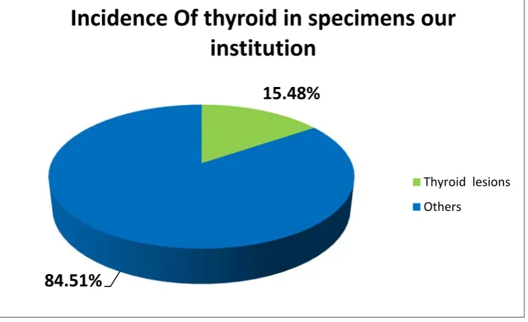
Table 1: Average incidence of thyroid specimens in our institution (N=9823)
Total specimens Thyroid specimens
9823 1521 (15.48%)
9823 specimens received in our institution during the study period were studied. Thyroid specimens were about 15.4% of the total specimens received.
Chart 1: Average incidence of thyroid specimens in our institution (N=9823)
15.48%
84.51%
Incidence Of thyroid in specimens our
institution
Thyroid lesions
50
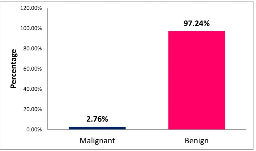
Table 2: Incidence of Benign and Malignant Thyroid lesion (N=1521)
Variable Frequency (Percentage)
Malignant 42(2.76%)
Benign 1479(97.24%)
Total 1521
The most common were benign thyroid lesion which contributed around 97% and malignancy about 2.67% among total 1521 specimens studied.
Chart 2: Incidence of benign and malignant thyroid lesion (N=1521)
2.76%
97.24%
0.00% 20.00% 40.00% 60.00% 80.00% 100.00% 120.00%
Malignant Benign
Pe
rc
e
n
ta
51
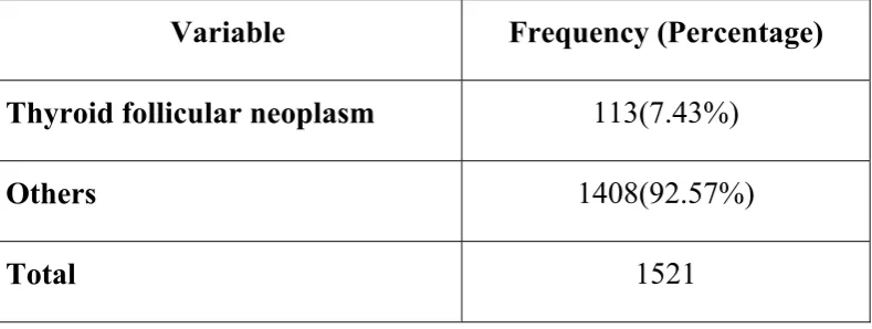
Table 3: Incidence of thyroid follicular neoplasm (N=1521)
Variable Frequency (Percentage)
Thyroid follicular neoplasm 113(7.43%)
Others 1408(92.57%)
Total 1521
Among the 1521 specimens studied, among that 7.43% were thyroid follicular neoplasm.
Chart 3: Incidence of thyroid follicular neoplasm (N=113)
92.57%
7.43%
Incidence of thyroid follicular neoplasms
Thyroid follicular neoplasms
52
Table 4: Incidence of malignant lesions in total follicular lesions (N=113)
Variable Frequency (Percentage)
Benign 95(84.07%)
Malignant 18(15.92%)
Total 113
Among 113 specimens of thyroid lesions, 84% of them were benign and 16% were malignant.
Chart 4: Incidence of malignant lesions in total follicular lesions (N=113)
84.07% 15.92%
0 20 40 60 80 100
53
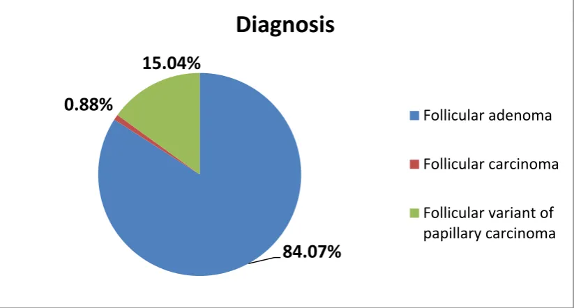
Table 5: Categorisation of thyroid follicular neoplasm (N=113)
Variable Frequency (Percentage)
Follicular adenoma 95(84.07%)
Follicular carcinoma 1(0.88%)
Follicular variant of papillary
carcinoma 17(15.04%)
Total 113
Among 113 thyroid lesion studied, 84% of were follicular adenoma, 15% were of follicular variant of papillary carcinoma and only 0.88% were follicular carcinoma.
Chart 5: Categorisation of thyroid follicular neoplasm (N=113)
84.07% 0.88%
15.04%
Diagnosis
Follicular adenoma
Follicular carcinoma
54
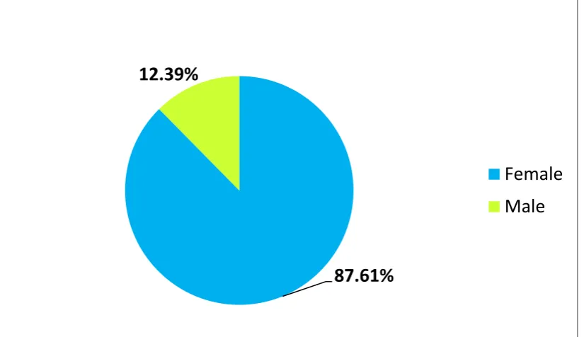
Table 6: Gender wise distribution of follicular neoplasm (N=113)
Variable Frequency (Percentage)
Male 14 (12.39%)
Female 99 (87.61%)
Total 113
Females contributed around 88% of total follicular neoplasm and males about 12%. The female male ratio was 7:1.
Chart 6: Gender wise distribution of follicular neoplasm (N=113)
87.61% 12.39%
55
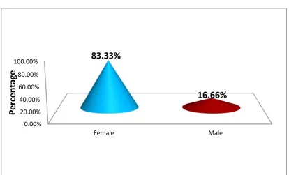
Table 7: Gender wise distribution of malignant follicular neoplasm (n=18)
Variable Frequency (Percentage)
Female 15(83.33%)
Male 3(16.66%)
Total 18
Among 18 cases of malignant follicular neoplasm, 84% were females and 16% were males.
Chart 7: Gender wise distribution of malignant follicular neoplasm (n=18)
0.00% 20.00% 40.00% 60.00% 80.00% 100.00%
Female Male
83.33%
16.66%
Pe
rc
e
n
ta
56
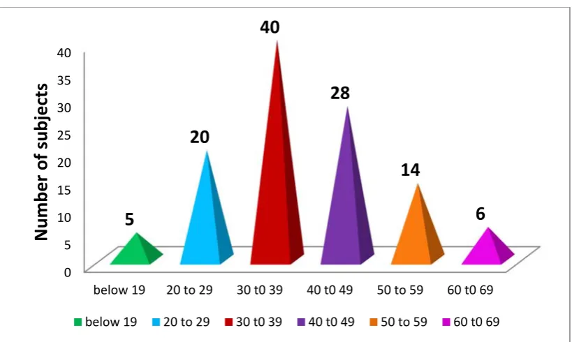
Table 8: Age wise distribution of thyroid follicular neoplasm (N=113)
Variable Frequency (Percentage)
Below 19 5(4.43%)
20 to 29 20(17.70%)
30 to 39 40(35.40%)
40 t0 49 28(24.77%)
50 to 59 14(12.39%)
60 to 69 6(5.31%)
Total 113
Among 113 cases of thyroid follicular neoplasm, 35% belonged to age group 30-39yrs, 25% of them were between 40-45 yrs, and 17% between 20-29 yrs, 12% were between 50-59 yrs. around 4.43% below 19 yrs and 5.31% between 60-69 yrs
Chart 8: Age wise distribution of thyroid follicular neoplasm (N=113)
0 5 10 15 20 25 30 35 40
below 19 20 to 29 30 t0 39 40 t0 49 50 to 59 60 t0 69
5 20 40 28 14 6 Number o f subjects
57
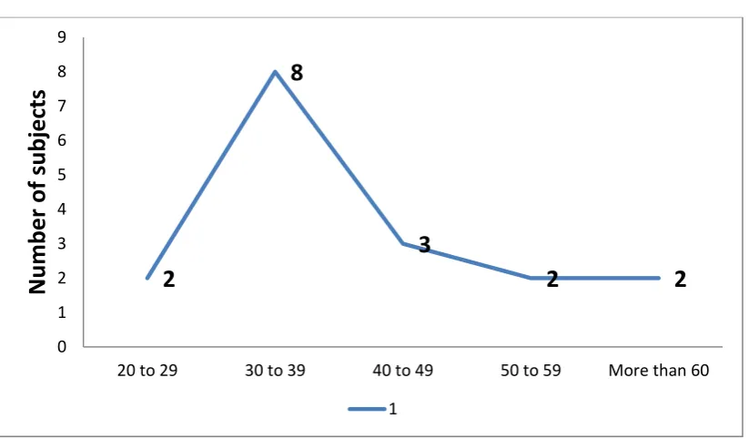
Table 9: Age wise distribution of malignant follicular lesions (n=18)
Variable Frequency (Percentage)
Less than 19 1(5.56%)
20 to 29 2(11.12%)
30 to 39 8(44.44%)
40 to 49 3(16.66%)
50 to 59 2(11.11%)
More than 60 2(11.11%)
Among 18 cases of malignant follicular neoplasm, 44% belonged to age group 30-39yrs, 17% of them were between 40-49 yrs, and 11% between 20-29 yrs, 50-59 yrs and more than 60 yrs respectively. Around 5.56% were below 19 yrs.
Chart 9: Age wise distribution of malignant follicular lesions (n=18)
2 8 3 2 2 0 1 2 3 4 5 6 7 8 9
20 to 29 30 to 39 40 to 49 50 to 59 More than 60
Number of subjects
58
Table 10: Clinical presentation of Thyroid follicular neoplasm (N=113)
Variable Frequency (Percentage)
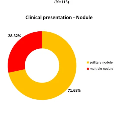
Solitary nodule 81(71.68%)
Multiple nodule 32 (28.32%)
About 71% of thyroid follicular neoplasm presented with solitary nodule and 28.32 % with multiple nodule.
Chart 10: Clinical presentation of Thyroid follicular neoplasm (N=113)
71.68% 28.32%
Clinical presentation - Nodule
soilitary nodule
59
Table 11: Surgical procedure done (N=113)
Variable Frequency (Percentage)
Total thyroidectomy 38(33.63%)
Hemi total thyroidectomy 74 (65.49%)
Near total thyroidectomy 1(0.88%)
Among 113 cases of thyroid lesion, 65% of them underwent hemi-total thyroidectomy, 33.63% of them underwent hemi-total thyroidectomy and 0.88% near total thyroidectomy.
Chart 11: Surgical procedure done (N=113)
33.63%
65.49%
0.88%
Procedure done
Total thyroidecdtomy
Hemi total thyroidectomy
60
Table 12: Variants of follicular Adenoma (n=95)
Variable Frequency (Percentage)
Simple /Normofollicular 81(85.27%)
FA with papillary hyperplasia 5(5.27%)
FA Fetal variant 3(3.15%)
FA Hurthle cell variant 4(4.21%)
FA colloid variant 2(2.10%)
Total 95
Among 95 cases variants of follicular adenoma, 85% of them was simple/normo follicular, 5.27% were FA with papillary hyperplasia, 4.21% FA hurthle cell variant, 3.15% FA fetal variant and 2.10% FA colloid variant.
Chart 12: Variants of Follicular Adenoma (n=95)
Simple /Normofillicular
FA with papillary hyperplasia
FA fetal variant
FA hurthle cell variant
61
Table 13 :The intensity of PSMA staining by immunohistochemistry in FTA, FTC and FVPC
Intensity score 0 1+ 2+ 3+ Total
Fischer’s exact test (df) p value Follicular
Adenoma 30 4 1 1 36
10.384
(3) 0.013
Follicular carcinoma and Follicular variant of papillary carcinoma
6 4 4 0 14
In the present study 83% of follicular adenoma had negative staining.11% of follicular adenoma had 1+ (faint reaction at high power).2.7 % of follicular adenoma had 2+ (moderate intesity at low power) and 2.7% of them had 3+ (strong intensity at low power).
Where as the malignant lesions (FC and FVPC) had 57% of negative staining ,14% of them were 1+ and 28% of them had 2+ staining.
Fischer exact test was used to calculate and the P values obtained were less than 0.05.
62
Table 14: The Extent of PSMA staining by immunohistochemistry in FTA, FC and FVPC
Extent
score Negative Minimal Moderate Diffuse Total
Fischer’s exact test
(df)
p value
Follicular
Adenoma 34 1 0 1 36
10.04
(4) 0.023 Follicular carcinoma and Follicular variant of papillary carcinoma
9 0 2 2 14
In the present study 86 % of the follicular adenoma had negative staining (0 to 9% of satined endothelial cells, 5.5 % had minimal staining (10 to 39% of stained endothelial cells ,1% had moderate staining (40 to 69% of stained endothelial cells) and 5% had diffuse staining (more than 70% of stained endothelial cells)
Where as for the malignant lesions (FC and FVPC) 64% showed negative staining and 14% had diffuse staining.
Fischer exact test was used to calculate and the P values obtained were less than 0.05.
63
[image:76.595.142.494.103.391.2]PHOTOGRAPHS
Figure 1 - Follicular adenoma – (H&E 100x) case 1581/18
[image:76.595.138.496.435.698.2]64
Figure 3 – Follicular adenoma: Well encapsulated single nodule with a thin capsule. Case 2255/18.
Figure 4 - Follicular adenoma –Hurthle cell variant (H&E 100x) case 674/18
[image:77.595.134.500.414.701.2]65
Figure 5- Hurtle cell variant of follicular adenoma (H&E 400x) case 2361/16
[image:78.595.134.500.426.693.2]66
Figure 7–Follicular variant of papillary carcinoma (H&E 100X) case 1835/18
[image:79.595.133.501.434.696.2]67
Figure 9 – Gross picture of follicular variant of papillary carcinoma. Case 507/17.
Figure 10 – follicular carcinoma thyroid (H&E 100x) case 1018/16
[image:80.595.119.519.394.673.2]68
Figure 11- follicular carcinoma showing capsular invasion (H&E 400x) case 1018/16
[image:81.595.123.512.393.653.2]69
Figure 13- Follicular adenoma - PSMA staining negative. Case 3411/16.
[image:82.595.124.510.381.662.2]70
DISCUSSION
Thyroid gland lesions having follicular growth pattern includes Follicular thyroid adenoma (FTA), follicular thyroid carcinoma (FTC) and follicular variant of Papillary Thyroid Carcinoma (FVPTC).Diagnosing the thyroid tumour pathology preoperatively would reduce the cost of management of thyroid nodules by eliminating diagnostic thyroidectomies which are being performed for benign thyroid nodules.
Even though FNAC is a widely recognised practice and a non expensive technique for diagnosing the lesion, we cannot differentiate between follicular adenoma and carcinoma in FNAC. Even in histomorphology, it is sometimes problematic for pathologist to diagnose a solitary encapsulated nodule with follicular histological pattern.
In this present study, out of 113 cases of thyroid follicular neoplasm, 95 cases were follicular adenomas, a case was follicular carcinoma and 17 cases were of follicular variant of papillary carcinoma. Thyroid follicular neoplasm showed an incidence of 7.4% which correlated with similar studies conducted in other institutions
71 Age incidence
Most of the cases of follicular adenoma were in the third decade. This correlated with the study conducted by Hiva saffer et al which also showed the highest incidence of follicular adenoma in the third decade of life (101) .The incidence of follicular carcinoma was common in fifth and
sixth decade. In the study done by Christopher et al the highest incidence of follicular adenoma was in the third decade and follicular carcinoma was in the sixth decade of which correlated with our study (102). The
incidence of follicular variant of papillary carcinoma was in the third decade of life.
[image:85.595.103.529.464.754.2]Table number 15 gives the age incidence comparative study Table 15
Age Incidence Comparison Study
Sl.no. Study Age incidence
1 Hiva Saffar et al Follicular adenoma -Third decade Follicular carcinoma –f