CORRELATION OF DOSE TO BONE MARROW WITH HEMATOLOGICAL TOXICITY AND MRI BASED ESTIMATION OF CONVERSION OF ACTIVE TO INACTIVE BONE MARROW IN LONG COURSE CHEMO-‐ RADIATION
FOR LOCALLY ADVANCED RECTAL CANCER
DEPARTMENT OF RADIOTHERAPY CHRISTIAN MEDICAL COLLEGE
VELLORE 632004
DISSERTATION SUBMITTED IN PARTIAL FULFILLMENT OF
MD BRANCH IX RADIOTHERAPY EXAMINATION APRIL 2015
I Jayant J Bhargav, a post graduate registrar in the department of Radiotherapy, Christian Medical College, hereby declare that the dissertation entitled “CORRELATION OF DOSE TO BONE MARROW WITH HEMATOLOGICAL TOXICITY AND MRI BASED ESTIMATION OF CONVERSION
OF ACTIVE TO INACTIVE BONE MARROW IN LONG COURSE CHEMO-‐
RADIATION FOR LOCALLY ADVANCED RECTAL CANCER ” is a bonafide work
done by me. This is being submitted to The Tamil Nadu Dr. M. G. R Medical University in partial fulfillment of the MD Radiotherapy (Branch IX) examination conducted in April 2015.
DR JAYANT J BHARGAV POST GRADUATE REGISTRAR DEPARTMENT OF RADIOTHERAPY CHRISTIAN MEDICAL COLLEGE
This is to certify that the dissertation entitled “CORRELATION OF DOSE TO BONE MARROW WITH HEMATOLOGICAL TOXICITY AND MRI BASED
ESTIMATION OF CONVERSION OF ACTIVE TO INACTIVE BONE MARROW IN
LONG COURSE CHEMO-‐ RADIATION FOR LOCALLY ADVANCED RECTAL
CANCER ” is a bonafide work done by Dr. Jayant J Bhargav, Post Graduate Registrar in the Department of Radiotherapy, Christian Medical College, Vellore during the period from June 2013 to April 2015 and is being submitted to The Tamil Nadu Dr. M. G. R Medical University in partial fulfillment of the MD Branch IX Radiotherapy examination conducted in April 2015.
Principal Head of the Department Christian Medical College Dr.Selvamani B
Vellore,India-‐632004 Professor & Head
This is to certify that the dissertation entitled “CORRELATION OF DOSE TO BONE MARROW WITH HEMATOLOGICAL TOXICITY AND MRI BASED
ESTIMATION OF CONVERSION OF ACTIVE TO INACTIVE BONE MARROW IN
LONG COURSE CHEMO-‐ RADIATION FOR LOCALLY ADVANCED RECTAL
CANCER ” is a bonafide work done by Dr. Jayant J Bhargav, Post Graduate Registrar in the Department of Radiotherapy, Christian Medical College, Vellore during the period from June 2013 to April 2015 and is being submitted to The Tamil Nadu Dr. M. G. R Medical University in partial fulfillment of the MD Branch IX Radiotherapy examination conducted in April 2015.
Guide
Dr Thomas Samuel Ram Professor
ACKNOWLEDGEMENT
I would like to express my heartfelt gratitude to my mentor and guide Dr
Thomas Samuel Ram without whose help , this work would not have been possible.
His ‘out of the box’ thinking never fails to impress and inspire me. His constant
support and guidance, his willingness to help at any time of the day(or night) are
things that I deeply appreciate.
I would like my express my sincere gratitude to Professor Dr Selvamani B ,
Head of Department, for being a limitless source of support and encouragement. Her
remarkable ability to multitask, her genuine interest in providing and maintaining the
highest quality of medical care in the clinic, is truly inspiring.
I am grateful to Dr Anuradha, Department of Radiodiagnosis, for her endless
patience and dedication. The seemingly endless hours of work she has put in, without
as much as even a frown, is something that I deeply appreciate.
I would like to thank Mr Timothy, Mr Shivashakthi, Department of Medical
physics and Dr Rajeev, Department of Radiodiagnosis, for their valuable
contributions.I would like to thank Dr B Antonisamy and his associates for providing
their statistical expertise.
Lastly, I would like to express my sincere gratitude to my parents and sister,
for their valuable support and encouragement.
Correlation of Dose to Bone Marrow with
Hematological toxicity AND MRI based
estimation of conversion of active to
inactive bone marrow in Long
course
Chemo-
radiation for Locally advanced
Rectal cancer.
Table of Contents
Introduction ... 1
Aims and Objectives ... 2
Review of literature ... 3
Epidemiology ... 3
Risk Factors ... 6
Anatomy ... 8
Management of rectal cancer ... 11
Bone marrow ... 36
Materials and methods ... 50
Study design ... 50
Inclusion criteria ... 52
Exclusion Criteria ... 52
Sample size estimation ... 52
IRB clearance ... 53
Work-‐up ... 54
3D Conformal therapy ... 55
Protocol for contouring of the entire pelvis on planning CT ... 56
Protocol for estimating marrow inactivation ... 58
Concurrent chemotherapy ... 62
Weekly assessment ... 62
Variables ... 62
Sources of Data ... 63
Statistical methods ... 65
Results ... 66
Patient Demography ... 67
Tumour characteristics ... 69
Stage characteristics ... 70
Dosimetric variables ... 71
Toxicity grading ... 71
Pre and post RT Volumes of bone marrow on MRI ... 78
Discussion ... 82
Conclusion ... 85
References ... 86
Enclosures ... 95
INFORMED CONSENT ... 95
PARTICIPANT INFORMATION SHEET ... 97
PATIENT INFORMATION SHEET IN HINDI ... 101
DATA COLLECTION SHEET ` ... 105
COURSE CHEMO- RADIATION FOR LOCALLY ADVANCED RECTAL CANCER
DEPARTMENT
: RADIOTHERAPYNAME OF THE CANDIDATE
: JAYANT J BHARGAVDEGREE AND SUBJECT
: MD RADIOTHERAPY(BRANCH IX)NAME OF THE GUIDE
: THOMAS SAMUEL RAMAims : To correlate the dose to bone marrow with the incidence and grade of hematological toxicity and to estimate the extent of inactivation of bone marrow in patients with locally advanced rectal cancer undergoing neo adjuvant long course chemoradiation.
Methods and materials: 20 patients with locally advanced rectal cancer were enrolled for the study after clearance from the institution review board. All the patients received preoperative long course radiotherapy using 3D conformal modality to a dose of 50.4Gy.They received concurrent chemotherapy with daily Capecitabine (825mg/m2). The entire pelvis was contoured on the simulation CT, the active marrow (red) bone marrow was delineated on both the pre radiotherapy as well as the post radiotherapy MRI of the pelvis on the T1 weighted images. Baseline and weekly blood investigations were recorded during the course of therapy. The dosimetric parameters such as V5, V10, V20, V30 and V40 were correlated with the incidence of Grade 3 or more hematological toxicity. The pre and post radiotherapy volumes of the active marrow and the extent (in percentage volume) of inactivation of bone marrow (red to yellow marrow conversion) due to LCCRT was also documented.The Shapiro Wilk/Mann Whitney test was used to correlate the bone marrow dose with toxicities and the paired T test was used to test the significance of conversion of active to inactive marrow.
Results: The incidence of grade 3 or more toxicity of hemoglobin correlated with V30 and V40 values(p value 0.02 and 0.0095 respectively). The toxicity grades of the other blood elements however did not show any correlation with any of the dosimetric variables. The median value of the pre radiotherapy active marrow was 346.21cc and the median of the post radiotherapy active marrow was 116.44cc.The percentage inactivation after therapy had a median value of 57.64% (range 38.98% - 83.39%)There was also a significant conversion of active to inactive bone marrow as detected on the MRI, the correlation of the pre and post neoadjuvant chemoradiotherapy marrow volumes was highly significant (p value <0.0001)
tissue toxicity
Introduction
Colorectal cancer is emerging as a major cancer burden with the increase in
incidence and mortality, both globally as well as in India (1). Neoadjuvant long course
chemoradiation therapy and Total Mesorectal Excision (TME) is the current standard
of care in locally advanced rectal cancer (2).Following surgery, patients receive
adjuvant chemotherapy based on the findings of the surgical pathology(3).It is well
known that both radiotherapy and chemotherapy leads to significant
myelosuppression. With the availability of CT based planning several authors have
attempted to document the dose to bone marrow and correlate the hematological
toxicity by studying the dose volume histogram(4) (5). There is not enough literature
at present evaluating the degree of myelosuppression and the extent of bone marrow
damage caused by neo adjuvant chemoradiotherapy. This study is undertaken to
estimate the incidence and degree of hematological toxicity, which could be, attributed
to chemoradiotherapy and objectively estimate the extent of bone marrow injury.
Aims and Objectives
-1. To correlate the dose of radiation received by the bone marrow(volume of the marrow
receiving a specified dose) and the incidence of grade 3-4 hematological toxicity in
patients undergoing Long course chemoradiation for locally advanced rectal cancer.
-2. To estimate the extent of inactivation of bone marrow caused following completion of
Review of literature
Epidemiology
Colorectal cancer continues to be a major global burden in terms of the incidence and
the morbidity. Worldwide, Colorectal cancer is the third most common malignancy
affecting men and the second most common malignancy affecting women(6).Globally
cancers of the anorectum constitute more than 40% of the total colorectal cancers
noted. Though the incidence of rectal cancer in India is much lesser than that in
developed world, it is on a definite increasing trend. In India, when both genders are
taken together, it ranks fifth in terms of incidence and sixth in terms of mortality(7). The estimated age standardized incidence rate (ASR) of rectal cancer in India is 7.19
and 5.08 per 100,000 in males and females respectively (1).
Of particular importance is the observation that there has been an increased number of
younger (with a mean age of 40-45 years) patients from West Bengal, the North
Eastern states as well as from Bangladesh, being diagnosed with colorectal cancer (8)
Figure 1: Worldwide incidence of colorectal cancers (ASR)
[image:23.595.90.500.373.593.2]Figure 3: Ten leading cancers in the Indian population
[image:24.595.139.490.82.291.2]
Risk Factors
The risk factors associated with rectal cancer can be grouped into two
categories
1)Non modifiable risk factors
a) Age: More than 90% of colorectal cancer occurs in patients aged 50 or more. The likelihood of diagnosis of rectal cancer progressively increases after the age of 40,and
sharply after the age of 50 (10).However, the number of younger patients being
diagnosed with colorectal cancer, especially in our country, has steadily been
increasing (1)
b) History of adenomatous polyps: Neoplastic polyps of the colorectum are precursor
lesions of colorectal cancer (11). Most of the sporadic colorectal cancers develop
from the pre existing villous or tubular adenomas. The development of a malignancy
from an adenoma has a long latency of about 10 years (12). Detection of and removal
of the precursor prior to malignant transformation may reduce the risk of invasive
colorectal cancer (13).
c) History of Inflammatory bowel disease: Ulcerative colitis and Crohn’s disease both
increase the risk of developing colorectal cancer later in the life (11). This warrants
d) Family history of Colorectal cancer or adenomatous polyps: There is a high risk
among first-degree family members of patients diagnosed with colorectal cancer and
adenomatous polyps. Approximately 20-25% of colorectal cancers are detected among
the first-degree family members. (14) (15)
d) Predisposition due to genetic syndromes: 5 and 10% of Colorectal malignancies are
detected in people with genetic syndromes, such as FAP and the syndromes of
Gardner, Lynch and Turcot.
2) Modifiable risk factors
Modifiable risk factors of colorectal cancers include smoking, physical inactivity,
Anatomy
There is considerable variation and ambiguity regarding the anatomical
definition of the rectum due to differing anatomical and surgical landmarks. For
surgical and oncological considerations the anatomy of the rectum is simplified as
follows:
The rectum is divided into three parts. The lower rectum is approximately 3 to
6cm from the anal verge. The mid rectum is from 5-6 cm to 8-10 cm. The upper
rectum is from 10 cm to 12-15 cm from the anal verge.
The location of the tumour is usually specified in terms of the distance from
the anal verge. Occasionally it may be specified based on the dentate line or the
anorectal ring. The reference anatomical landmark from which the measurements are
made, have to be clearly mentioned. Likewise, the method of measurement; per rectal
examination, colonoscopy, flexible endoscopy has to be mentioned.
The location of the tumour has considerable implications in the prognosis and
selection of appropriate therapy. The upper third of the rectum is envisaged by the
peritoneum anteriorly and laterally. The middle third is lined by the peritoneum on the
anterior aspect only. The lower third of the rectum, which is in close proximity to
The majority of the lymphatic drainage of the rectum passes along the superior
hemorrhoidal artery. The para rectal nodes above the level of the middle rectal-valve
drain along the superior hemorrhoidal lymphatic chain. The lymphatics below the
level of the middle rectal valve pass to nodes along the middle hemorrhoidal artery,
obturator fossa, hypogastric and common iliac arteries. The rectovaginal
[image:29.595.112.549.306.712.2]Management of rectal cancer
Suspected rectal growth
Complete history and physical examination
(including DRE and pelvic exam)
CBC
Biochemical profile
Renal profile
Liver function test
CEA
Imaging
EUS
MRI abdomen and
pelvis
CT abdomen and pelvis
Chest radiograph
*PET-CT(suspected
metastasis)
BIOPSY
Colonoscopy
Work-up
The work up of a patient with a suspected rectal growth includes a history,
physical examination including a per rectal examination, complete blood cell count,
liver function tests, renal function test and a baseline CEA.
Colonoscopy or barium enema to evaluate the large intestine for polyps,
synchronous growths is often needed.
Imaging studies include a CT or MRI of the abdomen and pelvis for accurate
delineation of the tumour as well as to rule out metastasis in other abdominal organs.
MRI of the pelvis with or without the endorectal coil is considered to be more superior
in visualization of the tumour extent compared to a CT pelvis (17).A PET-CT would
be warranted in some scenarios to help in exclusion of distant metastasis. It is,
however more useful in cases of a recurrent or a suspected recurrent growth (18).The
Endocrectal Ultrasound is considered to be the imaging modality of choice for
accurate T staging of the growth (19). A chest radiograph or a CT thorax is warranted
to rule out lung metastasis.
A biopsy of the growth is essential and may be done at the time of the
colonoscopy/sigmoidoscopy or as a guided procedure using endorectal ultrasound or
CT to help in localization of the tumour.
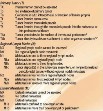
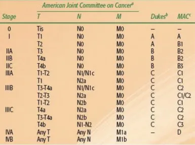
The staging of patients with rectal cancer is carried out based on the TNM staging
based on the American Joint Committee on Cancer (AJCC)/International Union
Against Cancer (UICC) staging systems (20).The older Dukes’s classification and the
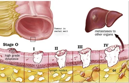
Figure 9: Representation of different stages of rectal cancer in terms of depth of
[image:34.595.90.505.148.416.2]Prognostic factors affecting outcomes
1) Tumour location
Tumors above the anorectum drain to the internal iliac lymph nodes
and have a propensity to metastasize to the liver via the portal
drainage; those below it drain into the nodes along the inferior rectal
and external iliac pathways and may metastasize to the lungs via the
caval drainage.
Overall, distal tumours have a worse prognosis than the proximal
growths(21)(22).
2) Tumour stage
Tumour staging based on the AJCC or the UICC systems remains the
dominant factor in determining prognosis(23).
3) Histopathological factors
Histology such as signet ring cell type or melanomas have a poorer
prognosis(24).Higher grade of the tumour (poorly
differentiated)tumours are associated with poorer
prognosis.Lymphovascular invasion is also considered to be an
independent factor and the presence of which indicates poorer
4) Tumour fixation
Tumours which are fixed tend to have poorer surgical outcomes which
in turn translates into poorer local control and survival
outcomes(27)(28)
5) Circumferential involvement
Circumferential involvement of the tumours may lead to partial or
complete luminal obstruction. These tumours are found to have a
higher incidence of lymph nodal metastasis and portend a poorer
prognosis. (29)(25)(30)
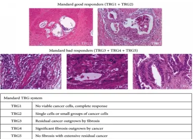
6) Degree of Tumour regression
The routine use of neo adjuvant therapy for locally advanced rectal
cancer has led to the development of grading systems based on the
extent of tumour regression.
Though different grading systems are used, the most commonly used
are the Mandard and the Dworak systems. Higher grades of regression
Therapeutic options
Surgery
Surgery has always been the mainstay of treatment in rectal cancers. Early
efforts at surgery mainly involved wide local excision of tumours. The surgical
options included:
Local Excision
a)Trans anal approach:
This approach offers the least morbidity among surgical options. Mainly employed
for tumours that are less than 8cm from the anal verge. Tumours that are more
proximal cannot be approached using this technique.
b)Trans-Sphincteric(York Mason) approach:
The entire anal sphincter is divided in the midline. Used for tumours near the
anorectal region.
A complete resection requires excision of the entire thickness of the growth into the
fat. The entire growth has to be removed in one uninterrupted specimen, so that the
pathologist is enabled to give a better description of the margins in relation to the
tumour. The major limitation of the above mentioned approaches is the fact that
lymph nodal sampling or excision is not possible. It was noted that that the incidence
of nodal metastasis in T1 lesions was in the range of 5-10%. But, for T2 it was noted
to be as high as 20-35% (35).This high rate of lymph nodal metastasis, makes local
excision unsuitable for T2 lesions. Local excision for even favourable T2 and rarely
T3 lesions followed by adjuvant therapy has yielded unfavourable results.
At present, local excision is recommended for small growths that are usually less than
four centimetres, less than 8-10 cm from anal verge, clinically T1 or occasionally,
favourable T2 lesions. They are usually well to moderately differentiated mobile
lesions that occupy less than 40% of the circumference. It is also not recommended
in case of adverse pathological findings like ulceration or lymphovascular invasion
(36) (37).
Other locally advanced rectal growths necessitate the need for different surgical
options.
Historically, excisions with generous 5cm margins both proximally and distally were
attempted. The intramural spread of tumour was rarely beyond 1.5cm. 2cm proximal
and distal margins were attempted with acceptable results. Some of the more recent
results, while offering the advantage and possibility to proceed with a sphincter
saving surgery (38) (39).
Low anterior resection
Low anterior resection (LAR) offers a sphincter sparing surgical excision, while not
compromising of the local or distant recurrence rates (40). The possibility of saving
the sphincter reduces with the more distal location of the tumour.
LAR is now being performed for upper third as well as the middle third and in some
lower third growths as well (41). A pre operative assessment of the sphincter tone,
body habitus and pelvic anatomy is a must for selecting the appropriate patient for the
procedure. The use of circular stapling devices and the need for modest 1-2cm
margins has vastly increased the adoption of this approach.
In patients planned to undergo LAR following neo adjuvant radiotherapy, it is
advisable to mobilize the splenic flexure so that an unirradiated loop of bowel may be
later used for the anastomosis. The advantage of the LAR, being the possibility of
sparing the sphincter, with a resultant better quality of life due to the lack of a
colostomy, is sometimes compromised by the post operative complications of poor
sphincter control, bowel urgency and frequency (42).
Abdominoperineal resection
The abdominoperineal resection (APR) has historically been the gold standard for
The anatomy of the pelvis, the proximity to the prostate or the vagina and the thin
mesorectum has a considerable bearing of the margins achieved following surgery
(43). APR in general has more morbidity compared to the LAR. It also suffers from
the fact that patients report a lower quality of life due to the presence of the
permanent colostomy (44).
Worldwide there has been a decrease in the adoption of this approach even for the
distal tumours (45).
Total Mesorectal excision
Following a standard LAR or APR the local recurrence rates were found to be in the
range of 15-30%. The high rate of recurrence is probably due to the fact that the
lateral spread of the tumour spread is not just at the level of the tumour, but all
through the mesorectum. The standard approaches did not address the same. It was
noted that local recurrence rates reduced when there was an enbloc removal of the
tumour along with the endopelvic fascia encompassing it. Total mesorectal excision
(TME) entails a sharp dissection along the plane to separate the visceral and the
parietal layers of the endopelvic fascia so that the entire rectal growth along with the
entire mesorectum is excised out in one uninterrupted gross specimen (46). Surgical
expertise has a considerable influence on the margins attained and the resultant local
and distant recurrence rates (47). This approach enables the attainment of a better
radial margin compared to the other surgical approaches. It is recommended that a
minimum of 12-15 nodes be excised for complete pathological staging (48). TME,
leak and delay in wound healing, leads to a higher rate of local control. This has since
then, become the standard approach in patients undergoing excision.
TME both open as well as using a laparoscopic approach resulted in similar
oncological outcomes (49).
Recent surgical advances employing extra levator APR, laparoscopic
approaches for TME, ultra low LAR and robotic LAR and TME are in different
phases of validation. The above approaches all have a common drawback in being
available only in a few centres worldwide, the adoption of the same in other centres
Adjuvant therapy
The local failure rates after surgery alone was as high as 25-30% (50).This
unacceptable rate of local failure necessitates the need for adjuvant therapies to
provide better local and distal control.
Surgery followed by radiotherapy alone
Post operative radiotherapy requires the use of larger radiation portals in order to
encompass the perineal scar in patients who have undergone an APR. Regardless of
the surgical approach, there were concerns of using radiotherapy alone due to the
larger small bowel volumes and the potentially hypoxic tumour bed leading to
perceived poorer outcomes.
Surgery alone versus surgery followed by adjuvant radiotherapy were compared; the
use of radiotherapy led to a reduced local failure rates, but had no effect on the
disease free survival or overall survival rates (50) (51).
Local failure rates after adjuvant radiotherapy alone, though marginally better, were
still unacceptably high.
Surgery alone versus surgery followed by adjuvant chemotherapy
Though, as a part of larger trial, the arm that employed post operative
In view of the above results, efforts were made to combine radiotherapy and
chemotherapy in the adjuvant setting.
Surgery alone versus surgery followed by chemoradiotherapy
As the treatment modality in one of the arms in large trials, adjuvant
chemoradiotherapy showed increase in the disease free survival as well as overall
survival (52) (53).Though the use of adjuvant combined chemoradiotherapy led to
significant improvements in disease free survival and overall survival, there was an
associated increase in the toxicity compared to the arms using adjuvant chemotherapy
or radiotherapy alone (52).
Postoperative chemoradiotherapy versus post operative radiotherapy alone
The post operative chemoradiotherapy arms in different trials all fared better
in terms of better loco regional control and overall survival (53).There was a
Neoadjuvant therapy
Neoadjuvant radiotherapy
The period in which the use of adjuvant therapy was being evaluated also
witnessed the adoption of neo adjuvant therapy predominantly in Europe. The
rationale for using neo adjuvant radiotherapy was the fact that radiotherapy could
provide the possibility of downstaging of tumour leading to better resection with
adequate margins or to the possibility of providing a sphincter sparing approach.
The usage of pre operative radiotherapy showed better local control and over all
survival rates. The early trials used a short course of hypofractionated radiotherapy
followed by surgery after a brief interval. The rates were consistently higher even on a
longer follow up (54).
The effect that the interval between the completion of radiotherapy and surgery, had
on the oncological outcomes was addressed by another trial that showed that a longer
interval allowed for significant downstaging with a higher pathological response rate
(55).Pre operative radiotherapy however, led to a higher incidence of complications
like bowel urgency, incontinence and fecal soiling.
The drawback of the above trials was the fact that the surgery performed was not the
optimal surgical approach as in TME. Preoperative radiotherapy followed by TME
compared to TME showed a lower local relapse in the pre operative radiotherapy arm,
albeit associated with higher toxicities (56).
The benefit of pre operative radiotherapy with fewer local recurrences with resultant
Pre operative chemoradiotherapy versus pre operative radiotherapy
Taking cues from the results of the combined modality arms in the adjuvant setting,
the same was attempted in the neo adjuvant setting. The trial showed that the
preoperative usage of chemoradiation resulted in higher pathological complete
response and lower local relapse rates, but with higher toxicity rates. There was no
benefit in terms of overall survival (57) (58).
Overall, preoperative chemoradiotherapy led to fewer local recurrences, higher rates
of pathological response compared to radiotherapy alone, while having no effect on
disease specific survival or overall survival. There was an increased incidence of
grade 3 or more toxicity (59).
The update of the CAO/ARO/AIO-94 trial after a median follow up of 11 years
showed the continued benefit of pre operative chemoradiation on local control. There
was however no improvement in the overall survival (60). The updated results of
another large trial conducted by the EORTC showed a similar benefit from pre
operative chemo radiotherapy followed by adjuvant chemotherapy (58) (61).
Neoadjuvant Long Course chemoradiation (LCCRT) followed by surgery and
adjuvant chemotherapy is now considered as the standard of care for all locally
advanced rectal cancers.
28 fractions along with 5-FU based chemotherapy) is more popular in the North
America. The two were compared in a trial, which showed that though there was a
significantly higher rate of pathological response rate and downstaging in the long
course regimen, this did not translate into higher rate of sphincter sparing surgery.
There was also no significant difference in the rates of toxicity, local recurrence or of
overall survival (62). There is no clear ‘better’ regimen as yet; reports on long-term
toxicities are still awaited.
Pre operative versus post operative therapy
Some of the early studies showed only a modest increase in disease free survival with
a trend towards increased overall survival in the pre operative chemoradiotherapy arm
(63).Some of the later phase III trials showed the benefit of pre operative
chemoradiotherapy(50.4Gy in 28 fractions with 5-FU based chemotherapy) in
reducing the number of pelvic recurrences, causing significant downstaging and in
increasing the number of sphincter sparing surgeries that could be performed. The
treatment, however had no advantages when it came to disease free survival or overall
survival. It was also noted that there was also lesser acute toxicity in the pre operative
arm with better compliance (64) .There was another trial that attempted to evaluate
pre operative short course radiotherapy(25 Gy in 5 fractions) versus post operative
chemoradiotherapy. It was found that the pre operative arm had lesser number of local
failures. There was also an increased disease free survival at 3 years although there
Choice of chemotherapy
Most of the early trials used 5-FU based chemotherapy along with radiation. The low
dose continuous infusion of 5-FU was found to be superior to the bolus 5-FU (66).
The other chemotherapeutic agents used in the neoadjuvant setting are:
1) Capecitabine
This is an oral fluoro pyrimidine. It requires the presence of the enzyme
thymidine phosphorylase to get converted into the active drug (5-FU) within the
tumour cells. The action of Capecitabine closely mimics that of protracted 5-FU
infusion. The studies comparing infusional 5-FU versus Capecitabine showed the
equivalence of the two drugs with respect to disease free and overall survival. The
toxicity profiles were slightly different in that the patients on Capecitabine
experienced hand foot skin reactions and proctitis, whereas the patients on infusional
5-FU had myelosuppression (67).
2) Oxaliplatin
The use of Oxaliplatin in combination with Capecitabine or 5-FU has been
evaluated. Recent trials have shown that the addition of Oxaliplatin increased the
incidence of grade 3 or more toxicity, while not improving the oncological outcomes
(68). The addition of Oxaliplatin to a modified 5-FU regimen resulted in higher rates
3)Irinotecan
Various groups have evaluated the combination of Irinotecan along with
infusional 5-FU or with Capecitabine. Though toxicity rates are slightly high, the
pathological complete response rate has been in the range of 25-30% (70).The
Locally advanced rectal cancer
Locally advanced rectal tumours are those that are
a) Fixed or adherent to surrounding structures
b) Of a size that is considered inoperable
c) Operable but of a size and character that may not yield adequate margins or have a
high probability of leaving behind micro metastatic disease
d)Lymph node positivity
As stated previously pre operative chemoradiotherapy resulted in better local
control, cancer specific survival and time to treatment failure. It also had a trend
towards better overall survival, although not significant in statistical terms (71).There
was an increase in the number of sphincter sparing surgeries that could be performed.
One of the few disadvantages of the approach is that these patients experienced
marginally more acute toxicity.
In cases where the risk of microscopic residual disease is high, even after
chemoradiotherapy, Intra operative radiotherapy may be utilized in an effort to
improve the local control. These are still in the experimental phase and require further
validation of results and toxicity prior to their routine use (72).
Radiotherapy in rectal cancer
Radiotherapy portals are designed to encompass all possible sites of local
recurrences. Recurrences are mainly noted in the pelvic soft tissue, pelvic nodes,
anastomotic site or at the perineum (73). Anterior recurrences were mainly noted on
in T4 tumours .The lymph nodal groups that are routinely included are the internal
iliac as well as the obturator groups. The external iliac group is included only in case
of anterior tumour extension or involvement of adjacent structures.
Conventional radiotherapy
Whole pelvic radiotherapy can either be delivered via the commonly used four fields
(Box technique) or the 3-field approach (Two lateral and a PA field).
Borders are as follows:
Whole pelvis
AP/PA fields
Superior: Sacral promontory (L5-S1) to encompass the attachment of the posterior peritoneum.
Lateral: 1.5 beyond the widest bony margins of the true pelvis to encompass both the possible lateral extension and the internal iliac chain.
contrast or an endoscopically placed clip. For post operative cases, in cases of post
LAR it is placed 3 cm beyond the region of the anastomosis, or in cases of post April
is placed beyond the anal verge to encompass the perineal scar.
The inguinal nodes are encompassed only in case of extension into the anal canal or
involvement of the anterior structures.
The para aortic nodes are not included in the portals as the involvement of these is
considered to be metastatic disease.
Lateral fields
Anterior: For T2 and T3 leisons-After giving generous margins from the growth it is generally placed at the posterior margin of the pubic symphysis to
encompass the internal iliac nodes.
For T4 lesions: Adequate margins from the growth including its anterior
extension, it is usually placed at the anterior margin of the pubic symphysis in order
to encompass the external iliac nodes.
Posterior: To encompass the entire sacral hollow
Superior and inferior: Margins same as that of AP/PA fields.
The boost field is framed to encompass the primary tumour with a 2 cm margin. The
Figure 11: The images on the left are a diagrammatic representation of the portals
[image:53.595.109.490.79.384.2]3D conformal radiotherapy
Conformal therapy offers the possibility of sparing normal tissues, while not
compromising on the dose delivered to the target volumes.
The GTV includes both the GTV-T and the GTV-N which is appreciated both
clinically as well as on imaging.
The CTV includes the entire mesorectum, presacral space as well as the obturator,
internal iliac groups. The common iliac and the external iliac are not routinely
included in all cases.
The PTV includes a symmetric or asymmetric expansion of the CTV to account for
organ motion and set up errors.
The OAR’s are routinely contoured and efforts made to limit to spare them well
below their threshold limits.
IMRT,though with its potential to reduce normal tissue dose and its resultant
advantages, has still not been approved for routine clinical use. Concerns of dose
heterogeneity as well as the possibility of geographical miss due to variations in the
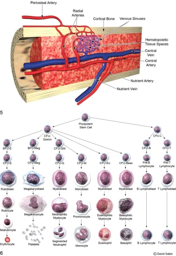
Bone marrow
Structure
The bone marrow is one of the largest organ systems in the body contributing
to about 5% of the body weight in adults. It is found in the central cavities of long and
axial bones. It is the major organ for hemopoiesis, also a primary lymphoid organ,
involved in the production of erythrocytes, granulocytes, monocytes, lymphocytes
and platelets. It consists of islands of hematopoietic tissue and adipocytes surrounded
by vascular sinuses, interspersed within the stromal meshwork of trabecular bone.
The hematopoietic tissue consists of blood cells with their precursors, adipocytes,
adventitial or barrier cells and macrophages. Hematopoiesis is a compartmentalized
process. Erythropoiesis takes place in distinct anatomical units referred to as the
‘erythroblastic islands’; granulopoiesis occurs in distinct islands too, albeit, not as
organized as the ones involved in erythropoiesis. Megakaryopoiesis, on the other
hand, occurs adjacent to the sinus endothelium. The hematopoietic cells reach the
bloodstream after entering into the venous sinuses. The platelets, however, are
released directly into the blood as cytoplasmic processes of megakaryocytes are in
direct relation to the sinus lumen.
Hematopoiesis though a continuous process can be separated into distinct stages. The
first stage involves pluripotent stem cells, which are uncommitted stem cells. These
stem cells maintain their numbers by a process of self-renewal. They also have the
series (multipotential stem cells). The third stage is when these committed stem cells
differentiate into lineage-specific progenitor cells.
Figure 12: Representation of the marrow with the pluripotent stem cell and the
[image:56.595.98.453.130.645.2]Figure 13: Diagrammatic representation of yellow and red marrow; cellular marrow
and adipocytes.
Every step in haemopoiesis; production, differentiation and maturation is under the
regulation of humoral factors. The humoral factors may either be general or be
specific for particular lineages. They may also be specific for either the early
non-committed cells higher up in the production process or to the more non-committed lineage
specific progenitor cells.
The distribution of active marrow in human adults is considerably different
from those in children. About 40% of the marrow is located in the pelvis, 10.9% in
the lumbar spine, 14.1% in the thoracic spine, 13.1% in the calvaria, 8.3% in the
Figure 14: Distribution of red marrow in adults
Figure 16: Distribution of active marrow in adults with corresponding site-specific
percentages
The fact that the cellularity of the bone marrow varies with age is well known. The
bone marrow of children is more cellular than that in adults. The cellularity is usually
expressed as the ratio of nucleated hematopoietic cells to fat cells. The ratio is about
[image:59.595.95.396.97.382.2]
Effect of radiation on bone marrow
The bone marrow is exquisitely sensitive to radiation. The bone marrow is one
of the organs that have been exhaustively studies by radiobiologists. The effects of
radiation on the bone marrow were described soon after the discovery of X rays. The
mechanisms or bone marrow destruction and the clinical implications of the same
were described in more detail while investigating the clinicopathological features of
acute radiation syndromes. The difference in kinetics and turnover of cells of different
lineages are reflected in the clinical and pathological features that are noticed
following radiation exposure. The same is also responsible for the differences noted in
attaining normal state kinetics in the period of recovery.
The peripheral blood picture of granulocytopenia, thrombocytopenia and anemia
partly reflect the early effects of the damage caused by radiation. The do not give an
indication of the extent of damage to the microenvironment, the reserve capacity of
the marrow or to the damage caused to the stem cells.
The pluripotent stem cells and blast cells are more radiosensitive than the mature post
mitotic cells. In both murine as well as in human models the most primitive stem cells
appear to be responsible for long-term hematopoiesis. The committed stem cells aid in
engraftment and the initial hematopoietic recovery. Invitro assays or explanted
stromal cells have been used to study radiation-induced injury to the
microenvironment.
2.5-doses, however, even large fields upto 20-40 Gy can be tolerated with minimum
residual effects. This is probably due to the capacity of stem cells from the
nonirradiated marrow and peripheral blood to seed into the bone that has been
irradiated. The process of recovery follows a characteristic pattern wherein the
platelets and granulocytes return to their normal values before the erythrocytes do.
Complete recovery of hematopoiesis is however seldom possible due to the
irreversible stromal and microvasculature injury. Reduced production of humoral
factors may also be a contributory cause.
The delayed effects of localized exposure, in addition to the dose, volume of
marrow irradiated and fractionation, also depends on age. Children appear to have a
Imaging of the bone marrow
1) Plain radiographs and Computed tomography: Though information about the structure of the bone is obtained, visualization of the bone marrow is not possible.
2) Nuclear medicine imaging: Functional imaging using the above pharmaceuticals, along with their appropriate targets, holds great promise in imaging of the functional
marrow. The common drawback is however, the poor spatial resolution with the
resultant lack of structural information (75).
Figure 17: Radiopharmaceuticals used in functional imaging of bone marrow
3) Magnetic resonance imaging: Of the limited radiological options available, MRI is the preferred modality for visualization of the marrow. Trabecular bone, water and fat
all have different MR signals. The relative contributions of the above three along with
The standard MR imaging protocol for visualization of the bone marrow may include coronal STIR images of the spine, pelvis and sagittal T1-‐w and fat-‐ suppressed, T2-‐w FSE or STIR sagittal images of the spine. Axial and contrast-‐ enhanced T1-‐w images are acquired in when appropriate(76)
T1-w images:
• Yellow marrow has a bright signal similar to that of subcutaneous fat • Water appears hypo intense
• The red marrow with 40% water, 40% fat and 20% protein appears darker than yellow marrow, with signal intensity slightly more than that of muscle.
T2-w images:
• Yellow marrow appears hypointense • Water appears bright
• The resultant image of the red marrow appears relatively unchanged, appears slightly more intense than the muscle.
• T2w-SPE with selective fat suppression offers a little more distinction between the yellow and the red marrow than a conventional T2-w image
STIR:
• Short inversion –Time inversion recovery with the advantage of eliminating signal from a selective tissue helps in visualization of the pathological area
against a backdrop of the uninvolved marrow. Coronal STIR images with a
large field of view helps in volumetric assessment a large area.
Contrast enhanced T1-w images:
• Useful when findings are not discernable on an unenhanced T1-w series. • Appropriate when diffuse marrow or meningeal involvement is expected
Imaging findings post radiotherapy
The changes observed post radiotherapy result in an increased T1-‐w signal with a sharp demarcation corresponding to the radiation portal.
The sinusoidal vasculature is affected post radiotherapy; the hematopoietic marrow is replaced by the fatty marrow with the resultant hypocellular bone marrow. There have been reports that at doses above 36 Gy, fatty replacement is permanent, with little chance of hematopoietic recovery. Below 30 Gy, however, these changes are likely to be reversible in 12–24 months (77) (78) .
Figure 18: T1-w images of the pelvis in coronal section (Pre Neoadjuvant
chemoradiotherapy) Note the hypointense areas representing the areas of red marrow
(indicated by arrows)
Figure 19: T1-w image of the pelvis in coronal section (Post Neoadjuvant
chemoradiotherapy); Note the hyperintensity in the sacrum and iliac crests indicating
[image:65.595.90.391.72.286.2] [image:65.595.90.409.375.633.2]Implications of bone marrow depletion
Approximately 51% of the active marrow is located in the lumbosacral spine
along with the pelvic girdle. This is of particular interest to the radiation oncologist,
as radiotherapy for the treatment of lower gastrointestinal, gyneaecological or
prostatic malignancies involves fields that would encompass the above mentioned
areas.
It is well known that both radiotherapy and chemotherapy have considerable
effects on the bone marrow in causing myelosuppression. The effects are of an even
greater significance in cases of radiotherapy along with concurrent chemotherapy, in
which case there is an additive effect of the individual bone marrow toxicities from
radiotherapy and chemotherapy. The resultant toxicity noticed during the course or
following completion of chemoradiotherapy is by no means insignificant.
It may lead to the following:
a) Degrees of hematological toxicity, which would warrant interrupting therapy.
b) Febrile or afebrile neutropenic illnesses with its associated morbidty, risk of
mortality and their associated cost of management.
e) Depletion of marrow reserves to an extent, which could compromise future
adjuvant therapy.
Of considerable concern in the management of lower gastrointestinal and
gynaecological malignancies is when the use of radiotherapy involving large pelvic
fields along with concurrent chemotherapy, has the potential to cause significant acute
and sub acute hematological toxicity. Peripheral blood investigations done during the
course and following completion of therapy reflect only the acute changes observed.
It does not, however, estimate the degree of damage caused to the marrow or its
reserve. Bone marrow biopsies are also unable to characterize the above changes
fully. Due to the paucity of methods to characterize or visualize the damage to the
bone marrow, efforts to reduce the dose the bone marrow were seldom made in the
past.
Initial efforts to reduce the dose to the active bone marrow involved using 4
field box techniques, placing manual shields to spare the ilium and the iliac crests.
With the advent of conformal and intensity modulated radiotherapy techniques,
multiple beams were placed along with appropriate shielding offered by customized
blocks, and multi leaf collimators resulting in significantly reduced doses to the
organs at risk (OAR’s). Further efforts involved the contouring of the entire pelvis on
the planning CT and limiting the dose to the OAR’s including the pelvis without
compromising the dose delivered to the target volumes. “Bone marrow sparing
IMRT” has been recently attempted both prospectively in the clinic as well as in
received by the pelvis. This translated into lesser hematological toxicities, lesser
number of emergency visits and lesser treatment breaks(84)(85)(86)(87).This
however resulted in gross over estimation of the amount of active marrow(88)(84).
With the developments of novel imaging modalities such as the FLT PET, FDG PET,
SPECT and the MRI (T1 sequences, DCE) efforts are now being made to contour the
active marrow alone such that more accurate estimates of the volume and their
tolerance may be obtained (89)(90). The above methods are however, still in the
experimental phase and not yet being used in routine clinical practice.
Materials and methods
Study design
Algorithm of study
Recruitment of participants of locally advanced rectal cancer undergoing Long course chemo radiation(based
on inclusion and exclusion criteria as mentioned)
Baseline blood investigations and MRI abdomen and Pelvis
Contour entire pelvis on planning CT scan Contour pelvic bone marrow on T1 MRI images
Long course chemoradiation:3D conformal Radiotherapy and concurrent chemotherapy with
Capecitabine
Weekly blood investigations during treatment:hemoglobin,total and differential count,platelet count,Serum creatinine,SGOT/SGPT
and Alkaline phosphatase
WEEK 0
Figure 20: Algorithm indicating the study design
For the study it was decided to recruit participants from the departments of Radiation
oncology (Unit-I) and surgery (Surgery-II) from Christian Medical college, Vellore.
A search from our departmental unit database was carried out for all patients with Document hematological and clinical toxicity
based on RTOG CTC (weekly during treatment)
Contour Pelvic bone marrow on MRI T1 images(on the MRI repeated prior to surgery-‐4
to 6 weeks following completion of LCCRT)
Correlate bone marrow dosimetric parameters(volume of bone receiving a particular dose) with incidence of >=grade
3 hematological toxicity(based on contouring of the entire pelvis on planning
CT)
Estimate extent of inactivation of bone marrow comparing both the pre RT MRI and
From the above broad search, participants were recruited based on the fulfilment of
inclusion criteria.
Inclusion criteria
• Locally advanced rectal cancer: clinical or radiological evidence of T3/T4 or N1 evidence; or that is and/or clinically bulky
• No evidence of distant metastasis
• Adenocarcinoma
• Considered or being considered for neo adjuvant Long course radiotherapy (3D conformal RT) along with concurrent chemotherapy
(concurrent Capecitabine)/or completed treatment as previously
mentioned
• Underwent pre and post RT MRI in our Institution
Exclusion Criteria
• History of prior pelvic malignancies
• History of prior radiation to the abdomen/pelvis
• Prior history of usage of chemotherapy/immunosuppressant
• Known case of myelodysplastic syndrome/myelofibrosis
• Participants with outside MRI films (either pre radiotherapy or post radiotherapy/pre surgery MRI) were not considered for the study (unless
the same were repeated at our hospital).
Sample size estimation
The sample size for the study was determined after discussion with the statistician.
N=(Z (1-a/2)+Z1-b))2. P1(1-P1).P2(1-P2) / (P1-P2)2
5% level of significance Z (1-a/2)=1.96
80%Power = Z1-b= 0.84
D=(P1-P2)2=Difference in the toxicity rate
A sample of size of 26(13 receiving less than the threshold dose and an equal number
receiving more than the same specified threshold) was needed to detect the difference
of 60% toxicity level between the groups with 80% power and 5% level of
significance using an uncorrected chi-square test
IRB clearance
Clearance from the institutional review board and ethical committee was obtained on
May 08,2014
The prospective patients were recruited into the study after taking their informed
consent (Enclosures 1-4)
However, due to unforeseen slow accrual of patients we sought the permission of the
IRB to modify the study design so that we could recruit retrospective patients as a part
Work-up
All patients had undergone a complete staging as well as a metastatic work up. A
baseline CBC including Haemoglobin, total count, differential count, platelet count,
renal and liver function tests, CEA, ECG, Echocardiograph, Chest radiograph, MRI of
the abdomen and pelvis was obtained for all patients. Height,Weight,baseline BSA
was calculated for all patients. Cardiology clearance was taken prior to starting
chemotherapy.
Other parameters that were recorded were:
Location of the tumour (upper, middle or lower rectum)
Proliferative or ulcerative tumour
Presence or absence of circumferential involvement
Presence or absence of anorectal involvement.
Staging of rectal cancer was based AJCC cancer staging manual 7th edition, 2010
The clinical and radiological findings of all patients were discussed in a multi
disciplinary tumour board comprising of Radiation Oncologists, Surgical Oncologists,
Medical Oncologists, Diagnostic and Radiologists .The consensus decision was to
offer Neoadjuvant radiotherapy using 3D conformal radiotherapy along with
concurrent chemotherapy with daily Capecitabine.
All the patients in this study were proposed to receive neo adjuvant long course
3D Conformal therapy
Position:
All patients were simulated and treated in the supine position. The forearms and
hands were placed over the chest or upper abdomen, based on the patient’s preference
Immobilization:
An immobilization device utilizing a vacuum assisted bag the VAC-LOC was made
for all patients.
3 Fiducial markers were placed on the patient’s body and their positions prior to CT
simulation were verified by aligning them using the in-room lasers.
Planning CT protocol
The simulation CT was acquired as follows:
Informed consents for using oral and IV contrast were taken.
Patients were asked to lie supine on the flat couch insert along with the vacloc and the
positions of the patient and the fiducials were verified.
Oral contrast was given to aid in visualization of the small bowel.
IV cannulas were placed and their patency verified.
The scout film was viewed to verify patient position.
IV contrast (1ml/Kg) was injected using a machine driven piston.
5mm cuts were acquired from the level of the diaphragm superiorly to the level of the
Target delineation
Volume delineation was carried out as follows:
GTV: The rectal growth visualized on imaging. Clinically or radiologically involved
nodes.
CTV: Areas of subclinical disease extension including the entire mesorectum,
presacral space and the internal iliac group of lymph nodes.
PTV: Symmetric or asymmetric expansion of the CTV to account for set up and
systematic errors.
Phase I: The above-mentioned PTV
Phase II: The tumour or the post operative site with a 3 cm margin. The nodal stations
were not routinely included in the phase II volume.
Protocol for contouring of the entire pelvis on planning CT
The entire pelvis and parts of the lumbosacral spine and proximal femurs including
the head and trochanters were contoured on the planning CT for all patients.
Contouring was carried out as follows:
The bone window was used.
Entirety of the bone from L5-S1 junction or from the level of the superior most
section including the PTV phase I, whichever was higher. The inferior extent was at
the lowermost level of the ischial tuberosity.
The dosimetric parameters relating the bone marrow such as V5, V10, V20, V30 and
Figure 21: Delineation of the Pelvis on the planning CT
[image:76.595.88.442.414.677.2]Protocol for estimating marrow inactivation
The active marrow (red marrow) was contoured on the MRI abdomen and pelvis on
the T1 weighted images along with the assistance of a senior radiologist.
The images that were acquired were reviewed by the radiologist. The active marrow
on both the pre radiotherapy MRI as well as the post radiotherapy MRI was contoured
by the radiologist as follows:
The yellow marrow has hyperintense signal intensity on T1; red marrow has a relative
hypointense signal intensity compared to yellow marrow but a higher intensity than
that of the muscle.
The marrow was delineated on the axial images; the superior and inferior limits being
the same as that used for contouring the bone on the planning CT. The summated
volumes of the active marrow on both the pre as well as the post treatment MRI’s
Figure 23: T1-w axial images of the pelvis. The hypointense areas were contoured
[image:78.595.93.412.72.316.2] [image:78.595.90.416.406.690.2]Plan evaluation
The plans were evaluated based on ICRU principles where in the target volume
coverage and distributions were reviewed while ensuring that the organs at risk
[image:79.595.91.505.181.458.2]received a dose that was within their tolerance limits.
Figure 26: DVH of both the target volumes as well as the organs at risk; note the blue
unbroken line representing the DVH of the pelvis (represented by the arrow)
Treatment delivery
Concurrent chemotherapy
All patients received concurrent chemotherapy with Capecitabine (825mg/m2) given
Monday to Friday (on the days of receiving radiotherapy)
The need, the benefits and side effects expected with radiotherapy and chemotherapy
were explained to patients and their informed consents taken.
Weekly assessment
Patients underwent a weekly assessment during the course of radiotherapy. Clinical
toxicities were documented as per the RTOG toxicity grading system. Weekly blood
investigations including Hemoglobin, total and differential counts, platelet count,
liver function tests and Serum creatinine were ordered.
Patients underwent a reassessment of their disease status 6 weeks following
completion of the Neoadjuvant treatment. Apart from the above-mentioned
investigations, they also underwent MRI of the pelvis to assess tumour downstaging.
Variables
The variables that were considered were:
1) Hematological toxicities-
Toxicities relating to Hemoglobin, Total leucocyte count, absolute
neutrophil count, lymphocyte count and platelet count were graded
separately according to the Cooperative group common toxicity criteria