Copyright © 2004, American Society for Microbiology. All Rights Reserved.
Insertion of Green Fluorescent Protein into Nonstructural Protein 5A
Allows Direct Visualization of Functional Hepatitis C
Virus Replication Complexes†
Darius Moradpour,
1,2Matthew J. Evans,
1,3Rainer Gosert,
2Zhenghong Yuan,
1‡ Hubert E. Blum,
2Stephen P. Goff,
4Brett D. Lindenbach,
1and Charles M. Rice
1*
Center for the Study of Hepatitis C, Laboratory of Virology and Infectious Disease, The Rockefeller University, New York, New York 100211; Department of Medicine II, University of Freiburg, D-79106 Freiburg, Germany2; and
Integrated Program in Cellular, Molecular, and Biophysical Studies3and Howard Hughes Medical Institute,
Department of Biochemistry and Biophysics, College of Physicians and Surgeons,4
Columbia University, New York, New York 10032 Received 29 December 2003/Accepted 28 March 2004
Hepatitis C virus (HCV) replicates its genome in a membrane-associated replication complex, composed of viral proteins, replicating RNA and altered cellular membranes. We describe here HCV replicons that allow the direct visualization of functional HCV replication complexes. Viable replicons selected from a library of Tn7 -mediated random insertions in the coding sequence of nonstructural protein 5A (NS5A) allowed the identifi-cation of two sites near the NS5A C terminus that tolerated insertion of heterologous sequences. Replicons encoding green fluorescent protein (GFP) at these locations were only moderately impaired for HCV RNA rep-lication. Expression of the NS5A-GFP fusion protein could be demonstrated by immunoblot, indicating that the GFP was retained during RNA replication and did not interfere with HCV polyprotein processing. More im-portantly, expression levels were robust enough to allow direct visualization of the fusion protein by fluores-cence microscopy. NS5A-GFP appeared as brightly fluorescing dot-like structures in the cytoplasm. By confocal laser scanning microscopy, NS5A-GFP colocalized with other HCV nonstructural proteins and nascent viral RNA, indicating that the dot-like structures, identified as membranous webs by electron microscopy, represent functional HCV replication complexes. These findings reveal an unexpected flexibility of the C-terminal domain of NS5A and provide tools for studying the formation and turnover of HCV replication complexes in living cells.
Hepatitis C virus (HCV) infection is a major cause of chronic hepatitis, liver cirrhosis, and hepatocellular carcinoma worldwide (37). A protective vaccine does not exist to date and therapeutic options are still limited. The virus contains a sin-gle-stranded 9.6-kb RNA genome of positive polarity that en-codes a polyprotein of about 3,000 amino acids (aa) (reviewed in references 27 and 33). The polyprotein precursor is co- and posttranslationally processed by cellular and viral proteases to yield the mature structural and nonstructural proteins. The structural proteins include the core protein, which forms the viral nucleocapsid, and the envelope glycoproteins E1 and E2. The nonstructural proteins NS2 through NS5B include the NS2-3 autoprotease and the NS3 serine protease, an RNA helicase located in the C-terminal region of NS3, the NS4A polypeptide, the NS4B and NS5A proteins, and the NS5B RNA-dependent RNA polymerase.
Similar to all positive-strand RNA viruses investigated thus far (reviewed in references 1 and 12), HCV forms a
mem-brane-associated replication complex, composed of viral pro-teins, replicating RNA, and altered cellular membranes (13, 16, 31, 44). Determinants for membrane association of the HCV nonstructural proteins have been mapped (reviewed in references 11 and 34), but the protein-protein interactions involved in formation of a functional HCV replication complex are poorly understood (10). A specific membrane alteration, termed the membranous web, was recently identified as a site of HCV RNA replication in Huh-7 cells harboring subgenomic HCV replicons (16). The membranous web could be induced by expression of NS4B alone and was similar to the “sponge-like inclusions” previously observed by electron microscopy in the liver of HCV-infected chimpanzees (13). Given the often close association with the endoplasmic reticulum (ER) and based on earlier studies demonstrating the colocalization of individually expressed HCV proteins with the ER (8, 20, 43, 46), as well as more recent data indicating that HCV RNA replication takes place in a compartment that sustains endogly-cosidase H-sensitive glycosylation (22), it is believed that the membranous web is derived from membranes of the ER (16). However, the steps involved in membranous web formation, the interplay between HCV translation and replication, and the turnover of HCV replication complexes are poorly under-stood. Such investigations would be enhanced by systems that allow tracking of functional HCV replication complexes in living cells.
* Corresponding author. Mailing address: Center for the Study of Hepatitis C, Laboratory of Virology and Infectious Disease, The Rock-efeller University, 1230 York Ave., New York, NY 10021. Phone: (212) 327-7046. Fax: (212) 327-7048. E-mail: [email protected].
† This study is dedicated to Walter Siegenthaler on the occasion of his 80th birthday.
‡ Present address: Shanghai Medical College, Fudan University, Shanghai 200032, China.
7400
on November 8, 2019 by guest
http://jvi.asm.org/
Toward this goal, we used transposon-mediated mutagenesis to identify sites in the HCV NS5A protein where exogenous sequences could be inserted with minimal effect on replicon function. Viable replicons harboring a green fluorescent pro-tein (GFP) inserted at two permissive sites in the C-terminal domain of NS5A allowed direct visualization of the NS5A-GFP fusion protein by fluorescence microscopy. NS5A-NS5A-GFP colocalized with other HCV nonstructural proteins and nas-cent viral RNA, indicating that the fusion protein is incorpo-rated into functional replication complexes. These results in-dicate that the C-terminal region of NS5A is highly flexible and provide a system for studying the assembly and disassembly of functional HCV replicases in living cells.
MATERIALS AND METHODS
Transposon library and plasmids.A library of replicon clones with random insertions were created in a blasticidin selectable Con1 HCV replicon plasmid harboring the S2204I adaptive mutation (5), pCon1/SG-Bsd(I) (15), by using the GPS-LS linker scanning system according to the manufacturer’s instructions (New England Biolabs, Beverly, Mass.). This TnsABC transposase-based system
allows essentially random and single-hit in vitro insertion of a Tn7-based
transprimer, a minimal transposable element, into a plasmid of interest (4). Plasmids carrying an insertion can be selected based on kanamycin resistance encoded within the transposon. To concentrate transposon insertions in a sub-region of the polyprotein, an EcoRI-to-MfeI fragment containing the C-terminal two-thirds of NS5A and about one-quarter of the NS5B N terminus was sub-cloned into pCon1/SG-Bsd(I) and replated on tetracycline and kanamycin to select for transposon containing replicon plasmids. A PmeI site, present near the transposon ends, was used to remove all but 10 bases of the insert, which, in addition to a five-base duplication of the target generated during the transposase reaction, results an insertion of 15 nucleotides or 5 aa.
Subsequently, a second transprimer, containing an ampicillin selectable marker followed by the FLAG sequence (amp-FLAG) (15), was cloned as a blunt-ended fragment into the NS5A transposon library after digestion with PmeI to liberate the original transprimer. Transformants were selected on agar plates containing both tetracycline and ampicillin to select for replicon clones with the amp-FLAG cassette randomly inserted within the region of interest.
The final stage of library construction entailed the removal of the majority of the amp-FLAG cassette leaving only the small FLAG linker. This was achieved by subjecting the pool of clones to DraI digestion, which cuts only within the ampicillin cassette, and religation. Since the FLAG sequence left behind will code for this epitope only in a single orientation and frame, stop codons were designed in all other possible translational frames. The presence of a stop codon in any non-FLAG coding insertion would prevent translation of the full comple-ment of viral proteins required for replication and would thus prevent selection of replicons not coding for the FLAG peptide after transfection into Huh-7 cells. Two permissive sites were identified, as described in Results, resulting in replicon constructs pCon1/SG-Bsd(I)/FlagI.1 and pCon1/SG-Bsd(I)/FlagI.6 (15). Subsequently, the GFP coding sequence was excised from a vector containing a human codon-optimized, red-shift variant GFP kindly provided by Brian Seed (Harvard University, Boston, Mass.) and cloned into the DraI sites of pCon1/ SG-Bsd(I)/FlagI.1 and pCon1/SG-Bsd(I)/FlagI.6 to yield pCon1-SG-Bsd(I)/ GFP-FLAGI.1 and pCon1-SG-Bsd(I)/GFP-FLAGI.6, respectively. The sites and the amino acid sequences flanking these insertions are illustrated in Fig. 1A.
Finally, the XhoI-MfeI fragment of pCon1/SG-Bsd(I)/GFP-FLAGI.1 and pCon1/SG-Bsd(I)/GFP-FLAGI.6, encompassing the domain of NS5A harboring the GFP insertions, was subcloned into the XhoI-MfeI sites of G418 selectable replicon constructs pCon1/SG-Neo(GIT) and pCon1/SG-Neo(I)/AflII. The GIT replicon construct contains two cell culture adaptive mutations in NS3 (E1202G and T1280I) and one in NS4B (K1846T) (15), identified by Lohmann et al. (29). The resulting plasmids were named pCon1/SG-Neo(GIT)/GFP-FLAGI.1 (GIT/ 5A-GFP-1) and pCon1/SG-Neo(GIT)/GFP-FLAGI.6 (GIT/5A-GFP-6), as well as pCon1/SG-Neo(I)/AflII/GFP-FLAGI.1 (I/5A-GFP-1), and pCon1/SG-Neo(I)/ AflII/GFP-FLAGI.6 (I/5A-GFP-6) (Fig. 1B). As a negative control, the XhoI-MfeI fragments were also subcloned into a replicon construct with substitutions inactivating the NS5B RNA-dependent RNA polymerase, pHCVrep1bBartMan
(pol⫺)/AvaII (5) to yield plasmids pCon1/SG-Neo(pol⫺)/GFP-FLAGI.1 (pol⫺/
5A-GFP-1) and pCon1/SG-Neo(pol⫺)/GFP-FLAGI.6 (pol⫺/5A-GFP-6) (Fig.
1B).
In vitro transcription and RNA electroporation.Plasmids were linearized with ScaI, and in vitro transcription was performed essentially as described previously (7). Transcripts were purified by using the RNeasy minikit (Qiagen, Valencia, Calif.) with an on-column DNase treatment by using the RNase-Free DNase Set (Qiagen).
Huh-7 or Huh-7.5 cells (7), a highly permissive, alpha interferon-cured Huh-7 human hepatocellular carcinoma cell line derivative, were transfected with in vitro-transcribed RNA by electroporation essentially as described previously (7).
In brief, RNA transcripts (1g) were mixed with 6⫻106washed cells in 0.4 ml
in a 2-mm gap cuvette and immediately pulsed (820 V, 99-s pulse length, five
pulses at 1-s intervals) by using a BTX ECM 830 square wave electroporation system (Genetronics, San Diego, Calif.). Cells were seeded into
100-mm-diam-eter dishes at 6⫻105, 6⫻104, and 6⫻103cells per dish, together with cells
transfected with pol⫺ RNA transcripts such that the total cell number was
maintained at 6⫻105cells per dish. At 72 h after plating, selection was started
with either 1,000g of G418 or 3g of blasticidin (Invitrogen, La Jolla, Calif.)/
ml, depending on the replicon resistance marker. Three weeks later, drug-resistant colonies were pooled and further expanded or fixed with 7% formal-dehyde, followed by staining with 1.25% crystal violet in 25% ethanol to facilitate colony counting.
Antibodies. Monoclonal antibodies (MAbs) 1B6 against NS3 (46), 4b-52 against NS4B (20) (kindly provided by Michinori Kohara, The Tokyo Metropol-itan Institute of Medical Science, Tokyo, Japan), 11H against NS5A (8) (kindly provided by Jan Albert Hellings, bioMe´rieux, Boxtel, The Netherlands), and 5B-3B1 against NS5B (32) have been described. MAb M2 against the FLAG epitope was from Sigma (St. Louis, Mo.) and MAb JL-8 against GFP from Clontech (Palo Alto, Calif.). A polyclonal antiserum against GFP was obtained from Molecular Probes (Eugene, Oreg.).
Immunoblot.Immunoblot was performed as described previously (35).
Confocal laser scanning microscopy (CLSM).Indirect immunofluorescence microscopy was performed essentially as described previously (35). In brief, cells grown as monolayers on glass coverslips were fixed with 2% paraformaldehyde, permeabilized with 0.05% saponin, and incubated with primary antibodies in phosphate-buffered saline containing 3% bovine serum albumin and 0.05% saponin. Bound primary antibody was revealed with a Rhodamine Red-X-conjugated secondary antibody, and nuclei were counterstained with TO-PRO-3 iodide (Molecular Probes). Coverslips were mounted in 50% glycerol in phosphate-buffered saline and examined by using a Zeiss LSM 510 microscope. Images were processed by using Adobe Photoshop 7.0.
BrUTP labeling.Newly synthesized viral RNA was labeled with
5-bromouri-dine 5⬘-triphosphate (BrUTP) during a 3-h incubation period in the presence of
5g of actinomycin D/ml to selectively inhibit cellular DNA-dependent RNA
synthesis as described previously (17). BrU-labeled viral RNA was detected with a MAb against BrdU (Bio Cell Consulting, Reinach, Switzerland), followed by a Cy3-conjugated secondary antibody. CLSM was performed as described above.
EM and IEM.For conventional electron microscopy (EM), cells were fixed in
2.5% glutaraldehyde and 2% OsO4 and embedded in Poly/Bed 812
(Poly-sciences, Warrington, Pa.) according to standard protocols. For immuno-EM (IEM), cells were fixed and embedded in LRGold (London Resin Co., London,
United Kingdom) at⫺20°C as described previously (13). Bound primary
anti-bodies were revealed with gold-conjugated secondary antianti-bodies (Amersham Pharmacia Biotech, Piscataway, N.J.) as described previously (13).
RESULTS
Identification of insertion sites in NS5A.To identify sites in NS5A permissive to the insertion of heterologous sequences, a library of replicon constructs containing randomly inserted FLAG epitope coding sequences in the NS5A/5B region was generated by using a Tn7transposon-mediated strategy. Ac-cording to the least-efficient stage of library construction, this library represented 25 random in-frame insertions throughout NS5A/5B. Huh-7 cells were electroporated with RNA tran-scribed from this library and subjected to drug selection to score for viable replication events. The library RNA exhibited an⬃12-fold reduction in colony-forming ability compared to the parental replicon, suggesting that a significant proportion of the population was impaired for replication, as expected.
Fourteen individual drug-resistant colonies were isolated
VOL. 78, 2004 VISUALIZATION OF FUNCTIONAL HCV REPLICASES 7401
on November 8, 2019 by guest
http://jvi.asm.org/
and expanded for additional examination. All but one clone expressed an NS5A-FLAG fusion protein, as determined by immunoblotting (data not shown). To determine insert loca-tions, the NS5A/5B region was amplified by reverse transcrip-tion-PCR (RT-PCR) from RNA purified from these clones, followed by sequence analysis. All clones that were positive for the FLAG epitope by immunoblot contained an insert close to or within a serine-rich region in the C-terminal domain of NS5A. The one clone that was negative for NS5A-FLAG by immunoblot contained no FLAG insert in NS5A/5B when
ex-amined by RT-PCR and sequence analysis. Three replicons contained an insert after amino acid position 2356 (aa 384 of NS5A), and 10 replicons contained an insert after amino acid position 2390 (aa 418 of NS5A).
HCV replicons harboring GFP insertions in the C-terminal domain of NS5A are viable. The GFP coding sequence was inserted into the two FLAG-permissive sites in replicon con-structs with cell culture adaptive changes in NS3 (E1202G and T1280I) and NS4B (K1846T) (GIT) (29) or in NS5A (S2204I) (5) (Fig. 1). RNA was in vitro transcribed from these plasmid
FIG. 1. Replicon constructs. (A) Sites and amino acid sequences of the GFP insertions in NS5A. (B) Replicon constructs. Cell culture adaptive mutations in NS3 and NS4B (GIT constructs) or NS5A (S2204I constructs) are marked by circles. The pol⫺ control constructs harbor an
inactivating GDD3AAG mutation in the NS5B RNA-dependent RNA polymerase.
on November 8, 2019 by guest
http://jvi.asm.org/
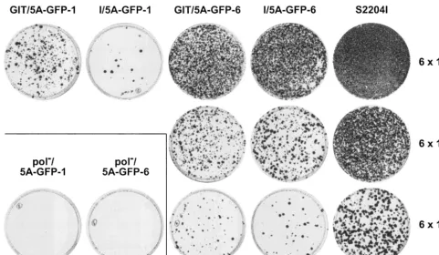
templates and electroporated into Huh-7.5 cells, followed by G418 selection. Three weeks later, G418-resistant colonies were pooled and expanded or stained with crystal violet, as shown in Fig. 2. Each of the four different constructs was viable, albeit with different colony formation efficiencies. Counting of G418-resistant colonies resulting from three inde-pendent electroporation experiments revealed a colony forma-tion efficiency of about 1 CFU/ng of replicon RNA for GIT/ 5A-GFP-1, 0.1 CFU/ng for I/5A-GFP-1, 10 CFU/ng for GIT/ 5A-GFP-6 and I/5A-GFP-6, and 250 CFU/ng for the unmod-ified parental GIT and S2204I replicons. Thus, replicons harboring the GFP insertion after aa 2390 (GIT/5A-GFP-6 and I/5A-GFP-6) were 10-fold (compared to GIT/5A-GFP-1) or 100-fold (compared to I/5A-GFP-1) more efficient in initi-ating HCV RNA replication than the constructs with GFP inserted after aa 2356. However, even for I/5A-GFP-1, G418-resistant cell populations or individual clones could be easily expanded, particularly if the G418 concentration was reduced to 400g/ml (data not shown). Efficiency of the parental rep-licon constructs GIT and S2204I was about 25-fold higher than that of the constructs harboring the GFP-6 insertion. As ex-pected, the pol⫺control constructs yielded no G418-resistant
colonies.
Taken together, initiation of HCV RNA replication by rep-licon constructs harboring a GFP insertion in NS5A was sur-prisingly efficient. In good accordance with the different num-ber of clones identified in the initial screen for permissive insertion sites with the FLAG epitope sequence as an insert (3
of 14 after aa 2356 and 10 of 14 after aa 2390), the more C-terminal insertion site was more tolerant of the GFP insert.
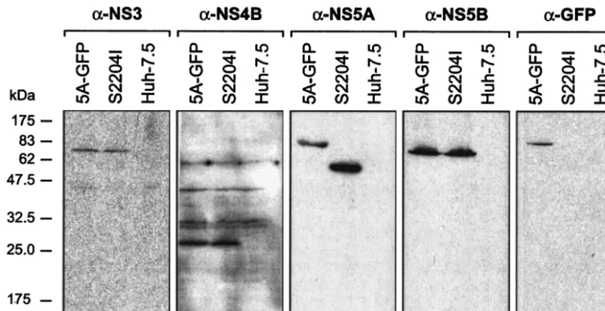
[image:4.603.51.532.68.347.2]GFP is retained in NS5A during RNA replication.Western blot analyses of I/5A-GFP-6 and S2204I replicon cells at pas-sage 10, as well as of naive Huh-7.5 cells as negative control, were performed to investigate whether the NS5A-GFP fusion protein was stable during HCV RNA replication. As shown in Fig. 3, a band of the expected molecular mass of ⬃85 kDa, corresponding to the NS5A-GFP fusion protein, was detected in I/5A-GFP-6 cells by using both MAbs directed against NS5A and against GFP. The unmodified NS5A protein of 56 to 58 kDa was detected in S2204I replicon cells. In addition, cor-rectly processed NS3, NS4B, and NS5B proteins of the ex-pected molecular masses of 70, 27, and 68 kDa, respectively, were detected in both I/5A-GFP-6 and S2204I cells. Analogous findings were obtained for the other replicon constructs. The phosphorylation status of NS5A-GFP was not investigated be-cause replicon constructs harboring the adaptive changes K1846T (present in the GIT constructs) or S2204I (present in the I constructs) are impaired for hyperphosphorylation (15). Taken together, these results demonstrate that the GFP moiety is retained in NS5A during RNA replication and does not interfere with HCV polyprotein processing. Cells have been maintained for 40 passages without any appreciable changes of their characteristics compared to the earlier pas-sages. In addition, RT-PCR and sequence analyses performed on selected clones at passages 8 to 18 revealed that the GFP insertion is stably retained (data not shown). Further studies
FIG. 2. HCV replicons harboring GFP insertions in the C-terminal region of NS5A are viable. RNA was in vitro transcribed from constructs GIT/5A-GFP-1, I/5A-GFP-1, GIT/5A-GFP-6, I/5A-GFP-6, and S2204I, as well as pol⫺/5A-GFP-1 and pol⫺/5A-GFP-6, and electroporated into
Huh-7.5 cells, followed by plating into 100-mm-diameter dishes at 6⫻105, 6⫻104, and 6⫻103cells per dish and G418 selection as described in Materials and Methods. G418-resistant colonies were stained with crystal violet after 3 weeks.
VOL. 78, 2004 VISUALIZATION OF FUNCTIONAL HCV REPLICASES 7403
on November 8, 2019 by guest
http://jvi.asm.org/
will be necessary to determine the stability of NS5A-GFP over more extended passaging.
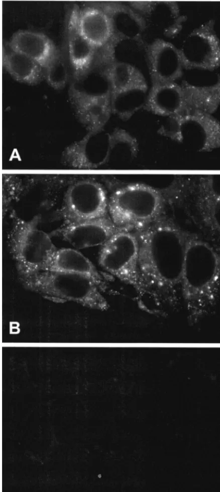
NS5A-GFP can be directly visualized by fluorescence mi-croscopy.Huh-7.5 cells harboring the different replicon con-structs were cultured on glass coverslips and examined by fluorescence microscopy. As shown in Fig. 4, the GFP cence was robust enough to be directly visualized by fluores-cence microscopy. The signal was found in the cytoplasm as brightly fluorescing dots and in a reticular staining pattern that surrounded the nucleus, extended throughout the cytoplasm, and included the nuclear membrane. As previously observed in Huh-7 cells harboring unmodified HCV replicons (16), there was some degree of heterogeneity in the intensity of staining of individual cells. No nuclear or plasma membrane staining was observed. This pattern was strikingly similar to the distribution of HCV nonstructural proteins in Huh-7 cells harboring sub-genomic HCV replicons, as determined by immunofluores-cence microscopy (16). In such cells, the dot-like structures harbor the viral nonstructural proteins and replicating RNA and, therefore, represent replication complexes, which appear as membranous webs by electron microscopy (16).
The fluorescence patterns of replicon constructs harboring insertions after aa 2356 and after aa 2390 were identical (data not shown). However, the dot-like structures were generally smaller and more dispersed throughout the cyto-plasm in replicons harboring the GIT adaptive changes (Fig. 4A) compared to the larger dots concentrated in the jux-tanuclear region found in replicons with the S2204I adaptive background (Fig. 4B). Further studies will investigate the basis for this interesting difference.
Time course analyses indicated that fluorescence was stron-gest in subconfluent cells 5 to 7 days postseeding but dropped rapidly once cells became fully confluent (data not shown).
This is in accordance with observations made previously in Huh-7 cells harboring subgenomic HCV replicons (41).
Taken together, the striking similarity of the size, distribu-tion, and morphology of the dot-like structures identified here with those previously identified as HCV RNA replication com-plexes suggests that the NS5A-GFP fusion protein is incorpo-rated into such complexes.
NS5A-GFP colocalizes with other HCV nonstructural pro-teins and nascent viral RNA.To further investigate whether the fluorescent dot-like structures represent replication com-plexes, we investigated their localization in relation to other HCV nonstructural proteins and nascent viral RNA. As ex-pected, CLSM revealed a perfect colocalization of GFP with the signal of a MAb directed against NS5A (Fig. 5A). More importantly, the NS5A-GFP fusion protein colocalized with NS3, as shown in Fig. 5B. Analogous findings were obtained with MAbs directed against NS4B and NS5B (data not shown). These results indicate that the NS5A-GFP fusion protein is incorporated into a multiprotein complex, together with the other HCV nonstructural proteins.
To conclusively identify sites of active RNA replication, we determined the site of viral RNA-dependent RNA synthesis by metabolic labeling with BrUTP in the presence of actinomycin D, followed by immunostaining with a MAb against BrdU and CLSM. As shown in Fig. 6, GFP (green in Fig. 6A) and the BrU signal (red in Fig. 6B) colocalized to the same cytoplasmic dot-like structures (yellow in Fig. 6C). The finding that NS5A-GFP colocalizes with newly synthesized viral RNA demon-strates that the dot-like structures represent the site of viral RNA synthesis and, therefore, active HCV RNA replication complexes.
Membranous webs represent the site of HCV RNA replica-tion.EM and IEM were performed to identify the
ultrastruc-FIG. 3. GFP is retained in NS5A during RNA replication. Lysates of Huh-7.5 cells harboring the replicon constructs I/5A-GFP-6 (5A-GFP) and S2204I, as well as lysates of naive Huh-7.5 cells, were separated by 12% sodium dodecyl sulfate-polyacrylamide gel electrophoresis, followed by immunoblotting with MAbs 1B6 against NS3, 4b-52 against NS4B, 11H against NS5A, 5B-3B1 against NS5B, or JL-8 against GFP. About 100 g of cellular protein was loaded per lane. Molecular mass standards in kilodaltons are indicated on the left.
on November 8, 2019 by guest
http://jvi.asm.org/
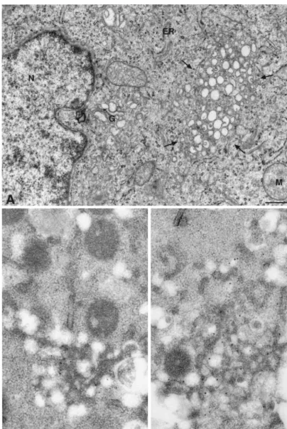
[image:5.603.74.510.69.293.2]tural equivalent of the dot-like structures harboring the NS5A-GFP fusion protein, other HCV nonstructural proteins, and replicating viral RNA, i.e., viral replication complexes. As shown in Fig. 7A, typical membranous webs, composed of small vesicles embedded in a membrane matrix, were found in cells harboring GFP replicon constructs. This specific mem-brane alteration was very similar to the membranous web pre-viously identified in U-2 OS human osteosarcoma-derived cell
lines inducibly expressing the HCV polyprotein (13) and in Huh-7 cells harboring unmodified HCV replicons (16). By double-labeling IEM with primary antibodies against GFP and NS5A or NS3 and secondary antibodies conjugated with 10- or 15-nm gold, the NS5A-GFP fusion protein and NS3 were found to strongly accumulate on membranous webs. Taken together, and in perfect agreement with a previous report (16), these results demonstrate that membranous webs are formed in Huh-7.5 cells harboring GFP replicons and represent the site of HCV RNA replication in these cells.
DISCUSSION
HCV replicons have allowed, for the first time, studies on efficient and genuine HCV RNA replication in vitro (5, 21, 30, 40; reviewed in reference 3). HCV nonstructural proteins 3 to 5B form a complex required for autonomous HCV RNA rep-lication. Since the original reports of functional genotype 1b replicons, replicons for genotype 1a (6, 18) and 2a (23), as well as derivatives expressing or activating the expression of easily quantifiable marker enzymes (luciferase,-lactamase, and se-creted alkaline phosphatase) in a separate cistron have been made to facilitate genetic studies, as well as drug screening and evaluation (24, 36, 47). Replicon-containing cells have also provided a source of membrane fractions containing crude replication complexes for biochemical studies (2, 19, 25).
In the present study, we describe replicons that allow direct visualization of functional HCV replication complexes in living cells. Central to this effort was the identification of sites in an essential replicase component permissive for insertion of GFP. We used Tn7-mediated in vitro transposition to create a pool of random insertions within the NS5A coding region (4). Only replicons with insertions that did not significantly disrupt the function of the viral protein could replicate and were selected. Two permissive sites in the C-terminal domain of NS5A were identified by this approach and were used to insert the GFP coding sequence. These constructs were only moderately im-paired for HCV RNA replication, revealing an unexpected flexibility of NS5A and of the HCV RNA replication machin-ery. Indeed, the length of NS5A was increased by 58% from 447 to 708 aa by the insertion (Fig. 1A). One possible expla-nation for why GFP was tolerated as an internal insertion is that the N and C termini are close to each other in the three-dimensional structure of GFP (38) and are, therefore, less likely to displace the surrounding domains of NS5A. Insertions of alternative heterologous sequences, including selectable markers and other fluorescent reporters, into the permissive sites identified here are currently being studied.
NS5A was chosen as a region for insertional mutagenesis because adaptive mutations (reviewed in reference 3), includ-ing a 47-aa deletion (5) and a 4-aa insertion (21), suggested a certain flexibility of this viral nonstructural protein. All FLAG epitope insertions identified in the initial random insertion screen mapped to one of two locations at the C-terminal do-main of NS5A. The more N-terminal, less frequently identified insertion site at amino acid position 2356 falls directly inside a cluster of serine residues. Interestingly, the C-terminal, more frequently identified insertion site at aa 2390 coincides with a 43-aa deletion (aa 2371 to 2413) recently identified as an adap-tive change in HeLa cells harboring a subgenomic HCV
rep-FIG. 4. NS5A-GFP can be directly visualized by fluorescence mi-croscopy. Huh-7.5 cells harboring the constructs GIT/5A-GFP-6 (A) and I/5A-GFP-6 (B) were analyzed by fluorescence microscopy at 5 days postseeding. (C) Cells harboring the parental S2204I replicon without GFP insertion served as the negative control.
VOL. 78, 2004 VISUALIZATION OF FUNCTIONAL HCV REPLICASES 7405
on November 8, 2019 by guest
http://jvi.asm.org/
[image:6.603.54.273.73.562.2]licon (48). Other reports have shown this region of the polyprotein to be somewhat malleable and perhaps dispens-able for replicon function (N. Appel and R. Bartenschlager, personal communication; T. Tellinghuisen and C. M. Rice, unpublished data). In addition, structure predictions indicate that whereas the⬃180 aa immediately following the N-termi-nal membrane-anchoring␣-helix of NS5A (8, 14) are likely to form a stable subdomain, the C-terminal half of NS5A has characteristics more typical of unfolded proteins that undergo “induced folding” by binding to their natural ligands (39).
Transposon insertion screens have been useful, among
[image:7.603.53.535.72.322.2]oth-ers, to identify genes important for herpesvirus (9) and cyto-megalovirus (45) propagation, as well as to map domains of the human immunodeficiency virus type 1 (HIV-1) genome (26), the Moloney murine leukemia virus envelope protein (42), and Gag protein (2a) required for production of infectious virus. Although the goal of our study was to create at least one functional replicon clone expressing a GFP-tagged version of NS5A, rather than a complete genetic footprint of NS5A, the random insertion approach appears to have been important in our success. Based on the complexity of the library and accord-ing to the least efficient step in the library clonaccord-ing (clonaccord-ing of
FIG. 5. NS5A-GFP colocalizes with other HCV nonstructural proteins. Huh-7.5 cells harboring replicon construct I/NS5A-GFP-6 were stained with MAb 11H against NS5A (A) or MAb 1B6 against NS3 (B), followed by CLSM as described in Materials and Methods. Nuclei were counterstained with TO-PRO-3 iodide.
FIG. 6. NS5A-GFP colocalizes with nascent HCV RNA. I/5A-GFP-1 replicon cells were metabolically labeled with BrUTP in the presence of actinomycin D, followed by CLSM. (A) GFP fluorescence. (B) Detection of newly synthesized, BrU-labeled viral RNA with a MAb against BrdU and a Cy3-conjugated secondary antibody. (C) The overlay demonstrates colocalization (yellow) of the NS5A-GFP fusion protein and nascent viral RNA. The border and the nucleus of a single cell are highlighted by thick and thin lines, respectively. Negative controls were identically treated naive Huh-7 cells. Comparable results were obtained in cells harboring replicons with the GIT adaptive background.
on November 8, 2019 by guest
http://jvi.asm.org/
[image:7.603.65.522.527.675.2]FIG. 7. Ultrastructure of GFP replicon cells and immunogold detection of GFP and HCV nonstructural proteins on the membranous web. (A) Huh-7.5 cells harboring replicon construct I/5A-GFP-1 were analyzed by EM at 5 days postseeding. A membranous web is marked by the arrows. Bar, 500 nm. ER, endoplasmic reticulum; G, Golgi apparatus; M, mitochondria; N, nucleus. The same cells were analyzed by double-label IEM with MAb JL-8 against GFP and the NS5A-specific polyclonal antiserum WU144, followed by 10- and 15-nm gold-conjugated secondary antibodies, respectively (B), or an anti-GFP polyclonal antiserum and MAb 1B6 against NS3, followed by 15- and 10-nm gold-conjugated secondary antibodies, respectively (C). GFP and HCV nonstructural proteins are found almost exclusively on the membranous web. Bars, 200 nm.
VOL. 78, 2004 VISUALIZATION OF FUNCTIONAL HCV REPLICASES 7407
on November 8, 2019 by guest
http://jvi.asm.org/
the amp-FLAG transprimer) (15), there should have been about 25 different in-frame FLAG fusions in the replicon li-brary. Thus, if replicons with only two different insertions were viable, then fewer than one of ten sites was compatible with RNA replication. Even the two insertion sites identified in our study differed by a factor of 10 or 100, depending on the adaptive background, in terms of efficiency of replication ini-tiation. In this context, we have also explored directed inser-tion at the C terminus of NS5B. It was shown previously that the addition of 19 heterologous amino acids to the C terminus of NS5B could yield, albeit at very low efficiency, viable repli-cons (22). However, GFP insertions in this position were not tolerated regardless of whether a stop codon was introduced between NS5B and the GFP (D. Moradpour, B. D. Linden-bach, and C. M. Rice, unpublished data). Thus, apart from disrupting functional protein domains such insertions may in-terfere with critical RNA elements in the HCV genome.
The replicons described here represent an attractive system for live cell imaging studies (28). Such studies have now suc-cessfully been initiated and should allow us to gain a dynamic view of the formation and turnover of HCV replication com-plexes in living cells. Moreover, the replicons described in the present study may be useful for the rapid identification of cell types or particular cells in a given population that are permis-sive for high levels of HCV RNA replication.
ACKNOWLEDGMENTS
We thank Alison North for assistance with CLSM; Elke Bieck and Anja Wahl for expert technical assistance; Denise Egger, Kurt Bienz, Benjamin Bu¨chele, and Benno Wo¨lk for helpful discussions; Brian Seed for the human codon optimized GFP sequence; Michinori Ko-hara for MAb 4b-52; and Jan Albert Hellings for MAb 11H.
D.M. gratefully acknowledges support from the Roche Research Foundation for sabbatical work at The Rockefeller University. This study was supported by Public Health Service grant CA57973, the Greenberg Medical Research Institute, and grant Mo 799/1-3 from the Deutsche Forschungsgemeinschaft.
REFERENCES
1. Ahlquist, P., A. O. Noueiry, W. M. Lee, D. B. Kushner, and B. T. Dye.2003. Host factors in positive-strand RNA virus genome replication. J. Virol.
77:8181–8186.
2. Ali, N., K. D. Tardif, and A. Siddiqui.2002. Cell-free replication of the
hepatitis C virus subgenomic replicon. J. Virol.76:12001–12007.
2a.Auerbach, M. R., C. Shu, A. Kaplan, and I. R. Singh.2003. Functional charaterization of a portion of the Moloney murine leukemia virus gag gene
by genetic footprinting. Proc. Natl. Acad. Sci. USA100:11678–11683.
3. Bartenschlager, R.2002. Hepatitis C virus replicons: potential for drug
development. Nat. Rev. Drug Discov.1:911–916.
4. Biery, M. C., F. J. Stewart, A. E. Stellwagen, E. A. Raleigh, and N. L. Craig.
2000. A simple in vitro Tn7-based transposition system with low target site
selectivity for genome and gene analysis. Nucleic Acids Res.28:1067–1077.
5. Blight, K. J., A. A. Kolykhalov, and C. M. Rice.2000. Efficient initiation of
HCV RNA replication in cell culture. Science290:1972–1974.
6. Blight, K. J., J. A. McKeating, J. Marcotrigiano, and C. M. Rice.2003. Efficient replication of hepatitis C virus genotype 1a RNAs in cell culture.
J. Virol.77:3181–3190.
7. Blight, K. J., J. A. McKeating, and C. M. Rice.2003. Highly permissive cell lines for subgenomic and genomic hepatitis C virus RNA replication. J.
Vi-rol.76:13001–13014.
8. Brass, V., E. Bieck, R. Montserret, B. Wo¨lk, J. A. Hellings, H. E. Blum, F. Penin, and D. Moradpour.2002. An aminoterminal amphipathic alpha-helix mediates membrane association of the hepatitis C virus nonstructural
pro-tein 5A. J. Biol. Chem.277:8130–8139.
9. Brune, W., C. Menard, U. Hobom, S. Odenbreit, M. Messerle, and U. H. Koszinowski.1999. Rapid identification of essential and nonessential
her-pesvirus genes by direct transposon mutagenesis. Nat. Biotechnol.17:360–
364.
10. Dimitrova, M., I. Imbert, M. P. Kieny, and C. Schuster.2003. Protein-protein interactions between hepatitis C virus nonstructural Protein-proteins. J.
Vi-rol.77:5401–5414.
11. Dubuisson, J., F. Penin, and D. Moradpour.2002. Interaction of hepatitis C
virus proteins with host cell membranes and lipids. Trends Cell Biol.12:517–
523.
12. Egger, D., R. Gosert, and K. Bienz.2002. Role of cellular structures in viral
RNA replication, p. 247–253.InB. Semler and E. Wimmer (ed.), Molecular
biology of picornaviruses. ASM Press, Washington, D.C.
13. Egger, D., B. Wo¨lk, R. Gosert, L. Bianchi, H. E. Blum, D. Moradpour, and K. Bienz.2002. Expression of hepatitis C virus proteins induces distinct membrane alterations including a candidate viral replication complex. J.
Vi-rol.76:5974–5984.
14. Elazar, M., K. H. Cheong, P. Liu, H. B. Greenberg, C. M. Rice, and J. S. Glenn.2003. Amphipathic helix-dependent localization of NS5A mediates
hepatitis C virus RNA replication. J. Virol.77:6055–6061.
15. Evans, M. J.2003. Mechanisms and regulation of HCV RNA replication. Ph.D. thesis. Columbia University, New York, N.Y.
16. Gosert, R., D. Egger, V. Lohmann, R. Bartenschlager, H. E. Blum, K. Bienz, and D. Moradpour.2003. Identification of the hepatitis C virus RNA repli-cation complex in Huh-7 cells harboring subgenomic replicons. J. Virol.
77:5487–5492.
17. Gosert, R., A. Kanjanahaluethai, D. Egger, K. Bienz, and S. C. Baker.2002. RNA replication of mouse hepatitis virus takes place at double-membrane
vesicles. J. Virol.76:3697–3708.
18. Gu, B., A. T. Gates, O. Isken, S. E. Behrens, and R. T. Sarisky.2003. Replication studies using genotype 1a subgenomic hepatitis C virus
repli-cons. J. Virol.77:5352–5359.
19. Hardy, R. W., J. Marcotrigiano, K. J. Blight, J. E. Majors, and C. M. Rice.
2003. Hepatitis C virus RNA synthesis in a cell-free system isolated from
replicon-containing hepatoma cells. J. Virol.77:2029–2037.
20. Hu¨gle, T., F. Fehrmann, E. Bieck, M. Kohara, H.-G. Kra¨usslich, C. M. Rice, H. E. Blum, and D. Moradpour.2001. The hepatitis C virus nonstructural protein 4B is an integral endoplasmic reticulum membrane protein. Virology
284:70–81.
21. Ikeda, M., M. Yi, K. Li, and S. M. Lemon.2002. Selectable subgenomic and genome-length dicistronic RNAs derived from an infectious molecular clone of the HCV-N strain of hepatitis C virus replicate efficiently in cultured
Huh7 cells. J. Virol.76:2997–3006.
22. Ivashkina, N., B. Wo¨lk, V. Lohmann, R. Bartenschlager, H. E. Blum, F. Penin, and D. Moradpour.2002. The hepatitis C virus RNA-dependent RNA polymerase membrane insertion sequence is a transmembrane
seg-ment. J. Virol.76:13088–13093.
23. Kato, T., T. Date, M. Miyamoto, A. Furusaka, K. Tokushige, M. Mizokami, and T. Wakita.2003. Efficient replication of the genotype 2a hepatitis C virus
subgenomic replicon. Gastroenterology125:1808–1817.
24. Krieger, N., V. Lohmann, and R. Bartenschlager.2001. Enhancement of hepatitis C virus RNA replication by cell culture-adaptive mutations. J.
Vi-rol.75:4614–4624.
25. Lai, V. C. H., S. Dempsey, J. Y. N. Lau, Z. Hong, and W. Zhong.2003. In vitro RNA replication directed by replicase complexes isolated from the
sub-genomic replicon cells of hepatitis C virus. J. Virol.77:2295–2300.
26. Laurent, L. C., M. N. Olsen, R. A. Crowley, H. Savilahti, and P. O. Brown.
2000. Functional characterization of the human immunodeficiency virus type
1 genome by genetic footprinting. J. Virol.74:2760–2769.
27. Lindenbach, B. D., and C. M. Rice.2001.Flaviviridae: the viruses and their
replication, p. 991–1041.InD. M. Knipe and P. M. Howley (ed.), Fields
virology, 4th ed. Lippincott/The Williams & Wilkins Co., Philadelphia, Pa. 28. Lippincott-Schwartz, J., and G. H. Patterson.2003. Development and use of
fluorescent protein markers in living cells. Science300:87–91.
29. Lohmann, V., S. Hoffmann, U. Herian, F. Penin, and R. Bartenschlager.
2003. Viral and cellular determinants of hepatitis C virus RNA replication in
cell culture. J. Virol.77:3007–3019.
30. Lohmann, V., F. Ko¨rner, J.-O. Koch, U. Herian, L. Theilmann, and R. Bartenschlager.1999. Replication of subgenomic hepatitis C virus RNAs in
a hepatoma cell line. Science285:110–113.
31. Miyanari, Y., M. Hijikata, M. Yamaji, M. Hosaka, H. Takahashi, and K. Shimotohno.2003. Hepatitis C virus nonstructural proteins in the probable membranous compartment function in viral RNA replication. J. Biol. Chem.
278:50301–50308.
32. Moradpour, D., E. Bieck, T. Hu¨gle, W. Wels, J. Z. Wu, Z. Hong, H. E. Blum, and R. Bartenschlager.2002. Functional properties of a monoclonal anti-body inhibiting the hepatitis C virus RNA-dependent RNA polymerase.
J. Biol. Chem.277:593–601.
33. Moradpour, D., V. Brass, R. Gosert, B. Wo¨lk, and H. E. Blum.2002.
Hep-atitis C: molecular virology and antiviral targets. Trends Mol. Med.8:476–
482.
34. Moradpour, D., R. Gosert, D. Egger, F. Penin, H. E. Blum, and K. Bienz.
2003. Membrane association of hepatitis C virus nonstructural proteins and identification of the membrane alteration that harbors the viral replication
complex. Antivir. Res.60:103–109.
35. Moradpour, D., P. Kary, C. M. Rice, and H. E. Blum.1998. Continuous human cell lines inducibly expressing hepatitis C virus structural and
non-structural proteins. Hepatology28:192–201.
36. Murray, E. M., J. A. Grobler, E. J. Markel, M. F. Pagnoni, G. Paonessa, A. J.
on November 8, 2019 by guest
http://jvi.asm.org/
Simon, and O. A. Flores.2003. Persistent replication of hepatitis C virus
replicons expressing the-lactamase reporter in subpopulations of highly
permissive Huh7 cells. J. Virol.77:2928–2935.
37. National Institutes of Health.2002. National Institutes of Health Consensus Development Conference Statement: management of hepatitis C: 2002.
Hepatology36(Suppl. 1):S2–S20.
38. Ormo¨, M., A. B. Cubitt, K. Kallio, L. A. Gross, R. Y. Tsien, and S. J. Remington.1996. Crystal structure of theAequorea victoriagreen fluorescent
protein. Science273:1392–1395.
39. Penin, F., J. Dubuisson, F. A. Rey, D. Moradpour, and J. M. Pawlotsky.2004.
Structural biology of hepatitis C virus. Hepatology39:5–19.
40. Pietschmann, T., V. Lohmann, A. Kaul, N. Krieger, G. Rinck, G. Rutter, D. Strand, and R. Bartenschlager.2002. Persistent and transient replication of
full-length hepatitis C virus genomes in cell culture. J. Virol.76:4008–4021.
41. Pietschmann, T., V. Lohmann, G. Rutter, K. Kurpanek, and R. Barten-schlager.2001. Characterization of cell lines carrying self-replicating
hepa-titis C virus RNAs. J. Virol.75:1252–1264.
42. Rothenberg, S. M., M. N. Olsen, L. C. Laurent, R. A. Crowley, and P. O. Brown.2001. Comprehensive mutational analysis of the Moloney murine
leukemia virus envelope protein. J. Virol.75:11851–11862.
43. Schmidt-Mende, J., E. Bieck, T. Hu¨gle, F. Penin, C. M. Rice, H. E. Blum, and D. Moradpour.2001. Determinants for membrane association of the
hepa-titis C virus RNA-dependent RNA polymerase. J. Biol. Chem.276:44052–
44063.
44. Shi, S. T., K. J. Lee, H. Aizaki, S. B. Hwang, and M. M. Lai.2003. Hepatitis C virus RNA replication occurs on a detergent-resistant membrane that
cofractionates with caveolin-2. J. Virol.77:4160–4168.
45. Smith, G. A., and L. W. Enquist.1999. Construction and transposon
mu-tagenesis inEscherichia coliof a full-length infectious clone of pseudorabies
virus, an alphaherpesvirus. J. Virol.73:6405–6414.
46. Wo¨lk, B., D. Sansonno, H.-G. Kra¨usslich, F. Dammacco, C. M. Rice, H. E. Blum, and D. Moradpour.2000. Subcellular localization, stability andtrans -cleavage competence of the hepatitis C virus NS3-NS4A complex expressed
in tetracycline-regulated cell lines. J. Virol.74:2293–2304.
47. Yi, M., F. Bodola, and S. M. Lemon.2002. Subgenomic hepatitis C virus replicons inducing expression of a secreted enzymatic reporter protein.
Vi-rology304:197–210.
48. Zhu, Q., J. T. Guo, and C. Seeger.2003. Replication of hepatitis C virus subgenomes in nonhepatic epithelial and mouse hepatoma cells. J. Virol.
77:9204–9210.
VOL. 78, 2004 VISUALIZATION OF FUNCTIONAL HCV REPLICASES 7409