A CLINICAL STUDY OF NON VENEREAL GENITAL DERMATOSES
Dissertation submitted to THE TAMILNADU
DR. M.G.R. MEDICAL UNIVERSITY CHENNAI – 600 032
APRIL - 2015.
In partial fulfillment of the regulations required for the award of M.D. DEGREE
IN
DERMATOLOGY, VENEREOLOGY AND LEPROLOGY BRANCH XII
DEPARTMENT OF DERMATOLOGY
DECLARATION
I Dr.A.N.M.Maalik babu solemnly declare that the dissertation entitled “A clinical study of non venereal genital dermatoses” was done by me in the Department of Dermatology and Venereology at Coimbatore Medical College Hospital during the period from August 2013 to July 2014 under the guidance & supervision of Dr.P.P.Ramasamy M.D.,D.D., Professor & Head of Department, Department of Dermatology and Venereology ,Coimbatore Medical College Hospital ,Coimbatore. The dissertation is submitted to Tamil nadu Dr.MGR Medical University,Chennai towards the partial fulfillment of the requirement for the award of M.D., degree in Dermatology, Venereology and Leprology. I have not submitted this dissertion on any previous occasion to any university for the award of any degree.
PLACE: Dr. A.N.M.Maalik babu
CERTIFICATE
This is to certify that the dissertation entitled “A clinical study of non venereal genital dermatoses ” is a record of bonafide work done by Dr.A.N.M.Maalik babu , Post graduate student in the Department of Dermatology , Venereology and Leprology , Coimbatore Medical College Hospital,Coimbatore under the guidance of Dr.P.P.Ramasamy M.D.,D.D., Professor & Head of Department, Department of Dermatology, Coimbatore Medical College Hospital ,Coimbatore in partial fulfillment of the regulations of the Tamilnadu DR.M.G.R Medical University, Chennai towards the award of M.D., degree (Branch XII) in Dermatology, Venereology and Leprology.
GUIDE,
Dr.P.P.Ramasamy M.D., D.D., Professor
Department of Dermatology,
Coimbatore Medical College & Hospital,
Dr.P.P. Ramasamy M.D., D.D.,
Professor & Head of the Department , Department of Dermatology,
Coimbatore Medical College & Hospital,
Dr.S.Revwathy MD.DGO.,DNB Dean,
Coimbatore Medical College & Hospital Coimbatore
Coimbatore Date:
Date:
ACKNOWLEDGEMENT
First of all, I Thank the Almighty for what I am today. I wish to thank the Dean of this institution, Dr.S.Revwathy MD.DGO.,DNB, for permitting me to undertake this study. I would like to thank my guide , Dr.P.P.Ramasamy M.D.,D.D., Professor and Head of Department of Dermatology, venereology and leprology from the bottom of my heart for his scholary advice and timely assistance during the preparation of the thesis.
I would like to express my gratitude to our Prof. Dr. K. Mahadevan, M.D, D.V., Department of STD for his support and guidance .
My special thanks to Dr.M.Revathy M.D., Assistant professor of Department of Dermatology for her constant encouragement and valuable suggestions during the period of this study.
I owe great debt of gratitude to Dr. R.Madhavan M.D.and Dr. S.Bharathi M.D., Assistant Professors in Department of Dermatology for their kind support and encouragement. I sincerely thank Dr. B.Eswaramoorthy M.D., and Dr.A.P.Balaji Assistant Professors in Department of Dermatology and Venereology for their valuable guidance and help.
grateful to all patients for their co-operation and participation in the study.
ABBREVIATIONS
LSA - Lichen simplex et atrophicans
GBHC - Gamma benzene hexachloride
ACD - Allergic contact dermatitis
LP - Lichen planus
KOH - Pottasium hydroxide
FDE - Fixed drug eruption
HSV1&2 - Herpes simplex virus 1 and 2
HPV - Human papilloma virus
HIV - Human immuno virus
PUVA - Psoralan ultraviolet A
UVB - Ultraviolet B
VIN - Vulval intra epithelial neoplasia
SCC - Squamous cell carcinoma
EMPD - Extra mammary paget’s disease
VDRL - Venereal disease research laboratory
BSF Jawans - Border security force Jawans
BMI - Body mass index
SJS - Stevens Johnson syndrome
LSC - Lichen simplex chronicus
NVGD - Non venereal genital dermatoses
BXO - Balanitis xerotica obliterans
VVC - Vulvo vaginal candidosis
MF - Mycosis fungoides
IHC - Immuno histo chemistry
HPE - Histopathological examination
H&E - Haematoxylin and Eosin
CONTENTS
S.NO. CONTENTS PAGE NO.
1 INTRODUCTION 1
2 AIM AND OBJECTIVE 2
3 REVIEW OF LITERATURE 3
4 MATERIALS AND METHODS 67
5 OBSERVATION AND RESULTS 69
6 DISCUSSION 97
7 SUMMARY 115
8 CONCLUSION 119
9 BIBLIOGRAPHY
10 APPENDICES
LIST OF TABLES :
Table No: Name of the table
Page No:
1 Age and Sex distribution of cases 70
2 Occupational status of the patients 72
3 List of Genital dermatoses observed 74
4 List Of Genital Dermatoses Found Exclusively In Men 76 5 List of genital dermatoses found in both sexes 77 6 List of Benign conditions and normal variants 79
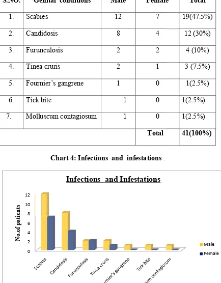
7 List of Infections and infestations 81
8 List of Inflammatory conditions 82
9 List of Miscellaneous conditions 84
10 Classification according to the site of involvement 84 11 List of dermatoses involved genitalia alone 86 12 List of Dermatoses involved both genitalia and skin 87 13 List of Dermatoses involved genitalia, oral mucosa and skin 88 14 Categorization according to the site of involvement in genitalia 89
15 List of associated skin disorders 90
LIST OF CHARTS :
Chart No: Name of the chart Page No:
1 Age and Sex distribution of cases 71 2 Occupational status of the patients 73 3 Benign conditions and normal variants 80
4 Infections and infestations 81
5 Inflammatory conditions 83
6 Categorization according to the site of involvement
85
LIST OF COLOR PLATES :
[image:14.595.101.399.195.428.2] [image:14.595.101.366.510.743.2]I.Benign conditions and normal variants
FIGURE NO: Genital Dermatoses 1 Pearly penile papule
2 Angiokeratoma of Fordyce 3 Phimosis
4 Para phimosis 5 Vulval skin tag 6 Bartholin’s cyst
II.Infections and infestations:
FIGURE NO: Genital Dermatoses
1 Nodular scabies
2 Tinea cruris
III.Inflammatory conditions:
FIGURE NO: Genital Dermatoses 1 Bullous FDE
2 Lichen planus
3a Zoon’s balanitis (clinical) 3b Zoon’s balanitis
(Histopathology) 4 Lichen nitidus 5 Pemphigus vulgaris 6 Psoriasis
7a EMF (Genital) 7b EMF(oral)
IV.Miscellaneous conditions:
FIGURE NO: Genital Dermatoses 1 Vitiligo
V.Interesting cases:
FIGURE NO: Genital Dermatoses
1 Tick bite
2 Lymphangiectases
3a Verrucous carcinoma(clinical) 3b Verrucous carcinoma(Histopathology) 4a Erythroderma (Mycosis fungoides)
4b MF( Histopathology)
[image:16.595.101.435.154.416.2]ABSTRACT
Background :
Most dermatological diseases generally occur elsewhere and also involve the genitalia.When other sites are involved the diagnosis is straightforward.If the lesion is present exclusively on genitalia,it is a difficult task for the treating doctor to differentiate nonvenereal from venereal genital lesions.
Aim and Objective:
To study the clinical pattern and prevalence of non venereal genital dermatoses
Materials and Methods:
Results:
A total of 150 cases (114 males,36 females) with non venereal genital dermatoses were encountered in our study.Prevalence of the non venerealgenital dermatoses in the study was 2.6 per 1000 cases.Male to Female ratio of patients in our study was 3.16 : 1.Majority of the patients were found in the age group of 33 to 42 years [42 (28%)].Commonest NVGD was found to be scabies occuring in 19 (12.6%) patients.pearly penile papule was found to be more common [ 10 (6.6%)] among benign conditions and normal variants.Among inflammatory conditions and miscellaneous groups, contact dermatitis and vitiligo were the commonest conditions respectively.One case of verrucous carcinoma of penis was seen.
Among four categories classified according to the site of involvement, Genitalia alone was found to be involved in more number of patients [87(58%)] Scrotum and Labium majora were the most common site of involvement in male and female genitalia respectively.
Conclusion:
treat with appropriate drugs.Scabies was the most common nonvenereal genital dermatosis in our study. Knowing about the prevalence , clinical and etiological characteristics of various nonvenereal genital dermatoses are helpful in arriving at a diagnosis and also creating awareness among patients to improve their personal hygiene and social habits.
INTRODUCTION
Non venereal genital dermatoses are often confused with venereal diseases which may be a cause for concern to the patients and also serve as a diagnostic dilemma to the physician.
Contrary to popular belief , all lesions occurring over the genitalia are not the manifestations of sexually transmitted disease.
These non –venereal disorders cause mental distress and guilt feeling in patients who believe that they have developed sexually transmitted disease .
Since non –venereal genital diseases include a wide variety of disorders , the identification & establishment of the nature of disease is a challenging venture.
AIM AND OBJECTIVE :
REVIEW OF LITERATURE
CLASSIFICATION :
Based on etiopathogenesis , Fitzpatrick and Gentry 3 classified Non venereal genital dermatoses in to the following categories:
A. Benign conditions and normal variants B. Congenital anomalies
C. Infections and infestations D. Inflammatory conditions E. Premalignant conditions F. Malignant conditions G. Miscellaneous lesions
Non venereal dermatoses of male genitalia
I. Benign conditions and Normal variants :
They are single or multiple asymptomatic lesions with varying sizes and morphology involving external genitalia of both sexes.
1. pearly penile papule 2. Fordyce spots
3. foreskin abnormalities 4. Angiokeratoma of Fordyce 5. Melanocytic nevi
7. Epitheloid hemangioma 8. Lymphangiectases
9. trauma induced lesions and artifact
1. Pearly Penile Papules:
They are 1-2mm , skin coloured or shiny papules arranged in a row or more than a row around coronal sulcus or proximal to it or spotted over the glans penis. It may be found in upto 50% of men 4,5 . Pearly penile papules are structural variants of angiofibroma. Differential diagnosis of this condition is condyloma accuminata. Histologically it resembles adenoma sebaceum,subungual fibroma and acquired acral angiofibroma.These papules are commonly seen over the genitalia of adolescent boys . Reassurance and counsel regarding the physiological nature of the condition is very much needed.
2. Fore skin abnormalities :
At birth usually foreskin adher with the underlying glans penis. Four percentage of boys will be able to retract the foreskin at
foreskin may vary from person to person and the foreskin can be shorter or longer .
Circumcision might be reflecting the racial, religious and cultural differences in any population . In world wide ,around 25% men have been circumcised7. During Infantile period circumcision seems to be unsafe and the situation will be reverse in later age groups. It protects the men from carcinoma penis, sexually transmitted infection including HIV and other inflammatory dermatoses like psoriasis, lichen sclerosis, seborrheic dermatitis, lichen planus. According to O’farrell et al wetness of penis is hardly seen in circumcised men, but it is the main cause for balanitis in 40% cases who have uncircumcised8
i . Phimosis (muzzling)
of phimosis is circumcision and subsequent treatment of underlying cause.
ii . Paraphimosis:
Inability to pull back the retracted prepuce over glans penis due to edema over prepuce is called paraphimosis . It is otherwise known as waisting or constrictive posthitis. Paraphimosis is mainly
due to trauma or improper handling of foreskin. But the other causes
like acute contact urticaria are also identified10 . Rickwood said that paraphimosis is mainly due to abuse and not the disease of foreskin. Treatment is mainly by puncturing the prepuce with hypodermic needle, manual pressure and ice packs over the swollen prepuce. For severe cases emergency dorsal slit should be performed .
3. Angiokeratoma of Fordyce:
are cryotherapy , ablation with electrocautery, CO2 laser and pulsed dye laser.
4. Fordyce spots :
They are ectopic sebaceous glands .They have present as multiple , discrete , asymptomatic , tiny yellowish papules over sub-preputial area of penis . we have to counsel the patients regarding the harmless nature of the condition.
5. Melanocytic naevi :
It is commonly seen over penis and more frequently in patients with atypical nevus syndrome. Multiple blue naevi, spitz naevi over glans penis have been described13,14.Epitheloid blue nevus is very rarely seen over genitlia. Half of the nevi located over glans penis and penile shaft with normal skin in between is called kissing or divided nevus.
6. Lymphangiectases
It may occur due to building up of lymph in superficial vessels which is usually a sequel of surgery or radiotherapy .
this condition. Topical antibacterial application is needed to prevent secondary bacterial infection. Excision, cryotherapy and laser therapy are the useful treatment options for these patients.
7. Genital Skin tag : (Men)
They are small flaps of skin which are attached to the skin surface through a stalk of flesh. Penile shaft,glans penis and scrotum are the common sites of genital skin tags in men. Usually it is seen in obese individuals .The incidence of genital skin tags is less in Indian population while comparing western population. They may easily become irritated by friction of clothing or rubbing and also infected frequently .
8. Epitheloid hemangioma :
9. Trauma induced lesion and artifact
i. Penile hematoma and fracture
Penile hematoma and fracture are rare even though penis is very vascular17,18.These patients usually present with pain, swelling , deformity of penis. It may lead to complications like urethral damage and urinary retention. The prognosis is usually good but Peyronie’s disease may develop over the lesion later .Injection of
drugs to improve erection may also leads to complication like hematoma.
ii. Sclerosing lymphangitis
It is otherwise known as Mondor’s phlebitis/ penile lymphocele/ penile lymphoedema. The Classical presentation is serpiginous mass over coronal sulcus which usually due to prolonged and frequent sexual intercourse . The main predisposing factor is the scar resulting from circumcision. Usually these condition resolve spontaneously but sometime surgical excision may be needed 19.
iii. Strangulation of the penis
rings (washers) and nuts placed over the penis by the patient for prolonging the duration of erection20,21 . Finally it may lead to pain, swelling, urethral fistula, pseudo ainhum, gangrene and amputation that were collectively called as Penile strangulation
Tourniquet syndrome. Foreign bodies like glass beads , small and smooth round stones are introduced under the skin of penis to increase erotic sensation that can lead to complication like abscess, fistulae and calculi. Oil or petroleum jelly introduced in to genital skin for enhancing sexual pleasure leads to paraffinoma.
iv. Dermatitis artefacta and mutilation
v. Lipogranuloma
Injection of mineral oil, petroleum jelly and silicone in to the penile skin for maintaining erection or enlarging penis may lead to paraffinoma or lipogranuloma. Usually these patients present with
deformity of penis and painful erections and inability to penetrate during intercourse 24.
II.Congenital abnormalities :
Defect in embryogenesis and sexual differentiation of genital organs resulting in the following conditions:
exstrophy-epispadias complex27. Hypospadias is the most common congenital abnormality of male external genitalia other than cryptorchidism .In this condition urethral opening seen over ventral surface of penis or scrotum The incidence is 3-5/1000 live male births. It may be due to failure of fusion of urethral folds. The reason could be mutation of MAMLD1 (CXorf6) gene. If the length of the stretched penis is less than 2.5 standard deviations for that age group is called Micropenis . Median raphe cysts is mainly due to anomalies in the development of urethral groove or by trapped epithelial cells in between the genital folds. Patient may present with cystic or nodular swellings over ventral penis. These lesions usually become traumatized or infected with staphylococci.
III. Infections and Infestations: 1. Scabies :
and the cure rate is 70%.If two doses at the interval of two weeks is taken by the patients ,cure rate will be increased to 95%.
2.Candidosis:
3.Staphylococcal infection :
Furuculosis (boil) is one of the commonest conditions due to Staphylococcus aureus in which extensive involvement of hair follicle (including follicular and peri follicular region) is seen .It may appear as a single or multiple lesions over mons pubis,root of the penis and scrotum .
It begins as a tender papule which becomes a indurated nodule within 2-3 days. Later it becomes necrotic and discharge pus from a point at the
follicular opening. Some of them do not discharge pus on the surface are called blind boils. Factors which may be responsible for its severity and increased frequency are diabetes mellitus and intake of systemic steroids. Topical 2% sodium fusidate or 2% mupirocin over the affected area ( 3-4 times a day) is effective in the treatment of furunculosis. If the lesion is not resolved with topical medication, systemic drugs like cloxacillin or dicloxacillin 250mg-500mg (sixth hourly) to be given for 5 days Rifampicin 600mg (stat)with tab.ciprofloxacin for 10 days followed by cloxacillin for 1-2 months are effective treatment regimen for recurrent furunculosis 33.
4.Tinea cruris:
5.Fournier’s gangrene:
In 1883 , five cases of spontaneous genital gangrene and ulceration were described by the Parisian dermatologist Alfred Fournier, but Baurienne is the one who first reported this condition 37 . The disease usually starts with urethral or appendageal infection.The causative organisms are mainly the resident urethral or lower gastrointestinal flora . Necrotizing vasculitis is the most distressing consequences of Fournier’s gangrene which may involve skin,subcutis,fascia and muscle. Exotoxin is the key factor in the development of necrotizing vasculitis.Painful and erythematous swelling over genitals and perianal region associated with suppuration is the classical presentation of this condition. Death can be avoided if treatment in the form of surgery is initiated early.
6.Tick bite :
Ticks are mainly the parasites of animals. Humans are only
7. Molluscum contagiosum
It is caused by molluscipox virus .The typical lesion is a firm,dome shaped waxy or pearly white papule with central umbilication. If a young man presents with genital molluscum contagiosum ,it is usually assumed to be sexually transmitted ,but this may not be always true 39.
8.Trichomycosis pubis :
It is the corynebacterium infection of pubic hair shafts.Usually these patients present with asymptomatic yellow,red or black small dirty looking concretions on the hair shafts40. These concretions are bacterial colonies. sweat may be discoloured over the affected area. This condition could be treated with topical antimicrobial cream like benzoic acid. Poor personal hygiene may be the most important predisposing factor.Regular use of anhydrous aluminum chloride is recommended.
9. Pthirus pubis (Pubic louse)
Transmission of pubic louse occur not only by sexual route but also by other mode like sharing of bed and upholstery of public transport 41
macules over lower abdomen and thighs due to altered blood pigment or reaction to louse’s saliva.Gamma benzene hexachloride in the form of lotion or cream is an effective form of therapy. It should be applied to the whole body (below neck) for 12 hours and then washed off . Intake of ivermectin in the dosage of 200µg/kg is also quite effective.
10.Streptococcal dermatitis
These patients present with dysuria , erythema and swelling over penis or with features of balanoposthitis. A case of bullous necrotic erysipelas of genitalia has been reported which is mainly due to Streptococcus pyogenes 42 .
11. Ecthyma gangrenosum:
It is caused by Pseudomonas aure.and affects mainly the acral and anogenital region .Penile lesion usually leads to complication like gangrene and the prognosis of this condition is poor.
12.Chronic Penile edema
present with chronic swelling over penis, foreskin, scrotum, buttock and thighs .The lesions may be warm and red in colour.There may be intercurrent attacks of cellulitis which needs systemic broad spectrum antibiotics. After controlling with systemic antibiotics ,surgical intervention can be tried in the form of circumcision.
13.Tuberculosis:
It rarely affects the penis44. modes of transmission include sexual transmission , contact with the infected clothing and it can occur secondary to tuberculosis elsewhere . Painless papules over glans penis which evolve into ulcers seen in patients with papulonecrotic tuberculid. These ulcers will heal with varioliform scars. Worm -eaten pattern of scars has also been described in papulonecrotic tuberculids45.Histopathology shows wedge shaped area of dermo epidermal necrosis surrounded by epithelioid cell granuloma.Lichen scrofulosorum is also rarely involved over genitalia. It responds well with standard antitubercular therapy.
14.Deep fungal infection:
Paracoccidiomycosis may be the cause of scrotal swelling and genital nodules and erosions in some cases 48.
15.Others:
In bacillary angiomatosis, the lesions are asymptomatic , single or multiple, violaceous or red firm non blanching vascular papules and nodules .It is the most important differential diagnosis of Kaposi’s sarcoma. Herpes zoster is also reported over scrotum and penis and the usual complications are constipation and urinary retention. Involvement of Hansen’s disease over male genitalia is rare , but some cases have been reported . 49 Amoebiasis can involve genitalia and these patients present with painful ulcers over the glans penis. Usually these patients present with urinary frequency, dysuria and retention 50. Buruli ulcer over penis and scrotum is mainly due to M. ulcerans which was rarely reported. Cutaneous leishmaniasis can also affect the genitalia 51.The
anogenital consequences of onchocerciasisare mainly due to involvement of the ileal crest and scrotum. Scrotal nodules (hanging groin) ,scrotal enlargement and leopard skin hypopigmentation are the classical lesions
IV.Inflammatory dermatoses: 1.Irritant contact dermatitis:
It is a local inflammatory reaction characterized by erythema,edema and erosion followed by the application of a substance some individuals exihibit increased sensitivity to some classes of irritants.Host factors influencing irritant contact dermatitis are age ,race,sex and pre existing dermatitis. Other contributing factors are friction ,maceration, over washing and anorectal or urological disease 54
. Birely has reported irritant contact dermatitis over glans in 72% patients and out of which 67% had a history of atopy 55.Common substances which lead to irritant contact dermatitis in genital area are soap,antiseptic solution and hair removal cream .Acute irritant contact dermatitis is treated by wet compresses followed by application of topical corticosteroids.In severely affected individuals ,systemic corticosteroids and antibiotics should be administered. In chronic irritant contact dermatitis ,regular application of emollients should be encouraged. Self treatment by the patient may increase the morbidity and also worsen the situation further.
There are acute and chronic phases in allergic contact dermatitis.In acute phase patient presents with itching, erythema, swelling, vesiculation over the genitalia,but in chronic cases lichenification might be seen over the involved area. Erythema and even angioedema due to contact urticaria by rubber constituents of condoms and gloves have been reported . Common contact sensitizers of genital ACD are rubber, contraceptives and medicaments. Avoidance of the allergens is the important measures in the management of Allergic contact dermatitis.Other treatment options are moisturizers, topical corticosteroids and oral antihistamine. If the lesions are severe and extensive ,systemic corticosteroids are the treatment of choice. Bauer et al have conducted patch test for all the patients with anogenital lesions and found that ACD is prevalent in 35% of patients 56
3. Balanoposthitis
organisms .Diabetes mellitus is an important predisposing factor for balanoposthitis 58.
4.lichen simplex chronicus:
5. Urticaria , Angioedema and dermographism:
Urticaria usually presents as generalized eruptions ,but in some patients it may present like unexplained genital itching. Stroking of the inner aspect of thigh will produce wheal along the line of trauma . Genital angioedema induced by lisinopril have also been reported 60.
6.Seborrheic dermatitis
It is an inflammatory disorder of the skin described by well defined erythematous plaque with greasy scales seen over scalp, face and flexural areas.In flexures,sweating and constant friction lead to fissures covered by crusts formed by serous exudates.Secondary infection by bacteria and candida are also quite common 61 .
7.Lichen planus:
It is the chronic inflammatory disease affecting skin ,nail ,oral and genital mucosa. Classical skin lesions are flat topped , pruritic, purple, polygonal, papules and plaques involving flexor aspect of wrist ,forearm and legs. The genital lesions particularly over glans penis are in an annular pattern .Plaque with central atrophy surrounded by a thin rim of active erythema is the classical lesion of annular lichen planus 62.Douglass and George reported that about 18% of total 114 patients with lichen planus had lesions over glans penis63 Erosive lichen planus over glans penis also reported64. Rarely squamous cell carcinoma may develop over chronic erosive lichen planus lesions.Potent topical corticosteroids and immunomodulators like topical tacrolimus are useful in the treatment of genital LP. Oral steroids has been used in severe cases. circumcision may be done in some chronic cases.
8.Lichen sclerosus et Atrophicus:
sclerotic constricting band connected distal to the prepuce in uncircumcised men which results in phimosis . On examination, lesions are atrophic white patches ,hemorrhagic blisters,erosion s and ulcerations over penis.The end stage is balanitis xerotica obliterans which is characterized by depigmented, contracted and fissured prepuce fixed over glans and not retractable by force.The etiopathogenesis of this condition is unknown but the association with HLADQ7 and spirochaete Borrelia have been reported in lichen sclerosus65. Histopathological picture shows hyperkeratosis ,follicular plugging epidermal atrophy,and hydrophic degeneration of basal layer.In dermis , edema and homogenization of collagen with lymphocytic infiltration. Campus et al have reported that lichen sclerosus constituted one third of all cases of carcinoma penis. squamous cell carcinoma is the most serious complication particularly with the involvement of glans penis.66 Treatment of choice is topical application of ultra potent corticosteroids. Topical Tacrolimus is an alternative for steroids.
9.Fixed drug eruption :
red ,swollen plaque with blisters or ulceration. Common sites of involvement are glans penis,oral mucosa ,hands and feet.Development of eruptive lesion may take 1-2 weeks after the first exposure of offending drug but the lesions will appear within few hours to days after subsequent exposure.Common causative drugs are sulfonamides, NSAIDs, tetracyclines, quinolones and phenytoin. The clinical pattern in FDE is specific for each drug. Treatment is mainly to stop the offending drug and to start oral or topical Corticosteroids.
10.Erythema multiforme:
It is an acute , relapsing skin disease characterized by symmetrical papular,urticarial and multiple target lesions involving limbs,palms and soles associated with oral and genital erosins.Etiolgical factors are HSV1 &2 infection, mycoplasma ,histoplasmosis and drugs like sulphonamides, barbiturates, penicillin and phenytoin 74.Usually this condition is self limiting .Oral antihistamine and topical steroids help in reducing pruritis.
11. Stevens Johnson syndrome :
cover less than 10% body surface area.Common offending drugs are sulfonamides, penicillins, quinolones and cephalosporins 69.Mucous membranes (oropharynx,eyes,genitalia and anus) are also involved .Management includes withdrawal of the offending drug, fluid replacement ,dressings and supportive care.
12.Psoriasis :
13.Erythroderma :
14. Plasma cell balanitis of zoon:
It is an idiopathic disorder of an elderly uncircumcised.But the evidence says that it may be due to the irritant response of retained smegma and urine. The typical lesions are usually asymptomatic, well defined,glistening,moist brown or red patches over the glans and prepuce.Cayenne pepper spots over the glans is the classical finding probably due to hemosiderin deposition 72.Histopathological picture shows epidermal atrophy with lozenge shaped keratinocytes and dense subepidermal infiltrate of plasma cells .This condition may improve with good hygiene of genitalia and topical steroids. But the treatment of choice is circumcision.
15.Lichen Nitidus:
16. Hailey-Hailey disease:
It is a rare , hereditary disorder (autosomal dominant) caused by mutation in ATP2C1 gene. It is otherwise known as benign familial chronic pemphigus . Patients present with recurrent eruption of vesicles and bulla over genital,axilla and groin area.The blisters rupture and lead to erosions .Painful fissures are seen in flexures which limit mobility .Aggravating factors for Hailey-Hailey disease are heat,UV exposure,friction and infections 73. Histopathological features are suprabasal acantholysis which is usually incomplete may give the dilapidated brick-wall appearance .Exacerbation of this condition is controlled by topical steroids and systemic antibiotics .Dapsone 100-200 mg/day is used in refractory cases.
17. Behcet’s disease :
topical steroids,dapsone,colchicines. Systemic steroids and azathioprine are indicated only in severe cases.
18.Pemphigus vulgaris
It is an autoimmune bullous disorder characterized by flaccid blisters or erosions over trunk , limbs ,glans penis,scrotum and oral mucosa.Nikolsky’s sign and bulla spread sign are positive. Histopathology shows intraepidermal blisters with acantholytic cells and basal cells arranged in row of tomb stone appearance, perivascular lymphocytic infiltration. Systemic steroids are the main stay of treatment.The dose of the prednisolone in mild to moderate cases, is 60-80 mg/day where as in severe cases the dose is 60-80-120mg/day. Tapering off the dose by 50% in every 2 weeks if 80-90% of the lesions healed.Dexamethasone-cyclophosphamide pulse therapy is recommended for severe and recalcitrant cases.
19.Peyronie’s disease :
disease are genetic predisposition, trauma , systemic vascular disease and diabetes mellitus. Spontaneous recovery may occur in 20-30% of cases76. Intra lesional corticosteroids, intralesional verapamil ,Nisbett’s operation,sildenafil (for erectile dysfunction) are the available treatment modalities .
20. Others:
Pemphigus vegetans is a variant of pemphigus vulgaris. Some cases of pemphigus vegetans with tender balanitis and moist vegetative plaque over genitalia have been reported 77. Cicatricial pemphigoid is a subepidermal blistering disorder which affects skin and mucous membrane . Early lesions of cicatricial pemphigoid are blisters over the penis which later erode and form an ulcer. These lesions may heal with multiple scars. Reiter’s disease is usually associated with HLA B27 and the common precipitating factors are non specific urethritis or amoebic dysentery. The classical features of this syndrome are arthritis, urethritis and conjunctivitis. It may affect penis (circinate balanitis), palms and soles ( keratoderma blenorrhagica). Genital lesions will usually respond to topical calcineurin inhibitors.
sequential lesions over groin and axilla which are pathognomonic of hidradenitis suppurativa. natal cleft and buttock are the other sites involved in hidrdenitis suppurativa. It is considered asan apocrine acne. squamous cell carcinoma may develop on chronic lesions 78.
Penile acne: The usual presentation is comedones,papules,pustules and nodules over shaft of the penis 79.These lesions might resolve with conventional treatment for acne. Hypereosinophilic syndrome : its usual presentation is orogenital ulceration , erythroderma and urticaria. Some cases of Wegener’s granulomatosis with penile ulceration and necrosis have been reported 80.
V.Benign tumours :
1. Mucoid cysts :
They are small,flesh coloured, mobile cystic papules or nodules with absence of punctum seen over penis .These lesions may present at
2.Verruciform xanthoma :
It is a painless,yellow brown ,verrucous ,sessile or papillary plaque lesion which commonly involves oral mucosa .Fewer than 20 cases of genital involvement have been reported 81 .Surgical excision is the
treatment of choice.
3.Miscellaneous benign lesions:
Keloid can complicate circumcision and trauma 82 . multiple syringomata benign lesion over genitalia may mimic genital warts or lichen planus .Other benign lesions rarely seen over the genitalia are naevus comedonicus, dermoid cyst, apocrine cystadenoma, dermatofibroma, composite adnexal tumour,reticulohistiocytoma, gaint
cell fibroblastoma,connective tissue naevi ,fibrous hamartoma of infancy,leiomyoma, varicosities /venous lakes,acquired capillary and cavernous hemangioma,glomus tumour,port wine stain and strawberry
nevus.
VI. Precancerous dermatoses and carcinoma in situ:
1.Squamous hyperplasia:
acanthosis ,hyperkeratosis with absence of cytological atypia 83 .It is the most common epithelial abnormality that may be found in association with squamous cell carcinoma of penis.
2.Penile horn :
It is a cutaneous horn which rarely affects penis 84.The underlying causes are micaceous balanitis,verrucous carcinoma and squamous cell carcinoma.The most important predisposing factor is chronic
inflammation and circumcision for long standing phimosis.The lesions may present as premalignant or malignant in one third of cases.
3.Porokeratosis:
Genital porokeratosis is rarely reported .Clinical presentation
might be annular raised double rimmed lesions involving natal cleft ,penis and scrotum. The differential diagnosis are granuloma annulare ,seborrheic keratosis and lichen planus. coronoid lamella is the
characteristic histopathological finding of porokeratosis.Topical 5-fluorouracil and imiquimod have been used in the treatment of porokeratosis 85.
4. Pseudoepitheliomatous micaceous and keratotic balanitis:
It is the rare disease affecting uncircumcised penis. The classical lesion is a thick,scaly,micaceous patch over glans penis. Multiple urinary streams while micturition may be seen due to perimeatal
5. Carcinoma in situ of penis :
They are erythroplasia of queyrat(EQ), bowen’s disease(BD) and Bowenoid papulosis.The classical lesions of EQ and BD are red shiny patches or plaques presenting over glans and prepuce of uncircumcised
penis.Bowenoid papulosis is an analogous of BD but it may be associated with HIV and HPV infection. Poor hygiene,collection of smegma, trauma, friction, heat, maceration, phimosis, inflammation,
smoking and dermatoses like lichen sclerosus are the potential factors in the development of carcinoma.
VII.Malignancy:
1.Verrucous carcinoma / Buschke-Lowenstein tumor :
It is a low grade, well differentiated squamous cell carcinoma . The lesion is polypoid or cauliflower like growth that involves penis. The tumour is well demarcated from the surrounding tissue and is locally
invasive . Maceration and secondary infection are frequent in uncircumcised patient. Several HPV types may be involved in the development of mixed tumour(both verrucous and squamous carcinoma) 87
.Histopathological picture is massive epidermal acanthosis and hyperplasia without significant atypia.Tumour shows deeper invaginations of well defined proliferative epithelium consist of clear pale keratinocytes.The prognosis is poor in untreated cases.Treatment
excision or cryotherapy.Mohs’ micrographic surgery,CO2 laser, radiotherapy and bleomycin are the other treatment options 88.
2. Carcinoma of penis :
Squamous cell carcinoma is the commonest neoplasm of penis.
World wide, incidence of carcinoma penis highest in Uganda and lowest in Israel 89.Phimosis,chronic balanoposthitis,LSA,chronic irritation and inflammation are the important risk factors in the development of
carcinoma penis. Powell et al 90 established that half of the patients with carcinoma penis had a clinical or histological evidence of lichen sclerosus .circumcision in later age groups does not give complete protection against the occurrence of carcinoma penis.Differential
diagnosis are basal cell carcinoma,pyoderma gangrenosum and Kaposi’s sarcoma.Histopathological picture of well differentiated tumour is mature keratinocytes with intercellular bridges and multiple horn pearls.
Undifferentiated tumors show high nuclear cytoplasmic ratio,few inter cellular bridges with dense lymphocytic infiltration in dermis. Mohs’micrographic surgery and laser are the treatment options.
3. Carcinoma of the scrotum:
Squamous cell carcinoma of the scrotum has been reported in chimney sweepers 91, mule spinners,Persian nomads and Indian jute oil processors. Other risk factors in the development of scrotal carcinoma are
keratoses.The clinical presentation of scrotal carcinoma is similar to that of carcinoma penis.
4. Malignant melanoma :
Malignant melanoma of penis is very rare( less than 100 cases
reported) 92. It accounts for about 1-1.5% of all malignancies of the penis and less than 0.15% of all melanomas 93,94,95.
5. Basal cell carcinoma is the most common type of skin cancer but the involvement of anogenital area is rare 96.
6.Other rare tumors occur over genital area are fibrosarcoma, hemangiopericytoma,leiomyosarcoma,malignant fibrous histiocytoma, dermatofibrosarcoma protuberans and spindle cell sarcoma Involvement
of Langherhan’s cell histiocytosis over penis is very rare.Mycosis fungoides may be confined to the genital region.Penile lymphoma may present as painless subcutaneous nodules, erythematous swelling,
phimosis and ulceration.
VIII.Miscellaneous conditions
1. Vitiligo
It is acquired ,progressive, melanocytopenia of unknown etiology, characterized by circumscribed achromic macules with leukotrichia.In male genitalia ,it is commonly seen over glans penis,prepuce and scrotum. Various hypothesis are proposed regarding etiopathogenesis.
factor reduction hypothesis. Associated skin disorders are canites, alopecia areata,psoriasis,atopic eczema,lichenplanus and discoid lupus erythematosis.Associated systemic disorders are hypothyroidism, diabetes mellitus,Grave’s disease and hyperparathyroidism. The
clinicians might not always observe the genital lesions and also the patient may not be aware of it 97 Topical usuage of imiquimod in the treatment of genital wart has been attributed in the development of penile
vitiligo 98 .Topical and oral treatment options for genital vitiligo are topical corticosteroids , topical Tacrolimus , oral corticosteroids, azathioprine and cyclophosphamide .
2.Sebaceous cyst:
It is a keratin containing cyst lined by epidermis .Patients present with 1-3cm dome shaped nodule which is freely mobile over the underlying structures.There may be central punctum and white cheesy
material may be expressed from it. It is commonly seen over scalp, face, neck and chest .The other sites of involvement in males are penis and scrotum. Histopathology shows ,cyst is lined by true epidermis composed
3.Scrotal calcinosis :
It is an idiopathic disorder in which solitary or multiple hard, smooth white papules or nodules are seen over scrotum. Rarely they may get infected after trauma. Occurrence of scrotal calcinosis was first
described by Hutchinson 99 and origin of these lesions are also a
debatable one . Some literature have suggested that they may arise from epidermoid cysts, millia,eccrine epithelial cyst. Metastatic calcinosis
of scrotum is mainly due to renal failure and secondary hyperparathyroidism. We have to reassure the patient regarding the benign nature of this condition. The treatment modalities available are Laser ablation and surgical excision.
4.Penile melanosis :
Patients present with pigmented macules over glans and shaft of the penis .Usually they are benign lesions but in case of acral lentiginous
melanoma, they may enlarge with irregular edges and multifocal pigmented patterns .Biopsy is needed to confirm the diagnosis.Some cases have been reported in association with PUVA therapy or diabetes 100
. Laser treatment is helpful to some extent.
Non venereal dermatoses of female external genitalia
I. Benign abnormalities and normal variants:
They are small red to blue-black colour papules found on labia majora. usually they are asymptomatic ,but it may increase in size and bleed during pregnancy.
ii) Sebaceous gland hyperplasia
This is the prominence of gland which directly open onto the surface.they are usually seen over inner aspect of labia majora and labia minora. While stretching the skin ,these glands look like yellow papules.
iii)Vestibular papillomatosis
It is considered as female equivalent of pearly penile papules .it may be filiform and soft frond like projections over vestibule and inner aspect of labium minora .
iv) Skin tag(Vulval ,vaginal)
These are multiple,flesh coloured,furrowed papules or long filliform lesions or pedunculated papules.common sites are neck,axilla
and groin.but it can occur over vulva or vagina.It may associated with colonic polyp, diabetes mellitus and acromegaly.
v) varicosities of the labial veins may be seen in pregnancy or in association with limb varicosities 101 .
vi) Trauma and artefact:
minora, narrowing of the introitus ,cauterization and applying corrosive material over genitalia are the some cultural Femal genital mutilations102.
vii) Factitial dermatitis:
Vulval trauma may occur due to self inflicted, accidental ,surgery
and obstetrical trauma. Sclerosing lymphogranuloma is an artefactually induced granuloma 103 .
II.Congenital and developmental abnormalities
i)Ambiguous genitalia
External genitalia which are discordant with the genotype is called ambiguous genitalia.
There are five main groups:
1) Male pseudohermaphrodite:
Patients are genetically males(46xy) but having female external genitalia
2) Female pseudohermaphrodite:
Patients are genetically females(46xx) but having male external genitalia
3) True hermaphrodite :
4) Pure gonadal dysgenesis:
It is a Progressive loss of germ cells in developing gonads .
5) Mixed gonadal dysgenesis:
It is an asymmetrical gonadal development that leads to an
unassigned sex differentiation.
ii) Labial problems:
Persistence of caudal elements of milk line in labium majora with
variable size and symmetry of the normal labium minora are the labial problems present since birth. There may be a marked hypertrophy of labium minora in some cases of neurofibromatoses104.
Labial adhesions may occur as a familial trait 105 or as a part of abnormal
sexual differentiation.In some cases , labial adhesion may be the late complication of lichen sclerosus .There is no need for any intervention for this problem unless there is any difficulty in micturition.
iii)Clitorial problems:
some times the clitoris may remain hypoplastic or absent because of failure in fusion of the genital tubercle106 .Hypertrophy of clitoris is
seen in congenital adrenal hyperplasia. Enlarged clitoris are also seen in Lawrence seip syndrome ( congenital generalized lipodystrophy).Clitoral tumours like hemangioma, lipoma, neurofibroma may mimic genital sexual ambiguity .There may be a buildup of keratinous debris seen
usually seen in lichen scerosus .Imperforate hymen occur either due to failure in generation of the epithelial cells or due to the scar formation.It is usually well recognized at puberty.
III.Infections and I nfestations:
(i) Bacterial infections:
1.Staphylococci
Staphylococcus aureus is the usual causative organism which
results in folliculitis, boils and abscess of the vulva.It is associated with the underlying problem like diabetes and immunosuppression. Pseudo folliculitis may occur after shaving or waxing and is mainly due to newly regrowing hairs which induce inflammatory reaction 107.
2.Streptococcal infections
Infection occurs at the site of trauma or any surgery. Commonest causative organism for vulval cellulitis is Group A β hemolytic
streptococci. Patients usually present with vulval edema,erythema and sometimes with vesicle or bulla
3.Bacterial gangrene:
4. Gram –negative bacterial infection:
Pseudomonas aeruginosa is the commonest organism isolated from urine of patients with cystitis,but it is not a cause of vulvovaginitis. There has been some reports of blue staining of napkins of infants with
pseudomonas infection108 . Trichomycosis affects axillary and pubic hairs .patients present with red,yellow and black nodules over hair shafts.
Corynebacterium Tenuis is the causative organism of Trichomycosis pubis and axillaris. Histological examination of these nodules reveals concretions of bacteria. Ulcers with greyish membrane over vulva seen in Corynebacterium diphtheria infection .but it is rare in developed countries.
5. Mycobacterial infection:
Mycobacterium tuberculosis affects vulva by hematogenous spread from distant foci or from upper genital tract or exogenous
infection contracted from sputum or by sexual intercourse.
Mycobacterium leprae may also affect female ganitalia but the involvement of vulva is rare.109 Loss of pubic hair seen in these patients.
Streptomyces,Actinomyces and Nocardia also affect female genitalia110.
6.Mycoplasma infection:
Mycoplasma hominis is rarely isolated from cases of Bartholin’s
patients usually present with fever,malaise and a tender swelling arising posterior to the origin of the labium minus.Other organisms involving in the bartholin’s abscess are Gonococcus and Chlamydia trachomatis.
(ii) Fungal infections:
1.Candidal vulvovaginitis: Candida albicans is the most common organism affecting vulva and vagina.Patients usually present with thick white curdy discharge which primarily arise from vagina that leads to
secondary vulvitis with well demarcated sheets of erythema over outer aspects of vulva .It may rarely extend in to the genitocrural folds and perianal skin.Beyond this edge ,multiple superficial small pustules .They are called satellite pustules. Pregnancy, diabetes,high dose of estrogen
oral contraceptive pills and broad spectrum antibiotics are the predisposing factors for candidiasis .
2. Dermatophyte infection:
It usually affects inguinal folds and perianal area but involvement of vulva is rare.The common causative organisms are Trichophyton rubrum and Epidermophyton floccosum.The lesions are erythematous
and scaly plaque with a spreading raised edge.Tinea incognito may also occur over perianal region and inguinal folds following application of topical steroid in the presence of dermatophyte infection.
In wide spread pityriasis versicolor infection ,there may be involvement of vulva112 .Both white and black piedra affect vulva which results in nodules along the hair shafts.White piedra may synergistically act with corynebacterium.
4.Protozoal infection:
Leishmania tropica causes cutaneous,mucocutaneous and visceral forms but vulva affected mainly in the cutaneous form .Schistosoma
haematobium may also affect vulva leading to ulceration and scarring 113.
(iii) Viral infection:
Vulval lesions may occur as a part of generalized viral infection. Molluscum contagiosum is not necessarily transmitted by sexual
activity and it can be acquired by non sexual route also.Herpes zoster involving vulva was also reported.
(iv) Malakoplakia of vulva:
It is the granulomatous response to the infections like Escherichia coli,Pseudomonas and Staphylococcus aureus.Malakoplakia usually affects urinary or gastrointestinal tract but it can also affect vagina,vulva
and perineum.The lesions are plaques,ulcers,nodules and sinuses. Underlying etiological factors are malignancy ,dermatomyositis ,lupus erythematosus,rheumatoid arthritis and organ transplantation 114,115.
IV.Inflammatory dermatoses:
It is mainly due to contact of vulval skin with vaginal discharge and urine which may compromise the barrier function and leads to irritant contact dermatitis116.Other contact irritants are cleansing agent,bubble baths,disinfectants,perfumes ,deodorants and medicaments.
vulval edema may develop after constant rubbing of the lesion.
2.Allergic contact urticaria:
Latex and semen are the most common causes of contact urticaria
in the vulvovaginal area 117.Fixed drugeruption due to seminal fluid has been reported.Seminal fluid usually induces an immediate ( type 1) reaction.
3. Allergic contact dermatitis:
Occurrence of contact dermatitis over vulva is extremely rare.High incidence of both vulval and perianal contact dermatitis is mainly due to application of topical medications over perianal skin. High incidence of
positive patch test in patients with both anal and genital dermatoses has been reported118 .There have been many reports regarding allergic contact dermatitis by using condoms ,sanitary napkin, intrauterine contraceptive
devices and vaginal tampons.
4.Lichen planus :
The lesions over vulva may occur as a part of generalized LP or in isolation. About 20% of the generalized LP patients will have genital
to the vagina is the classical morphology of genital lichen planus lesion.Occurance of Wickham’s striae in genital LP is rare.In severe cases the lesions may involve the labia minora and clitoris.combined oral and vulval disease occur in 53%-73% of cases 120.
Other clinical forms of LP:
Pigmented flexural LP: Usually it involves mons pubis, inguinal and genitocrural folds with characteristic morphology of brown pigmented
patches which resemble melanocytic naevi
Vulvo vaginal –gingival lichen planus:
The lesions are very similar to mucous membrane pemphigoid.It usually affects inner aspect of labia minora,vagina and vestibule.These
patients present with heavy vaginal discharge,dysuria,dyspareunia and post coital bleeding. Vaginal lesions are velvety red erosions which are friable and bleeds on touch.vaginal synechiae and adhesions are the
complications, that leads to vaginal stenosis.
5.Lichen sclerosus et atrophicans:
It is a chronic inflammatory skin disorder affecting any part of the
skin that cause substantial discomfort and morbidity. It is otherwise known as lichen albus,hypoplastic dystrophy and kraurosis vulvae. The first case was reported by Hallopeau in 1887 121.The etiology is still unknown but the association between autoimmunity and lichen sclerosus
genetic(HLA-DQ7 and to lesser extent HLA-DQ8,9), infections (pleomorphic acid fast bacilli,spirochaetes and human papilloma virus) and local factors like trauma,constant friction. It commonly affect anogenital region(85-98%) of the patients .The other sites of
involvement are inner thigh,submammary area,shoulders and wrist.Patients usually present with intractable pruritis over genitalia and also with other features like soreness of vulva dysuria and dyspareunia.
Classical lesion is flattened atrophic plaque which may become confluent extending around the vulva and perianal skin in a figure of eight configuration.There may be edema, purpura,bullae and erosion over the lesion.severe scarring and fusion of labia may be seen at end stage of
lichen sclerosus. Differential diagnosis are lichen simplex chronicus,lichen planus,candidial vulvitis. Histopathological study shows epidermal atrophy,basal cell degeneration, pale staining homogenous
zone in papillary dermis due to edema and a band like inflammatory infiltrate with macrophages,mast cells and plasma cells.squamous hyperplasia may be seen due to chronic pruritis.Treatment is mainly with
topical application of potent corticosteroid,tacrolimus and emollients. Oral antihistamine to be given for controlling pruritis.Surgical management (vulvectomy)is only indicated in recurrent disease. Other surgical methods are dissection of buried clitoris,division of fused labia
squmous cell carcinoma over the lesion of lichen sclerosus.So ,long term followup of these patients is necessary
6. Seborrhoeic dermatitis:
It usually involves inguinal, genitocrural folds,labium majora,mons
pubis and perianal skin.We have to look for similar lesions over scalp and axilla .The differential diagnosis are psoriasis, Tinea cruris and eczema.
7.Psoriasis:
Flexural psoriasis is most commonly seen over anogenital skin. Genitocrural folds,mons pubis ,outer aspect of labia majora and natal cleft are the other common sites of involvement in flexural psoriasis.Clinical
lesion is a well defined erythematous plaque over genitocrural fold with absence of silvery scales.Rarely scarring may occur over vulva .
8.Bullous disorders:
pemphigus vulgaris, bullous pemphigoid and cicatricial pemphigoid can affect female genitalia. It may occur either as a part of general lesion or in isolation 122. Usually at the time of presentation ,we
can see only erosions or crusts in pemphigus vulgaris.But in ullous pemphigoid ,patients present with intact tense blisters over normal or erythematous skin.All mucosal sites should be examined and looked for blisters or erosions .Systemic steroids,cyclophophamide,azathioprine are
nicotinamide are particularly useful in the treatment of bullous pemphigoid.
9.Reiter’s disease :
Circinate vulvitis is much rarer than circinate balanitis 123. The
lesions are ulcerative and scaly plaques.Histopathological findings are hyperkeratosis, parakeratosis,absence of granular layer and collections of polymorphs in epidermis with normal dermis.
10.Zoon’s vulvitis :
It is a reaction pattern of an inflammatory condition.The lesions were first described by garnier and zoon124.The lesions are erythematous patches with a glazed appearance .plasma cell dermal infiltrate and
absence of cytological atypia are the characteristic histopathological findings .Treatment is topical application of potent steroid.If inflammation has subsided , topical 5% lidocaine can be applied over the
lesion to reduce the burning sensation .
11.Chronic vulval purpura :
In it, purpuric patches are seen over vestibule due to deposition of
hemosiderin .It is usually associated with lichen aureus 125
12.Crohn’s disease :
Anogenital lesions is either a direct extension from active intestinal disease or a metastatic spread.It occurs in approximately 30% of patients
ulceration,abscess,sinus and fistula formation. sometimes they present with vulval edema .
13.Erythema multiforme:
Genital lesions are always associated with oral lesions but rarely
associated with the lesions over acral area.The lesions are more frequently seen over vulva and vagina. The complications are vaginal stenosis and adenosis .
14.Fixed drug eruption:
FDE is more commonly seen over penis than over the vulva126. Common offending drugs are paracetamol, fluconazole and tetracycline.
15.Toxic epidermal necrolysis:
The initial lesions are dusky or erythematous macules with darker purpuric centers which later present as epidermal separation in a sheet .
16.Hidradenitis suppurativa:
It is a chronic ,suppurative and cicatricial follicular disease that primarily affects apocrine glands bearing areas such as anogenital and axilla.It is not a true infectious process .The bacterial infection like
streptococcus,staphylococcus are found in only 50% of active lesions. Streptococcus milleri is frequently involved in anogenital infections.
Exacerbation of the above conditions may occur due to friction,infection and irritants
V.Benign tumours:
Fibromas arise from deeper connective tissue structures like the
structures surrounding introitus and perineal body.Lipomas usually develop from the fatty tissue of labium majora and also from clitoris 127. Lymphangioma circumscriptum is characterized by thin walled vesicles is
usually seen over vulval region 128. Cavernous hemangioma may present as soft compressible mass which mainly involves labia minora and vulva. Melanocytic naevi of genitalia generally having similar clinical and histological features as naevi at other sites of the body but
vulval naevi in premenopausal women is an exception because of having atypical histological129 features.Epidermal cysts of the vulva may be single or multiple and develop from epithelial implants . Most common
site of involvement is labia majora. Steatocystoma is a solitary cyst predominantly involves vulva.Calcified nodules have been described at the site of involvement130. Syringomas are adenomas which develop
from acrosyringium of eccrine sweat duct.Vulval syringomas are multiple, bilateral and symmetrical lesions. They are Usually asymptomatic and do not require any treatment. Papillary hidradenoma are sweat gland adenomas with apocrine differentiation .They involve
firm asymptomatic, single papule or nodule that most commonly seen over labia majora,interlabial sulcus and lateral surface of labia minora.Sometimes multiple lesions may develop along the sides of vulva.Usually the covering epithelium remains intact but in some areas it
become ulcerated.Malignant change like apocrine carcinoma and adenosquamous carcinoma have been reported 131.Fox-Fordyce disease: presents as multiple itchy skin coloured papules over the mons
pubis,labia majora and axilla .These lesions are due to blockage of apocrine duct with retention of sweat.They appear around the time of puberty and it may disappear in pregnancy .Mucinous cysts :They are found in the vestibule mainly due to obstruction of mucus secreting
glands.Common site of involvement is vulva. Bartholin’s duct tumour: They are better considered as examples of hyperplasia or hamartoma of Bartholin’s duct and are very rarely reported in literature.132
Neurofibroma and neurofibromatosis: In one case series ,18% had vulval lesions as a part of generalized neurofibromatosis or in isolation133. Localized neurofibromatosis of the female genital tract has also been
described.Glomus tumour: It may involve labia minora which leads to dyspareunia 134. Leiomyoma: patients usually present with well defined ,painless,non tender nodule or swelling in the labia .It is not associated with uterine leiomyomas.They may enlarge during pregnancy.A clitoral