A STUDY ON NOSOCOMIAL INFECTION IN
INTENSIVE MEDICAL CARE UNIT-INCIDENCE,
PATTERN AND ETIOLOGY
DISSERTATION
ON
M.D. DEGREE EXAMINATION
BRANCH I
(GENERAL MEDICINE)
THE TAMILNADU Dr. M.G.R. MEDICAL UNIVERSITY
CHENNAI – TAMILNADU.
TIRUNELVELI MEDICAL COLLEGE
TIRUNELVELI
CERTIFICATE
This is to certify that this dissertation titled “A STUDY ON
NOSOCOMIAL INFECTION IN INTENSIVE MEDICAL CARE
UNIT-INCIDENCE, PATTERN AND ETIOLOGY” is the bonafide
Original work done by Dr. R.RAMARAJ, submitted as partial fulfillment
for the requirements of M.D. Degree Examinations, General Medicine
(Branch I) to be held in APRIL 2014.The period of study was from
September 2012 to August 2013.
PROF. Dr. S.ALAGESAN, M.D., D.M (NEURO). PROF. Dr. R. GEETHARANI, M.D.
Additional Professor of Medicine, Professor and H.O.D
Unit Chief M V Department of Medicine
Department of Medicine Tirunelveli Medical College Hospital
Tirunelveli Medical College Hospital Tirunelveli.
Tirunelveli.
THE DEAN
ACKNOWLEDGEMENT
I am extremely grateful to The DEAN, Tirunelveli Medical College,
for granting me permission to do this dissertation work in Tirunelveli
Medical college Hospital, Tirunelveli.
I express my sincere gratitude to the professor and the Head of the
Department of Medicine Prof. Dr.R.GEETHARANI, M.D., for her
valuable support and guidance in preparing this dissertation.
Iam greatly indebted to my Unit chief Prof. Dr.S.ALAGESAN. M.D,D.M.
(Neuro) who inspired, encouraged and guided me in every step of this study.
.
Iam thankful to Assistant Professors of my unit, Dr.A.Prince
Prabhakaran M.D, Dr. T.Grashia, M.D., Dr.S.Manikandan,M.D, for
their guidance and help throughout this work.
I thank the Microbiology,Biochemistry Departments for their help in
investigation aspects.
I express my gratitude to all the patients who participated in this study.
DECLARATION
I Dr.R.Ramaraj, solemnly declare that dissertation titled “A STUDY
ON NOSOCOMIAL INFECTION IN INTENSIVE MEDICAL CARE
UNIT-INCIDENCE,PATTERN AND ETIOLOGY” is a bonafide work done
by me at Government Tirunelveli medical college and Hospital from
September 2012 to August 2013 under the guidance and supervision of my
unit chief PROF.DR.S.ALAGESAN,M.D,D,M(NEURO), Professor of
medicine.
This Dissertation is submitted to Tamilnadu DR.MGR.Medical
University, towards partial fulfillment of requirement for the award of M.D.
Degree (Branch-I) in GENERAL MEDICINE.
PLACE:Tirunelveli
DATE:
CONTENTS
Page No.
1. INTRODUCTION 1
2. AIM OF THE STUDY 4
3. REVIEW OF LITERATURE 5
4. MATERIALS AND METHODS 48
5. RESULTS AND OBSERVATIONS 51 6. DISCUSSION 76 7. CONCLUSION 81
BIBLIOGRAPHY 82
PROFORMA 92
MASTER CHART 95
ABSTRACT
A STUDY ON NOSOCOMIAL INFECTION IN INTENSIVE MEDICAL CARE
UNIT-INCIDENCE,PATTERN AND ETIOLOGY.
R.RAMARAJ, FINAL YEAR POST GRADUATE,M.D.GENERAL
MEDICINE,TIRUNELVELI MEDICAL COLLEGE HOPITAL,TIRUNELVELI.
BAKGROUND:
Nosocomial infection is defined as an infection which develops 48 hours after admission to hospital and which was not incubating at the time of
admission.The nosocomial infection results in increase in hospital stay
time,increased morbidity and mortality.The aim of this study was to find the incidence,Etiology of nosocomial infection in intensive medical care unit in Tirunelveli medical college hospital.It was conducted in 200 patients from September 2012 to august 2013.
METHODS:
All patients admitted in IMCU in Tirunelveli Medical college Hospital and stayed in the IMCU for more than 48 hours were included in the study.Data was included in a proforma and analysed using Epidemiological Information Package 2010 developed by Centre for disease control,Atlanta.
RESULTS:
During the study period out of 200 patients 16 patients developed Nosocomial infection.So the incidence of Nosocomial infection was 8%.The most common Nosocomial infection was urinary tract infection(5.5%) followed by respiratory infection in 2% and blood stream infection in 0.5%.The most common
organism causing Nosocomial infection was Klebsiella(5%),E.Coli(2%) and pseudomonas(1%).
CONCLUSION:
commonest followed by respiratory and blood stream infection.Gram negative organisms were the most common cause in this study.
KEY WORDS:
1
INTRODUCTION
Nosocomial infection is defined as an infection which develops 48
hours after hospital admission or within 48 hours after being discharged[1,36]
and the infectious agent or toxin should not be incubating at the time of
admission.The risk of nosocomial infection is 5 to 10 times higher in
intensive medical care unit than in general wards[5,38].The nosocomial infection is more common in elderly,immunosuppression,diabetics,renal
failure,family members with MDR organisms1. After admission, a patient's
flora acquires the characteristics of surrounding bacterial pool. Most
infections which become clinically evident after 48 hours of hospitalization
are hospital-acquired. Infections which occur after the discharge of the
patient from the hospital is healthcare-associated if the organisms were
acquired during the hospital stay.
Hospital-based programs of prevention,control and surveillance of
nosocomial infections are in place since the 1950s.[2] The Study on the
Efficacy of Nosocomial Infection Control Project (SENIC) in 1970s showed
that nosocomial rates could be reduced by 32% if infection surveillance
were coupled with appropriate infection control programs.[3] In 2005, the
National Healthcare Safety Network (NHSN) was started in United states to
integrate and succeed previous surveillance systems at the Centers for
Disease Control and Prevention (CDC): National Nosocomial Infections
2
Surveillance System for Healthcare Workers (NaSH)[4]. Both developed and
resource-poor countries are faced with the burden of healthcare-associated
infections. In a World Health Organization (WHO) cooperative study (55
hospitals in 14 countries from four WHO regions), about 8.7% of
hospitalized patients had nosocomial infections.[6]
A 6-year surveillance study from 2002-2007 involving intensive care
units (ICUs) in Latin America, Asia, Africa, and Europe, using CDC's NNIS
definitions, showed higher rates of central-line associated blood stream
infections , ventilator associated pneumonias and catheter-associated urinary
tract infections than those of comparable United States ICUs.[7]
Patients are treated better in hospitals than in other places.But
presence of a large number of patients under the same roof facilitate the
spread of infection from one person to another. Infections in hospitals
existed even in ancient times. Nosocomial infections in this era of powerful
antibiotics still are important consequence of hospitalization.A minimum
4% of patients are discharged from the hospital after acquiring infections
based on underlying disease of the patient, hospital size and numerous
other factors. Nosocomial infection places a huge burden on the patient and
country.It prolongs the hospital stay of the patient.So it affects the economy
of the patient’s family as the patient and his family members could not go to
3
The nosocomial infection can be prevented by maintaining asepsis in
the concerned ward.Hand washing of the health personnel is the most
important factor. In addition maintaining strict asepsis during urinary
catheterization, during intubation, during insertion of vascular catheter is
very important.
In this study we want to find the incidence of nosocomial infection
and the organisms causing it so that appropriate precautionary measures
could be taken. Also the empirical antibiotics could be given to cover these
4
AIM OF THE STUDY
1) To find incidence of Nosocomial infection in those patients
admitted in intensive medical care unit in Tirunelveli Medical college
hospital.
2) To find the etiological agents in such infections.
3) To determine the incidence of specific type of nosocomial
5
REVIEW OF LITERATURE
DEFINITION
The term nosocomial infection is now known as hospital acquired
infection (HAI) and expanded to health care associated infections (HCAI). It
includes infections acquired in institutions other than the acute-care facilities
(e.g. nursing homes) during hospital stay but not diagnosed till discharge and
through outpatient care such as day surgery, dialysis, or those on home
parenteral therapy. It is defined as a disease condition resulting from the
presence of an infectious agent or its toxin which was not present or
incubating at the time of admission to hospital. Usually the infection
becomes evident 48 hours or more after admission1.
The common sites of infection are:
Respiratory tract
Blood stream
Urinary tract
Surgical site.
HISTORICAL MILESTONES:
Egyptian papyrus written in 3000 B.C gives details of hospital related
infections.The absence of data regarding hospital related infections
before this period does not mean absence of infection before this
6
In Ayurveda (600 B.C) there is detailed description about hospital
acquired infections and how to prevent or minimize them. There is
also description about segregation of infective patients from normal
persons.
The great physician Charaka and pioneer of many surgeries Sushruta
have written about the prevention of infection in clinical practice.
The Herodatus records describe about the conditions of hospitals in
Rome and Greek in 1000 to 600 B.C give evidence about the
infections.
Hippocrates in 400 B.C also mentioned about importance of hospital
acquired infections and the means to prevent them.
For several centuries the westerners believed that the cause for the
disease is the contagion and disease may spread by wind and various
air currents.
It was found that certain drugs had the ability to prevent or check the
progress of infection.
In 1856 Louis Pasteur found that some bacteria was the reason for the
fermentation of wine which can be prevented by heating during which the
microorganisms were killed.In 1864 he proved that many such
micro-organisms existed in the atmosphere.. In a famous lecture to Acadimiede
Medicine in 1873, Louis Pasteur said that “If I had the honour of being a
7
cleaning my hands with the greatest care would only use sponges previously
raised to a heat of 1300-1500 Fahrenheit. I would still have to fear germs
suspended in the air, and surrounding the bed of the patient.”
The presently famous work of Semmelweiss on causes for puerperal
sepsis was not accepted during 1861. He found that puerperal sepsis was
more common with doctors who examined patients after doing autopsy.
Semmelweiss proposed that morbid matter were transferred to the hands of
doctors from cadavers or other patients.This was responsible for the
disease transmission. A drastic reduction in rate of infection was achieved by
hand-washing with chlorinated lime.
Florence Nightingale noted in the book “Notes on Hospitals”-
“It may seem a strange principle to enunciate as the very requirement
in a Hospital that it should do the sick no harm.”
The real rate of mortality and morbidity in large city hospitals is
higher for same type of diseases than in patients getting treatment out of the
hospital.
Florence Nightingale did not accept germ theory of disease. She gave
guidelines regarding nursing care, design of the hospital and personal
hygiene.
In 1869 Simpson in his “The sequelae of amputation” found that in
8
As per Lister’s theory of antisepsis wound packing of compound
fracture by carbolic acid and sterilization of instruments,suture materials
reduce the rate of infection. Decontamination of hands and is an important
aseptic procedure.
Gustao Neubar introduced the use of protective mask and sterile gown
during surgery in the year 1883.Halsted used rubber gloves during surgery
for the first time in 1890.Von Bergman introduced steam sterilisation in
1896 .Use of the mask and gloves increased decreased infection rate during
and after the surgery and improved the success rate of surgery.
Flugge established that tuberculosis spread by aerial and droplet
spread in 1897. Hutinel found the isolation technique of diphtheria and
many other bacteria in 1894.
In the 20th century the prevention of infection during and after surgery
by aseptic techniques gained importance and this was given more importance
than the antibiotic use.. AdequateVentilation of the operation theatre was
given paramount importance.
THE ERA OF ANTIBIOTICS
The discovery of penicillin reduced severe infection and sepsis caused
by many bacteria including Staphylococcus aureus.Many streptococcal
infections were prevented or treated effectively.So both severe infections and
9
Resistance to penicillin and other antibiotics emerged subsequently which
resulted in severe infections and sepsis by S.Aureus.Air borne,dust borne
mode of spread of infection were studied.Spread of infection through the
infected hand of hospital workers and relatives were also studied. S.Aureus
infections began to decrease due to the use of newer powerful and broad
spectrum antibiotics.After the decrease in incidence of gram positive
infections,infections due to gram-negative bacteria began to occur in more
patients;Many outbreaks occurred due to the gram negative bacteria like
Klebsiella and E.coli. Pseudomonas aeuroginosa also caused a lot of
infctions particularly in hospitals.If a particular group of antibiotic is used
regularly in a community then organisms which are resistant to that
particular antibiotic began to emerge.
The Burden
The worldwide nosocomial infection rate ranges from 6% to
15%.[37]In Asia it ranges from 4% to 48% of which 45% to 65% are lower
respiratory tract infections. Highest prevalence occurs in intensive care units
(ICUs), in acute surgical and orthopedic wards. In a surveillance conducted
in 12 ICUs in India, the rate of HCAI was 4.9% and 9.6 per 1000 ICU days.
Healthcare-associated infections result in excess length of stay, mortality and
health care costs. In 2002 an estimated 1.7 million healthcare-associated
10
March 2009, the CDC released a report estimating overall annual direct
medical costs of healthcare-associated infections that ranged from $28-45
billion.[11]Nosocomial infections occur in both adult and pediatric patients. Bloodstream infections, followed by pneumonia and urinary tract infections
are the most common nosocomial infections in children; Urinary tract
infections are the most common healthcare-associated infections in
adults.[12] Among pediatric patients, children younger than 1 year, babies
with extremely low birth weight (≤1000 g) and children in the PICU or
NICU have higher rates of healthcare-associated infections.[13,14] Ninety-one
percent of bloodstream infections were in patients with central intravenous
lines (CVL), 95% of pneumonia cases were in patients undergoing
mechanical ventilation, and 77% of urinary tract infections were in patients
with urinary tract catheters.[12]
The commonest organisms were:
Pseudomonas
Acinetobacter
Staphylococcus aureus
Methicillin resistant S.Aureus (MRSA)
Enterobacteriaceae
Candida species
11 Stenotrophomonas.
Common Sources of Infection
Causative organisms may be present on the skin, nose, mouth,
gastrointestinal tract, or vagina of the patient. They may be acquired from
external sources like health-care personnel, visitors, hospital equipments,
medical devices, or the health-care environment. Most infections are of
bacterial etiology, though fungal and viral infections may occur in
immunosuppressed patients and those already on broad-spectrum antibiotics.
HOSPITAL ACQUIRED PNEUMONIA (HAP)
Definitions
Pneumonia occurring 48 hours or more after admission and which was
not incubating at the time of admission is HAP. Intubation and mechanical
ventilation (MV) is associated with 20-fold increase in risk of developing
pneumonia.
Ventilator associated pneumonia (VAP) is pneumonia in a person who
has a device to assist respiration through an endotracheal tube or
tracheostomy tube for a period of at least 48 hours before the onset of
infection. VAP represents 80% of episode of HAP. Mortality in VAP due to
Pseudomonas and Staphylococcus is very high.
Health care associated pneumonia (HCAP) [31] is defined as pneumonia in
12
1. Hospitalization in an acute care hospital for >2 days within the last 90
days.
2. Residence in a nursing home or long-term care facility within the last
90 days.
3. Receive outpatient intravenous antibiotics or chemotherapy or home
wound care in last 30 days.
4. Attended a hospital clinic or haemodialysis clinic in the last 30 days.
5. Has a family member with known multi-drug resistant pathogens.
SYMPTOMS:
1) Cough
2) Breathlessness
3) Sputum production
4) Pleuritic chest pain
5) Elevated body temperature.
Symptoms can be absent or moderate in older patients.
Chest X-ray may give clue to etiology:
1) Interstitial pneumonia caused by intracellular pathogens.
2) Lobar pneumonia may be caused by S.Pneumoniae.
CXR allows for staging of severity according to localization and
number of involved lobes.
CXR also helps to detect complications:
13
-Cavitations
-Acute respiratory distress syndrome
CT scan:
-Cavitations in Tuberculosis
-Halo or crescent sign in aspergillosis of neutropenia patients.
Causative Organisms
HCAP may be early onset, that is within 4 days of hospitalization or
late onset, beyond 4 days.
The organisms causing early infections are:
Moraxella catarrhalis,
Haemophilus influenza
S.Pneumoniae
Viruses
Late onset HCAP are caused by:
Gram-negative bacteria
Staphylococcus aureus
Viruses
Yeasts
Fungi
14 Pneumocystis carinii.
Late onset pathogens often are multi-drug resistant (MDR).[33] Over
80% of nosocomial pneumonias are caused by Gram-negative bacteria. Now
Acinetobacter is the organism which is of great concern.
In India, 38% of HAP are caused by Acinetobacter, Pseudomonas
species(20%), Klebsiella pneumonia (23%) and MRSA (5%). Forty eight
percent of VAP and 2.3% HAP are caused by MDR organisms (Table1),
while 7.3% are ploymicrobial.[34,35] In most ICUs, MRSA although present is
not as big a problem as in the western world.
Table 1: Risk Factors for Multidrug Resistant(MDR) Infections[18]
Regular dialysis
Immunosuppression
Heart disease
Renal failure
Hepatic failure
High incidence of antibiotic resistance in the community
Presence of a family member with MDR organism
Table 2: Risk Factors for HAP and VAP [15,16]
Male
15
Pre-existing diseases-pulmonary, diabetes, dialysis
Immuno suppression
Presence of intubation
Enteral feeding
Mechanical ventilation
Supine position
APACHE II score > 15
Previous use of antibiotic for > 2weeks
Multi-organ failure
Reintubation due to failed weaning
Use of paralytics, sedative
Length of ICU stay
Diagnosis of HAP or VAP is made in the presence of progressive
radiographic infiltrates or pleural effusion and at least 2 of the 4
clinical signs of infection –
Fever>380C,
Purulent secretions,
Leucocytosis or Leucopaenia,
Decreasing oxygenation.
Blood cultures are rarely positive.Positive pleural effusion culture is
considered as specific.However spread of infection to pleural space is rare.
16
to find organisms causing pneumonia.Microscopy and culture of sputum or
endotracheal aspirates are associated with a high percentage of false positive
results because of colonization of upper respiratory tract or trachea
-bronchial tree. If culture of endotracheal secretions is sterile in a patient with
no change in antimicrobial therapy within the last 72 hours Ventilator
associated pneumonia can be ruled out with high probability.
-Negative predictive value>90%.
-Extra pulmonary infectious process must be evaluated.
Management
1. Identification of pulmonary infection is the first step.
2. Appropriate culture is required.
3. Semi-quantitative or quantitative cultures of lower respiratory tract
should be performed if HAP or VAP is suspected. Endotracheal
aspirates, bronchoalveloar lavage (BAL), protected specimen brush
(PSB) are required to isolate organisms. A quantitative endotracheal
culture or non-bronchoscopic BAL is more relevant in the Indian
set-up. Recent start or change of antibiotics in the preceding 24 to 72
hours may give rise to false negative reports.
4. A broad-spectrum antibiotic should be started at the earliest in all
clinically unstable patients regardless of culture reports as delay is
associated with increased mortality. Choice of empirical antibiotics is
17
and resistance patterns. Broad spectrum antibiotics covering
Gram-negative and Gram-positive organisms are usually started. A
re-evaluation is done at 48 to 72 hours. Once culture sensitivity reports
are available de-escalation may be done.
A clinical pulmonary infection score (CPIS), based on temperature, total
leukocyte count, chest radiographic findings, respiratory secretions,
endotracheal aspirate cultures and oxygenation status has been developed to
predict presence of VAP. If CPIS is less than 6 both at baseline and at 72
hours, most clinicians would safely allow stopping antibiotics.
Guidelines for initial empiric antibiotic treatment:
If no risk factors for MDR pathogens and early onset VAP
(duration of hospitalization less than 5 days) we may give
monotherapy or limited spectrum antibiotic.
In patients with late onset (>5 days) or with risk factors for
MDR pathogens a broad spectrum antibiotic or a combination
of antibiotics should be given.
Initial choice should take in to account:
Patient characteristics
Underlying diseases
Contraindications to certain antibiotics.
18
Once the culture results are available change the broad spectrum
antibiotic to a narrow spectrum to which the organism is susceptible. This
prevents the development of resistance.
Duration of therapy:
If aminoglycosides are used treatment may be stopped after 7 days. No
clear consensus has been reached as to the duration of antimicrobial therapy
for ventilator-associated pneumonia (VAP). Many experts treat for 14-21
days. However, shorter course of antibiotic therapy (about 1 wk) may be
adequate therapy for some cases.[17]
Response to therapy:
Improvement is usually apparent after 48 to 72 hours of antibiotic
therapy. Fever and hypoxemia are the best indicators for monitoring
treatment.
Temperature becomes less than 38. C or
Pao2/Fio2 becomes more than 250 within 72 hours of adequate
treatment.
BLOOD STREAM INFECTION (BSI)
Epidemiology
Primary blood stream infections are identified by growth of
19
from one or more blood cultures. Skin contaminants like coagulase
Staphylococcus or Diphtheroids are considered causative of BSI, if more
than one blood culture is positive along with presence of systemic signs and
symptoms of infection like fever, chills, and hypotension. An alternative
focus of infection should be absent.
CATHETER ASSOCIATED BLOOD STREAM INFECTIONS:
Catheter associated blood stream infections (CABSI) is said to be
present if fever occurs during and up to 48 hours after removal of central
venous catheter or arterial catheter but diagnosis does not require growth of
same organism from the blood and the catheter.
Catheter related blood stream infection (CRBSI)
Diagnosis of CRBSI requires growth of same organism quantitative or
semi-quantitative from the blood as well as the catheter.
CRBSI is seen in 5% patients with indwelling vascular uncoated
catheter and almost 2 to 5 infections per 1000 catheter days. All lines arterial
or central venous are risky. The incidence of CRBSI increase with the
duration of catheterization,[26,27,28] number of ports, and number of
manipulations. Mortality may be almost 8% in Staphylococcus aureus
bacteraemia. Fever, hypotension, purulence at exit site, blocked lumen, all
may herald CRBSI. BSI due to short peripheral intravenous catheters is very
low but phlebitis is very common. Line removal should be considered if the
20
aureus, Candida species, or mycobacteria; if the patient is critically ill; if the
bacteremia does not clear in 48-72 hours; if symptoms of bloodstream
infection persist beyond 48-72 hours; and if noninfectious valvular heart
disease, endocarditis, metastatic infection, or septic thrombophlebitis is
present.[17]
In a report from north India, incidence of CRBSI was 19.4%.
Organisms causing nosocomial BSIs were Pseudomonas (33% episodes),
and Acinetobacter, Escherichia coli, Candida species, coagulase-negative
Staphylococci and S.Aureus.
PATHOGENESIS:
First step is the colonization of the catheter. For non-cuffed catheters
skin insertion site is the source of colonization. For cuffed catheters lumen of
the hub is the primary source of entry. Micro organisms are introduced via
the hand of the medical personnel while manipulating the hub. Colonization
is universal after insertion of a central venous catheter but is independent of
catheter related infection.
Second step in pathogenesis is the formation of biofilm of
extracellular polysaccharide rich slimy material by organisms. It promotes
adhesiveness of bacteria to the surface of the catheter. Also resists
antibiotics.
Femoral catheterization is associated with a higher rate of infection
21
Transparent occlusive dressings produce a warm environment. So they
are associated with a high rate of infection than gauze dressing of the
catheter.
Clinical manifestations:
1) Local manifestations
2) Systemic manifestations
Local manifestations:
Erythema
Edema
Tenderness
Purulent discharge
SYSTEMIC MANIFESTATIONS:
Fever and chills
Hypotension
Hyperventilation
Altered mental status
Nausea and vomiting
Abdominal pain
Diarrhea
22
Purulent drainage from the catheter exit site or erythema, tenderness
and swelling within 2 cm of the catheter exit site and colonization of the
catheter if removed.
Port-pocket infection:
Erythema or necrosis of the skin or subcutaneous tissue either over or
around the reservoir of the implanted catheter and colonization of the
catheter if removed.
Tunnel infection:
Erythema, tenderness and induration of the tissues above the catheter
and more than 2 cm from the exit site and colonization of the catheter if
removed.
Diagnosis
BSI is identified by the growth of pathogenic bacteria or fungi (that
are not related to another site of infection) from one or more blood cultures
drawn from peripheral veins. At least two sets of blood cultures must be
drawn in each instance. Three sets may be needed to establish continuous
bacteraemia.
Different methods of diagnosing CRBSI have been described. Some
require removal of the catheter (qualitative, semi-quantitative and
quantitative cultures) while some can be done while retaining the catheter in
place(qualitative or quantitative blood cultures from catheter). The best
23
another from the peripheral venous blood and the different time to culture
positivity is noted. If central line sample shows positivity 2 hours earlier than
the peripheral culture, it is a CRBSI.
CATHETER SPARING DIAGNOTIC METHODS:
Paired blood cultures simultaneously from the central vein and
peripheral vein.
Both blood samples drawn less than 10 minutes apart with the
same volume of blood.
CVL/PERIPHERALRATIO of CFU of 5:1 represents true
infection.
Acridine orange cytospin technique:
Positive test indicates presence of bacteria. It is a rapid
diagnostic test. It takes only 30 minutes for this test.
Catheter-drawn quantitative blood culture is the method in which
a single quantitative blood culture is drawn from central venous
catheter. Cutoff of 100 CFU/ml establishes the diagnosis. Major
drawback is that it cannot distinguish between CRBSI and high
grade bacteremia.
DIAGNOSTIC METHODS REQUIRING CATHETER REMOVAL:
24
It is the international reference diagnostic method. Consists of
rolling a 3 to 5 cm section of the distal tip of the central venous
catheter over a agar plate.Cutoff of >15 CFU defines catheter
colonization.
QUANTITATIVE CATHETER CULTURES:
Involves flushing a catheter segment in a broth with a target of
retrieving organisms from both surfaces of catheter. Threshold of
>1000 CFU correlated best with colonization.
STAIN AND MICROSCOPY RAPID DIAGNOTIC
TECHNIQUES:
It includes staining the removed catheter segments and subsequent
fields indicate colonization.
Acridine orange staining is used for rapid diagnosis in which
fluorescence is indicative of positivity.
PREVENTIVE STRATEGY:
Central venous catheters should be used only if medically
necessary and should be removed as early as possible.
Hand washing
Maximal sterile barriers during insertion.
Cutaneous antiseptics with chlorhexidine.
25
ANTIMICROBIAL CATHETER LOCK SOLUTIONS:
It involves flushing catheter lumen and then filling with 2 to 3 ml of a
combination of anti-coagulant and a anti-microbial agent. Dwell time varies
between 20 to 24 hours. Not possible if catheter has to be used. It is used in
catheters which have to be kept for more than 30 days. Combination of
vancomycin and heparin with or without ciprofloxacin is used. Minocycline
and EDTA can also be used.
ANTIMICROBIAL IMPREGNATION OF CATHETERS:
Consists of impregnation of outer or inner surface of catheters with
antibiotics.Slow release of antimicrobials will prevent initial colonization
and biofilm formation. Concern has been expressed regarding development
of resistant organisms in these patients.
Management
Management includes:
Confirming the source of infection
Determining the choice of antimicrobials
Determining the duration of therapy
Deciding whether to remove the catheters
Catheter should be removed if :
CRBSI is suspected
26 Haemodynamically unstable
Organ dysfunction
Fungal sepsis
MDR organisms
Once the diagnosis is confirmed.
Routine replacement over a guide wire is not recommended. Empirical
antibiotics should be started in seriously ill patients according to the local
microbiological flora and this may require a change according to the culture
sensitivity reports. Duration of antibiotics is tailored according to the
causative organism and by the presence or absence of any complication.
Fungal sepsis should be considered in patients at risk like in those with prior
antibiotic exposure, parenteral nutrition,[29,30] abdominal surgeries, and
immune compromised host.
SURGICAL SITE INFECTION (SSI)
Epidemiology
In India incidence of postoperative infections in hospitals varies from
10% to 25%. Wound infections affect nearly 20% of post-operative cases
.These occur due to close contact of medical and paramedical staff with the
patient at various stages of treatment. In a north Indian hospital, incidence of
wound infection in post-operative elective surgeries ranged from 11% to
27
Surgical patients are at risk of infection for many reasons. Surgery is
inherently invasive. It creates portal of entry in natural epithelial barriers for
pathogens to invade the host. Surgical illness is immune suppressive
(trauma, burns, malignant tumors).There may be therapeutic immune
suppression following solid organ transplantation.
During surgery patients may be given general anesthesia. These
patients will have:
Period of reduced consciousness during emergence
Risk of pulmonary aspiration of gastric contents
Nosocomial pneumonia occurs more frequently among surgical
patients than comparably ill medical patients.
CONTROL OF BLOOD SUGAR:
Hyperglycemia is deleterious to host immune function. Poor
peri-operative control of blood sugar increases the risk of infection and worsens
outcome from sepsis. Blood sugar value >200 mg/dl any time on first
postoperative day increases the risk of surgical site infection 4 times. Blood
glucose level should be maintained below 140 mg/dl. Some studies show
that it decreases the mortality by 20 to 40%.There is less incidence of
nosocomial infection and less organ dysfunction.
BLOOD TRANSFUSION:
Blood transfusion increases the risk of infection. Transfusion exerts
28 Presentation of leucocyte antigens.
Induction of shift to T-Helper 2 phenotype
Leucocytes depleted red blood cell transfusion does not decrease the
risk of infection.
Stored blood leads to loss of 2,3-diphospho glycerate and adenosine
triphosphate.This leads to loss of membrane deformability.This causes
disruption of nutrient blood flow and impaired oxygen offloading.Thus
blood transfusion does not increase oxygen delivery to critically ill patients
with sepsis.It may increase the risk of organ dysfunction.
Table3: Factors Determining Nosocomial Wound Infection
Factors related to surgical procedures:
Pre-operative shaving-1 day before operation,
Type of surgery
Anesthesia
Wound drains
Tissue damage
Blood loss
Host factors:
Age
Immunity
29 Nutrition
Obesity
Antibiotic.
Diagnosis and Management
Signs of wound infection are:
Local redness
Swelling
Wound discharge
Fever
In severe cases shock and organ dysfunction.
Appropriate culture from wound, drain and blood should be sent and
empirical antibiotics started. Prevention of SSI includes treating infections
harboured by the host before surgery, good antiseptic precautions, and
antibiotic prophylaxis within 1 hour of surgery, hair clipping rather removal
and optimum post-operative care including good glycemic control.
URINARY TRACT INFECTIONS (UTI)
UTI’s in hospital are mostly due to urological manipulation or the
presence of indwelling catheters.
Risk for UTI is high in:
30 Diabetics
Elderly
Peripartum period
Prolonged Duration of catheterization.
Catheter-Associated Urinary Tract infections (CAUTI)
A diagnosis of CAUTI is confirmed when a patient meets one of the
two criteria. The first is when a patient with a urinary catheter has one or
more of the following symptoms with no other recognized cause:
Fever (temperature>380C),
Urgency or suprapubic tenderness with
Culture-positive urine showing more than 105 colony-forming
units per ml, with no more than two microorganisms isolated.
The second criterion is when a patient with a urinary catheter has at
least two of the following criteria with no other recognized cause:
Positive dipstick analysis for leucocyte esterase or nitrate,
Pyuria (>10 leucocytes per ml of urine),
Organism seen on gram-stain or physician diagnosis of urinary
tract infection.
In a report from India, 24% of nosocomial infections were due
to UTI and all had indwelling catheters. In another study age
31
UTI.[46] Commonest isolated pathogen is E.coli, others include
Enterobacter, S.epidermidis, S.aureus, and Serratia.
Pathophysiology:
Except for distal urethra the urinary tract is normally sterile.
Resistance to UTI is due to:
Exposure to uropathogenic bacteria.
Age
Hormonal status
Urine flow
Insertion of a urinary catheter allows organisms to access the
bladder.Catheter induces an inflammation in the urethra.Allows bacteria to
ascend in space between urethral mucosa and catheter.
Catheter allows formation of biofilm.It consists of adherent
organisms,extracellular products,host components deposited on catheter
surfaces.Biofilm protects organism from antimicrobials and host immune
response.
Ascending route of infection is common in women due to their short
urethra.
Internal route of infection through lumen of catheter is due to reflux of
pathogens from drainage system in to bladder.It also occurs when the
drainage system fails to close or with contamination of urine in the collecting
32 MICROBIOLOGY:
Common organisms which cause UTI are:
Escherichia coli
Pseudomonas aeuroginosa
Enterococci
Poly microbial infections in few cases (5 to 12%).
In IMCU gram negative organisms cause more than 70% of cases.
IMPACT OF UTI IN IMCU:
Nosocomial UTI is responsible for significant morbidity to the
patients. But the urinary tract is the source of sepsis in only 10 to 14% of
cases far less than the lung.
Urosepsis is inflammation of the upper urinary tract which causes
seeding of the blood with bacteria which causes local and distant destruction
of tissues.
PREVENTION OF UTI:
Reducing the duration of catheterization is the most important step in
prevention of UTI.
Indwelling catheters are to be used only when necessary
Sterile techniques are to be used during catheterization
Closed system of drainage is to be used
33 Irrigation is to be avoided.
URINARY DRAINAGE SYSTEM:
Maintenance of a closed drainage system is good method for
prevention.
Hand washing should be performed immediately before and after any
manipulation of the catheter site or apparatus.
If small volume of fresh urine is needed for investigation the distal end
of the catheter or the sampling port should be cleaned with a
disinfectant and urine should be aspirated with a clean syringe.
Large volumes of urine should be should be obtained aseptically from
the drainage bag.
Unobstructed flow should be maintained.
Catheter and collection tube should be prevented from kinking.
Collecting bag should be emptied regularly using a separate collecting
container for each patient.
Poorly functioning or obstructed catheter should be irrigated or
replaced.
Collection bags should be kept below the level of the bladder.
Indwelling catheter should not be arbitrarily changed at fixed
intervals.
34
Silver alloy catheters reduce the incidence of symptomatic UTI.
Catheters coated with minocycline and rifampin or nitofurantoin
reduces bacteriuria.
Meatal care:
Twice daily meatal care does not reduce rate of infection.Vesical
irrigation with antibiotics is not recommended as it does not reduce infection
rate.The organisms also become more resistant.
Alternatives to urinary catheter:
Condom catheters
Suprapubic catheterization
Intermittent urethral catheterization.
Suprapubic catheterization is advantageous as compared to
indwelling catheters with respect to bacteriuria,recatheterisation
and discomfort.
Management:
Asymptomatic bacteriuria in catheterized patients is not to be treated.
Symptomatic UTI should be treated.
Antibiotics selected should have good tissue penetration,minimal side
35
High urinary levels should be present for an adequate period to
eliminate the organisms.Renal concentration of cephalosporins
remained higher than minimal inhibitory concentration for the most
common bacteria. B-lactam antibiotics have a low pka,poor lipid
solubility and penetrate poorly in to prostate.Good penetration in to
prostate tissue has been demonstrated with
aminoglycosides,fluoroquinolones,sulfonamides,nitrofurantoin.Renal
toxic drugs should be avoided.
TREATMENT OF COMPLICATED UTI:
Antibiotics should be started within the first hour after taking
culture samples.
Empirical therapy should include one or more antibiotics
presumed to have activity against the presumed organism.
For septic shock a combination of b-lactam with
anti-pseudomonal activity and a fluoroquinolone should be used.
TRACHEOBRONCHITIS
It is a very common problem, characterized by at least 2 of the 4,
namely fever, cough, new or increased sputum production, rhonchi, or
wheezing and at least one of the following: positive culture obtained by deep
tracheal aspire or bronchoscopy or positive antigen on respiratory secretions
36 SINUSITIS
This entity is often overlooked in febrile patients especially when
nasogastric or nasotracheal tubes are present. Apart from imaging, aspiration
of affected sinus is necessary to diagnose the causative organism.
GASTROINTESTINAL INFECTIONS IN THE IMCU:
10 TO 30% of nososcomial diarrhea is due to clostridium
difficile.Other causes are:
Antibiotics
Chemotherapeutic agents
Proton pump inhibitors
Tube feeding
Laxatives
Idiopathic
Empirical treatment is advised in severe cases while lab tests are pending.
Other pathogens are:
Rota virus
Noro virus
Salmonella species
37
They are at risk of developing opportunistic infections.Use of various
chemotherapeutic agents and immune modulators also predispose to
diarrhea.Organisms which cause diarrhea in this group are:
Clostridium difficile
Salmonella enterica
Noroo virus
Cryptosporidium
Isospora
Cyclospora
Cytomegalovirus
Mycobacterium avium intracellulare
Rapid diagnostic tests:
Direct stool examination for ova,cysts,parasites
Stool test for clostridium difficile toxin
PCR test for cytomegalovirus,herpes virus
Stool and blood cultures
If these tests do not provide specific diagnosis endoscopy and mucosal
biopsy are done to find the etiology.
38
In hospitalized patients C.difficile is one of the most important causes
of diarrhea. Illness ranges from mild watery stool to life-threatening colitis
and toxic megacolon.
The identifiable risk factors for this include previous antibiotic
treatment chemotherapeutics, immune suppressives, surgery, exposure to
gastric acid suppressants, low immunity and advanced age. Metronidazole
and oral vancomycin are the drugs of choice.
MANAGEMENT:
Correct dehydration.
Anti microbials
Dehydration:
Mild:
3 to 5% loss in body weight.Patients have increased thirst and slightly
dry mucous membrane.Treated with ORS 50 ml/kg over first 2 to 4 hours.
Moderate:
6 to 9 % loss in body weight.Patients have loss of skin turgor,dry
mucous membranes, tenting of skin.Treatment is with ORS 100 ml/kg over
first 2 to 4 hours.
Severe dehydration:
>10% of loss in body weight. Patients have lethargy, altered
39
decreased capillary refill time. Treatment is with immediate IV fluid
replacement with 20 ml/kg of ringers lactate solution to restore perfusion and
mental status. Continue with 100 ml/kg ORS.
Empirical antimicrobial treatment.
PREVENTION AND CONTROL OF NOSOCOMIAL INFECTIONS
A hospital infection control committee comprising of a senior
microbiologist, intensivisit, physician and surgeon is essential to prevent and
control HAI. A central sterile supply department (CSSD) should be involved
in dealing with sterile equipment and stores. Periodic surveillance of
infections is important. Microorganisms, sensitivity patterns, antibiotic use,
outcomes, all must be audited. Antibiotic policy should be formulated and
revised regularly for effective therapy.
STRATEGIES TO BE ADOPTED TO COMBAT HCAI
1. Environmental factors:
Adequate bed-space ratio
Identifying infected zones
Proper disposal of biomedical wastes in protocol containers
Ensure food hygiene
Routine checking of potable, dialysis water
Ventilation strategies for operating theatres, isolation areas
40
2. Specific standard precautions for all patients in health care settings
as recommended by Centers for Disease Control and Prevention:[32]
Hand hygiene[40] with alcohol based rubs is to be performed
after examining each patient, before and after every
procedure or handling patient’s body fluids. In suspected
C.difficile infection hands are to be washed with soap and
water.
Respiratory and cough etiquettes are to be followed.
Mask, eye protection or face shield is to be worn for
procedures which might involve splashes.
N95 or higher masks for diseases transmitted by respiratory
aerosols like tuberculosis, some viruses.
Gloves are to be used where recommended. Masks and
gowns are to be worn while handling patients infected with
Acinetobacter, MRSA or MDR pathogens.
Appropriate handling of soiled linen and equipment and
disinfection of environmental surfaces.
Used needles are not to be bent, broken by hand or recapped.
For patient resuscitation, a mouthpiece, resuscitation bag are
41
For injected medications, single dose vials are preferable to
multiple dose vials.
With the better availability of technology, India also faces the
problems of HAI with its attendant emergence of MDR pathogens. As a
consequence of these the outcome in the form of patient survival and cost of
therapy is worrying. Strict infection control policies and judicious use of
antibiotic will be the cornerstone of combating this problem.
PREVIOUS STUDIES:
1)Nosocomial infections in intensive care unit-Martin langer,Ida
salvo,Massimo mussico.
2)A Study on incidence of nosocomial infections in a university
hospital.-L.Ortona, G.Federico,M.Fantoni.Study was carried in a 1800 bed hospital
over 9 months period.Nosocomial infections were 6.5% per 100
discharges.UTI was the most common.E.coli,proteus,klebsiella were the
causative organisms.Catheterisation was the most important risk factor.
3)Risk factors and outcome of nosocomial infections.-results of a matched
case control study of ICU patients. April 1998. Emmanualegirou
,FrancoisStephen,Ananovoara.Studied about the relation between underlying
disease,severity of illness,therapeutic drugs and incidence of nosocomial
42
4)Nosocomial blood stream infections in US hospitals-A prospective
nationwide surveillance.24000 blood stream infections were recorded in 49
hospitals over a 7 year period.Gram positive organisms were responsible for
65% cases,gram negative organisms were responsible for 25% cases,fungi
9.5%.
5)Prospective incidence study of nosocomial infections in a paediatric
intensive care unit—in 2003.PonsM,Serra M.15% patients had nosocomial
infections.51% patients had bacteremia,19% patients had UTI,17% had
respiratory infections.
6)Study on the efficacy of nosocomial infection control(SENIC project) in
1998 by Hughes M.Evaluated the nosocomial infection control programmes
in US.32% of infections were preventable.
6)Alexis M Elward, et al 55 - Washington – prospective study 2000 - rates,
risk factors, and outcomes of ventilator-associated pneumonia in pediatric
intensive care unit . The incidence of ventilator-associated pneumonia was
3.3% and 5.1% in mechanically ventilated patients. The most common
organisms were Pseudomonas aeruginosa (29.4%), Klebsiella pneumoniae
(14.7%), Staphylococcus aureus (11.8%), yeast (8.8%), Haemophilus
influenza (8.8%), Streptococcus pneumoniae (5.9%). Multiple factors were
analysed for risk factors.
Ventilator-associated pneumonia was associated with the following
43
PICU, the presence of a central line, multiple central venous catheters,
Bronchoscopy.
Patients with VAP had higher mortality rate (20% vs 7%) which
approached statistical significance.
7)Emad H. Ibrahim, et al 58 from Washington did a prospective cohort
study identify the occurrence of ventilator-associated pneumonia in a
community hospital, and to determine the risk factors for VAP and the
influence of VAP on patient outcomes in a nonteaching institution.
Eight hundred eighty patients received mechanical ventilation and
comprised the study cohort. One hundred thirty-two patients (15.0%) who
received mechanical ventilation acquired VAP during their ICU stay.Patients
with VAP were also statistically more likely to require reintubation,
tracheostomy, multiple central venous lines, and to receive treatment with
histamine type-2 receptor antagonists or sucralfate.Newman CD.Catheter
related blood stream infections in the paediatric intensive care unit. 2006
8)GastmeierP,GeffersC,BrandtC. Effectiveness of a nation-wide
nosocomial infection surveillance system for reducing nosocomial
infections.
9)IYAD I.AL RUN-Community acquired urinary tract infection causing
microorganisms among paraplegic patients in Gaza.E.Coli was the most
common organism causing community acquired UTI.Urogenic bladder and
44
10)YUAN,YUAN-Incidence and factors associated with nosocomial
infection in a intensive care unit of an urban hospital in china.The infection
rate was 6.5%.
11)Rahim baghei.2007.An epidemiological study of nosocomial infections
in the patients admitted in the intensive care unit in the urmia imam reza
hospital.The incidence of nosocomial infection was 8.5%.Most common
infections were pneumonia,UTI.Most common organism causing pneumonia
was pseudomonas aeuroginosa.Urinary tract infection was caused by E.Coli.
12)Ritesh agarval -2005.Epidemiology,risk factors,outcome of nosocomial
infections in a respiratory intensive care unit in North India.33% patients had
infection.23% patients had pneumonia while 7.5% had bacteremia,1.5% had
UTI.The most common organisms were Acinetobacter(34%),Pseudomonas
aeuroginosa (23%),Escherichia coli(15%).
13)Akash deep-2004-Clinical and microbiological profile of nosocomial
infections in a paediatric intensive care unit.The rate of nosocomial
infections was 27%.The incidence of urinary,respiratory,blood stream
infections were 56%,34%,10%.Klebsiella was the most common organism.
14)Mehta.A.-2007-Device associated Nosocomial infection rates in
intensive care units in seven cities.Health care associated infection occurred
in 9%.Blood stream associated infection occurred in 7.92 1000 ICU
days.VAP occurred in 9 per 1000 ICU days.UTI occurred in 1.4 per 1000
45
15)Mohamed saleem-2012-Prevalence of nosocomial infections in surgical
wards in a tertiary care hospital in lucknow.20% had nosocomial infection.
Older patients had increased infection than younger age.Escherichia coli was
the most common organism followed by staphylococcus and
acinetobacter,pseudomonas aeuroginosa,klebsiella.
16)Umesh.S.Kamat-2009-Epidemiology of hospital acquired Urinary tract
infection in a medical college hospital in Goa.Overall infection rate was 8%.
33% of catheterised patients had UTI. E.Coli, pseudomonas, klebsiella,
46 STUDY JUSTIFICATION
Almost all patients admitted in Intensive Medical Care Unit (IMCU) of our
hospital is in critical condition. Many patients develop nosocomial
infection. The causative agents and risk factors vary in each IMCU.
Nosocomial infection increases morbidity and mortality in critically ill
Patients. This increases the duration of stay, need for prolonged antibiotic
administration and increased utilisation of hospital resources. Many studies
on nosocomial infection had been done in western countries. There has been
limited data from developing countries especially India. There are no prior
studies from our institute on nosocomial infection. Hence it was decided to
study the incidence and etiological agents of nosocomial infection in our
IMCU. The results of the study will be helpful in finding the etiological
agents and help in formulating antibiotic policy for nosocomial infection.
This can reduce irrational use of antibiotics and subsequently prevent
47
MATERIALS AND METHODS
Materials and methods:
Selection criterion:
All patients admitted in IMCU in Tirunelveli medical college hospital
for more than 48 hours.
Total number of patients under study- 200
Period of Study:
All the patients admitted as inpatients in Intensive medical care unit
in Tirunelveli Medical College Hospital, during the period of
September2012 – November 2013 were included in this study.
STUDY DESIGN:
Prospective study.
Geographic distribution:
Geographic distribution of the patients were predominantly from rural
areas of Tirunelveli, Tenkasi, Tuticorin Districts.
Exclusion criterion:
All patients admitted in IMCU for less than 48 hours.
Patients with evidence of sepsis at admission.
Patients with proven pre-existing infection.
48
1. Repeated cultures could not be performed in IMCU as patients were
shifted to medicine ward.
Selection and study of this patients were done as mentioned in the
proforma.
METHODOLOGY:
All the patients were asked a thorough and detailed history and
general and systemic examination were done.Incidence,rate of infection, also
known as cumulative incidence rate method, is to measure the frequency of
new cases of nosocomial infections occurred in a given time. Since the
measurement data and the method required are easy to be collected and
calculated, it was widely used by many articles.[21,22,23] However, the
weakness of this method is that it does not consider the time of
hospitalization as well as some other risk factors that would influence the
incidence rate. As indicated by researches, the Nosocomial infection
incidence rate was nearly zero in the first day of admission, significantly
increased after 1 week’s stay, peaked during 4 to 7 weeks’ stay, then
dropped as the time went on.[24,25]
After careful clinical examination of the patients all were submitted to the
following investigations.
49 a) Complete blood count.
b) Blood-Sugar, Urea, Creatinine.
c) Liver function tests
d) Urine analysis.
II. CULTURE
URINE
SPUTUM
BLOOD
STOOL
All the culture samples were delivered to laboratory in a sterile manner
immediately.
III. CHEST X RAY.
Collection of samples:
URINE:
10 to 20 ml of mid-stream urine was collected in a sterile, dry, clean,
wide necked bottle by explaining to the patient to avoid contamination i.e a
clean catch sample. The bottles were labelled with name, date and time of
collection of sample. The sample was sent to the lab immediately and
processed.
From catheterized patients urine sample was collected by disinfecting
the wall of catheter at its juncture with the drainage tube.Urine was aspirated
50 BLOOD:
For blood culture the veni puncture site was washed with soap,rinsed
with sterile water,cleaned with a swab of 70% alcohol and then dried.Blood
was drawn with sterile 10 ml syringe and transferred to blood culture bottle
containing thioglycate broth,tryptic soya broth and the bottles were gently
rotated to ensure mixing of blood with the broth.The whole procedure is
done in an aseptic manner to avoid contamination.Blood was drawn from
two separate sites and two samples were sent.Catheter tip was also sent in a
culture bottle.
SPUTUM:
Sputum was collected after the patient rinses his mouth with sterile
distilled water to remove excessive saliva and food debris.The patients were
asked to cough deeply and the expectorated sputum was collected in a
sterilized screw capped open mouth containers.The suction material from the
endotracheal tube was also collected in a sterile container and sent to the lab
51
RESULTS AND OBSERVATIONS
Statistical analysis:
The information collected regarding all the selected cases were
recorded in a Master Chart. Data analysis was done with the help of
computer using Epidemiological Information Package (EPI 2010)
developed by Centre for Disease Control, Atlanta.
Using this software range, frequencies, percentages, means, standard
deviations, chi square and 'p' values were calculated. Yate’s corrected chi
square test was used to test the significance of difference between qualitative
variables. A 'p' value less than 0.05 is taken to denote significant
52 RESULTS AND OBSERVATION
I. Relation between age and infection:
The total number of patients included in the study was 200 during the
period of 2012 – 2013 in Tirunelveli Medical College and Hospital.
The Age and infection distributions were compiled in tabular columns
as follows:
Age group
Cases
No %
Up to 20 years 19 9.5
21-30 years 29 14.5
31-40 years 26 13.0
41-50 years 37 18.5
51-60 years 45 22.5
61-70 years 34 17.0
>70 yrs 10 5.0
Total 200 100
Range 13 - 80 years
Mean 46.0 years
53
The above table describes the number of patients admitted in relation
with age of patient. The mean age of admission of patients in IMCU was 46
years.37% of patients were below 40 years of age and 63% of patients were
above 40 years. 19 patients were below 20 years of age,29 patients were
between 21 to 30 years of age,26 patients were between 31 to 40 years of
age,37 patients were between 41 to 50 years of age,45 patients were between
51 to 60 years age,34 patients were between 61 to 70 years and 10 patients
were more than 70 years of age.The most number of patients admitted were
in the age group of 51 to 60 years of age -22.5%.As in all medical wards old
age people are admitted more than younger people in IMCU.This is because
old age people have decreased immunity,associated diseases like
diabetes,hypertension,Coronary heart disease,Carcinoma. Also they may be
smokers and alcoholics.The rate of recovery is good in young patients
compared to old age patients. Our study reveals that more patients were
54
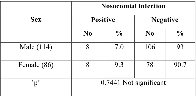
55 II Sex distribution
The total number of patients included in the study was 200 during the
period of 2012 – 2013 in Tirunelveli Medical College and Hospital.114 were
male and 86 were female patients.The Age, Sex distributions were compiled
in tabular column as follows:
Sex Cases
No %
Male 114 57
Female 86 43
Total 200 100
Total male patients were 114 and female patients were 86 in this
study. 57% of the patients were male and 43% were female.More male
57
SIGNIFICANCE OF RISK FACTORS
Diabetes mellitus and Nosocomial infection
Out of the 200 patients in our study 30 patients had previous history of
hypertension and 42 patients had previous history of diabetes out of which
12 patients had both.
Column1 Column2
DIABETES 42
HYPERTENSION 30
BOTH
DIABETES,HT 12
TOTAL
PATIENTS 200
DM 10%
NON DM 90%
58
RISK FACTOR
NUMBER OF PATIENTS
DM 4