“
COMPARISON OF GLASGOW COMA SCALE AND QTCINTERVAL FOR PREDICTING OUTCOMES IN ORGANOPHOSPHATE POISONING
”
Submitted in Partial Fulfilment of Requirements for
M.D.DEGREE EXAMINATION
BRANCH -1 INTERNAL MEDICINE
THE TAMIL NADU DR.M.G.R.MEDICAL UNIVERSITY CHENNAI.
INSTITUTE OF INTERNAL MEDICINE MADRAS MEDICAL COLLEGE
CERTIFICATE
This is to certify that the dissertation titled “COMPARISON OF
GLASGOW COMA SCALE AND QTC INTERVAL FOR
PREDICTING OUTCOMES IN ORGANOPHOSPHATE
POISONING” is the bonafide original work of Dr. MANOJ KUMAR
in partial fulfilment of the requirements for M.D. Branch – I (General Medicine) Examination of the Tamilnadu DR. M.G.R. Medical University to be held in APRIL 2016. The period of study was from March 2015 to September 2015.
PROF. K. SRINIVASAGALUM.D.
Director and Professor Institute of Internal Medicine
Madras Medical College &
Rajiv Gandhi Government General Hospital Chennai – 600 003.
Dr. VIMALA M.D. DEAN
Madras Medical College &
DECLARATION
I solemnly declare that dissertation titled “COMPARISON OF
GLASGOW COMA SCALE AND QTC INTERVAL FOR
PREDICTING OUTCOMES IN ORGANOPHOSPHATE
POISONING” is a bonafide work done by me at Madras Medical
College and Rajiv Gandhi Government General Hospital, Chennai – 3 during March 2015 to September 2015 under the guidance and supervision of my chief Prof. K. Srinivasagalu M.D. , Director and Professor of Medicine at Madras Medical College and Rajiv Gandhi Government General Hospital, Chennai.
This dissertation is submitted to the Tamilnadu Dr. M.G.R. Medical University, towards the partial fulfilment of requirement for the award of M.D. Degree (Branch – I ) in General Medicine – APRIL
2016.
Dr. MANOJ KUMAR
Post Graduate MD – General Medicine Institute of Internal Medicine
Madras Medical College. Place: Chennai
ACKNOWLEDGEMENT
I owe my thanks to the Dean, Madras Medical College and Rajiv Gandhi Government General Hospital, Chennai – 3, Prof. Vimala M.D., for allowing me to avail the facilities needed for my dissertation work.
I am grateful to my beloved mentor and guide,
Prof. K. Srinivasagalu, Director and Professor, Institute of Internal
Medicine, Madras Medical College and Rajiv Gandhi Government General Hospital for motivation, advice and valuable criticism, which enabled me to complete my work.
I am extremely thankful to Prof. S. Raghunanthanan M.D., Professor of Toxicology and Poison Control Center for allowing me to avail the facilities and guiding me through the study.
I am extremely thankful to my Assistant Professors Dr. D.K.
Sivakumar M.D., Dr. Balamanikandan M.D., Dr. Kalpana M.D., for
their guidance and encouragement.
I am also thankful to all my unit colleagues Dr. Barath Raj Kidambi, Dr. Velvizhi M., Dr. Sujatha N.K.. and my junior colleagues for their full co operation in this study and my sincere thanks to all the patients and their families who co-operated for this study.
CONTENTS
Sl. No.
TITLE
Page No.
1.
INTRODUCTION
1
2.
AIMS AND OBJECTIVES
3
3.
REVIEW OF LITERATURE
4
4.
MATERIALS AND METHODS
67
5.
OBSERVATION AND RESULTS
71
6.
DISCUSSION
82
7.
CONCLUSION
85
BIBLIOGRAPHY
ANNEXURES
ABBREVIATIONS
PROFORMA
ETHICS COMMITTEE APPROVAL ORDER
TUNITIN – PLAGIARISM SCREEN SHOT
DIGITAL RECEIPT
PATIENT INFORMATION SHEET (ENGLISH&TAMIL)
PATIENT CONSENT FORM (ENGLISH & TAMIL)
AIMS
AND
REVIEW
OF
MATERIALS
AND
OBSERVATION
AND
1
INTRODUCTION
Organic phosphorus compounds (OPC) groups of cholinesterase inhibiting insecticides that most commonly produces toxicity in humans. In clinical practice all insecticides with a ‘P’ atom in their molecular structure that possesses cholinesterase inhibition are considered as OPC’s,but there are other compounds also possessing the P atom in their molecule that also have property of cholinesterase inhibition (e.g.:phosphoric acids and phosphonates). Some contain thioesters (e.g.:parathion), others are vinyl esters.
All those that have property of cholinesterase-inhibition (anticholinesterases) that contain phosphorus atom in their molecule will be collectively called as OPC’s. OPC’s act by inhibiting the enzyme acetylcholinesterase thereby increasing the acetylcholine levels in the nicotinic and muscarinic receptors. This increased Ach will produce the features of cholinergic excess syndrome1
2
Most of the OPC poisoned patients are managed in ICU settings. And the new advanced treatment modalities have resulted in increased survival in these patients. Such measures are also prolong the in-hospital stay and increases the hospital expenses. So there is a need for prognostication of these patients and also for avoiding expensive procedures and treatments.
The Glasgow Coma Scale is an effective bed side tool to assess the conscious level of any patient. Although it was initially devised for assessing patients with head injury, there is ample evidence to advocate its use for patients presenting with single or mixed poisonings. The QTC
interval is an emerging marker of severity in acute OP poisoning. Multiple studies have indeed demonstrated an association between QT interval and patient outcomes in acute OP poisoning.
Thus the need for study arises due to the convergence of following points: 1. To identify the predictors of mortality in acute OP poisoned
patients.
2. To evaluate and compare the performance of Glasgow Coma Scale and QTC interval in predicting the outcomes of patients poisoned
3
AIMS AND OBJECTIVES
The aim of our study is to compare the Glasgow Coma Scale and the QTC
4
REVIEW OF LITERATURE
THE GLOBAL PROBLEM
Globally, more people are killed each year by acute poisoning of anticholinesterase insecticides than by any other xenobiotic. In Asia alone , around 2 lakh people succumb to toxic organic phosphorus insecticides, mostly from deliberate self ingestion. Around 3000 to 6000 ventilators are required in Asia alone at any given point of time to provide ventilator support to these poisoned patients4. When a poisoning with organophosphate occurs, it often requires intensive care along with long hospital stays, significantly adding to the healthcare expenditure in the high prevalence areas like India.
5
A pesticide is equivalent to a pharmaceutical agent. There dozens of classes of pesticides, each with different uses, different mechanism of action and therefore exert different toxic effects on non target hosts like humans. The pesticides are commonly classified according the species upon which they act on. The four major classes are as follows:
• Insecticides
• Herbicides
• Fungicides
• Rodenticides
Other classes include- acaricides, larvicides and molluscides. By convention, plant growth factors and plant hormones are also classified under the category of pesticides.
6 HISTORY AND EPIDEMIOLOGY
Tetraethylpyrophosphate was the first potent synthetic organophosphorus compound. It was synthesised by Clermont in the year 18545. Later in 1932, Lange and Kruegger reported choking and blurring of vision after inhaling fumes of dimethyl and diethyl phosphorofluoridates. Following this report, Schrader began working on these compounds in Germany, initially for use as insecticides but later on for use in war. Schrader and his colleagues synthesised hundreds of compounds, including the popular insecticide parathion and various other nerve agents used in war, like sarin, soman and tabun. Since that time, more than 50,000 compounds have been synthesised as insectides. More than a dozen such compounds are now being produced commercially.
7
The anticholinesterase compounds are broadly grouped according to their toxicities by the world health organisation’s classification of insecticides into five groups. They are as follows:
• Class Ia: “extremely hazardous”
• Class Ib: “highly hazardous”
• Class II: “moderately hazardous”
• Class III: “slightly hazardous”
• Class IV: “active ingredients unlikely to produce acute hazard in normal use.”
WHO RECOMMENDED CLASSIFICATION OF PESTICIDES BY
HAZARD8
8
9
An estimated one million to two million poisonings due to organophosphates occur in Asia region alone. This is based on the following data. The case fatality ratio in poisoning with OPs is around 10% to 20% and about two lakh patients die due to organophosphate poisoning every year. Respiratory failure remains the most common cause of death in this patient group. It is estimated that OP poisoned patients require on an average, 1.1-2.3 million days of ventilator support. This means that about 3140 to 6280 ventilators are being used every year only to provide ventilator support to patients developing respiratory failure secondary to OP poisoning.
10
presenting with acute parathion poisoning was 40%. In a developing country like India, where healthcare facilities are sparse, very few patients survive acute Organophosphate poisoning.
The easy availability of the highly toxic organophosphates in India coupled with the lack of adequate health care facilities especially in the rural areas, make acute poisonings with organophosphate insecticides highly fatal.
Organophosphates are the most lethal pesticides that are in household and agricultural use today. The acute OP poisoning is also one of the most lethal poisonings in the world today. Strict regulation of the manufacture and sale of these compounds is necessary to prevent accidental and suicidal poisonings. Such a measure would substantially reduce the mortality and morbidity and lowering the health care expenditure of the State.
PHARMACOLOGY
11
DIAGRAM SHOWING THE GENERAL CHEMICAL
STRUCTURE OF AN ORGANOPHOSPHORUS COMPOUND
12
CLASSIFICATION OF OP ACCORDING TO THE LEAVING
GROUP
13
Group 4: widest of all groups. All the available OPs today belong to this group. They are usually a dimethoxy or a diethoxy compound.
The organophosphates can be direct acting or indirect acting.
Direct acting OPs:
The “oxons”. They do not require activation. They directly bind to and inhibit the acetyl cholinesterase enzyme.
Indirect acting OPs:
The “thion”. They are inactive prodrugs. They are activated by desulfuration to produce the active oxon form. This activation reaction occurs in the intestinal mucosa or in the liver after absorption.9
Mechanism of action:
Normally the acetyl choline released into the neuromuscular junction is metabolised by acetyl cholinesterase to choline and acetic acid.
14
nature of the OP compound. The in vitro half life for spontaneous reactivation of human acetylcholinesterase for dimethoxy compounds is o.7 to 0.86 hours, whereas for the diethoxy compounds it is between 31 and 57 hours. Thus spontaneous reactivation of the AChE enzyme is faster with dimethoxy OPs.
15
can be regained only through De novo synthesis of the enzyme. Therefore early diagnosis and prompt administration of oximes is crucial in improving the overall outcome of the patient.
The OPs also vary in their lipid solubility, the rate of conversion from thion to oxon, rate of AChE inhibiton and the degree of inhibition of the butyrylcholinesterase activity in the plasma. Some of the OP may not be classified under dimethoxy or diethoxy group. They contain an alkyl or aryl group linked to the phosphorus atom by a sulphur atom instead of an oxygen atom. These compounds exhibit very rapid aging compared to the other two.
16
PHARMACOLOGIC PROPERTIES OF DIMETHOXY
17
18
Normal metabolism of
acetylcholine by
acetylcholinesterase
19
PHARMACOKINETICS AND TOXICOKINETICS
Routes of entry
Organophosphates are well absorbed from lungs, gastrointestinal tract, mucous membranes, and conjunctiva after an exposure by oral ingestion, inhalation of fumes or direct contact. OPs are absorbed through albeit to a limited extent. But in exposure to toxic agents , cutaneous absorption itself may be rapid enough to be lethal. Topical absorption is increased in the presence of cuts in the skin, dermatitis and in areas with high environmental temperature.
Absorption and distribution
The time to attain peak plasma concentration following an acute poisoning is unknown. Studies done in human volunteers , using very low doses of chlorpyrifos showed that the Cmax can be about 6 hours following
ingestion. But in patients ingesting large quantities or in cases of poisoning with the fast acting thions, symptoms occur in matter of minutes suggesting the absorption in such cases is rapid.
20
accumulated into the brown fat and the salivary glands; high concentrations were seen in kidneys, liver and adipose tissue. Gradually the adipose tissue went on to accumulate the highest concentration of the insecticide. The parathion was measurable in blood upto 48 days post exposure due to slow release from the adipose tissue.
The slow release of the lipophilic compounds from their can cause the cholinergic crisis to recur days after the acute exposure. This is more likely with compounds fenthion and diclofenthion which have high fat solubility. Dimethoate, methamidophos, and oxydemeton methyl are OP compounds that have low fat solubility; therefore they have smaller volume of distribution and have high serum concentrations after exposure.
Metabolism
21 Role of Paraoxonase
Paraoxonases are enzymes that hydrolyses the active oxon metabolites of some OPs. Many studies were done to determine the relationship between human paraoxonase activity and their susceptibility to acute and chronic OP poisoning12. The activity of this enzyme differs significantly among different species. In animal studies, exogenous administration of the paraoxonase enzyme resulted in protection against toxic effects of OPs, whereas Paraoxonase knockout mice showed increased susceptibility to the toxicity of OPs11. Many authors opine that the polymorphisms in human paraoxonase enzyme may be the reason for the interindividual variations in susceptibility to OPs.
PATHOPHYSIOLOGY
22
receptors- resulting in depolarisation and propagation of the action potential.
Organic phosphorus insecticides inhibit the carboxylic ester hydrolases in the body. It includes:
• Acetylcholinesterase
• Butyrylcholinesterases
• Plasma and hepatic carboxylesterases
• Paraoxonases
• Chymotrypsin and other non specific proteases.
As stated earlier, the acetylcholinesterase hydrolyses acetylcholine into choline and acetyl molecules. The choline is taken back by the presynaptic nerve terminal for resynthesis of the acetylcholine in the vesicles. The AChE is found in human nervous tissue, skeletal muscle NMJ and the red blood cells. The AChE activity in the RBC correlates with the enzyme activity in the nervous system, in cases of acute OP poisoning.
23
The inhibition of the acetylcholinesterase is thought to account for all of the manifestations of the OP poisonings. However, clinical implication of the inhibition of the other enzymes is not clearly understood.
In addition, it is worthwhile to note that people ingest commercial formulations and not the pure compounds. EACh OP compound is sold as an emulsifiable liquid. The active OP compound is mixed in an organic solvent such xylene or cyclohexanone1 and a surfactant. This combination is optimised by a company for eACh OP compound it manufactures. Therefore, there will be difference in the same OP compound sold by two companies and in two different OPs sold by the same company.
The clinical effect of ingestion of these coformulants is not clearly studied and is uncertain. The solvent themselves have low toxicities. However, there is a report of death of a minipig within minutes of exposure to dimethoate formulated with xylene solvent. The RBC cholinesterase was reduced by only 30%, indicating a possible non AChE related mechanism. Early work has shown that toxicity of dimethoate can be altered by changing its solvent.
24
produce a chemical pneumonitis. This aspiration pneumonia is not responsive to oximes or atropine.
25 CLINICAL MANIFESTATIONS
The clinical features of acute toxicity result from the presence of excess acetylcholine at the muscarinic and nicotinic cholinergic receptors , resulting in their overstimulation. The clinical presentation is one of a cholinergic excess.
THE AUTONOMIC MANIFESTATIONS ARE:
1. Salivation
2. Lacrimation
3. Urination
4. Diarrhoea
5. Gastric hypersecretion
6. Emesis
7. Bronchospasm
8. Bronchorrhea (can mimic pulmonary edema)15
9. Miosis
Of the above mentioned features , miosis is the most commonly encountered sign. Bronchorrhea can be so profuse that patients “drown” in their own secretions. These are features of muscarinic stimulation.
26
relaxant succinylcholine. There are initial fasciculations of muscle fibres followed by paralysis. This effect is described as the most reliable sign of parathion toxicity. Cranial nerve paralysis is uncommon. A rare patient may present only with the nicotinic features of paralysis without other attend cholinergic symptoms. Extrapyramidal system involvement is uncommon; if present , it persists for several days following resolution of the cholinergic features.
Sometimes muscarinic signs may be absent or may not be clinically predominant. The parasympathetic effect may be offset by the excessive stimulation of the nicotinic receptors of the sympathetic nervous system(resulting in excess catecholamine release). Such patient may present with mydriasis, bronchodilation, tachycardia and urinary retention.
The onset of symptoms will vary depending on the
• Route of exposure
• Degree of exposure
o Patients ingesting large quantities of compounds will have a rapid inhibition of the AChE enzyme, consequently they exhibit toxicity earlier- as early as 5 minutes after consumption.
27
• The OP compound consumed.
o The oxon OPs are direct acting agents. Consequently , patients become symptomatic very soon after ingestion. There is a case report of a man who died within fifteen minutes of Mevinphos ingestion.
o Some thion OPs are rapidly converted to their active form. classical example is parathion. Patients become unconscious within minutes after consumption of parathion.
o Lipid solubility also affects the onset. The more lipid soluble agents like Fenthion are rapidly redistributed to fat stores. The onset of symptoms is delayed – respiratory failure after Fenthion ingestion can occur 24 hours following ingestion. The less lipophilic agents manifest their toxicity much earlier.
28
CENTRAL NERVOUS SYSTEM EFFECTS OF OPs:
Many patients are awake and alert and they complain of ,
• Anxiety
• Restlessness
• Insomnia
• Headache
• Dizziness
• Blurred vision
• Depression
• Tremors
• And many other non specific symptoms.
29 METABOLIC EFFECTS:
The increased adrenergic drive results in the following:-
• Leucocytosis15,17 – the increased adrenergic stimulation results in demarginalisation of white blood cells.
• Glycogenolysis and Ketosis – the excess adrenergic drive causes glycogenolysis resulting in hyperglycemia and ketosis simulating ketoacidosis18,19. However the disturbances in glucose metabolism are uncommon. A study conducted recently showed that out of 79 patients poisoned with OP or carbamate only six patients showed hyperglycemia. In an older study, 7 out of 105 patients had hyperglycemia. It is possible that the effect on glucose metabolism may be specific effects of individual compouds rather than all OPs in general. Further studies with individual compounds are needed to study the association .
30
• Elevation of liver enzymes also occurs secondary to acute OP poisoning.
CARDIOVASCULAR MANIFESTATIONS:
The cardiovascular manifestations reflects the mixed effects the autonomic nervous system and also the effects of OP induced hypoxia and hypovolemia.
• Heart rate-bradycardia is seen relatively few patients. Patients who had received atropine before reaching the hospital may exhibit tachycardia.
• QT interval-There are several reports in literature describing the prolongation of the QT interval and its associated arrhythmias in patients with acute OP poisoning. This may be confounded by the presence of hypoxia and hypotension secondary the effect of OP poisoning. Majority of patients with QT prolongation succumb to dysrrthmias.
31
32 RESPIRATORY COMPLICATIONS:
• Bronchorrhea and bronchoconstriction – occurs secondary to muscarinic stimulation. They respond to adequate atropine therapy.
• Weakness of intercostal muscles and diaphragm resulting in respiratory failure. Respiratory failure is the most common cause of death in acute OP poisonings.
• Loss of central respiratory drive.
The respiratory failure secondary to muscle paralysis does not respond to atropinisation. Patients need adequate ventilator support till the muscle power is regained.
Aspiration of the coformulated pesticides results in aspiration pneumonia due to the hydrocarbon in the coformulants. The hydrocarbon causes aspiration pneumonia, chemical pneumonitis and acute respiratory distress syndrome.
DELAYED SYNDROMES
• INTERMEDIATE SYNDROME
33
INTERMEDIATE SYNDROME
A syndrome of delayed muscle weakness causing respiratory failure , without cholinergic features was first reported in the year 197424 and later in 1987.
“Intermediate Syndrome” is defined as the muscle weakness that occurs 24 to 96 hours following OP poisoning and after resolution of the cholinergic crisis.25
It typically involves the proximal muscle groups especially the neck flexors, cranial nerves and can progress to cause respiratory failure that can last for several weeks. Consciousness is not impaired unless there is hypoxia or a pneumonia. Weakness of neck flexion is often the first sign. This can be easily assessed at the bed side by asking the patient to lift his head off the pillow. Recognising this syndrome is very important because an apparently well patient can suddenly progress to respiratory failure and even death, if not promptly treated.
34
Many case reports and small case series around the globe have shown that the intermediate syndrome is more with certain OP compounds than others. They are malathion, fenthion, methylparathion and parathion. Two large cohort studies have shown that IMS is more common with fenthion poisoning than with malathion, chlorpyrifos or fenitrothion poisoning.
The IMS is just one manifestation of the OP induced NMJ dysfunction in the peripheral nerves29. The relative incidence and timing of the IMS depends on the rapidity and quantity of the Acetylcholinesterase inhibition. Fat insoluble OPs like dimethoate have very high blood concentrations , therefore the enzyme inhibition is also intense. Hence, the NMJ dysfunction develops earlier, even before the cholinergic crisis resolves. Fat soluble compounds like fenthion, have lower blood levels with large redistribution. They cause protracted inhibition of the AChE enzyme. Thus the respiratory failure and the NMJ dysfunction with fenthion occurs later27.
35
therapy may not be able to prevent occurrence of intermediate syndrome. Overall, the dysfunction of the NMJ may be secondary to inadequate oxime therapy( as in chlorpyrifos poisoning) or because of the unresponsiveness of the AChE inhibition to oximes(as in case of malathion poisoning).
DIAGNOSIS OF INTERMEDIATE SYNDROME
1. Clinical examination remains the most reliable way to diagnose
IMS.
2. Electromyography (EMG) shows tetanic fade in IMS, indicating
both pre and post synaptic involvement. More recent works have shown that decrement – increment phenomenon occurs in IMS. This is seen prior to the development of neurological features and respiratory failure28.
TREATMENT
• Treatment is mainly supportive, airway management and mechanical ventilator support for respiratory failure.
36
unlikely to produce any benefit if administered after the onset of NMJ dysfunction.
• The duration of IMS is typically 5 to 18 days31.
ORGANIC PHOPHORUS-INDUCED DELAYED NEUROPATHY
(OPIDN)
Peripheral neuropathies can occur following chronic exposure to organophosphates or after an acute poisoning. OPIDN occurs to inhibition of an enzyme in the nervous tissue known as the NTE(neuropathy target esterase) , now identified as the lysophospholipase(lysoPLA). OPs inhibit the enzyme by phosphorylation. The lysoPLA is involved in degrading phosphatidylcholine of the endoplasmic reticulum. The loss of the NTE-lysoPLA activity results in accumulation of the membrane phospholipids in the nerve tissue, affecting the axonal transport and glial-axonal interactions.
37
the predominant involvement of distal large fibres; axonal degeneration occurs followed by demyelination33.
Patients present with pain and weakness of the distal muscles which can progress to paralysis. Pyramidal tract signs occur weeks to months after the onset. The administration of atropine or pralidoxime does not have any effect on the course of the illness. EMGs and Nerve Conduction studies are useful in diagnosing the condition and also to differentiate it from other similar diseases like Guillian-Barre syndrome. Recovery is variable, takes months to years. Patients often have residual neurologic deficits.
CHRONIC TOXICITY
38
detect chronic exposure. Workers in contact with OP should have their baseline BuChE levels tested for future comparison and monitoring38,39 .
40 BEHAVIORAL TOXICITY:
Acute or chronic exposure to OPCs also associated withbehavioural toxicity. Symptoms include confusion, anxiety, psychosis,depression, drowsiness, fatigue, and irritability. Electroencephalographicchanges may be seen and lasts for weeks41. Morphologic changes in thebasal ganglia of one child identified with single photos emissioncomputed tomography scan (SPECT) following poisoning. Studies haveshown a cognitive processing deficit after acute OPCs self-poisoninglasting for at least six months and this is not found in matched populationswho had poisoned themselves with Acetaminophen42,43. So far there is noclear evidence for neuro-psychiatric deficiencies resulting fromsubclinical exposure to OPCs.
DIAGNOSTIC TESTING
41
of a critically ill patient presenting with a cholinergic syndrome must not await the confirmation of the diagnosis.
Laboratory tests that are used for the diagnosis of OP poisoning are:
• Measuring the insecticide levels or their active metabolites in the biologic tissues.46,47,48
• Estimation of cholinesterase activity in the plasma or blood.
Although tests are available to detect OP compounds in blood and urine, their results are not available immediately. The normal reference range for many pesticide compounds have not been established. So, the diagnosis of anticholinesterase poisoning solely relies on the measurement of cholinesterase activity.
CHOLINESTERASE ACTIVITY
The two cholinesterases that are commonly measured are:
• Butyrylcholinesterase (BuChE, plasma cholinesterase, EC
3.1.1.8)
o It is produced by the liver and secreted into the blood.
o It metabolises xenobiotics like succinylcholine and cocaine.
• Red blood cell cholinesterase(AChE 3.1.1.7)
42
o They two enzymes differ in their posttranslational modification. The RBC enzyme is a monomer bound to the GPI-linked proteins of the cell membrane. Whereas, the neuronal enzyme is in secretory form. It binds with other enzyme molecules to form dimers and tetramers that attach to the post synaptic neuronal membrane.
o The RBC AChE activity tends to correlate accurately with the enzyme activity at the NMJ.49
There are significant interindividual and inter OP variability with respect to the degree and duration with the organophosphates inhibit the AChE. After an acute exposure , the BuChE levels fall first followed by the RBC cholinesterase levels. In most patients both the enzyme levels will be low by the time they present to the hospital. There are interspecies variabilities in BuChE and AChE and this makes interpretations of animal models very difficult. The concentration of AChE enzyme in human plasma is very low. All studies that cite serum AChE activity are likely measuring the Butyrylcholinesterase levels.
BUTYRYLCHOLINESTERASE
43
• Hereditary deficiency of BuChE
• Malnutrition
• Liver parenchymal disease
• Chronic debilitating illnesses
• Iron deficiency anemia.
The reference range for this enzyme is wide. So a patient with high normal baseline levels might have the enzyme levels within the reference range despite a fall in levels after an exposure to OPs. The day to day variability in enzyme levels is also high, about 20% in healthy individuals. The inhibition of BuChE varies between OPs and it does not have any clinical effects. So, the BuChE levels on admission donot predict the outcome of the patients.
RED CELL ACETYLCHOLINESTERASE
44
Normal values 600 to 700 mU/µmol Hb. A study in Caucasians noted a mean value of 651+/- 18 mU/µmol Hb.
After an episode of poisoning the red cell AChE levels take several weeks to recover, in the absence of oxime therapy. On an average it takes 66 days for the enzyme levels to normalize following a severe inhibition (assuming no therapy with oximes). Patients can have a normal NMJ function, no cholinergic features , yet a low red cell cholinesterase levels; this is because the neuronal AChE recovers faster than the red cell enzyme. Therefore in subacute poisoning a low red cell AChE alone cannot be used to predict actual time of onset or the duration of exposure. The red cell cholinesterase levels can reduce in other conditions like pernicious anemia, treatment with anitdepressants or with antimalarials.
MEHOD OF BLOOD SAMPLE COLLECTION
• Fluoride containing tubes should not be used. Fluoride permanently inactivates the enzyme, resulting in falsely low values.
• Specimens for estimation of red cell AChE must collected in EDTA containing tubes.
• Specimens for BuChE estimation can be drawn into plain tubes , the enzyme is estimated in the serum.
45
1:20 or 1:100 at bed side and is then rapidly frozen. Such preparations are not necessary for BuChE estimation.
PROTEIN ADDUCTS
Researches are underway to identify tests to detect OP exposure many weeks after the event. Newer techniques like spectrophotometry are being employed to identify phosphorylated proteins in blood like albumin or BuChE52.
CNS , central nervous system. RBC , red blood cell.
46 ATROPINE CHALLENGE
Atropine may be helpful in diagnosing a patient presenting with cholinergic crisis when there is no clear history of excessive exposure to OP or carbamates. If an individual is not exposed to significant quantities of insecticide then a test dose of atropine,1 mg should be able to produce classical antimuscarinic signs- tachycardia, dry mucous membranes and mydriasis. Conversely, if the cholinergic features persist despite the test dose of atropine, then it strongly suggests the presence of an anticholinesterase poison15. However, in cases with mild poisoning, the test dose of 1mg of atropine might be able to produce antimuscarinic signs. Hence, the reversal of cholinergic signs doesn’t completely rule out a OP poisoning.
ELECTROMYOGRAM STUDIES
47
DIFFERENTIAL DIAGNOSIS
The differential diagnoses of a cholinergic poisoning fall into three main categories
1. The first group comprises the insecticidal and non-insecticidal cholinesterase inhibitors. This includes pyridostigmine overdose seen in patients with myasthenia gravis. These patients have low levels of BuChE and AChE.
2. Second category includes the cholimimetics. They directly stimulate the nicotinic and the muscarinic receptors. They have no effect on the AChE enzyme activity. The BuChE and red cell AChE levels should normal in these cases. Examples include preparations of methacholine, pilocarpine, bethanechol. Non therapeutic agents like muscarinic mushrooms can be cholimimetic.
48
TABLE SHOWING CATEGORIES OF CHOLINERGIC
49 MANAGEMENT
GENERAL PRINCIPLES
Most common cause of death in OP poisoning is due to respiratory failure.
Respiratory failure in OP poisoning occurs due to the following reasons
o Muscarinic effects causing bronchospasm, bronchorrhea, bradycardia and hypotension.
o Nicotinic effects resulting in paralysis of the diaphragm and the intercostal muscles.
o Aspiration of the OP and coformulants causing aspiration pneumonia.
o Loss of central respiratory drive.
Therefore, the primary goal of management is protecting the airway, maintaining the circulation and providing ventilator support.
This is achieved by antagonising the muscuranic effects
Airway maintainence is best achieved by endotracheal intubation and initiating mechanical ventilation if necessary.
Intubation is indicated in the following situations
o Patients who are comatose
50
o Patients who are unable to deal with the copious secretions. If a paralyzing agent is needed, then it is recommended to use a non depolarizing agent53,54. Succinyl choline is degraded by plasma pseudocholineasterase, and hence its degradation will be reduced in case anticholinesterase poisoning resulting prolonged paralysis.
ANTIMUSCARINIC THERAPY
Atropine is a competitive antagonist of acetylcholine at the muscarinic receptor. It reverses the muscarinic effects like,
• Miosis
• Salivation
• Lacrimation
• Diarrhea
• Vomiting
• Bronchospasm
• Bronchorrhea
• Bradycardia
Dosing of atropine:
51
For children, 0.05 mg/kg per dose is used with a minimum of 0.1 mg per dose.
Signs of atropinisation:
• Mydriasis
• Tachycardia
• Dry skin and mucous membranes
• Absent or decreased bowel sounds
• No bronchospasm
Since the goal of muscarinic blockade is to stabilise the cardiorespiratory status, the more practical end points for atropinisation shall be,
1. Pulse rate >80 bpm
2. Systolic BP >90 mm of Hg 3. No bronchospasm
4. Clear lungs on auscultation.
All the above parameters can be rapidly and easily assessed
Atropine infusion:
52
Patients should be periodically monitored for signs of under or over atropinisation and boluses of atropine are administered or the infusion rate is halved accordingly.
Atropine infusions are particularly needed in cases of severe poisoning by fat soluble OPs that continue to release from the fat stores. There is case report stating that such infusion has been used as long as 32 days.55
Large doses of atropine are required to control the lung secretions in severe cases. On an average, such patients may require 40 mg of atropine. Sometimes even 1000mg of atropine may be needed in 24 hrs56. there are literature reports that say the a total cumulative dose 11000mg of atropine was used in a patient.
Atropine does not reverse the nicotinic effects. So the patients must regularly monitored for the development of proximal muscle weakness. If it occurs, sixth hourly monitoring of tidal volume is recommended for early diagnosis and treatment of respiratory failure.
Excessive administration of atropine results in toxicity. Signs toxicity are:
• Tachycardia
• Hyperthermia
• Absent bowel sounds
53
• Mydriasis
When these signs are present, the dose of atropine must be reduced. It is to be noted that tachycardia per se is not a contraindication for atropine because it may be due to many other causes like aspiration pneumonia, hypotension, hypoxia.
Some patients may be develop CNS features of muscarinic blockade while the systemic cholinergic features may persist. In such circumstances, glycopyrrolate can be used instead of atropine. The dosing is similar to atropine, it is administered as 1 to 2 mg boluses that are repeated when required. Glycopyrrolate has a quaternary ammonium structure which prevents it from entering the central nervous system. As with atropine, much higher doses may be required in patients with severe poisoning. There is one randomized controlled trial that compared effectiveness of atropine with glycopyrrolate in ICU settings, but it was too small to produce any significant differences57.
When the supply of atropine gets exhausted, topical preparations of atropine and other antimuscarinic agents like diphenhydramine may be used.
OXIMES
54
reducing the nicotinic and muscarinic stimulation thereby providing symptomatic relief. Oxime administration rapidly increases the RBC AChE levels paralleling the rise in neuronal AChE levels.
The currently available oximes are: 1. Monopyridium oximes
a. Pralidoxime (2-PAM) 2. Bispyridium oximes
a. Obidoxime
57 Dosing of oximes:
A recent clinical trial in India demonstrated that high dose oxime therapy was able to reduce the duration of mechanical ventilation and improve the mortality in patients with moderate poisoning.
2 g of pralidoxime58 mixed in 100 ml of 0.9% saline is infused over 30 minutes. This is followed by a continuous infusion of 8-10 mg /kg/hr infusion.
The infusion is continued till there is no atropine requirement for 12 to 24 hours. It is important to monitor for recrudescence of cholinergic symptoms following cessation of oxime therapy. If it should occur then oxime therapy can be repeated for another 24 hours.
Lipophilic OPs cause delayed onset symptoms. In such cases trial evidence favours a delayed initiation of oxime therapy. This approach reverses the cholinergic signs and is able to prevent respiratory failure.
Monitoring of therapy can be done by:
• Measurements of OP levels in blood and urine.
• Serial measurements of plasma cholinesterase
• Incubating patient patient serum with normal RBC to check for inhibition of the enzyme in the normal RBC.
58
DIAGRAM SHOWING MECHANISM OF ACTION OF OXIMES
59 Adverse effects of oximes:
Therapeutic dose of oximes donot cause any significant reactions. Tachycardia, blurring of vision, rise in diastolic blood pressure, sudden cardiac arrest and laryngospasm may occur on rapid I.V. administration. Oximes are classified under category C in pregnancy. Oxime administration to pregnant women protects both the mother and the fetus, in the event of a poisoning.
BENZODIAZEPINES
Although animal studies have demonstrated benefit on treating with benzodiazepines along with oximes59, to date no human studies were able to demonstrate a benefit with the same. Therefore routine administration of benzodiazepines is not recommended in humans. They can be used to treat seizures, aid intubation and to sedate agitated patients.
DECONTAMINATION
1. Cutaneous absorption of OPCs necessitates removal of allclothing as soon as possible.
60
3. Skin should be triple washed with water, soap, and water, and rinsed again with water. Although alcohol-based soaps are sometimes recommended to dissolve hydrocarbons, these products can be difficult to find, and expeditious skin cleansing should be the primary goal.
4. Cutaneous absorption can also result from contact with OPCs andcompounds in vomitus and diarrhoea if the initial exposure was by ingestion.
5. Oily insecticides may be difficult to remove from thick or long hair, even with repeated shampooing, hence shaving scalp hair may be necessary.
6. Exposed leather clothing or products should be discarded because decontamination is very difficult once impregnation has occurred. 7. In some military institutions the cholinesterase impregnated sponge
is found to be effective in cutaneous OPCs
61
QT INTERVAL
The QT interval is the interval measured in the electrocardiogram. It extends from the beginning of the QRS complex to the end of the T wave. The QT interval incudes the ventricular activation and recovery phases. Thus , it corresponds to the entire duration of the ventricular action potential.
Diagram showing a magnified long axis view point of various
62
However there exist a few practical difficulties in measuring the QT interval. They are as follows:
Difficulty in identifying the beginning of the QRS and the end of T wave.
The timing of the QRS complexes varies between the precordial leads.
The QT interval has to be adjusted for age, QRS duration, sex and most importantly the heart rate62.
The variation in the timing of the QRS complexes and the T wave among the leads can result in a variation of about 50 milliseconds to 65 milliseconds in the QT interval measurement. Automated ECG machines calculate the QT interval in all the leads and finally compute an average of the readings. While calculating the QT interval manually, one must choose the lead where the QRS begins early and the T wave ends late- typically the leads V2 and V3 and also a lead where the U waves are not
prominent like the lead aVr.
QT INTERVAL CORRECTION FOR HEART RATE
QT interval varies markedly with heart rate. As the heart rate increases, the QT interval shortens. In the year 1920, Bazett described a formula the correction of the influence heart rate on the QT interval.
63
Where the QT interval and the R-R interval are measured in seconds. Even after this correction, the Bazett’s formula tends to overestimate the QT interval resulting in erroneous diagnosis of a prolonged QT interval. The variation may be as much as 30% , as estimated in various studies. A newer formula has been described to make the QT interval insensitive to the heart rate62,
QTC= QT+1.75*(HR-60)
QTc - corrected QT interval
HR – heart rate.
All units measured in milliseconds.
The ACC/AHA have prescribed the upper limit for QTc as 460 ms
in females and 450 ms in males while the lower limit has been fixed at 390 ms. Apart from variations with HR, it also varies with age, time of the day and even the season. Older individuals have cardiac myocyte hypertrophy (compared to younger individuals) and therefore have a longer ventricular activation and recovery times, culminating in longer QT intervals.
64
QT INTERVAL IN ACUTE OP POISONING66, 67, 71
The relationship between QT interval and acute OP poisoning is still uncertain. Baydin et al had reported that 35.4% of patients admitted with OP poisoning had prolonged QTC intervals. Their study, however,
found a negative correlation between the QTC interval and serum
cholinesterase levels. Yurumez et al analysed the hospital records of 85 patients admitted with OP poisoning and found that 47 patients i.e. 56% had a prolonged QT interval. However, the 2 patients who died in the study had normal QTC intervals.
Akdur et al found that 53.6% of patients with acute OP poisoning had prolonged QT interval, but they could not find a relationship between the prolonged QT interval and the outcome of the patients. In Taiwan, Liu-SH et al had studied the QT interval of 118 patients and they compared the mortality in the patients with normal QTC interval with that
in patients with prolonged QTC intervals. They found a higher mortality
in patients who had prolonged QTC intervals. Thus, they finally
concluded that the QTc interval helps to predict outcomes after acute
organophosphate poisoning.
65
GLASGOW COMA SCALE
The Glasgow Coma Scale , abbreviated as GCS, was initially described as a bed side tool to assess the mental status of patients admitted with head trauma. It served as a valuable tool in triaging head trauma patients brought to the casualty. Further it was a good predictor of outcome in those patients.
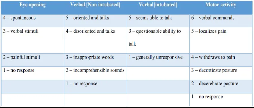
The GCS is a score calculated using three parameters: Eye opening response – scored from 1 to 4 Best verbal response – scored from 1 to 5 Best motor response – scored from 1 to 6.
[image:81.595.109.535.492.674.2]The minimum score for a patient is 3 and the maximum is 15. Total score is calculated by adding the scores of the three components described above.
66
Apart from head trauma , GCS is also used to assess the conscious level of patients in anesthesia, in those presenting in an obtunded state to assess the need for emergent airway management , monitoring the progress of unconscious patients. It is an established indication that a patient with a GCS score of less than 7 needs intubation for airway protection.
Ezadi Mood et al in 2011 showed that admission GCS score can be validated for the assessment of patients with mixed poisoning. A poisoned patient can a have rapid changes in conscious levels owing to number of factors thereby making the utility of GCS questionable; but numerous studies have demonstrated the usefulness of GCS and its components in predicting the severity and the outcomes in poisoned patients72. Grmec and colleagues demonstrated that the GCS scores in OP poisoned patients was lower among the non survivors compared to the survivors70. Akdur et al had compared the effectiveness of Poison Severity Score and GCS in acute OP poisoning and concluded that both the parameters were reliable predictors of patient outcome64.
67
MATERIALS AND METHODS
SELECTION OF PATIENTS
Patients admitted to Rajiv Gandhi Government General hospital Toxicology Unit are selected based on the inclusion criteria. Any patient with a clear history of consumption of an organophosphorus insecticide was included in the study. Patients presenting with acute cholinergic crisis with a characteristic smell of OP in the stomach wash, patients whose gastric content analysis confirms OP consumption were also included in the study. Since the study design is prospective as well as retrospective observational one, the patient records of the Rajiv Gandhi Government General Hospital were also included.
STUDY CENTER
Toxicology Unit , Rajiv Gandhi Government General Hospital, Park town, Chennai- 600003.
DURATION OF STUDY
6 months
STUDY DESIGN
68
SAMPLE SIZE
50 patients
DATA COLLECTION AND METHODS
Patients admitted to the toxicology unit are subjected to thorough history taking and clinical examination. Routine blood investigations are taken as per the institution protocol. ECG is also taken. Retrospective data are obtained from the case records in the medical records department.
INCLUSION CRITERIA
1. Any patient with known or suspected organophosphate poisoning , identified as below
a. History of consumption of an OPC insecticide
b. Classical clinical features of cholinergic crisis – miosis, hypersalivation, fasciculations, characteristic odour of stomach wash.
c. Gastric analysis positive for OP.
69
EXCLUSION CRITERIA
1. Patients who have consumed other poisons with the opc. 2. Patients who have consumed alcohol.
3. Patients with pre existing cardiac disease or chronic lung disease 4. Coma due to trauma.
5. Patients with hypoglycemia or hypothermia. 6. Patients with liver cell failure, renal failure
METHODOLOGY
Patients admitted with organophosphate poisoning- selected for study based on the inclusion and exclusion criteria. A detailed physical examination is done with special emphasis on the GCS of the patient on admission. All patients were subjected to routine blood tests – CBC, RFT and blood sugar levels and ECG. QTc interval is calculated based on admission ECG, using the Bazett’s formula.
QTC=QT/√(R-R interval)
Patients were divided into two groups –
• Group A includes patients without respiratory failure and
• Group B includes patients with respiratory failure.
STATISTICAL METHODS
The statistical analysis is done using SPSS software. valueobtained is analysed using the SPSS software
SPONSORSHIP
FLOW CHART DESCRIBING THE
TOTAL NO OF PATIENTS n=50
APPLYING INCLUSION AND
EXCLUSION CRITERIAS
OBTAINING THE NECESSARY
CLINICAL AND LAB
PARAMETERS
CALCULATING GCS AND QTc
INTERVAL
70 STATISTICAL METHODS
The statistical analysis is done using SPSS software. valueobtained is analysed using the SPSS software
SPONSORSHIP –
None ;CONFLICT OF INTEREST
FLOW CHART DESCRIBING THE
METHOD OF STUDY
TOTAL NO OF PATIENTS n=50
APPLYING INCLUSION AND
EXCLUSION CRITERIAS
OBTAINING THE NECESSARY
CLINICAL AND LAB
PARAMETERS
CALCULATING GCS AND QTc
INTERVAL
COMPARING THE PARAMETERS
BETWEEN THE TWO GROUPS
The statistical analysis is done using SPSS software. The ‘p’
CONFLICT OF INTEREST
–None
FLOW CHART DESCRIBING THE
SEX WISE DISTRIBUTION
SL. NO. NUMBER PERCENTAGE
0 5 10 15 20 25 30 35 40
71
SEX WISE DISTRIBUTION
MALE FEMALE 38 12 76% 24%
MALE FEMALE
SEX DISTRIBUTION
72
AGE WISE DISTRIBUTION
SL. NO. AGE GROUP(yrs.) No. of Patients
1.
15 – 30 yrs
22
2.
30 – 45 yrs
11
3.
45 – 60 yrs
14
4.
60 – 75 yrs
3
73
HISTOGRAM SHOWING THE AGE
DISTRIBUTION AMONG THE STUDY
POPULATION
0 0.05 0.1 0.15 0.2 0.25 0.3 0.35 0.4 0.450 10 20 30 40 50 60 70 80 90 100
74
TABLE SHOWING THE COMPOUNDS CONSUMED BY THE
PATIENTS.
SL. NO.
COMPOUND CONSUMED
NO. OF PATIENTS
1
DIMETHOATE
6
2
MONOCROTOPHOS
9
3
PHENTHOATE
4
4
TRIZOPHOS
7
5
METHYLPARATHION
9
6
QUINOLPHOS
2
7
CHLORPYRIPHOS
3
8
FENTHION
4
9
DICHLORVAS
2
GRAPH SHOWING THE DISTRIBUTION OF
The most commonly consumed organophosphate compounds in our study were Monocrotophos and Methylparathion. The second most common compound 0 1 2 3 4 5 6 7 8 9 10 75
GRAPH SHOWING THE DISTRIBUTION OF
COMPOUNDS
The most commonly consumed organophosphate compounds in our study were Monocrotophos and Methylparathion. The second most common compound was Dimethoate.
GRAPH SHOWING THE DISTRIBUTION OF
76
COMPARISON OF GCS BETWEEN TWO GROUPS
GROUP A
GROUP B
MEAN
12.3636
6.5294
STANDARD
DEVIATION
2.1333
1.588
Group A – patients without respiratory failure.
Group B – patients with respiratory failure.
GRAPH COMPARING THE GLASGOW COMA SCALE
BETWEEN THE TWO GROUPS
77
GRAPH COMPARING THE GLASGOW COMA SCALE
BETWEEN THE TWO GROUPS
1
12.3636
6.5294
MEAN GCS
GROUP A GROUP B
GRAPH COMPARING THE GLASGOW COMA SCALE
78
COMPARISON OF THE QTc INTERVAL AMONG THE
TWO GROUPS
GROUP A
GROUP B
MEAN QTc INTERVAL
IN SECONDS
0.4477
0.4967
STANDARD
DEVIATION
0.01
0.007
Group A – includes patients without respiratory failure.
Group B – includes patients with respiratory.
79
GRAPH SHOWING THE QTc INTERVALS IN THE TWO
PATIENT GROUPS
0.4477
0.4967
0.4200 0.4300 0.4400 0.4500 0.4600 0.4700 0.4800 0.4900 0.5000 0.5100
GROUP A
COMPARISON OF GCS SCORE AMONG SURVIVORS
GCS SCORE
The difference in GCS score among survivors and non survivors was significant with a
based on student t test for two independent variables.)
0 2 4 6 8 10 12
GCS IN NON SURVIVORS
COMPARISON OF GCS AMONG SURVIVORS
80
COMPARISON OF GCS SCORE AMONG SURVIVORS
AND NON SURVIVORS
SURVIVORS
NON
SURVIVORS
11.069
±
3.08
5.8571
The difference in GCS score among survivors and non survivors was significant with a p value of <0.001. (the p value was calculated based on student t test for two independent variables.)
GCS IN NON SURVIVORS GCS IN SURVIVORS
COMPARISON OF GCS AMONG SURVIVORS
AND NON SURVIVORS
COMPARISON OF GCS SCORE AMONG SURVIVORS
SURVIVORS
5.8571
±1.77The difference in GCS score among survivors and non survivors . (the p value was calculated
COMPARISON OF QTc INTERVAL
SURVIVORS AND NON SURVIVORS
QTc INTERVAL
MEAN VALUE
The difference in the QTc intervals between the survivors and non survivors is significant in our study with a
value is calculated by the student t test for two independent variables.)
0.4300 0.4400 0.4500 0.4600 0.4700 0.4800 0.4900 0.5000
QTC IN SURVIVORS
COMPARISON OF QTc INTERVAL AMONG
SURVIVORS AND NON SURVIVORS
81
COMPARISON OF QTc INTERVAL AMONG
SURVIVORS AND NON SURVIVORS
SURVIVORS
SURVIVORS
QTc INTERVAL
MEAN VALUE
0.4590 seconds
0.4981 seconds
The difference in the QTc intervals between the survivors and non survivors is significant in our study with a p value of <0.001
value is calculated by the student t test for two independent variables.)
QTC IN SURVIVORS QTc IN NON SURVIVORS
COMPARISON OF QTc INTERVAL AMONG
SURVIVORS AND NON SURVIVORS
AMONG
SURVIVORS AND NON SURVIVORS
NON
SURVIVORS
0.4981 seconds
The difference in the QTc intervals between the survivors and non
p value of <0.001.( the p
value is calculated by the student t test for two independent variables.)
82
DISCUSSION
This is a prospective and retrospective observational study conducted at the Toxicology unit at Rajiv Gandhi Government General Hospital, Chennai. Patients who presented with a history of OP ingestion or with a documented evidence of OP poisoning were included in our study. A total of 50 patients who satisfied our inclusion criteria were included. Most of the severely poisoned patients were referred from the nearby government hospitals for intensive care management. Informed consent was obtained from the prospective study patients. The data for the retrospective study was obtained from the records of the Rajiv Gandhi Government Hospital.
83
GCS and QTc interval are calculated on admission. Many studies have quoted the usefulness of the above two parameters in predicting the development of respiratory failure. Patients with poisonings can have rapid changes in conscious level, thereby raising doubts regarding the usefulness of Glasgow Coma Scale in these patients. However, Ezadi Mood and colleagues demonstrated that GCS score on admission and its components can be used for patients with mixed poisoning to predict severity.
Okhan Akdur and et al compared the effectiveness of Poison Severity Score , Glasgow Coma Scale and QTc interval in predicting the outcomes in OP poisoned patients. They found a correlation between GCS and PSS in identifying patients with severe poisoning. However, the prolongation of QT interval did not correlate with the severity in their study.
In our study we found that the mean GCS score in patients with respiratory failure was significantly lower than in the patients without respiratory failure, with a p value of <0.001.
The mean QTc interval was prolonged in patients who had respiratory failure compared to those without respiratory failure. The difference was significant with a p value of <0.001.
84
We found the mean GCS score of the non survivors was significantly lower than the survivors. The p value was <0.001.
The mean QTc interval was longer among the non survivors compared to the survivors, which was significant with a p value of <0.001.
LIMITATIONS OF THE STUDY:
1. A multicentre study with a larger patient group will be needed to confirm the usefulness of GCS and QTC interval in predicting
outcomes in patients with acute OP poisoning.
2. The studies should also be conducted in different population groups, in order to confirm their predictive value.
3. Serum magnesium was not measured in the patients with prolonged QTC intervals.
85
CONCLUSION
In our study we thus conclude that Glasgow Coma Scale and heart rate corrected QT interval measured from admission ECG are useful predictors of respiratory failure and mortality in patients with acute OP poisoning. The patients who developed respiratory failure had prolonged QTC intervals and lower GCS scores and the same was also seen with the
patients who succumbed to the poisoning. Hence, in patients with acute OP poisoning, a low GCS score and a prolonged QTC interval on