1
A CLINICAL STUDY OF FOCUSED ABDOMINAL SONOGRAPHY FOR TRAUMA (FAST) IN BLUNT ABDOMINAL TRAUMA
A Dissertation submitted to
The Tamilnadu DR.M.G.R. Medical University, Guindy, Chennai
In partial fulfillment for the degree of
MASTER OF SURGERY In
GENERAL SURGERY APRIL 2015
DEPARTMENT OF GENERAL SURGERY TIRUNELVELI MEDICAL COLLEGE
2
DECLARATION BY THE CANDIDATE
I hereby declare that the dissertation entitled “CLINICAL STUDY
OF FOCUSED ABDOMINAL SONOGRAPHY FOR TRAUMA (FAST) IN
BLUNT ABDOMINAL TRAUMA” is a bonafide and genuine research work
carried out by me under the guidance of Dr.R.MAHESWARI M.S. Professor,
Department of General Surgery, Tirunelveli Medical
College, Tirunelveli.
Dr. K. PRAKASH M.B.B.S
Postgraduate in General Surgery,
Tirunelveli Medical College,
Tirunelveli.
Date:
3
CERTIFICATE BY THE GUIDE
This is to certify that the dissertation entitled “CLINICAL STUDY OF
FOCUSED ABDOMINAL SONOGRAPHY FOR TRAUMA (FAST) IN
BLUNT ABDOMINAL TRAUMA” is a bonafide research work done by
Dr. K. PRAKASH in fulfilment of the requirement for the degree of Master
of Surgery in General Surgery
Dr.R.MAHESWARI M.S.
Professor of General Surgery,
Tirunelveli Medical College,
Tirunelveli.
Date:
4
ENDORSEMENT BY
THE HEAD OF THE DEPARTMENT, DEAN
This is to certify that the dissertation entitled “CLINICAL STUDY OF
FOCUSED ABDOMINAL SONOGRAM FOR TRAUMA (FAST) IN BLUNT
ABDOMINAL TRAUMA” is a bonafide and genuine research work carried out
by Dr.K.PRAKASH under the guidance of Dr.R.MAHESWARI M.S.
Professor, Department of General Surgery, Tirunelveli Medical College,
Tirunelveli.
Dr.K.RAJENDRAN M.S. Dr.Thulasiraman M.S (Ortho)
Professor and Head, Dean
Department of General Surgery, Tirunelveli Medical College,
Tirunelveli Medical College, Tirunelveli.
Tirunelveli.
Date: Date:
5
COPYRIGHT
Declaration by the candidate
I hereby declare that The Tamil Nadu Dr.M.G.R. Medical
University,Chennai shall have the rights to preserve, use and disseminate
this dissertation in print or electronic format for academic/research
purpose.
Dr. K.PRAKASH M.B.B.S,
Postgraduate in General Surgery,
Tirunelveli Medical College,
Tirunelveli.
Date :
6
ACKNOWLEDGEMENT
I express my deep sense of gratitude and indebtedness to my respected
teacher and guide Dr.R.MAHESWARI M.S. Professor,Department of General
Surgery,Tirunelveli Medical College,Tirunelveli, whose valuable guidance and
constant help have gone a long way in the preparation of this dissertation.
I express my sincere thanks to Professors Dr.K.Rajendran, Dr.Pandy,
Dr.Varadarajan, Dr.Alex Arthur Edward, Dr.Sridhar, Dr.Manokaran,Dr.Anto
for their valuable advice and support.
I am also thankful to Assistant Professors Dr.K.J.P.Selvi,
Dr.Sivanupandian, Dr.Nagalakshmi for their help.
I also thank Professor Dr. Nellaiappan and faculty members of
Department of Radiology for their guidance.
I express my thanks to all of the staff members of the Department OF
General Surgery and all my Postgraduates colleagues and friends for their help
during my study and preparation of this dissertation and also for their
co-operation.
I always remember my family members for their everlasting blessings
and encouragement.
Lastly, I express my thanks to my patients without whom this study
would not have been possible.
Dr.K.Prakash M.B.B.S,
Postgraduate in General Surgery,
Tirunelveli Medical College,
Tirunelveli.
Date :
7
ABSTRACT
The aims of study to evaluate the ability of FAST in BAT
And correlation with laparotomy findings as in India BAT is leading cause of
Mortality and morbidity hence to improve surgeon‟s precision to arrive early
Intervention
.
Methods:
The present clinical study is a prospective study conducted on 100 patients
with history of blunt abdominal trauma.
Inclusion criteria:
- All patients with blunt abdominal trauma irrespective of age and sex.
Exclusion criteria:
- Penetrating abdominal injuries.
8
USG with 3.5 -12 MHz transducer used this study
Results:
In this study a total number of 100 patients of BAT were evaluated by FAST
Road traffic accident was the major cause for BAT. FAST was able to identify
free fluid or Haemoperitoneum, and solid organ injuries (lacerations,
contusions, hematomas and rupture)
The overall sensitivity of FAST in evaluation of BAT was 90.9%and
specificity was 75%.
The sensitivity of in detecting free fluid in our study was 90%.
Amongst the solid organs; spleen, liver, and kidney were the most commonly
injured with incidence of 17%, 10% and 3% respectively.
Interpretation and conclusion:
Thus the ability of FAST to accurately detect the presence of free fluid and to
9 Key words:
10 CONTENTS
S.NO TITLE PAGE NO
1 INTRODUCTION 11
2 AIM OF THE STUDY 16
3 REVIEW OF LITERATURE 18
4 METHODOLOGY 77
5 OBSERVATION AND RESULTS 83
6 DISCUSSION 95
7 SUMMARY 104
8 CONCLUSIONS 107
9 BIBLIOGRAPHY 109
ANNEXURES:
I. PROFORMA 120
II. MASTER CHART 123
11
12
INTRODUCTION
Blunt abdominal trauma constitutes those cases where there is injury to on or
more viscera without any external penetrating injury.
The most common causes of blunt abdominal trauma (BAT) are: Road traffic
accidents, assault injuries to the abdomen, accidental fall, fall of heavy objects
over the abdomen, crush and blast injuries.
Since the world population has increased, the incidence of road traffic accidents
and assaults has increased. This has led to the popular quote “civilization and
violence seem to advance hand in hand”.
Blunt abdominal trauma is a frequent diagnostic problem in a patient with
multiple injuries. Any delay in diagnosis substantially increases morbidity and
mortality in trauma patients due to bleeding from solid organ or vascular injury,
or infection from perforation of a hollow viscus. Physical examination is often
unreliable especially when there is associated head injury, spinal cord injury, or
drug ingestion and intra-abdominal injuries may be missed in 16 to 45% of
patients. [1, 2, 3]
The amount of imaging used to evaluate a trauma victim must be inversely
proportional to severity of injuries, so that diagnostic studies do not interfere
with resuscitation.
The most important pre-operative step in patients with BAT is to Ascertain the
13
It is clear advantage to the operating surgeon if the same test is sensitive enough
for citing the organ of injury, especially when conservative approach towards
trauma is being popularized today.
Root et al was the one who first introduced a procedure called Diagnostic
Peritoneal Lavage (DPL) in the year 1965.[4,5] It is sensitive in detecting the
presence of any free fluid including intraperitoneal haemorrhage, but it is
invasive and has a significant rate of non-therapeutic laparotomies.
Computed tomography (CT) scan is non-invasive and accurate but it is costly,
time consuming, requires injection of contrast, exposure to radiation and patient
transport thereby limiting its use.[6,7]
With FAST (Focussed Abdominal Sonography for Trauma) it is possible to
evaluate and help in the management of patients with Blunt Abdominal Trauma.
In recent years USG has taken big leaps in accuracy and acceptance by the
clinical community since it is easy to perform, quick, cost-effective, and
non-invasive, no ionizing radiation or toxic contrast material is needed and can be
repeated as often as required. [8, 9, 10]
FAST combines the advantages of DPL (Fast) with those of CT (Non-invasive
and accurate). [11]
However, evidence exists that the sensitivity and specificity of FAST also
depends on the expertise of the radiographer. FAST results have been shown to
14
Unnecessary deaths and complications can be minimized by improved
Resuscitation, evaluation and treatment.
Rapid resuscitation is necessary to save the unstable but salvageable patient
with abdominal trauma. Accurate diagnosis and avoidance of needless surgery
is an important goal of evaluation. „As the surgeon directs these activities he
must seek the answers to two questions. First, does the patient need an
abdominal operation?
Second, will the patient tolerate the time required for diagnostic manoeuvres
before surgery is performed? However, most avoidable deaths result from
failure to resuscitate and operate on surgically correctable injuries.
When the diagnosis is in doubt and clinical judgment suggests surgery,
exploration provides definitive treatment as well as diagnosis; moreover, the
risks of negative exploration have become acceptable.
The new techniques and diagnostic tools available ate important in the
management of abdominal trauma. These improved methods, however, still
depend on experience and clinical judgment for application and determination
15
This study is based on 100 cases of abdominal injuries, admitted to our
hospital, and outlines the role of FAST in the evaluation of BAT and to assess
the diagnostic validity of FAST imaging, thus helping the surgeons in making
16
17
AIM OF THE STUDY
To evaluate the ability of FAST in detecting hemoperitoneum and solid
organ injury in BAT.
Correlation of laparotomy findings with FAST data to predict its
18
19
REVIEW OF LITERATURE
Ultrasound:
Ultrasound is defined as the sound above the range of human hearing, i.e.,
above the frequency of 20,000 Hz (normal human hearing frequency range
being between 20 Hz to 20,000 Hz).
History of ultrasound:
In 1794, the existence of ultrasound was first demonstrated by Spellanizine on
bats. [13]
In 1880, the use of diagnostic ultrasound based on the piezoelectric effect was
discovered by Pierre and Jacques curie. [14]
In 1912, ultrasound was used to search the sunken Titanic in North Atlantic but
the attempt was unsuccessful. [15]
In 1915, Langevia of France, was, however, the first to use it for detection and
destruction of „submarines‟ during First World War when it was named
20
In 1929, Russian scientist Sergel SoKolven emphasized the potential
importance of SONAR and is regarded as father of ultrasound.
In 1930, it was used in industries for detection of flaw in metals and metallic
structures like beams, bridges etc. [13]
In 1942, Dussik did the first medical use of US for visualization of cerebral
ventricles, a procedure he termed as „hyper phonogram‟. But his technique was
crude and it used very high-energy ultrasound, which was positively harmful for
tissues. [13]
Professor Ian Donald of Glasgow firmly reintroduced US in medicine with
several modifications to make it easy and safe. He designed a control scanner,
which is still used. He is regarded as the father of modern ultrasound. [13]
HISTORICAL PERSPECTIVE IN BAT:
Aristotle was the first one to have recorded visceral injury from blunt abdominal
trauma. Hippocrates and Galen are said to have given apt description about
abdominal injury. A sharp blow over the region of spleen was used as a method
21
Most of the lessons that we have learnt about abdominal injury have been
gained through military experience. Winston Churchill had pointed out, „War is
an epidemic of trauma‟.
Prior to the introduction of DPL in 1965, the diagnosis of intraperitoneal
injuries from blunt trauma was almost entirely based on physical examination,
hemodynamic parameters and serial hematocrits. Not surprisingly this resulted
in a high rate of surgical misdiagnosis.
DPL may however still be indicated as a complementary investigation after US
to elucidate the nature of the fluid, if there is any suspicion of bowel rupture or
to assess the possibility of occult pancreatic trauma.[16]
Gray scale imaging technique was introduced in 1972, with the development of
B scanners (static). And so, evaluation of BAT started. With the introduction of
real time bright (B) scans in the1980s, a more extensive and comparative study
of BAT has been carried out since then.
When it was first proposed, the trauma USG examination consisted of a single
intercostal, subcostal longitudinal view of the pouch of Morrison. If a black
stripe was identified between the liver and the kidney, it was interpreted as a
positive study. Sonographers did no checking of parenchymal organ injury. [17,
22
In a study performed for ultrasonography of epigastric injuries after blunt
trauma, they found that pancreas and second part of the duodenum are most
commonly involved in blunt epigastric injuries. They observed that USG is very
helpful to demonstrate these lesions and thus help in clinical management. [19]
In a study for evaluation of upper abdominal trauma in children, 100 children
with BAT were evaluated prospectively and the roles of CT scan, liver-spleen
scintigraphy and USG were compared. They concluded that CT is more
accurate in demonstrating traumatic lesions, but ultrasonography is equally
accurate in demonstrating free intraperitoneal fluid, easy to operate and can be
done at bedside in highly critical patients.[20]
In an examination of blunt abdominal trauma and acutely ill patients with
ultrasonography and CT, they found that USG plays an important role in the
evaluation of patients having BAT or presenting with renal failure or bleeding
during pregnancy. They concluded that USG and CT are invaluable diagnostic
tools in the radiological evaluation of traumatic patient. [21]
In a study of childhood blunt abdominal trauma by using FAST as the
initial diagnostic modality, during a 5 year retrospective study, a total of 170
children with BAT were investigated with ultrasonography, intravenous
23
compared with clinical outcome and laparotomy. During the study period,
FAST became the first line screening tool and was combined with
intravenous urography in suspected renal trauma. Inspite of permanent
accessibility of CT, it was used only in complex diagnostic problem or in
multiple injuries. [22]
Comparing the role of diagnostic peritoneal lavage and FAST in blunt
abdominal trauma, the reliability of FAST and DPL in assessing the need for
immediate surgical intervention in BAT was examined in 71 patients as a
prospective study. The study suggested that the performance of FAST as a
screening method is justifiable. DPL is a complimentary examination which is
indicated in cases with equivocal clinical or sonographic examination. Thus
both FAST and diagnostic peritoneal lavage are not competing procedures but
rather complimentary in the evaluation of patients with BAT. [23]
In a study of the reliability of ultrasonographic detection of haemoperitoneum
in blunt abdominal trauma in a prospective study of 72 patients, they concluded
that FAST is a quick, safe screening method in the evaluation of BAT to
detect haemoperitoneum and FAST might take over a great part of the role of
diagnostic peritoneal lavage. [24]
The reliability of FAST in detecting haemoperitoneum in patients with
multiple trauma was evaluated prospectively in 291 patients. According to
24
not only provides a rapid diagnosis for blunt abdominal trauma but can also be
used repeatedly with a high reliability to follow the patients‟ course and FAST
meets these conditions. They confirm that FAST has a high specificity and
sensitivity in the diagnosis of BAT. In their department FAST has replaced
DPL. DPL is time honoured and is reserved for selected cases only. [25]
The validity of FAST in the evaluation of patients with BAT was investigated in
a prospective study. 140 patients with suspected BAT were included in the
study. They found that FAST is a suitable test for screening patients with BAT,
since it is highly sensitive and specific, complication free and easy to master.[26]
Evidence exists that appropriate training is very important in determining the
sensitivity and specificity of FAST. Sonographic results have been shown to be
highly variable and dependent on technical expertise of the examiner. They
pointed out that differences in ultrasonographic detection of free fluid or
parenchymal organ injury varied with the experience of the examiner. The
sensitivity of sonographic detection of free fluid varied from 96 to 100%
depending on the examiner‟s experience; sensitivity of detection of
parenchymal organ injury varied from 36 to 45%, depending on examiner
25
In an examination of the practical value of FAST in blunt abdominal trauma in
children, 259 children admitted with BAT underwent FAST. The authors found
that compared with CT, FAST is more versatile, easier to perform in children
and more cost effective, even with addition of a functional imaging modality
like Doppler study or intravenous urography. [27]
In a prospective comparison of diagnostic peritoneal lavage, CT and
FAST for the diagnosis of blunt abdominal trauma, a prospective study
comparing the accuracy of diagnostic peritoneal lavage, CT and
ultrasonography screening was carried out. Patients with stable vital signs
following their initial resuscitation received both CT and FAST. DPL was then
done. The sensitivity, specificity and accuracy of these procedures were
detected. The author suggested avoiding overestimation of the capability of US
and finding that FAST, diagnostic peritoneal lavage and CT are complimentary
and important in the diagnosis of blunt abdominal trauma. [28]
In a study carried out for ultrasonographic evaluation of haemoperitoneum
during resuscitation, to assess the need for therapeutic laparotomy in patients
with blunt abdominal trauma, FAST and its suitable scoring pattern were used
26
The study suggested FAST as an initial rapid screening procedure in patients
with BAT and is useful for trauma surgeons in decision making during
resuscitation. [29]
In a study evaluated for the role of FAST in the initial assessment of BAT in
1000 patients it was concluded that emergency US may be used as the initial
diagnostic modality for suspected blunt abdominal trauma, as FAST combines
the advantages of DPL (Fast and accurate) with those of CT (Non - invasive and
accurate). [11]
In a study design to evaluate the overall diagnostic value of FAST, including
identification of individual organ injuries in 1,239 patients seen during a 15yrs
period it was concluded that FAST is reliable for the detection of injuries and
the identification of solid organ injuries but it has poor sensitivity for intestinal
injuries. [30]
In the assessment of the role of FAST for detection of bowel and mesenteric
injury from blunt trauma in a prospective study for 3 years, it was concluded
that, free fluid in the abdomen is not detected in the majority of patients with
isolated bowel and mesenteric injury. For clinical suspicion of bowel and
mesenteric injury, observation, serial physical abdominal examination, and CT
may be helpful in diagnosing this condition. [10]
27
576 USG‟s, to determine the diagnostic capacity of US in the evaluation of
BAT, they concluded that emergency USG is highly reliable and may replace
CT scan and DPL as the initial diagnostic modality in the evaluation of most
patients with BAT. They recommended the following algorithm for the
evaluation of BAT [5]:
In a prospective study in 744 consecutive children to assess the accuracy of
FAST in the detection of both haemoperitoneum and parenchymal organ
Injury in children, they concluded that FAST for blunt trauma in children is
highly accurate and specific, but moderately sensitive, for detection of
intra-abdominal injury.
In an evaluation of FAST for indirect (with free fluid analysis only) and direct
(with free fluid and parenchymal analysis) detection of organ injury in patients
28
prospective study conducted during a 22 month period at a level I emergency
trauma centre, they concluded that FAST is highly sensitive for the
detection of free intraperitoneal fluid but not sensitive for the identification of
organ injuries. In haemodynamically stable patients, the value of FAST is
mainly limited by the large percentage of organ injuries that are not associated
with free fluid. [31]
In a retrospective study of 604 patients with BAT examined by US and CT for a
period of 14 years by 2 groups on their experience with US, it was concluded
that the sensitivity of US decreased, but however, FAST was found to enable
experienced examiners to detect and classify parenchymal injuries efficiently,
despite disadvantage in detecting superficial and vascular injury. FAST should
[image:29.595.72.349.521.724.2]be used to explore not only free fluid but also solid organ injury. [32]
29
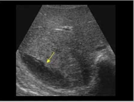
Fig1: a) free fluid in Morrison‟s pouch
30
Fig 2: free fluid in Morrison‟s pouch
[image:31.595.72.424.422.733.2]31
ULTRASOUND FEATURES
The FAST findings in blunt abdominal trauma can broadly be divided into:
a. Free fluid in abdominal cavity.
b. Visceral injuries.
FREE FLUID IN ABDOMINAL CAVITY:
Free fluid typically appears as a hypo echoic region within the peritoneal cavity
or pelvis and is usually linear or triangular in shape. The shape of the fluid
depends on its compression by the surrounding structures, like it is linear when
present between liver and kidney and the fluid that surrounds the bowel often
appears triangular. Fluid often accumulates at the site of injury and then flows
throughout the abdomen and into the pelvis. At the site of injury the blood may
appear echogenic as it forms a clot adjacent to the injured organ. [33]
Free intraperitoneal fluid most often collects in the most dependent portions of
the abdominal cavity. The common sites to detect intraperitoneal free fluid are
as follows:
PELVIS:
This is one of the dependent parts of the peritoneal cavity and so free
fluid usually accumulates in pouch of Douglas or in the perivesical space.
Sonographic sensitivity for the detection of free fluid could be improved by
having a full bladder, which acts as an acoustic window to detect even small
32
MORRISON‟S POUCH OR HEPATORENAL RECESS:
This space is between liver and right kidney.
FLANKS:
Free fluid can be detected earlier in flanks in right and left paracolic gutters.
It is more common on right side than left. This can be located by changing
position of the patient and transducer. Free fluid will accumulate at the site of
visceral lesion.
The capability of FAST to detect minimum amount of free fluid significantly
varies according to the authors.
Free fluid in the abdomen may represent blood, intestinal contents, extravasated
unopacified urine, or fluid instilled during diagnostic peritoneal lavage. [34] Free
intraperitoneal fluid of whatever origin appears similar on FAST. Thus
sonologically, fresh unclotted blood cannot be distinguished from bile or urine.
The initial focus of sonographic examination was a single view of the
hepatorenal fossa (Morison‟s pouch). It was soon realized that a more
comprehensive examination of the abdomen improved detection of free fluid,
however. This included examination of upper quadrants, the paracolic gutters,
and pelvis. [33]
The standard FAST procedure consisted of views of four areas obtained with
34
• Pelvic fluid (women of child bearing age).
• Loops of fluid filled bowel.
• Incomplete / empty bladder.
• Echogenic clot.
• Contained injury.
In a patient whose status was haemodynamically unstable, if haemoperitoneum
is present on FAST, then laparotomy is mandatory. Due to the above pitfalls,
when FAST becomes positive for haemoperitoneum, it indicates the presence of
injuries that are significant, but the absence of detected haemoperitoneum does
not exclude injury. In spite of all that, the use of a FAST protocol examination
(Focused Assessment with Sonographic examination of the Trauma patient) for
visualising abdominal quadrants for free fluid is an irreplaceable tool in the
initial and rapid evaluation of the acutely injured patient. [36]
FREE FLUID SCORING SYSTEMS [33]:
Scoring systems have been developed to help stratify patients into groups who
may or may not require laparotomy. Others have stratified patients based on
either the amount of free fluid in one location or the number of locations in
which free fluid was detected. For instance, Sirlin et al, described a scoring
system based on the location of the fluid was described by Sirlin et al. For each
35
Higher the score, higher the injury rate and the greater the need for laparotomy.
Others have advocated scoring systems based on the number of free fluid sites
or the vertical height of free fluid.
A common theme would be, more the amount of free fluid, the greater the
likelihood of injury or the need for surgical intervention.
In almost all recent studies of the use of FAST for detection of free fluid in
patients with BAT, the specificity of FAST is high. In some cases FAST may
detect small amounts of free fluid that are not visualized with CT.
SOLID ORGAN INJURY:
Initially FAST was used in detecting organ injuries in the 1970s, but more
recent studies focused on the detection of free fluid. A few recent studies have
demonstrated the ability of sonography to detect parenchymal organ
abnormalities directly.
A sensitivity rate of 41.4% for the direct detection of solid organ injuries by
sonography was reported by Rothlin et al. [33], [37] a sensitivity rate of 41%
detection in solid organ Injuries was also reported by Mc Gahan et al. [9]
Polletti et al showed a sensitivity rate of 41% for direct demonstration of organ
33
Fig 6: Probe positions in FAST
More recently in an article by Hahn et al, patients with proven intra
Abdominal injuries after BAT were evaluated and it was demonstrated that the
finding of free fluid with FAST was important. [33, 35] They showed that
examination of Morrison‟s pouch had the highest detection rate of free fluid in
their study (66%), whereas free fluid was detected 56% of the time in the upper
quadrants, 48% of the time in the paracolic gutters, and 36% of the time in the
pelvis, so examination of all the areas was important.
Pitfalls in the examination of abdomen for free fluid. [33]
• Pre-existing fluid (ascites)
36
SONOGRAPHIC APPEARANCE OF SOLID ORGAN INJURIES [33]:
Much of the work on sonographic classification and appearance of solid organ
injuries have been performed by McGahan et al and Richards et al. When
identified, acute solid organ injuries are often echogenic on sonography. Splenic
injuries are identified by a diffuse heterogeneous echogenic pattern. Hepatic
injuries show a discrete hyper echoic or diffuse hyper echoic pattern. Renal
injuries are echogenic, with a disorganized appearance that occurs with severe
renal lacerations.
SPLENIC INJURIES:
37
Fig 8: Splenic hilar tear
In blunt abdominal trauma, the most commonly injured organ is the spleen
which accounts for nearly 40% of all solid organ injuries. Potential factors that
can be responsible for splenic injuries are fractured ribs, intra-abdominal
compression and its rich blood supply and a relatively unprotected area. Almost
20% of patients with left sided lower rib fractures are associated with splenic
trauma, although fractures are not associated with 60 % of splenic injuries.
There is increased susceptibility to trauma in the spleen in persons with
splenomegaly or splenic infections such as infectious mononucleosis. [36]
38
HAEMATOMA:
This can be:
a. Sub capsular haematoma.
39
c. Perisplenic haematoma.
It is important to consider the timing of FAST relative to the trauma.
Immediately after the trauma, the hematoma is liquid and can easily be
differentiated from splenic parenchyma. However, after the blood clots and for
the next 24 to 48 hours, the echogenicity of the blood clot around the spleen
may closely resemble the echogenicity of normal splenic parenchyma albeit
slightly enlarged. Later, the blood reliquefies and the diagnosis becomes easy
again. [38]
With time, one may clearly see the subcapsular hematoma differentiated from
the pericapsular, walled-off hematoma by the capsule itself. The splenic capsule
is very thin and is mostly not visualized separately from adjacent fluid. In such
cases, an important clue to the location of the hematoma is provided by the
shape of the fluid collection. If the collection is crescentic and conforms to the
contour of the spleen, then a subcapsular hematoma should be suspected. But
40
Intraparenchymal or subcapsular hematoma initially appears only as an
Inhomogeneous area in the otherwise uniform splenic parenchyma.
Subsequently, the hematoma may resolve and repeat scans show the cyst at the
site of the original injury.
LACERATION:
A small peripheral laceration appears as a linear echo poor defect. With
transection, a more extensive plane of fluid may be seen traversing the entire
thickness of splenic pulp. [16, 39]
A shattered Spleen usually results from complex lacerations that are
interconnected.
RUPTURE:
A large laceration can be caused by severe injury, through and through the
splenic parenchyma and cause acute rupture of the spleen.
Late splenic rupture is defined as bleeding occurring due to injury to the spleen
that occurs 48 hours after the blunt abdominal trauma. Subcapsular splenic
haematomas which eventually ruptures is responsible for most of these cases.
Lysis of the clot and the resultant osmotic shift of fluid due to high oncotic
pressure or the initial pressure of the haematoma by the organs surrounding the
41
CONTUSION:
Focal areas of inhomogeneity or altered echo pattern represent contusion. With
time it will become more fluid and well defined haematoma is formed.
Splenic cyst: They are generally organized haematoma. They appear as
sonolucent cystic collection.
Free intraperitoneal fluid or left pleural effusions are usually seen with splenic
injuries.
HEALING PATTERN:
The nature and size of initial lesiondetermines the pattern of splenic healing and
the time it takes. Structural splenic recovery may or may not be associated with
resolution of sonographic appearances. On FAST, the spleen usually does not
change back to the previous normal appearance, a fibrous band is represented
by a residual echogenic band. A haematoma that is resolving is sometimes
results in small cysts. Sometimes a foci of calcification can also develop. [36]
STAGING, GRADING OR CLASSIFICATION CRITERIA [40]:
• Grading may be misleading; minor injuries can also go on to devastating
delayed bleed.
O Grade 1: Subcapsular haematoma or laceration < 1 cm.
O Grade 2: Subcapsular haematoma or laceration 1-3 cm.
O Grade 3: Capsular disruption; haematoma > 3 cm; parenchymal haematoma
42
O Grade 4A: Active parenchymal or subcapsular bleeding, pseudo aneurysm
or Arteriovenous fistula; shattered spleen.
O Grade 4B: Active intraperitoneal bleed.
The need for surgery in general is usually associated with a higher grade or
score of injury. This is demonstrated by most of the classification systems. But
in certain cases, several patients with even low grades of injury may
subsequently require emergency surgery.
In addition to all this, a look back of the patient records using such methods that
demonstrate that many patients with high-grade injuries have also been
successfully managed by conservative approach. [36]
[image:43.595.73.397.456.702.2]LIVER INJURIES:
43
Fig 10: subscapular hematoma liver
Liver occupies the second position behind the spleen in organs injured most
commonly in blunt abdominal trauma, this accounts for more than 20 -30 % of
all abdominal trauma. The Right lobe injury is four times more common than
the left lobe, the main reason for which is the larger size of the right lobe.
Injuries to the liver are accompanied by other injuries in over 50% of cases.
Splenic injuries are associated with usually left lobe injuries (45%) or
pancreatic trauma may also be involved, whereas rib injuries (33%) or adrenal
trauma most commonly associated with right lobe injuries. [36]
44
Various lesions encountered in blunt liver trauma are:
LACERATIONS:
Lacerations are present as sonolucent bands traversing the capsule (capsular
tear) or through and through the liver parenchyma (central laceration). This is
the most common type of injury paralleling branches of the right and middle
hepatic veins and the anterior and posterior branches of right portal vein.[41],[42]
HAEMATOMAS:
Subcapsular haematomas occur in potential space between liver parenchyma
and liver capsule as lentiform or curvilinear fluid collections :
Initially - anechoic
After 24 hrs - echogenic
After 4 -5 days - hypoechoic
After 1- 4 wks - internal echoes and septations develop within haematoma. [43]
Intrahepatic haematomas are surrounded by liver parenchyma. With an acute
bleed, the pattern tends to be echogenic. The sonographic findings of acute
trauma to the liver (<24hrs) was evaluated by Van Sonnenberg et al and was
determined that fresh hemorrhage was echogenic. On serial follow up scans,
there may be a significant increase in the cystic component before there is a
45
CONTUSIONS:
Contusions appear as inhomogeneous or altered echo texture in the liver. Large
contusions may lead to formation of haematomas.
The liver has a dual blood supply which is the main reason why the liver is
protected from any localised infarctions in the absence of any gross
parenchymal disruption. [36]
STAGING, GRADING OR CLASSIFICATION CRITERIA [43]:
•Clinical classification based on “American Association for Surgery of Trauma”
(AAST).
O Grade I
haematoma: Less than 10% surface area
tear, less than 1 cm depth of parenchyma.
O Grade II
haematoma: 10-50% surface area
ter.
46
O Grade III
Subcapsular or parenchymal haematoma.
panding.
tion: Parenchymal fracture more than 3 cm deep.
O Grade IV
-75% of hepatic lobe or
1-3 Couinaud segments within single lobe.
O Grade V
c lobe or >
3 Couinaud segments within a single lobe.
hepatic venous injuries (retro hepatic venacava, major
hepatic veins).
O Grade VI
vascular: Hepatic avulsion
While the classification of liver injuries may in certain conditions predict the
47
commonly managed by a conservative approach in recent times. This has
reduced the need for a highly specific staging.
[image:48.595.73.436.227.508.2]RENAL LESIONS:
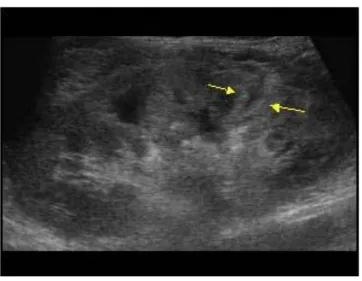
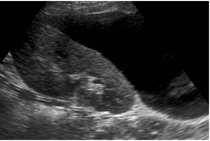
Fig 11: FAST showing Renal Laceration
Renal injuries are mostly caused by blunt abdominal trauma. Such injuries one
usually mild and heal without specific therapy. [43] Serious renal injury is often
associated with damage to other organs such as liver, spleen, bowel, pancreas or
chest. Multiorgan involvement occurs in 20% of those with blunt trauma. The
degree of renal injury varies considerably in blunt abdominal trauma.
48
RADIOLOGICAL CLASSIFICATION OF RENAL INJURIES: [43]
• GRADE I: 75-85% of all renal injuries.
O Minor injury (contusion; intrarenal or subcapsular haematoma)
O Minor laceration + limited perinephric haematoma.
No extension to collecting system or medulla.
• GRADE II: 10% of all renal injuries.
O Major Injury (major cortical laceration and extension to medulla and
collecting system).
O With or without urine extravasation or segmental renal infarct.
• GRADE III: 5% of cases
Catastrophic injury (multiple renal lacerations and vascular injury involving
renal pedicle).
• GRADE IV: rare
Ureter - pelvic junction injury - complete transection or laceration.
Subcapsular or perinephric haematomas may be present in any of these
categories. Any combination of these injuries may be seen in a single kidney.
49
vascular lesions (e.g., renal cell carcinomas, angiomyolipomas) also predisposes
to injury.
Ultrasonography is useful in detecting internal lesions as well as perirenal
pathology in blunt kidney trauma.
HAEMATOMAS [45], [46]:
Sonographic appearance of haematoma, changes with age or duration of
process. When fresh, or in early stages, it appears as a sonolucent or anechoic
fluid mass. In later stages, coarse clumps of strongly echogenic material are
identified in previously echo free collection, which represent clot material.
Internal haematoma, usually appear as a circumscribed disturbance of the echo
structure within the kidney. Depending upon the age of haematoma the
disturbance in the echotexture may be either more hypoechoic or hyperechoic
than the surrounding kidney tissue.
Subcapsular haematomas appear as small sickle shaped sonolucent areas inside
the renal capsule. Blood clots in collecting system appear as scattered
hypoechoic or hyperechoic areas. The pelvi- calyceal system is usually dilated.
LACERATIONS [47]:
Appear as linear band shaped hypoechoic or echodense zone in the sonogram,
or as a deformity of the contour of the kidney in this area. Parenchymal
50
appear as disruption of the central pelvic echoes, with a sonolucent band or area
in a plane at right angles to it, in transverse scan (also called fractured kidney).
SHATTERED KIDNEY[46] :
A complete disruption of the renal parenchyma with displacement of
fragments can easily be diagnosed by sonography and is always associated with
accumulation of fluid in the renal bed. Shattering of kidney can be
sonographically identified by the irregular density of the parenchymal structure
in a large part of the kidney together with movement of fragments. Normal
kidney structures cannot be identified.
In cases of injury to renal vascular pedicle the sonographic appearance of the
kidney can be completely normal.
PERINEPHRIC LESIONS: This can be of the following types:
a. EXTRA-CAPSULAR HAEMATOMA [46]:
Perirenal, pararenal and retroperitoneal haematomas are also sickle shaped
or crescent shaped, but lie outside the renal capsule or in the retroperitoneal
space. The kidney may be displaced by the haematoma.
b. URINOMA:
Perirenal collection of urine after trauma is called urinoma. Perirenal
51
PANCREATIC INJURIES:
Pancreatic injuries in blunt abdominal injuries is rarer when compared to other
solid organs, it is encountered in only 3 - 12% of blunt abdominal trauma. The
main cause of injuries is the Compression of the body and the neck of the
pancreas against the vertebral column by either steering wheels or seat belts in
adults and bicycle handlebars in children. [36]
Acute traumatic injury to the pancreas includes contusion with oedema,
laceration, haemorrhage and transection, which almost always occurs in the
body of pancreas. [19]
The ultrasonography findings with pancreatic trauma depend on the extent of
trauma. With contusion, a focal hypoechoic mass or diffuse hypoechoic
enlargement of the gland may be seen. With laceration or rupture, fluid may be
demonstrated in the retroperitoneum or intraperitoneal space. Traumatic
pseudocyst formation is known to occur in 30-60% of patients. [48] On
ultrasonography, pseudocysts appear as well defined anechoic masses with
associated distal acoustic enhancement. The most common sites for pseudocysts
are lesser sac, anterior or posterior pararenal space. A significant number of
cysts will resolve spontaneously and as such should be followed on
52
ADRENAL INJURIES:
Relatively uncommon. In BAT, usually unilateral in 80% of cases (Right -
85% and left - 15%) and bilateral in 20% of cases. [43]
Ultrasound initially shows an echogenic adrenal mass that may contain a bright
central region. Later the mass will become hypoechoic or anechoic centrally and
with time, may shrink. With progressive decrease in size the mass may again
become more echogenic and calcifications often develop. Alternatively, a
pseudocyst with all the ultrasound characteristics of a simple cyst may be left
behind. [49]
HOLLOW VISCUS:
BOWEL AND MESENTERIC INJURIES:
Bowel and mesenteric injuries are involved in approximately up to 5% of all
blunt abdominal trauma cases, as a result, they are relatively rare. [36]
The best treatment for mesenteric and bowel injuries is early surgical
management. As a result the rapid detection of bowel injuries is of paramount
importance. This is different from management of other organs where
conservative management can be practised. Morbidity and mortality is increased
by any delay in diagnosis and management. [36]
Injuries occurring due to deceleration can cause shearing forces at the points of
fixation of the bowel, such as, the ligament of Treitz, the retroperitoneal
53
present. These shearing stresses can cause tears of the mesentery or injuries of
the bowel wall. Restraining forces usually causes an increase in the intraluminal
bowel pressure that results in the direct rupture of the bowel. Blunt injury to the
colon is relatively rare. [36]
The various types of traumatic bowel lesions include:
CONTUSION:
Appears sonologically as increase in thickness of wall of the injured loop with
ileus localized to the area of injury.
PERFORATION:
Localized heterogeneous collection adjacent to the contused segment is an
indirect pointer to the diagnosis. However, it is difficult to localise the exact site
of perforation. Free intraperitoneal fluid may be the only finding present. If
large in amount floating bowel loops are seen with hypoperistalsis or
aperistalsis. [48]
INTRAMURAL HAEMATOMA:
Haematoma is usually localized to the submucosa if haematomas large, diffuse
gut wall thickening may be seen on sonograms.
MESENTERIC HAEMATOMA:
Bleeding from small mesenteric vessels will lead to a haematoma formation that
involves mesentery alone or extends to bowel wall. The ultrasonography
54
bleeding and examination. In most cases appearances are those of anechoic
mass due to unclotted blood. As the haematoma becomes organized internal
echoes are found and a complex mass results. [50]
Due to extensive gas, bowel injuries are difficult to identify on
FAST.[30] The absence of any solid organ injury, with presence of
Intraperitoneal free fluid should alert the sonologist to associated bowel injury.
[34, 36]
URINARY BLADDER INJURIES:
[image:55.595.72.491.392.674.2]55
(b) Echogenic blood in urinary bladder
Bladder injuries are classified into extra peritoneal and intraperitoneal types, but
these categories may be further subdivided as follows [51]:
Type I: Bladder contusion with incomplete or non-perforating tear of the
bladder.
Type II: Intraperitoneal rupture.
Type III: Interstitial rupture defined as mural defect without extravasation.
Type IV:
a. Simple extra peritoneal rupture with extravasation limited to perivesical
space.
b. Complex intraperitoneal rupture with extravasation outside the perivesical
space.
56
On FAST, free intraperitoneal fluid or free fluid in extra peritoneal
Perivesical space is noted in intraperitoneal or extra peritoneal rupture of
bladder respectively. Sometimes “bladder within a bladder” appearance is
noted, since hypoechoic urine outlines inner bladder wall and hypoechoic blood
and urine outlines outer bladder wall. On ultrasonography, echogenic masses
representing blood clot in the bladder can be detected.
However, sonography is usually not helpful in the assessment of these injuries
except to identify large fluid collection or free intraperitoneal fluid. [44]
Cystography is highly useful in detecting bladder rupture.
Many examiners incorporate the subcostal view of the heart as a portion of
FAST scan. This is useful for diagnosing a pericardial effusion. Finally cardiac
tears or ruptures of the heart may be noted on sonography.
Thus in the future, sonography will probably prove to be a more useful tool in
57
Fig 13: Zone I Retroperitoneal Hematoma
[image:58.595.71.289.405.695.2]58
MECHANISM, PATTERNS AND PATHOPHYSIOLOGY OF VARIOUS
BLUNT ABDOMINAL INJURIES
Obtaining history of mechanism of injury is of great importance in blunt
trauma abdomen. [52]
.. Some 50-60% of closed injuries are due to road accidents and most victims
are occupants of the vehicle, but 15% are on a motor cycle or pedal cycle and
15% are pedestrians. Other causes are falls, accidents at work or in the home,
assaults and seat-belt injuries. [53]
.. Injuries to the left side, with involvement of spleen are more common as is
rupture of the duodenum (closed loop due to seat belt) in occupants of left hand
seat of a car, whereas liver injuries are more common in right sided occupants.
[54]
PATHOPHYSIOLOGY OF BLUNT TRAUMA ABDOMEN [55]:
LIVER [55]
The liver is one of the most commonly injured organ in all patients with
abdominal trauma. It can be injured in both blunt as well as penetrating injuries
59
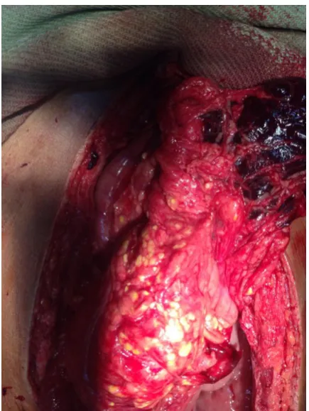
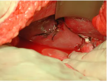
Fig 15: Liver laceration on laparotomy
[image:60.595.72.441.427.708.2]60
Although its location in the right upper quadrant and costal margin afford some
protection, the position assumed at the time of injury may make it possible for
injurious forces applied to the other quadrants of the abdomen to reach and
produce deleterious effects on this organ. Death is usually due to haemorrhage.
The mechanism of liver injuries are direct blows, which causes compression
between lower ribs on the right and the spine, or shearing at fixed points,
secondary to decelaration in blunt abdominal trauma.
Blunt trauma to the liver may cause a disruption of the hepatic substance
beneath an intact Glisson's capsule. Bleeding beneath the capsule can result in
subcapsular haematoma. The haematoma may be small and contained or
continue to expand. The capsule may be stripped from a large surface area of
the liver.
HEPATIC INJURIES: [56]
LIVER INJURY SCALE:
Grade Injury description
GRADE I
Haematoma
Laceration
Subcapsular, non expanding, < 10% surface area
61
GRADE II
Haematoma
Laceration
Subcapsular, non expanding, 10-50% surface area
Capsular tear, active bleeding, 1-3cm parenchymal depth, < 10cm in length
GRADE III
Haematoma
Laceration
Subcapsular, > 50% surface area or expanding, ruptured
subcapsular haematoma with active bleeding, intraparenchymal
haematoma > 10cm or expanding > 3cm parenchymal depth
GRADE IV
Laceration
Parenchymal disruption involving 25-75% of hepatic lobe
\GRADE V
Laceration
Parenchymal disruption involving > 75% of hepatic lobe
Juxtahepatic venous injuries, i.e. retrohepatic vena cava / major hepatic veins
\GRADE VI
62
[image:63.595.74.392.136.381.2]SPLEEN:
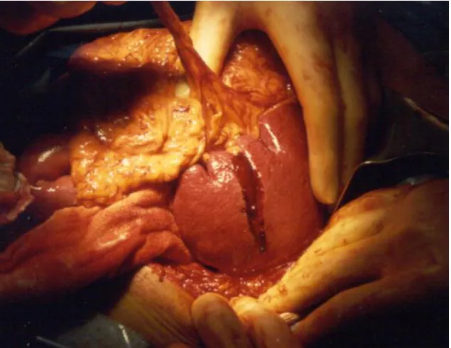
Fig 17: Splenic laceration
The spleen is the most commonly injured organ in patients who have suffered
blunt abdominal trauma. Although the normal spleen is relatively well protected
by the ribcage posteriorly and laterally and softly cushioned by surrounding
organs medially and anteriorly, it is one of the most frequently injured organs in
cases of blunt abdominal trauma.
The mechanism of injury is compression between the anterior abdominal wall
or anterior chest wall and the posterior rib cage on the left or by compression
between the perietes and vertebral column. The relative mobility of the spleen at
its pedicle subjects it to sudden and forceful dislodgement from the peritoneal
63
tears. In some patients, fractures occur along the major segmental arteries of the
spleen.
Traumatic rupture of the spleen is relatively common and not infrequently is
the sole critical injury produced by blunt trauma to the left anterior or lateral
thoracoabdominal wall.[57]
GRADE INJURY DESCRIPTION[58]:
GRADE I
Haematoma Subcapsular, non-expanding, <10 % surface area,
Laceration Capsular tear, non bleeding, <1cm parenchymal depth.
GRADE II
Haematoma Sub capsular, non expanding, 10-50% surface area.
Laceration Capscular tear, active bleeding.
GRADE III
Haematoma Subcapscular, >50 % surface area or expanding Ruptured
subcapscular haematoma, active bleeding
Intraparenchymal haematoma, >2 cm or expanding
GRADE IV.
Laceration > 3 cm parenchymal depth or involving trabecular vessels
GRADE V.
Laceration - completely shattered spleen
64
SPLENIC INJURY IN CHILDREN
Approximately half of the injuries occur during play or athletic activities
Whereas the remainder result from vehicular trauma. Abuse or neglect is
important to consider in children under 4 years of age when a reliable history is
not available. [59]
GASTRIC INJURIES:
The stomach is located in the intrathoracic portion of the abdomen and so it is
protected from injury by the overlying rib cage. It is loosely suspended in the
abdomen by the gastrohepatic ligament superiorly, the gastrocolic ligament
inferiorly, and by its attachment to the spleen laterally. It is relatively fixed at
the gastroesophageal junction and the retroperitoneal duodenum‟
Blunt injuries to the stomach are uncommon and have been reported in less than
2% of cases of blunt abdominal injury. Most blunt gastric injuries have been
observed after rapid deceleration motor vehicle accidents and in these cases are
often combined with significant associated injuries to the liver, spleen,
duodenum, pancreas and diaphragm. Relatively minor but focused blows, such
as those caused by bicycle handle bars, can also produce full-thickness gastric
65
BILIARY INJURIES [61]:
Extra hepatic biliary injuries after blunt trauma are uncommon. Gall bladder
avulsion can occur with high energy blunt trauma. Shearing forces can avulse
the gallbladder from its fossa and may leave it attached only by the cystic duct
and vascular structures. This has been termed traumatic cholecystectomy and
these patients are at high risk of volvulous of the gall bladder if the organ is not
removed.
Traumatic cholecystitis occurs when direct contusion to the gall bladder or
hemobilia from liver injury fills the gall bladder with blood. This blood
stagnates, clots and cannot be expelled from the gall bladder. The cystic duct
becomes blocked, and acute distension results.
EXTRAHEPATIC BILIARY TRAUMA:
The most common site of duct disruption from blunt forces is along the
superior border of the duodenum as the duct enters the pancreas. Shear forces
across the duct seem to be the most likely explanation, and avulsion of the duct
from the papilla is possible. [62]
SMALL BOWEL
Small bowel is the most commonly involved viscera in abdominal trauma.
The mechanism of injury to the small bowel are crushing injury, shearing injury
66
violent force is directly applied to the abdomen it crushes the intestines between
the force and lumbosacral spine causing crushing type of injury [63]. The second
mechanism is tearing or shearing of the bowel and its mesentery at points of
fixation. Tears are common near the ligament of Treitz and ileocaecal valve
where the short mesentery serves as tethering point. Reports have demonstrated
vertical decelerations, by falls or jumps from heights cause shearing intestinal
injuries
[image:67.595.72.433.386.688.2].
67
Fig 19: Mesenteric tear with bowel injury
[image:68.595.72.459.378.668.2]68
Fig 21: Traumatic bowel perforation
[image:69.595.72.509.437.705.2]69
COLON AND RECTUM: [64]
Colorectal injuries are more common in penetrating abdominal trauma than in
Blunt abdominal trauma. The mechanism of injury to the colon and rectum are
crushing injury, bursting injury and shearing injury. When violent force is
applied directly to the abdomen the intestines are crushed against the vertebral
column.
Bursting injuries are caused by a sudden blow against a distended loop.
Shearing injury occurs with sudden deceleration.
[image:70.595.74.438.457.730.2]URINARY TRACT
70
Urologic injury is usually not suspected initially unless the patient has gross
haematuria. Injury to the urinary tract can be caused by both blunt as well as
penetrating abdominal trauma. Although the normal kidney is afforded some
protection by its mobility and its partly intrathoracic position, the kidney is
often found fractured because of its tension within a thin capsule. The kidneys
that are ectopic, malrotated or fused are more vulnerable because of their
intraabdominal locations. Owing to the greater mobility of kidney in females as
well as to the somewhat more sheltered life of women and possibly to the
protection afforded by the wearing of corsetts the relative incidence of injuries
in adult males and females is 10:1.
• “Kidney is the most commonly injured organ in the urogenital” system. [65]
• “Renal injuries from blunt trauma occur consequent to upper abdominal injury
and rapid deceleration”.
CLASSIFICATION OF RENAL INJURY
I. MAJOR RENAL INJURIES
a. Renal pedicle injury
b. Deep parenchymal injury with an intact capsule
c. Deep parenchymal injury with a disrupted capsule
d. Shattered kidney with an intact capsule
e. Shattered kidney with a disrupted capsules
71
II. MINOR RENAL INJURIES
a. Contusion
b. Shallow cortical laceration
c. Forniceal disruption
Renal trauma of severe magnitude is seldom seen as an isolated injury,
therefore, unless a life threatening situation occurs, this type of trauma can
usually be treated with excellent results by conservative measure.
[image:72.595.74.522.393.729.2]BLADDER INJURIES
72
Bladder injuries most commonly results from external violence is blunt trauma
to the abdomen, mostly from crushing injuries to the bony pelvis, motor vehicle
accidents but also from falls or blows to the abdomen. The full bladder is
especially vulnerable to a deceleration injury.
In motor vehicle accidents, injuries are seen in passengers wearing seat belts
when the force of the collision may focus on the abdomen and thus the full
bladder, in the unrestrained child (or less commonly, the adults) who is thrown
by the impact against an unyielding object, or secondary to a pelvic fracture. [66]
CLINICAL FEATURES OF DIFFERENT BLUNT ABDOMINAL INJURIES
SPLENIC TRAUMA [52]:
These are two classes
1) RAPID DEATH:
Spleen has been avulsed or severely damaged by either a run-over or a blast
injury. The patient is admitted exsanguinated and dies before the
resuscitation can be begun or a laparotomy performed.
2) SHOCK:
The largest group presents with shock, signs of rupture in about three quarters
of all cases. The patient shows variable signs of hypovolemia and there is
evidence that points to a serious intra-abdominal problem. There may be
external evidence of damage to the left upper quadrant and if chest x-ray has
73
The abdomen may be slightly distended. Abdominal rigidity is variable, ranging
from generalized rigidity to that localized to left upper quadrant and extending
towards the flank. Tenderness is likewise variable.
DELAYED RUPTURE:
In instances many months after the putative injury, it has been described but the
peak of occurrence is within a few days.
LIVER INJURIES [52]:
Major hepatic injuries are easy to detect because of position of trauma,
profound hypotension and marked abdominal distension. A presence of
unexplained hypovolemia occurring with a bloody abdominal paracentesis,
contusion of the right upper quadrant or lower anterior chest, referred pain to
the right shoulder and signs of peritoneal irritation in the right upper quadrant
clinches the diagnosis of liver injury.
In closed injury there is much similarity between the presentations of rupture of
the liver and rupture of the spleen. The three types- early death in spite of
attempts at resuscitation, gradual development of signs of intra-abdominal
disaster and frank delayed rupture – are all seen but, by comparison with the
spleen, the last is rare.
Shoulder pain is not as common as in ruptured spleen but is clearly a helpful
indicator when it occurs. Apart from symptoms and signs of peritoneal
irritation, which is very nonspecific, detection of fractures to overlying ribs may
74
SMALL INTESTINE INJURY:
Any injury to the lower thorax, abdomen, or pelvis suggests the possibility of
small bowel injury. The diagnosis of small bowel injury is based upon
abdominal pain, signs of peritoneal irritation on physical examination, a positive
abdominal tap, plain X-Ray abdomen in erect posture showing gas under
diaphragm and ileus. But one must remember: „A negative peritoneal tap and
normal X-Ray do not by any means rule out small bowel damage, particularly
during the first 24 hours after the injury.[67]
Closed injury presents as peritoneal irritation, peritoneal lavage is helpful in
making decisions but may not detect small perforations.
DUODENAL RUPTURE [52]:
a) INTRAPERITONEAL RUPTURES:
These produce the syndrome of rupture of the gut. The pain may be greater than
usual, but this is not a reliable indicator.
b) RETROPERITONEAL RUPTURES:
There may be a significant latent period in which thepatient feels quite well.
However within a matter of some hours or at the most a day, severe pain in the
epigastrium and back begins and is associated with intractable vomiting, the
75
RUPTURE OF THE LARGE INTESTINE:
Nothing distinguishes colon rupture from that of rupture of other segments of
the gut except for the delayed variety of closed caecal rupture where the blow is
followed by a latent period, which in turn gives way to the right iliac fossa
symptoms and signs, culminating all too often in catastrophic faecal peritonitis.
INJURIES TO THE PANCREAS:
They are rare and are found only in 1-2% cases of all the abdominal injuries.
Pancreas is rarely injured because of its anatomical position which provides it
with shelter from other abdominal viscera. The injuries are often diagnosed on
exploration for traumatic abdomen with associated visceral injuries because the
pancreas is surrounded by major abdominal organs and blood vessels. The
opening of the lesser sac is very important to rule out the possibility of
pancreatic injury. [68] Absolute criteria do not exist for the firm preoperative
diagnosis of pancreatic injury. Gambil and Mason believed that the
determination of the urine amylase is a more reliable index of
pancreatic injury than the serum amylase. [69]
RENAL INJURIES: [65]
• Gross or microscopic haematuria is mostly seen.
•” These patients often have profuse abdominal tenderness, lower rib fractures,
76
• A abdominal mass that is palpable and with associated shock may indicate a
rapidly developing retroperitoneal haematoma “from a major renal parenchymal
or renal vascular injury”.
Haematuria may not be seen, and in that case diagnosis must be confirmed by
imaging study prompted by a higher level of suspicion.
URINARY BLADDER INJURIES: [66]
Signs and symptoms of rupture of bladder rupture are mostly not specific. The
patient may complain of suprapubic pain or relate that he attempted to urinate
and could not.
Tenderness is present in the suprapubic area and bowel sounds are absent,
especially if it is an intraperitoneal rupture.
77