“EVALUATION OF MRCP IN BILIARY OBSTRUCTION WITH ERCP, HISTOPATHOLOGICAL CORRELATION”
DISSERTATION SUBMITTED TO
THE TAMIL NADU Dr. M.G.R MEDICAL UNIVERSITY, CHENNAI IN PARTIAL FULFILLMENT OF THE REGULATIONS FOR THE
AWARD OF DEGREE OF M.D IN RADIODIAGNOSIS.
BY
DR. SITARA GOPLAKRISHNAN
GUIDE
DR. B. DEVANAND
DEPARTMENT OF RADIOLOGY
PSG INSTITIUTE OF MEDICAL SCIENCES AND RESEASRCH PEELAMEDU, COIMBATORE – 641004
“EVALUATION OF MRCP IN BILIARY OBSTRUCTION WITH ERCP, HISTOPATHOLOGICAL CORRELATION”
DISSERTATION SUBMITTED TO
THE TAMIL NADU Dr. M.G.R MEDICAL UNIVERSITY, CHENNAI IN PARTIAL FULFILLMENT OF THE REGULATIONS FOR THE
AWARD OF DEGREE OF M.D IN RADIODIAGNOSIS.
BY
DR. SITARA GOPALAKRISHNAN
GUIDE
DR. B. DEVANAND
DEPARTMENT OF RADIOLOGY
PSG INSTITIUTE OF MEDICAL SCIENCES AND RESEASRCH PEELAMEDU, COIMBATORE – 641004
CERTIFICATE
PSG INSTITIUTE OF MEDICAL SCIENCES AND RESEASRCH COIMBATORE
This is to certify that the Dissertation work entitled “Evaluation of MRCP in biliary obstruction with ERCP, Histopathological correlation” is the bonafide work of Dr. Sitara Gopalakrishnan in the department of Radiodiagnosis, PSG Institute of Medical Sciences and Research, Coimbatore in partial fulfillment of the regulations for the award of degree of M.D in Radiodiagnosis.
Dr. B. Devanand Professor
Department of Radiodiadnosis PSG IMS & R
Place: Coimbatore Date:
Dr. S. Ramalingam Principal
ACKNOWLEDGEMENT
Foremost, I would like to express my sincere gratitude to my professor and guide Dr. B. Devanand for his ever friendly co-operation which was present throughout the preparation of this work. This work would not have been possible without his guidance, support and encouragement. Dr. B. Devanand will always be a key inspiration to me.
I would like to thank Dr.S. Ramalingam Principal of PSG medical college for providing me with the opportunity and resources to accomplish my research work.
I would like to thank and express my sincere gratitude Dr. Prerna garg ratanlal, Assistant Professor for providing me with the motivation to complete my research work and helping me with the statistical analysis and proof reading, enabling me to complete this study. Dr. P. Prerna garg ratanlal’s knowledge in field of research and command over the English language has immensely helped in framing and formulating this thesis. She was very supportive right from the beginning to the end stages of my research work and helping me battle minor indifferences and providing me with valuable practical tips which were valuable in completing the work.
I would like to thank Dr.L.Venkata krishnana, Professor and Head, Department of Gastroenterology, who has been kind in guiding me through the entire process of the research work. I would also like to extend my heartfelt thanks to all the faculty of Gastroenterology, for helping me in providing ERCP data.
I would like to thank my fellow postgraduates and my dear friends Dr V.B Arun kumar and Dr.S. Sukithra for their immense support during the entire period of my study and for providing me the valuable help and support, and for making my college life unforgettable. They have helped me in various aspects of the study, aiding completion of my thesis work.
My special thanks to my friend Dr. Pradeep Rangaswamy for helping me in statistics and in framing and formulating my thesis. He has helped me in various aspects of the study, aiding completion of my thesis work. Without his immeasurable help it would not have been possible to complete my thesis.
I am very thankful and grateful to my family. Words cannot express how grateful I am to my father Mr.K.G.Gopalakrishnan, my mother Mrs.K.Shyamala, my sister Ms.Thushara and, for all the sacrifices that they have made on my behalf. Your prayers for me was what sustained me this far. My exceptional gratefulness goes forever to my grandmother, Mrs.Sharadha, whose blessings and guidance has brought me a long way in the journey of life.
ABSTRACT
AIM: To evaluate the efficacy of MRCP as a diagnostic tool as compared to ERCP in diagnosing extra-hepatic biliary abnormalities like choledocholithiasis, strictures and malignancies in diagnosing other ancillary findings like biliary ductal dilatation and abnormalities of pancreatic duct. Histopathological correlation is made if available.
MATERIALS AND METHODS: A prospective study of 50 consecutive patients with biliary obstruction referred for MRCP with subsequent assessment by ERCP/ histopathology were included.
CONCLUSION: We concluded that MRCP has high diagnostic accuracy and is equivalent to ERCP in diagnosing IHBR strictures and CBD tumors. In cases of CBD strictures and CBD stones MRCP is comparable to ERCP. In our study MRCP shows increased sensitivity in detecting ampullary tumors than ERCP. Owing to a small study population, results for other biliary pathology were inconclusive.
TABLE OF CONTENTS
S. NO CONTENT PAGE NO
1. INTRODUCTION 1
2. AIMS AND OBJECTIVES 5
3 MATERIALS AND METHODS 7
4. REVIEW OF LITERATURE 14
5. OBSERVATION AND RESULTS 39
6. DISCUSSION 76
7. SUMMARY 85
8. CONCULSION 88
9. LIMITATIONS & RECOMMENDATIONS 91
10. IMAGES 93
11. BIBLIOGRAPHY 109
12. ANNEXURES 118
2
INTRODUCTION
Obstructive jaundice is caused by biliary obstruction, which is due to blockage in any of the ducts that carry bile from liver to gall bladder and then to the small intestine. The causes of obstructive jaundice are classified as intra-hepatic or extra intra-hepatic. Hepatitis, cirrhosis and hepato-cellular carcinoma are the commonest intrahepatic causes. Extra hepatic causes are divided into intra-ductal and extraductal etiologies. Neoplasm, choledocholithiasis, biliary strictures and primary sclerosing cholangitis form the major causes of intraductal obstruction. Extraductal causes include compression of biliary channels by neoplasms, pancreatitis, and cystic duct stones (Mirzzi’s syndrome). The commonest cause of Obstructive jaundice is stones.
Obstruction of the biliary tract with different benign and malignant processes is a common clinical problem which requires accurate localization and discrimination of pathology.
Once cholestasis is detected clinically or biochemically patients are usually referred for USG abdomen as the primary imaging investigation.
3
An excellent clue in finding biliary obstruction in ultrasound has been ductal dilatation. However, ultrasound cannot identify the cause and level of obstruction accurately. Moreover, it is an operator dependent procedure and due to overlying bowel gas or obesity, it can provide only suboptimal imaging of retroperitoneal structures. 1
Hence arose the need for a non-invasive, radiation free, non-operator dependent, multiplanar imaging modality which can help not only to identify the obstruction but also demonstrate the nature of the pathology. Magnetic resonance cholangiopancreatography is one such modality which was initially introduced in the year 1991 and has been improved since then, to the point where it is considered at par with Endoscopic retrograde cholangioancreatography.
Endoscopic retrograde cholangiopancreatography has been considered as gold standard in diagnosing bile disorders, but it is invasive and requires endoscopic cannulation, sedation, the use of ionizing radiation. In addition, ERCP may be associated with significant complications such as hemorrhage, sepsis, pancreatitis, bile leak and mortality.
4
5
6
AIMS AND OBJECTIVES
To evaluate the efficacy of MRCP as a diagnostic tool as compared to ERCP in diagnosing extra-hepatic biliary abnormalities like choledocholithiasis, strictures and malignancies.
To evaluate efficacy of MRCP in diagnosing ancillary findings like gall stone disease, biliary ductal dilatation and abnormalities of the pancreatic duct.
7
8
MATERIAL & METHODS
A prospective study of 50 consecutive patients with biliary obstruction referred for MRCP with subsequent assessment by ERCP/ histopathology were included.
The study was conducted at MRI and ERCP units in a tertiary care center. MRI scanner used was, 1.5-T scanner (Magneton Avanto, Siemens, Erlangen, Germany) using an in-house using 16 channel body coil.
STUDY DESIGN
COMPARISON OF MRCP FINDINGS WITH ERCP
ERCP –HISTOPATHOLOGY
MRCP
9
INCLUSION CRITERIA:
All consecutive patients undergoing Magnetic resonance cholangiopancreatography with features of biliary obstruction & subsequently assessed by Endoscopic retrograde cholangiopancreatography / histopathology.
EXCLUSION CRITERIA:
Patients having a contraindication for MRI – like cardiac pacemaker/cochlear implant/non MR compatible clips used on brain aneurysms/claustrophobia.
Patients lost to follow up.
10
Pre-procedural preparation included six hour duration of fasting which helps in promoting filling of gall bladder. Usually, it takes about twenty minutes to complete the full examination.
Following sequences were obtained- 1. Localizer in 3 planes
2. Axial T1 In- phase trufisp 3. Axial T2 HASTE fat sat 4. Coronal T2 HASTE fat sat 5. Thick Slab HASTE
6. 3D Volume Respiratory triggered MRCP, in two planes (coronal section in plane with CBD, and axial section in plane with pancreatic duct).
11
Table 1. Parameters used in various sequences Parameters T2W
breath hold HASTE 3D T2W FSE with respiratory triggering T2W breath hold HASTE fat saturated thick slab T1W sequence Breath hold In phase TRUFI sequence with respiratory triggering TR/TE(ms) 2000/92 3748/702 4500/792 174/7.15 4.0/1.71
Number of averages
1 1 1 1 1
Flip angle 180 180 180 70 70
FOV 400x325 261x261 349x349 400x299 400x300 Matrix size 320x260 320x320 384x384 288x216 320x240
Slice thickness(mm)
6mm 10mm 40mm 6mm 4mm
Slice gap(mm) 7.2mm 0mm NA 40mm 0mm Number of
slices
30 19 1 20mm 30
Acquisition plane
Axial Coronal oblique
coronal Axial Axial, coronal Half-Fourier
factor
5/8 Phase encoding:7 /8 Phase encoding off Phase encoding off Phase encoding off Receiver bandwidth
473 200 277 230 504
12
An axial and coronal fat sat 2D breath-hold HASTE sequence was acquired covering the entire liver, up on to duodenal ampulla .This was usually performed using two breath-hold /respiratory triggered acquisitions.
3D respiratory-triggered heavily T2-weighted FSE sequences are performed in coronal plane and also in the axial oblique plane. Respiratory triggering was attained by using a navigator sequence which applies a MR pre-pulse so that respiratory motion can be monitored. The navigator was placed on the edge of diaphragm on the coronal localizer and sagittal localizer. Triggering of image acquisition was done when the diaphragm interface with the lung falls into a pre specified window of acceptance. In such a way the imaging slice from a consistent position was achieved.
13
From this volume of data, MIP reformats were obtained. Highest signal intensity pixels along a ray which was perpendicular to the plane of projection is only displayed. In this way fluid and bile-filled structures are highlighted strongly. If needed MIP reformats were obtained in planes such as coronal and sagittal oblique.
Additionally high resolution radial slice acquisitions, thick slab (4 cm) fat saturated HASTE sequence were performed if required.
T1 weighted in phase GRE images and TRUFI images acquired in axial and coronal planes. Contrast enhanced T1 W fat saturated images were acquired if needed.
MRCP was performed prior to ERCP and the results were evaluated by two radiologists with 5yrs and 20 yrs of experience and their consensus was obtained. ERCP was performed by well trained and experienced endoscopist. Cholangiograms were obtained and compared with MRCP findings.
14
15
REVIEW OF LITERATURE
BILIARY SYSTEM
Multiple channels carry bile from the hepatic parenchyma to the
duodenum. This constitutes the biliary tree.
The biliary tree can be classified as intra hepatic and extra hepatic bile ducts.3
INTRAHEPATIC BILE DUCTS:
All these biliary canaliculi unite to form segmental bile ducts and their drainage occurs in the following pattern:
The right posterior duct which courses more horizontally drains the segments VI and VII. The segments V and VIII drain into more vertically oriented right anterior duct.
16
The ducts of the left hepatic lobe are more anterior than those of the right lobe. It is important, particularly when contrast cholangiogram is performed because contrast may not opacify independent ducts.
EXTRAHEPATIC BILIARY SYSTEM:
The common bile duct is formed by the cystic duct from the gallbladder joining with the common hepatic duct. The initial course of the common bile duct courses is along the free edge of lesser omentum, from where it travels posteriorly and reaches the pancreas and duodenum. The ampulla of Vater is formed by common bile duct joining with the main pancreatic duct. The major duodenal papillae in D2 segment of duodenum is drained by the Ampulla of Vater.5
17
MRCP:
Magnetic resonance cholangiopancreatography is a non-invasive imaging technique that uses magnetic resonance imaging as a means to visualize the biliary duct and the pancreatic duct, without the need for contrast. This was introduced in 1991, and since then, the spatial resolution of this technique has improved considerably.
MRCP functions with heavily T2-weighted sequences and highlights static fluids or fluid filled slow moving structures like the pancreatic and bile duct. The ducts will appear hyper intense. Coronal T2-weighted images viewed as thin collimation images (3-5 mm), and it is subsequently processed to obtain a MIP (maximum intensity projection) image which is a cholangiogram like projectional image. Coronal thick slab (30 to 50 mm) can also be obtained, to produce the same effect in a very short time (<5 seconds). Usually both these are obtained to achieve higher accuracy.
Latest magnetic resonance equipment allow us to perform MRCP as fast as within fifteen minutes. This is because of the availability of ultra-fast sequences. At present, MRCP is performed widely as a modality of primary imaging, used in assessing conditions like obstructive jaundice and other pancreatico-biliary duct abnormalities that may be benign or malignant.6
18
ERCP:
Endoscopic retrograde cholangiopancreatography (ERCP) is a technique that combines endoscopy and fluoroscopy for diagnosing and treating biliary or pancreatic ductal abnormalities. An endoscope with a camera and light at the end is inserted through the esophagus, to the stomach and to the duodenum. Contrast media is injected in to the biliary and pancreatic ducts which are visualized on X-rays. ERCP additionally offers an advantage of treatment once the diagnosis is established. Stenting, sphincetrotomy or gall stone removal can be performed in the same sitting. Biopsies can also be taken for tissue diagnosis.
19
Coakley FV et al assessed the efficacy of MRCP in imaging pancreatic and biliary duct pathology. The study explains the procedure of Magnetic resonance cholangio-pancreatography (MRCP) and its uses. He quotes that “MRCP is used to visualize fluid in the biliary and pancreatic ducts as high signal intensity on T2-weighted sequence and it is the newest modality for biliary and pancreatic duct imaging”.
MRCP is already known for its usefulness in an array of pancreatic and biliary diseases that include various congenital variants, post-cholecystectomy disorders, chronic pancreatitis, neoplastic duct obstruction and choledocholithiasis. Despite MRCP being a technique that constantly evolves, it still reached a developmental stage where its usefulness has been proven clinically which can be compared to the clinical accuracy of other conventional cholangiography techniques.
With further development, MRCP is believed to have the potential in replacing diagnostic ERCP as the main modality for imaging the pancreatic duct and biliary duct.7
20
abnormalities. MRCP has now extremely improved in its diagnostic efficacy with its increased resolution and reliability to an extent that it can replace diagnostic ERCP.
The MRCP results were analyzed in patients with pancreatic and biliary ductal abnormalities, and these findings were compared with surgical findings or with the finding of ERCP.
There were 150 patients who had undergone MRCP by various techniques were evaluated. According to the reason for referral, the patients were classified into 4 groups. There were 65 patients with Obstructive jaundice, 25 patients with acute/chronic pancreatitis, 20 patients were screened prior to laparoscopic cholecystectomy were 20 whereas 40 patients had failed ERCP.
The results were favoring MRCP as it correctly identified the biliary obstruction level in 58 patients out of the total 61 patients. It was possible to characterize the nature of stricture whether it was benign or malignant in 30 patients out of 32 patients. Morphology of pancreatic duct was also assessed by MRCP duct in 13 out of 15 patients in whom ERCP has detected ductal changes.8
21
It was first used for biliary duct disorders and then subsequently was used for diagnosing pancreatic disorders. CT, MRCP, MRI and endoscopic ultrasonography gives a diagnosis in maximum number of people with pancreatic diseases and also helps patients and physicians to evade complications of ERCP.
However, pancreatic endotherapy may also be beneficial in a selected patients with pancreatic disorders so that they can avoid complex surgery and also prevents them from chronic medication use. Pancreatic cyst drainage, pancreatic sphincterotomy and pancreatic stenting have been proved to be some of the most challenging and effective endoscopic interventions. These procedures should be performed by well-trained and experienced therapeutic endoscopists taking extreme caution.
Huang LY et al assessed the application of ERCP in biliary ductal and pancreatic diseases. He included a total number of 2075 patients who underwent ERCP and their results were analyzed retrospectively. He also calculated the complications and the therapeutic effect of ERCP. In 64 cases, diagnostic ERCP was performed and the procedure was successful, in the remaining 2011 cases therapeutic ERCP was performed, and the success rate came up to 94.6%.
22
therapeutic tool for biliary ductal and pancreatic disease. Therapeutic ERCP has advantages like it is a minimally invasive procedure, safe and acts as an effective treatment method for biliary-pancreatic diseases.10
According to Ueno E MRCP accurately demonstrates the normal as well as pancreatic ductal abnormalities which also include the congenital anomalies of pancreatic duct and biliary tree.
MRCP was performed in 162 patients with pancreatic abnormalities which includes congenital anomalies of bile duct and pancreatic duct. ERCP was performed in 93 patients and their results were compared. Features such as visualizing the pancreatic duct and its branches, stenosis, filling defects and the presence or absence of ductal dilatation were documented.
They concluded by saying that when comparing MRCP with ERCP, MRCP overestimated the MPD stenosis, meanwhile it underestimated findings such as pancreatic duct filling defects in pancreatitis and dilatation of the branches.
23
diagnose the pancreatic duct abnormalities and also the congenital anomalies of the biliary tree and pancreatic duct.11
Vitellas KM et al in his study proved MRCP as an equally comparable investigation tool in diagnosing extrahepatic bile duct abnormalities when compared with invasive diagnostic endoscopic retrograde cholangio-pancreatography (ERCP).
In MRCP, calculi appears as dark filling defects in a hyperintense fluid background. Due to sclerosing cholangitis, bening structures appear as multifocal segments of dilatation and narrowing or bile ducts of normal-caliber, which produces a classic beaded appearance. Features such as bile duct dilatation and pancreatic duct dilatation in MRCP is highly in favor of malignancy of pancreatic head.
24
In patients in whom biliary-enteric anastomoses is present, MRCP is considered to be the imaging investigation of choice in the evaluation of any patient with suspected pancreatico-biliary disease. Another significant MRCP’s use, is its capability in demonstrating the abnormal anatomy of the bile duct before performing cholecystectomy. MRCP has also been found to be accurate in identification of pancreatic divisum.12
According to Albert JG et al after the introduction of endoscopic retrograde cholangio-pancreatography (ERCP) in the year 1970, gastroenterologists have acquired a vast spectrum of diagnostic and therapeutic options in the biliary ductal and pancreatic system. In present scenario MRCP has significantly replaced diagnostic ERCP and thereby it avoids complications which may occur in endoscopic technique.13
25
Out of the total study population of 73 patients, 42 patients had benign biliary disease, out of which 23 patients had primary sclerosing cholangitis; 9 patients were diagnosed with malignant biliary disease; and the remaining 22 patients had normal biliary ducts. MRC was obtained in 73/73 (100%), and invasive cholangiography (2 PTC's, 68 ERCP's) procedures in 70/73. Using findings of ERCP/PTC as the standard, MRC was assessed and diagnostic accuracy was found to be greater than 90% in the diagnosis of biliary ductal dilatation, bile duct stones, biliary obstruction, normal biliary tree and primary sclerosing cholangitis.
Therefore he proved MRC has significant diagnostic accuracy of 90% in diagnosing biliary abnormalities compared to the invasive cholangiography in which diagnostic accuracy came up to 97%. Thus the study concluded that MRC has extremely high diagnostic accuracy in diagnosing biliary abnormalities. Owing to the noninvasive nature of MRC, it is even more advantageous over the invasive cholangiography where the major intention of the procedure is to arrive at a diagnosis.14
26
pancreatic ductal dilatation or obstruction and periductal masses. MRCP showed 74.6% sensitivity and 83.5% specificity in detecting choledocholithiasis, 85.4% sensitivity and 87.4% specificity in detecting strictures, 85.9% sensitivity and 91.2% specificity in detecting obstruction, 92.4% sensitivity and 93.5% specificity in detecting ductal dilatation, and 90.8% sensitivity and 92.6% specificity in detecting periductal masses.
The study shows MRCP to have increased false negative results in case of choledocholithiasis and strictures and increased false positive results in case of ductal dilatation and periductal mass when compared with ERCP. Therefore, it cannot be used as a substitute, thereby ERCP remains the gold standard investigation for better visualization of the biliary tree and pancreatic duct.15
Magnetic resonance cholangiopancreatography was assessed by Liang C et al in patients with malignant obstructive jaundice. They proved the diagnostic efficacy in determining the location and extent of obstruction using MRCP was higher and the specificity was 82.9%.
27
in identifying the location and also the extent of obstruction. They concluded saying that MRCP had significant role in diagnosing malignant obstructive jaundice.16
Miletić D et al in their study assessed that Magnetic resonance cholangiopancreatography (MRCP) had extremely high sensitivity and specificity in demonstrating the level and identifying the presence of biliary duct obstruction. One major weakness of MRCP is its inability to offer therapeutic interventions. Other significant advantage of MR cholangiography is that it can also be used for detecting the resectability of a malignant neoplasm such as hilar cholangiocarcinoma by assessing the proximal extent of disease where ERCP may not be successful.17
28
retrograde cholangiopancreatography and 1patient underwent percutaneous transhepatic cholangiography.
Biliary ductal abnormalities such as presence of dilatation, strictures, and intraductal abnormalities were assessed. MRC was evaluated comparing with direct cholangiography as the gold standard and the sensitivity and specificity images were obtained in total number of 44 patients. Sensitivity for diagnosing biliary ductal dilatation (n = 27), bile duct strictures (n = 10), and intraductal pathologies (n = 7) were calculated and it was 96.3%, 90%, and 100%. In addition, MRC showed 16 of 17 patients with normal bile ducts (specificity, 94.1%). Therefore, they opined that MRC has extremely high sensitivity and specificity in evaluating the biliary tree. Based on this study, the efficiency of MRC using 3D FSE found to be sufficiently high to suggest it as a diagnostic imaging modality in evaluating biliary tract abnormalities.18
Eva C Kaltenthaler et al evaluated Magnetic resonance cholangiopancreatography (MRCP) and proved it to be an effective alternative diagnostic tool to endoscopic retrograde cholangiopancreatography (ERCP) for identifying biliary obstruction.
29
dilatation (5 studies) obstruction (3 studies). Three of the 18 studies reported with choledocholithiasis were removed from the analysis due to inadequate data, or study design differences.
The sensitivity was calculated for the 15 studies of choledocholithiasis and it ranged from 0.50 to 1.00 while specificity which was calculated to range from 0.83 to 1.00. Significant heterogeneity was found across the 15 studies so the sensitivities and specificities and the results were summarized by a Receiver Operating Characteristic (ROC) curve. The sensitivity was calculated and it ranged from 0.81 to 0.94 and specificity ranged from 0.92 to 1.00 for malignancy.19
30
According to a study done by Norero E et al MRCP has a higher accuracy in diagnosing choledocholithiasis. Retrospective review of patients with suspected choledocholithiasis were studied with MRCP and other confirmatory test such as ERCP, surgical common bile duct exploration or transcystic cholangiography. 125 patients were included in the study. According to their study sensitivity was found to be 97%, specificity 74% positive predictive value 89%, negative predictive value 90% and hence the accuracy of MRCP was 90% for the diagnosis of choledocholithiasis.21
31
Hekimoglu K et al assessed the diagnostic efficacy of few new MR sequences in MRCP and its comparison with ERCP along with review of current literatures. A total number of two hundred and sixty nine patients were included in this study. Among the 269 patients, there were 25 males and 145 females, whose mean age ranges from 23-92. The MRCP procedure was performed before the ERCP in all cases. All MRCP studies were performed with new MR technique using a heavily T2-weighted turbo spin echo (TSE) sequence.
The TSE sequence is one of the most widely used multiplanar 3-D MR technique currently, has high spatial resolution and fast imaging capacity. The study participants were divided into four main groups; group I consist of normal, group II includes stone disease, group III includes tumor and others into group IV. Group I comprises of 228 patients who had a normal pancreatic duct and biliary tree on both the investigations MRCP and ERCP.
32
According to Halefoglu AM et al MRCP has a much lower complication rate when compared to ERCP. As already proved ERCP is considered as the gold standard investigation for biliary tract imaging but is less invasive and is also associated with complications.24
Howard K et al assessed MRCP’s value in diagnosing biliary abnormalities in post cholecystectomy patients. According to their study, MRCP’s accuracy is comparable to ERCP. MRCP was found to be effective and also cost-effective when compared to ERCP, especially in patients for whom the suspected disease prevalence is low, thereby avoiding further intervention.
The results of their study proved that MRCP was dominant over ERCP. The results showed that, there was 59% likelihood that MRCP was cost saving for whom there is a low to moderate probability of common bile duct stones. It was seen that, with a higher adjusted quality survival, there was 83% chance of MRCP being more effective and it also had a better cost-effectiveness ratio.25
33
diagnosing malignancy it came up to 81% and 100%. These results calculated and were equivalent to ERCP (91% and 92% for any abnormality, and 93% and 94% for malignant diseases).26
According to Adamek HE et al, MRCP and ERCP have similar sensitivity in pancreatic carcinoma detection. In cases of suspected pancreatic carcinomas MRCP has the potential to prevent unnecessary common bile duct and pancreatic duct explorations, where it is highly unlikely to perform interventional endoscopic therapy. A total of 124 patients constituted the study.
Of the 124 patients, pancreatic carcinomas were detected in 30% of patients, chronic pancreatitis was found in 46% patients, neoplastic pancreatic diseases were seen in 14%, clear pancreatic ducts were seen in 10% of the study population. In diagnosing pancreatic cancer, MRCP’s sensitivity was 84% along with a specificity of 97%. The sensitivity of ERCP was 70% and specificity was found to be 94%.27
34
abnormalities and strictures were documented and clinical correlation of diagnosis was made.
Among the 78 patients included in the study, images from MRCP was collected for 76 patients. The sensitivity of MRCP in detecting normal bile ducts was found to be 71%, bile duct dilatation was 83%, and stricture was 85%, correct stricture location in 77% and bile duct calculi was 80%. The positive predictive value of MRCP for normal bile ducts was 62%, bile duct dilatation was 91%, strictures was 100%, correct stricture location was 91% and bile duct calculi was 100%.
The MRCP’s sensitivity in detecting benign strictures was 50% and malignant strictures 80%. The sensitivity of MRCP in normal pancreatic ducts recognition was 33%, dilation was 62%, strictures was 76% and correct location was 66%, benign strictures was 87% and malignant strictures was 60% and for pancreatic duct stones it was 60%. The positive predictive values of MRCP normal pancreatic ducts recognition was 50%, dilation was 100%, strictures was 87% and correct location was 100%, benign strictures was 87% and malignant strictures was 75% and for pancreatic duct stones it was 100%. Thus he concluded that MRCP is equivalent to ERCP in terms of contributing diagnostic information in many patients. Whenever inadequate results are obtained in established techniques, it is necessary for MRCP to be used.28
35
Siddique K et al assessed a prospective study with 60 patients. The history, physical examination and other investigations like ERCP, MRCP, USG abdomen, CT-Scan and histopathology were analyzed. Out of those 60 patients, 40 were male and 20 were female. Malignant obstructive jaundice was seen in 34 patients while 26 had benign etiology. Commonest causes for benign and malignant obstructive jaundice were found. Commonest malignancy was Carcinoma of the pancreatic head 18/60 (30%) followed by Carcinoma gall bladder 8/60 (13.33%), cholangiocarcinoma 7/60 (11.66%), and periampullary carcinoma 1/60 (1.66%). Choledocholithiasis 21/60 (35%) was the commonest benign cause followed by stricture of common bile duct 3/60 (5%) and acute pancreatitis 2/60 (3.33%).29
36
Mosler P et al assessed the diagnostic accuracy of mangnetic resonance cholangiopancreaticography in identifying pancreatic divisum. ERCP is considered as gold standard method in the diagnosis of pancreas divisum. They assessed patients who had undergone both the investigations of ERCP and MRCP. Pancreas divisum was detected by ERCP in 19 patients out of the total 113 patients, whereas the same was detected in 14 patients by MRCP. MRCP was also found to have provide three false positive results.
Thus MRCP had 73.3% sensitivity, 96.8% specificity, and 82.4% positive predictive value, 94.8% was its negative predictive value. The sensitivity of MRCP in diagnosing pancreas divisum was around 73.3% which is comparatively lesser, as previous studies had reported MRCP’s sensitivity to be as high as 100%.30
37
89%. The results prove that the 3 methods of imaging are complementary to each other.31
In the study "A prospective comparison of magnetic resonance cholangiopancreatography with endoscopic retrograde cholangiopancreatography in the evaluation of patients with suspected biliary tract disease "conducted by Varghese JC, Farrell MA, Courtney G et 100 patients underwent MRCP, in whom direct cholangiographic correlation such as ERCP, n = 98; PTC, n = 9; intraoperative cholangiography, n = 3 was used for comparison.
38
39
40
RESULTS
The results of the study are computed and tabulated below. I. AGE
Table no 2 showing the age distribution of the study population. AGE GROUPS
(in years)
FREQUENCY PERCENTAGE (%)
15 to 25 3 6
26 to 35 5 10
36 to 45 4 8
46 to 55 16 32
56 to 65 10 20
66 to 75 5 10
75 to 90 7 14
Total 50 100
41
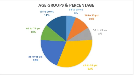
Figure no 4: Age groups & Percentage.
The above pie chart shows the percentage of the study population according to age group.
15 to 25 yrs 6%
26 to 35 yrs 10%
36 to 45 yrs 8%
46 to 55 yrs 32% 56 to 65 yrs
20%
66 to 75 yrs 10%
75 to 90 yrs 14%
42
Figure no 5: Age groups & Frequency.
Figure no 5 is a histogram showing age in groups and the number of patients in the study population.
0 2 4 6 8 10 12 14 16 18
15 to 25 yrs 26 to 35 yrs 36 to 45 yrs 46 to 55 yrs 56 to 65 yrs 66 to 75 yrs 75 to 90 yrs
43
[image:51.595.86.372.437.672.2]II. SEX
Table no 3: Gender distribution.
SEX FREQUENCY PERCENT
Male 28 56
Female 22 44
Total 50 100
The above table shows that 56% of the study population were Males, and 44% were females.
Figure no 6: Sex of study population.
44
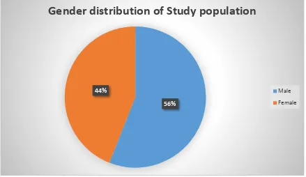
Figure no 7: Gender & percentage.
The above pie chart shows the percentage of the study population, with 44% being males and 56% being females.
56% 44%
Gender distribution of Study population
Male
45
III. PATHOLOGY DETECTED IN STUDY POPULATION. Table no 4: Pathologies detected.
S.NO PATHOLOGY FREQUENCY PERCENTAGE
1 IHBR strictures 3 6
2 Bile Duct Stones 28 56
3 Common Bile duct Strictures 14 28
4 Bile duct dilatation 30 60
5 Bile duct Tumor 6 12
6 Gall Bladder stones 4 6
7 Pancreatic duct Dilatation 2 4 8 Pancreatic duct variants 1 2
9 Ampulla stones 2 4
10 Ampulla strictures 2 4
11 Ampulla tumor 4 8
The above table shows the frequency and percentage of the various pathologies detected among the study population.
46
Figure no 8: Frequency of pathologies detected.
The above histogram shows the number of patients in which pathologies were detected in the study population.
0 5 10 15 20 25 30 35
No. of patients
47
IV. IHBR STRICTURES
Table no 5 showing frequency and percentage of IHBR strictures
The above table shows that out of the total study population, 6% had IHBR strictures, which were diagnosed by both ERCP and MRCP and was later confirmed by Histopathological examination.
PARAMETER FREQUENCY PERCENTAGE (%)
No IHBR stricture 47 94
ERCP & MRCP 3 6
48
Table no 6: Diagnostic efficacy of MRCP for IHBR strictures
SENSITIVITY (%)
SPECIFICITY (%)
POSITIVE PREDICTIVE
VALUE (%)
NEGATIVE PREDICTIVE
VALUE (%)
MRCP 100 100 100 100
ERCP 100 100 100 100
49
V. BILE DUCT STONES
Table no 7: Frequency and percentage of Bile Duct Stones.
From the above table, it is observed that MRCP detected bile duct stones in 48% of the study population along with ERCP. ERCP alone detected bile duct stones in 10%.
PARAMETER FREQUENCY PERCENTAGE (%)
No bile duct stones 21 42
ERCP 5 10
ERCP & MRCP 24 48
50
Figure no. 9: Bile duct stones and its frequency
The above histogram shows the frequency of bile duct stones detected with ERCP and ERCP with MRCP.
Figure no. 10: Bile duct stones & percentage.
The above pie chart shows the percentage of bile duct stones detected by ERCP and MRCP.
42%
10% 48%
Bile duct stones & Percentage
No bile duct stones
ERCP
51
Table no 8: Diagnostic efficacy of MRCP and ERCP for Bile Duct Stones
SENSITIVITY (%)
SPECIFICITY (%)
POSITIVE PREDICTIVE
VALUE (%)
NEGATIVE PREDICTIVE
VALUE (%)
MRCP 87.5 100 100 84.6
ERCP 100 100 100 100
In detecting Bile duct stones, it is observed that MRCP has sensitivity and
52
VI. COMMON BILE DUCT STRICTURES
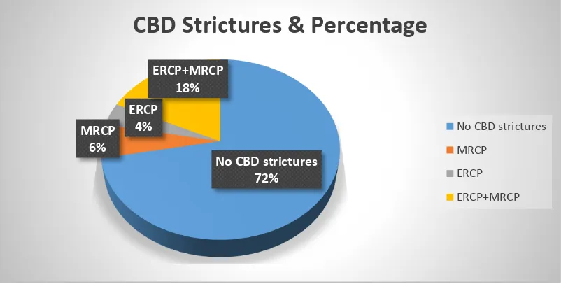
Table no 9: Frequency and percentage of Common bile duct Strictures
ERCP detected bile duct strictures in a total of 11 patients out of which MRCP detected CBD strictures in 9 patients. MRCP alone detected CBD strictures in 3 patients which were false positive.
PARAMETER FREQUENCY PERCENTAGE (%)
No CBD strictures 36 72
MRCP 3 6
ERCP 2 4
ERCP+MRCP 9 18
53
Figure no. 11: CBD strictures and percentage.
The above pie chart shows the percentage of CBD strictures detected by ERCP, MRCP and ERCP+MRCP. ERCP alone detected 4%; MRCP detected 6% of the study population. ERCP+MRCP detected BCD strictures in 18% of the study population.
No CBD strictures 72% MRCP
6%
ERCP 4%
ERCP+MRCP 18%
CBD Strictures & Percentage
No CBD strictures
MRCP
ERCP
54
Table no 10: Diagnostic efficacy of MRCP, ERCP for Common bile duct Strictures
SENSITIVITY (%)
SPECIFICITY (%)
POSITIVE PREDICTIVE
VALUE (%)
NEGATIVE PREDICTIVE
VALUE (%)
MRCP 84.6 92.3 78.5 94.7
ERCP 100 100 100 100
55
VII. BILE DUCT DILATATION
Table no 11: Frequency and percentage of Bile duct dilatation
Above table shows that out of 50 patients ERCP and MRCP together detected bile duct dilatation in 30 patients.
PARAMETER FREQUENCY PERCENTAGE (%)
No Bile duct dilatation 20 40
ERCP & MRCP 30 60
56
Figure no. 12: Bile duct dilatation & percentage.
The above pie chart describes bile duct dilation detected by ERCP+MRCP. 40%
60%
Bile duct dilatation & Percentage
No Bile duct dilatation
57
Table No 12: Diagnostic efficacy of MRCP, ERCP for Bile duct Dilatation
SENSITIVITY (%)
SPECIFICITY (%)
POSITIVE PREDICTIVE
VALUE (%)
NEGATIVE PREDICTIVE
VALUE (%)
MRCP 100 100 100 100
ERCP 100 100 100 100
58
VIII. COMMON BILE DUCT TUMOR
Table no: 13: Frequency and percentage of Common bile duct Tumor
Above table shows out of 50 patients ERCP and MRCP together detected CBD tumors in 6 patients.
PARAMETER FREQUENCY PERCENTAGE (%)
No CBD tumors 44 88
ERCP+MRCP 6 12
59
Table no: 14 Diagnostic efficacy of MRCP, ERCP for Common bile duct tumor
SENSITIVITY (%)
SPECIFICITY (%)
POSITIVE PREDICTIVE
VALUE (%)
NEGATIVE PREDICTIVE
VALUE (%)
MRCP 100 100 100 100
ERCP 100 100 100 100
60
IX. GALL BLADDER STONES
Table no 15: Frequency and percentage of Gall Bladder stones
It can be observed that gall bladder stones were detected in one patient by MRCP alone and in two patients by ERCP and MRCP.
PARAMETER FREQUENCY PERCENTAGE (%)
Nil pathology 47 94
MRCP 1 2
ERCP+MRCP 2 4
61
Figure no. 13: Gall bladder stones and frequency
62
Table no 16: Diagnostic efficacy of MRCP, ERCP for Gall bladder stones
SENSITIVITY (%)
SPECIFICITY (%)
POSITIVE PREDICTIVE
VALUE (%)
NEGATIVE PREDICTIVE
VALUE (%)
MRCP 100 97.9
66.6 100
ERCP 100 100 100 100
63
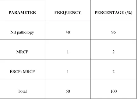
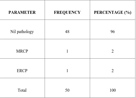
X. PANCREATIC DUCT DILATATION
Table no 17: Frequency and percentage of Pancreatic duct Dilatation
MRCP had detected pancreatic duct dilatation in one patient, whereas ERCP and MRCP; both were found to be positive for pancreatic ductal dilatation in another patient.
PARAMETER FREQUENCY PERCENTAGE (%)
Nil pathology 48 96
MRCP 1 2
ERCP+MRCP 1 2
64
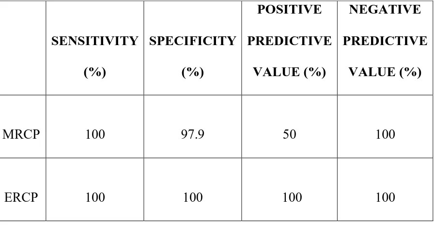
Table no 18: Diagnostic efficacy of MRCP, ERCP for Pancreatic duct dilatation
SENSITIVITY (%)
SPECIFICITY (%)
POSITIVE PREDICTIVE
VALUE (%)
NEGATIVE PREDICTIVE
VALUE (%)
MRCP 100 97.9 50 100
ERCP 100 100 100 100
65
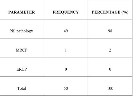
[image:73.595.74.525.242.568.2]XI. PANCREATIC DUCT VARIANTS
Table no. 19: Frequency and percentage of Pancreatic duct variants
Pancreatic duct variant was detected in one patient by MRCP. ERCP did not detect any variant of pancreatic duct.
PARAMETER FREQUENCY PERCENTAGE (%)
Nil pathology 49 98
MRCP 1 2
ERCP 0 0
66
Table no 20: Diagnostic efficacy of MRCP, ERCP for Pancreatic duct variants
SENSITIVITY (%)
SPECIFICITY (%)
POSITIVE PREDICTIVE
VALUE (%)
NEGATIVE PREDICTIVE
VALUE (%)
MRCP 0 98 0 100
67
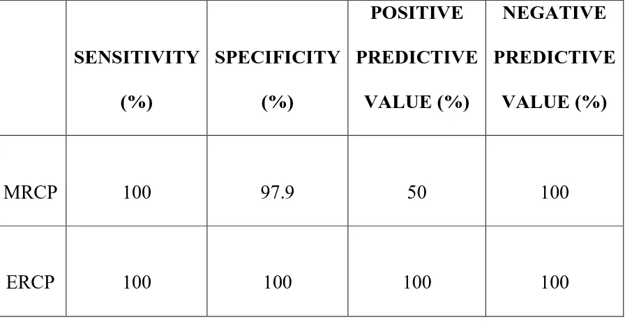
[image:75.595.74.525.242.568.2]XII. AMPULLA STONES
Table no 21: Frequency and percentage of Ampulla stones
The above table shows that ampulla stones were detected by MRCP in one patient. ERCP and MRCP both had detected ampulla stones in another patient.
PARAMETER FREQUENCY PERCENTAGE (%)
Nil pathology 48 96
MRCP 1 2
ERCP+MRCP 1 2
68
Table no 22: Diagnostic efficacy of MRCP, ERCP for Ampulla stones
SENSITIVITY (%)
SPECIFICITY (%)
POSITIVE PREDICTIVE
VALUE (%)
NEGATIVE PREDICTIVE
VALUE (%)
MRCP 100 97.9 50 100
ERCP 100 100 100 100
69
[image:77.595.74.525.242.568.2]XIII. AMPULLA STRICTURES
Table no 23: Frequency and percentage of Ampulla strictures
The above table shows that ampulla strictures were detected by MRCP and ERCP in one patient each.
PARAMETER FREQUENCY PERCENTAGE (%)
Nil pathology 48 96
MRCP 1 2
ERCP 1 2
70
Table no 24: Diagnostic efficacy of MRCP, ERCP for Ampulla strictures
SENSITIVITY (%)
SPECIFICITY (%)
POSITIVE PREDICTIVE
VALUE (%)
NEGATIVE PREDICTIVE
VALUE (%)
MRCP 0 97.9 0 97.9
ERCP 100 100 100 100
71
[image:79.595.61.538.158.622.2]XIV. AMPULLA TUMOR
Table no 25: Frequency and percentage of Ampulla tumor
The above table shows that ampulla tumor was detected in one patient each by MRCP and ERCP however was not confirmed by histopathology. MRCP and ERCP detected in one patient each, which was confirmed to be positive by histopathological examination.
PARAMETER FREQUENCY PERCENTAGE (%)
Nil pathology 46 92
ERCP 1 2
MRCP 1 2
MRCP + Histopathology 1 2
ERCP + Histopathology 1 2
72
Table no 26: Diagnostic efficacy of MRCP, ERCPfor Ampulla tumors
PARAMETER SENSITIVITY (%)
SPECIFICITY (%)
POSITIVE PREDICTIVE
VALUE (%)
NEGATIVE PREDICTIVE
VALUE (%)
MRCP 50 97.9 50 97.9
ERCP 50 97.9 50 97.9
Histopathology 100 100 100 100
73
XV. SUMMARY OF SENSITIVITY AND SPECIFICITY
Table no 27: Summary of Diagnostic efficacy for various pathologies.
The above table shows sensitivity and specificity of ERCP, MRCP and Histopathology in detecting various pathologies.
PARAMETER
MRCP ERCP Histopathology
Sensitivity (%) Specificity (%) Sensitivity (%) Specificity (%) Sensitivity (%) Specificity (%) IHBR
Strictures 100 100 100 100 100 100
Bile Duct
Stones 87.5 100 100 100
Strictures 84.6 92.3 100 100
Dilatation 100 100 100 100
Tumour 100 100 100 100 100 100
Gall Bladder
Stones 100 97.9 100 100
Pancreatic Duct
Dilatation 100 97.9 100 100
Variants 0 98
Ampulla
Stones 100 97.9 100 100
Strictures 0 97.9 100 100
74
Figure no. 14: Sensitivity of MRCP & ERCP
The above histogram shows the sensitivity of MRCP and ERCP for various pathologies
0 20 40 60 80 100 120
SENSITIVITY OF MRCP & ERCP
75
Figure no. 15: Specificity of MRCP and ERCP
The above histogram shows the specificity of MRCP and ERCP in detecting various pathologies shown above.
88 90 92 94 96 98 100 102
SPECIFICITY OF MRCP & ERCP
76
77
DISCUSSION
The aim of our study was to assess the diagnostic efficacy of MRCP as a diagnostic imaging modality, compared to invasive ERCP in the diagnosis of bile duct abnormalities, using specificity, sensitivity and positive and negative predictive values. If the results favor MRCP then diagnostic ERCP could be completely avoided and MRCP can be used as the investigation of choice for diagnosing biliary abnormalities while ERCP shall be reserved only for therapeutic purpose solely.
78
Most surgeons today would refer patients for MRCP if in doubt about the presence or absence of CBD stones on USG abdomen, when malignancy is a consideration, when ERCP is contraindicated or unsuccessful or when previous surgery such as biliary enteric anastamosis precludes ERCP. Though ERCP is considered the gold standard due to its high accuracy rate and its therapeutic value it can still be avoided for diagnostic purpose owing to its complications and operator dependency factors. MRCP has a few advantages over ERCP ; it is noninvasive ,is cheaper, radiation free, requires no anesthesia, is less operator dependent, allows better visualization of ducts proximal to an obstruction and when combined with conventional T1- and T2-weighted sequences allows detection of extraductal disease.
79
Currently, MRCP has poorer resolution than direct cholangiography and can miss small stones (<4 mm), small ampullary lesions, primary sclerosing cholangitis, and strictures of the ducts. MRCP also has difficulty in visualizing small stones in the pancreatic duct. Obstructing stones are generally easier to identify than non-obstructing stones (especially if smaller than the thickness of the acquired image slices). Small stones may not be distinguishable from sludge, mucin or even blood. Stones >4 mm are easily identified.34
In our study group of 50 patients (28 males and 22 females), age ranging from 15 to 56 years, we evaluated various causes of biliary ductal obstruction. Commonest of the findings were choledocholithiasis (56%), malignancy (12%) and stricture (28%). In our study choledocholithiasis was the commonest beingn and ampullary malignancy to be the most common malignant cause of biliary obstruction which is consistent with most of the studies carried out worldwide. Most authors have found biliary obstruction to be commoner in women35 which was not the case in our study.
80
In our study 3 patients had IHBR strictures on MRCP which were confirmed on ERCP. Thus MRCP showed 100% sensitivity and specificity in detecting IHBR strictures. According to C Bhatt et al36 there was no much difference in sensitivity and specificity of MRCP in detecting intrahepatic biliary strictures. Strictures were seen as narrowing of biliary radicles with upstream dilatation, Fig (4). The location and extent of stricture correlated with ERCP in all cases.
About 28 of 50 patients had CBD stones. It was observed that MRCP has sensitivity and specificity of 87.5% and 100% respectively in detecting bile duct stones whereas sensitivity and specificity of ERCP was 100% considering ERCP is gold standard, which is consistent with the study conducted by Verma D et al which showed sensitivities of EUS and MRCP for the detection of choledocholithiasis to be 93% and 85%.37 Stones on MRCP as well as ERCP are seen as filling defects in lumen of CBD with or without dilatation of CBD and IHBR (Figure 16).The location and number of stones found on MRCP was found to correlate with ERCP in all cases. About 14 patients had CBD strictures, which were seen as narrowing of CBD with upstream dilatation (Figure17).
81
It was observed in our study that the sensitivity of MRCP was 84% and specificity was 92% in detecting CBD strictures Figure 18). According to the study conducted by Nyree Griffin et al reported sensitivity of 91-100%.38 So the sensitivity attained in our study was slightly lower than Nyree Griffin et al but comparable to other studies and fall within the overall sensitivity range of studies carried out worldwide.
In our study MRCP showed sensitivity and specificity 100 % for CBD tumors which was higher than the study conducted by Pamos S et al39 where the sensitivity and specificity was 100 and 83.3% respectively. Additionally MRCP was found to be better in delineating the extent of tumor and extra biliary extension. As other sequences could simultaneously be acquired resectability and nodal status could also be assessed. ERCP images showed the level of block however the proximal extent and involvement of adjacent structures could not be evaluated.
82
According to the study conducted by Chen WX et al40 sensitivity and specificity in detection of ampullary carcinoma was 100% for ERCP and 26.83% for MRCP, while in our study it was 50 % and 97.9% for both. They found significant difference between MRCP and ERCP accuracy rate hence recommended ERCP in detecting ampullary carcinoma whereas our study which is in more in favor of MRCP. However owing to small sample result is inconclusive. MRCP however should score over ERCP as it offers additional advantage of simultaneous cross-sectional imaging.
About 60% patients were detected to have biliary radical dilatation on MRCP as well as ERCP. Thus MRCP had 100% sensitivity and specificity in detecting biliary duct dilatation compared to ERCP. According to Angulo et al diagnostic accuracy of MRCP was found to be greater than 90% in diagnosing biliary duct dilatation14which is slightly lesser than the results of our study.
Out of 50 patients 6% had gall bladder stones in whom MRCP sensitivity and specificity was 100% and 97.9%. According to Calvo MM41 the sensitivity of MRCP in detecting cholelithiaisis was 97.7% which is comparable with the results of our study. On MRCP stones were seen as filling defects in gall bladder.
83
100% and 97.9%. According to Meng Z the sensitivity of MRCP in detecting pancreatic duct dilatation was 72.7%.42
There were 2 patients who were detected to have ampullary stones and its sensitivity, specificity was 100% and 97.9% compared to ERCP. No major studies evaluating ampullary stones could be found, mostly as all cases of ampullary stones may be included in choledocholithiasis. However, no significant conclusion can be derived as the sample size is small.
Ampullary stricture was found in two patients, sensitivity and specificity was 0% and 97.9%. The false positive rates of MRCP were high; hence ERCP should be considered modality of choice for ampullary stricture.
84
Thus to conclude, MRCP has high sensitivity for CBD and intra-hepatic biliary radical abnormalities such as stones, strictures and malignancies. MRCP may have slightly lower sensitivity for stone detection, but still to avoid unnecessary diagnostic ERCP; in cases with suspicion (clinical/CBD -IHBR dilatation on USG) of choledocholithiasis/ampullary stones, MRCP is recommended.
85
86
SUMMARY
This was our prospective study of 50 patients the various common causes of obstructive jaundice were assessed with MRCP and the diagnostic efficacy of MRCP was evaluated comparing with ERCP and histopathology if available.
Of the 50 patients 6% had IHBR strictures, 56% had CBD stones, 28% had CBD strictures, 12% had CBD tumors, 4% had ampullary stones, 4% had ampullary strictures and 8% had ampullary tumor.
In our study 3 patients had IHBR strictures where MRCP showed 100% sensitivity and specificity in detecting IHBR strictures. Of the 50 patients 28 had CBD stones, it is observed that MRCP has sensitivity and specificity of 87.5% and 100% respectively in detecting Bile duct stones, whereas sensitivity and specificity of ERCP was 100% each.
87
However owing to inadequate study population results are inconclusive. There is significant difference between MRCP and ERCP accuracy rate in detection of ampullary carcinoma. Therefore their study recommends ERCP in detecting ampullary carcinoma whereas our study which is more in favor of MRCP.
There were 60% patients who were detected with biliary duct dilatation in ERCP which was equally detected in MRCP. Thus MRCP had 100% sensitivity and specificity in detecting biliary duct dilatation compared to ERCP.
Out of 50 patients 6% had gall bladder stones in whom MRCP sensitivity and specificity detecting gall bladder stones came up to 100% and 97.9% and was found to comparable with ERCP.
88
89
CONCLUSION
The aim of our study was to evaluate the diagnostic efficacy of MRCP compared to ERCP as a diagnostic our institution, using specificity, sensitivity, and positive and negative predictive values.
We conclude that MRCP has high diagnostic accuracy and is equivalent to ERCP in diagnosing (IHBR and CBD) abnormalities like IHBR strictures and CBD tumors.
In cases of CBD strictures, CBD stones and ampullary stones, MRCP is comparable to ERCP.
For ampullary tumors sensitivity of MRCP and ERCP was equivalent, which may be fallacious considering the small sample size.
90
Ancillary findings like intrahepatic biliary radical dilatation and gall stone disease are well demonstrated on MRCP.
MRCP as the method of choice for the diagnostic imaging of biliary and ERCP is reserved for therapeutic intervention in this setting as the commoner pathologies (stones ,strictures and malignancies in upper biliary tract) can be easily identified with high specificity with MRCP.
In cases wherein histopathology were available, that is in cases of tumors, MRCP showed high specificity as well as sensitivity.
91
LIMITATIONS AND
92
LIMITATIONS
Small study population, owing to difficulty in collecting patients who underwent both ERCP and MRCP, as most of the patients after
ultrasound are directly subjected to ERCP.
RECOMMENDATIONS
We would propose that all cases of obstructive jaundice should undergo MRCP so that ERCP can be used only for therapeutic purpose.
MRCP should be performed in cases of CBD / pancreatic duct dilatation (when the clinical data and USG abdomen are inconclusive) to RULE OUT presence of stones, strictures and malignancies to avoid complications of diagnostic ERCP.
MRCP should be performed in cases of suspected malignancies in hilar region, CBD and ampulla as it provides ancillary information as discussed.
93
94
[image:102.595.73.519.209.551.2]IMAGES
95
96
Fig .15 ERCP
ERCP
PANCREATIC DUCT
Cystic duct
IHBR
[image:104.595.132.536.239.518.2]CBD
97
Figure:19 CBD stones is seen as filling defects in ERCP and MRCP
98
Figure:19 CBD stones is seen as filling defects in ERCP and
MRCP
99
Figure20:CBD stricrtures seen as narrowing of the duct in ERCP and MRCP
100
Figure: 19. Thick slab image showing CBD stricture
101
[image:109.595.100.551.171.558.2]102
Figure no:21 CBD TUMOR : Moderate dilatation of the intrahepatic biliary channels.
There is abrupt cut of off the biliary channels at the site of the
formation of the common hepatic duct. The confluence of the right anterior
sectoral duct could not be visualized. Post contrast shows mild poorly defined
enhancement at the confluence.
103
104
105
Figure:22 Mass lesion in the region of ampulla with upstream dilatation of CBD.
106
107
108
109
[image:117.595.76.568.133.401.2]Figure:22 Mass lesion in the region of ampulla with upstream dilatation of CBD
110
Figure:22 shows features of ampullary tumor in ERCP with
upstream dilatation of CBD
111
112
BIBLIOGRAPHY
1. Tse F, Barkun JS, Romagnuolo J, Friedman G, Bornstein JD, Barkun AN. Nonoperative imaging techniques in suspected biliary tract obstruction. HPB (Oxford). 2006;8(6):409-25.
2. Hurter D, De Vries C, Potgieter P, Barry R, Botha F, Joubert G. Accuracy of MRCP compared to ERCP in the diagnosis of bile duct disorders. South African Journal of Radiology. 2008;12(1):14-22.
3. Bashir O. Biliary tree anatomy [Internet]. 2014 [cited 2014 Sep 20]. Available from: http://radiopaedia.org/articles/biliary-tree-anatomy.
4. Gray H, Standring S, Ellis H, Berkovitz B. Gray's anatomy. 1st ed. Edinburgh: Elsevier Churchill Livingstone; 2008.
5. Brant W, Helms C. Fundamentals of diagnostic radiology. 1st ed. Philadelphia: Lippincott, Williams & Wilkins; 2007.
6. Maccioni F, Martinelli M, Al Ansari N, Kagarmanova A, De Marco V, Zippi M, Marini M. Magnetic resonance cholangiography: past, present and future: a review. Eur Rev Med Pharmacol Sci. 2010 Aug;14(8):721-5. 7. Coakley FV, Schwartz LH. Magnetic resonance