Copyright © 2001, American Society for Microbiology. All Rights Reserved.
Adeno-Associated Virus Type 2-Mediated Gene Transfer:
Role of Cellular FKBP52 Protein in Transgene Expression
KEYUN QING,
1,2,3JONATHAN HANSEN,
1,2,3KIRSTEN A. WEIGEL-KELLEY,
1,2,3MENGQUN TAN,
1,2,3SHANGZHEN ZHOU,
4ANDARUN SRIVASTAVA
1,2,3,5*
Department of Microbiology & Immunology,
1Walther Oncology Center,
2Walther Cancer Institute,
3and Division of
Hematology/Oncology,
5Department of Medicine, Indiana University School of Medicine,
Indianapolis, Indiana 46202, and Avigen, Inc., Alameda, California 94501
4Received 27 February 2001/Accepted 22 June 2001
Although adeno-associated virus type 2 (AAV) has gained attention as a potentially useful vector for human
gene therapy, the transduction efficiencies of AAV vectors vary greatly in different cells and tissues in vitro and
in vivo. We have documented that a cellular tyrosine phosphoprotein, designated the single-stranded
D-sequence-binding protein (ssD-BP), plays a crucial role in AAV-mediated transgene expression (K. Y. Qing,
X.-S. Wang, D. M. Kube, S. Ponnazhagan, A. Bajpai, and A. Srivastava, Proc. Natl. Acad. Sci. USA 94:10879–
10884, 1997). We have documented a strong correlation between the phosphorylation state of ssD-BP and AAV
transduction efficiency in vitro as well as in vivo (K. Y. Qing, B. Khuntrirat, C. Mah, D. M. Kube, X.-S. Wang,
S. Ponnazhagan, S. Z. Zhou, V. J. Dwarki, M. C. Yoder, and A. Srivastava, J. Virol
.
72:1593–1599, 1998). We
have also established that the ssD-BP is phosphorylated by epidermal growth factor receptor protein tyrosine
kinase and that the tyrosine-phosphorylated form, but not the dephosphorylated form, of ssD-BP prevents AAV
second-strand DNA synthesis and, consequently, results in a significant inhibition of AAV-mediated transgene
expression (C. Mah, K. Y. Qing, B. Khuntrirat, S. Ponnazhagan, X.-S. Wang, D. M. Kube, M. C. Yoder, and
A. Srivastava, J. Virol
.
72:9835–9841, 1998). Here, we report that a partial amino acid sequence of ssD-BP
purified from HeLa cells is identical to a portion of a cellular protein that binds the immunosuppressant drug
FK506, termed the FK506-binding protein 52 (FKBP52). FKBP52 was purified by using a prokaryotic
expres-sion plasmid containing the human cDNA. The purified protein could be phosphorylated at both tyrosine and
serine or threonine residues, and only the phosphorylated forms of FKBP52 were shown to interact with the
AAV single-stranded D-sequence probe. Furthermore, in in vitro DNA replication assays,
tyrosine-phospho-rylated FKBP52 inhibited AAV second-strand DNA synthesis by greater than 90%. Serine- or
threonine-phosphorylated FKBP52 caused
⬇
40% inhibition, whereas dephosphorylated FKBP52 had no effect on AAV
second-strand DNA synthesis. Deliberate overexpression of FKBP52 effectively reduced the extent of tyrosine
phosphorylation of the protein, resulting in a significant increase in AAV-mediated transgene expression in
human and murine cell lines. These studies corroborate the idea that the phosphorylation status of the cellular
FKBP52 protein correlates strongly with AAV transduction efficiency, which may have important implications
for the optimal use of AAV vectors in human gene therapy.
Adeno-associated virus type 2 (AAV) is a small,
nonpatho-genic, single-stranded DNA-containing virus which requires
coinfection with a helper virus, usually adenovirus, for its
op-timal replication (1, 31). In the absence of coinfection with the
helper virus, the wild-type AAV establishes a latent infection
in which the viral genome integrates into human chromosomal
DNA in a site-specific manner (22, 23, 45). The nonpathogenic
nature of AAV coupled with the remarkable site specificity of
integration prompted the development of recombinant AAV
vectors for gene transfer and gene therapy. Although
recom-binant AAV genomes do not appear to integrate site
specifi-cally, AAV vectors have been successfully used for gene
deliv-ery to a wide variety of cells and tissues in vitro and in vivo (2,
3, 11, 12, 16–19, 21, 32–36, 47, 48, 51, 55, 57), as well as in
phase I clinical trials for gene therapy of cystic fibrosis and
hemophilia B (11, 18). However, the transduction efficiencies
of AAV vectors vary greatly in different cell types. Studies from
two independent laboratories have suggested that following
infection, the viral second-strand DNA synthesis is a
rate-limiting step in efficient transduction by AAV vectors (8, 9).
We have documented that a host cell protein, designated the
single-stranded D-sequence binding protein (ssD-BP),
inter-acts specifically and preferentially with the D sequence within
the inverted terminal repeat (ITR) at the 3
⬘
end of the AAV
genome and, in its tyrosine-phosphorylated form, prevents
vi-ral second-strand DNA synthesis, resulting in inhibition of
AAV-mediated transgene expression. ssD-BP is
phosphory-lated at tyrosine residues by the epidermal growth factor
re-ceptor protein tyrosine kinase (EGFR-PTK), and the
phos-phorylation state of ssD-BP correlates with the AAV
transduction efficiency in established and primary human cells
in vitro and in murine tissues in vivo (25, 26, 40, 42, 52, 53).
Despite the crucial role that ssD-BP plays in AAV-mediated
transgene expression, its identity has remained unknown.
In this report, we present data on the purification and
char-acterization of ssD-BP. The partial amino acid sequence of this
protein, purified to homogeneity from HeLa cells, revealed
100% homology to a cellular protein, termed FK506-binding
* Corresponding author. Mailing address: Department of
Microbi-ology & ImmunMicrobi-ology, Indiana University School of Medicine, Medical
Science Building Room 257, 635 Barnhill Dr., Indianapolis, IN
46202-5120. Phone: (317) 274-2194. Fax: (317) 274-4090. E-mail: asrivast
@iupui.edu.
8968
on November 9, 2019 by guest
http://jvi.asm.org/
protein 52 (FKBP52), which binds the immunosuppressant
drug FK506. This 52-kDa protein, which has also been shown
to be a chaperone protein, is ubiquitous, is phosphorylated,
and localizes predominantly to the nucleus, properties that
are shared with ssD-BP. The purified recombinant human
FKBP52 protein could be phosphorylated by both casein
ki-nase II (CK II) and EGFR-PTK. The purified protein was also
shown to interact with the AAV single-stranded D-sequence
probe by electrophoretic mobility shift assays (EMSA).
Fur-thermore, in in vitro DNA replication assays,
EGFR-PTK-phosphorylated FKBP52 inhibited AAV second-strand DNA
synthesis by greater than 90%. CK II-phosphorylated FKBP52
caused
⬇
40% inhibition, whereas unphosphorylated FKBP52
had no effect on AAV second-strand DNA synthesis.
Deliber-ate overexpression of FKBP52 led to a reduction in tyrosine
phosphorylation of the protein, which resulted in a significant
increase in AAV-mediated transgene expression in human and
murine cell lines. These studies corroborate the idea that the
cellular FKBP52 protein is a crucial determinant of AAV
transduction efficiency, which in turn may have important
im-plications for the optimal use of AAV vectors in human gene
therapy.
MATERIALS AND METHODS
Cells, viruses, plasmids, and antibodies.The human cervical carcinoma cell line HeLa, the adenovirus-transformed human embryonic kidney cell line 293, and the murine fibroblast cell line NIH 3T3 were obtained from the American Type Culture Collection (Manassas, Va.) and maintained as monolayer cultures in Iscove’s modified Dulbecco’s medium (IMDM) supplemented with 10% fetal bovine serum and 1% (by volume) 100⫻stock solution of antibiotics (10,000 U of penicillin plus 10,000g of streptomycin). Human AAV and adenovirus type 2 stocks were kindly supplied by Kenneth I. Berns (University of Florida, Gaines-ville) and Kenneth H. Fife (Indiana University School of Medicine, Indianapo-lis), respectively. The recombinant plasmids pQE-30 and pCMVwere obtained from Qiagen (Valencia, Calif.) and Clontech (Palo Alto, Calif.), respectively. Antibodies specific for human FKBP52 (goat polyclonal immunoglobulin G [IgG]) and human1integrin (mouse monoclonal IgG1) were purchased from
Santa Cruz Biotechnology (Santa Cruz, Calif.) and Chemicon Corp. (Temecula, Calif.), respectively.
Preparation of WCEs.Whole-cell extracts (WCEs) from HeLa, 293, and NIH 3T3 cells were prepared according to the method described by Muller (30). The total protein concentration was determined by the Bio-Rad Laboratories (Her-cules, Calif.) protein assay kit, and the extracts were frozen in liquid N2and
stored at⫺70°C.
EMSA were performed as described previously (42, 53). Briefly, DNA-binding reactions were performed in a volume of 20l with 2g of poly(dI-dC), 2g of bovine serum albumin, and 12% glycerol in HEPES buffer (pH 7.9). Ten micro-grams of proteins from each WCE were preincubated for 10 min at 25°C fol-lowed by the addition of 10,000 cpm of32P-labeled D(⫺)-sequence synthetic
oligonucleotide (5⬘-AGGAACCCCTAGTGATGGAG-3⬘) to the reaction mix-ture. The binding reaction was allowed to proceed for 30 min at 25°C. In some experiments, specific (FKBP52) and nonspecific (1integrin) antibodies were
also used in supershift assays as described previously (15, 20) with the following modifications. Briefly, the reaction mixture was incubated with 2g of antibody on ice for 1 h and then at 25°C for 15 min. In some experiments, WCEs were also immunoprecipitated with anti-FKBP52 antibody, and supernatants and resus-pended pellets were used in EMSA as previously described (44). Bound com-plexes were separated from the unbound probe on low-ionic-strength 4% poly-acrylamide gels using Tris-glycine-EDTA buffer (pH 8.5) containing 50 mM Tris-HCl, 380 mM glycine, and 2 mM EDTA. Following electrophoresis, the gel was dried in vacuo and autoradiographed with Kodak X-Omat film at⫺70°C.
Purification of ssD-BP.WCE was prepared as described above from⬇2.6⫻ 1010HeLa cells purchased from the National Cell Culture Center (Washington,
D.C.). All steps were carried out at 4°C. The WCE was subjected to ultrafiltra-tion on Centricon columns (Amicon, Beverly, Mass.) to remove proteins with masses of less than 30 kDa. The rest of the WCE was fractionated on a Sephacryl S-200 HR column (Sigma, St. Louis, Mo.). The column bed volume was 100 ml,
which was equilibrated with 2 bed volumes of buffer A (20 mM Tris-HCl [pH 7.5], 50 mM NaCl, 1 mM MgCl2, 0.5% NP-40, 0.5 mM phenylmethylsulfonyl
fluoride, 0.5 mM dithiothreitol). After the sample was loaded, the column was washed and eluted with 2 bed volumes of buffer A. Four-milliliter fractions were collected, and the protein concentration was determined. All fractions containing proteins were used in EMSA with the AAV D-sequence probe as described above. All positive fractions were pooled and fractionated by anion-exchange chromatography on a DE-52 column. The column bed volume was 50 ml, which was equilibrated with buffer A. After the sample was loaded, the column was washed with 1 bed volume of buffer A followed by elution with a continuous NaCl concentration gradient (50 to 500 mM). Two-milliliter fractions were col-lected, and the protein concentration was determined. All fractions containing proteins were dialyzed overnight against buffer A, followed by EMSA. All pos-itive fractions were pooled and subjected to chromatography using a nonspecific ssDNA-agarose column (Life Technologies, Rockville, Md.). The column bed volume was 2 ml, which was equilibrated with buffer A. After the sample was loaded, the column was washed with 1 bed volume of buffer A followed by elution with a stepwise NaCl concentration gradient (100 mM to 1 M). Two-milliliter fractions were collected and dialyzed overnight against buffer A, fol-lowed by EMSA. All positive eluates containing the ssD-BP were incubated with the ssD sequence-ligated streptavidin magnetic particles (Boehringer Mann-heim, Indianapolis, Ind.) according to the instructions provided by the vendor. The bound ssD-BP was eluted from the particles and electrophoresed on pre-parative sodium dodecyl sulfate (SDS)–10% polyacrylamide gels. A single pro-tein band with a mass of⬇52 kDa was excised and shipped to the Harvard Microchemistry Facility, Harvard University (Cambridge, Mass.) for mass spec-trometry and protein microsequencing analyses.
Expression and purification of the recombinant human FKBP52 protein from
Escherichia coli. A prokaryotic expression plasmid containing the human FKBP52 gene was generated by PCR amplification from a HeLa cell Marathon-ready cDNA library (Clontech) with the following primer pair: 5⬘primer, GAT GACAGCCGAGGAGATGAAGGCGACCGA, and 3⬘primer, GTTATGCTT CTGTCTCCACCTGAGACTGGC. The sequence of the PCR product was confirmed by sequencing and then inserted into the pQE-30 prokaryotic expres-sion vector. The recombinant FKBP52 protein was produced and purified using the His tag purification system and Ni2⫹affinity chromatography (Qiagen)
ac-cording to the instructions provided by the vendor. A eukaryotic expression plasmid containing the human FKBP52 cDNA gene under the control of the cytomegalovirus (CMV) immediate-early promoter was also constructed by stan-dard methods as described previously (41).
In vitro phosphorylation assays.In vitro phosphorylation by EGFR-PTK was carried out as previously described by Weber et al. (54) and Cybulsky et al. (4) with the following modifications. The complete reaction mixture contained 1g of the purified FKBP52 protein, 20 mM HEPES, 4 mM MgCl2, 10 mM MnCl2,
50 mM NaOV, 200M ATP, 10Ci (0.37 mBq) of [␥-32P]ATP, and 1 U (15,000
U/mg) of purified EGFR-PTK (CalBiochem, La Jolla, Calif.) with all appropri-ate controls. In vitro phosphorylation by CK II (CalBiochem) was carried out as previously described by McElhinny et al. (28) and Russo et al. (43). Briefly, the complete reaction mixture contained 1g of the FKBP52 protein expressed in and purified from bacterial cultures, 20 mM Tris-HCl, 50 mM KCl, 10 mM MgCl2, 50 mM NaOV, 200M ATP, 10Ci (0.37 mBq) of [␥-32P]ATP, and 100
U (300,000 U/mg) of CK II. The reaction mixtures were incubated at 30°C for 1 h and electrophoresed on SDS–10% polyacrylamide gels. The gels were dried in vacuo followed by autoradiography using Kodak X-Omat film at⫺70°C. In some experiments, in vitro phosphorylation assays with EGFR-PTK and CK II were also carried out without the addition of [␥-32P]ATP. The tyrosine- and serine- or
threonine-phosphorylated FKBP52 proteins were then used in EMSA with the radiolabeled D(⫺) probe as described above and in in vitro DNA replication assays as described below.
In vitro DNA replication assays.The appropriate AAV DNA substrate con-taining the 3⬘hairpin structure was prepared and labeled with [␥-32P]ATP (3,000
Ci/mmol) by using T4 polynucleotide kinase as described previously (44). To assess the effect of ssD-BP on AAV DNA replication (second-strand DNA synthesis), 20 ng of the unphosphorylated, EGFR-PTK-phosphorylated, or CK II-phosphorylated form of the purified FKBP52 protein was added to the end-labeled 3⬘-hairpin ITR and all four unlabeled deoxynucleoside triphosphates and incubated for 15 min at 25°C prior to adding the Klenow enzyme. After the Klenow enzyme was added, the reaction mixtures were incubated at 37°C for 30 min and then electrophoresed on 6% polyacrylamide gels. The gels were dried and autoradiographed at⫺70°C as described above.
Western blot analyses.To determine the levels of human FKBP52 in trans-fected cell lines, approximately 2⫻106cells were seeded in culture dishes, and
24 h later, WCEs were prepared by an SDS lysis procedure described elsewhere
on November 9, 2019 by guest
http://jvi.asm.org/
(10). Total protein concentrations were determined using the Bio-Rad protein assay kit, and 30g of protein was separated by SDS-polyacrylamide gel elec-trophoresis on a 10% polyacrylamide gel. After transfer to an Immobilon-P membrane (Millipore, Bedford, Mass.), the membrane was blocked for 1 h at 25°C with 1⫻Tris-buffered saline (TBS; 20 mM Tris-HCl, pH 7.5, 150 mM NaCl), 0.05% Tween 20, and 5% nonfat dry milk (TBST-milk), incubated with a 1:200 dilution of anti-FKBP52 antibody in TBST-milk for 1 h at 25°C, and then washed three times in TBST. Following incubation with a 1:2,000 dilution of horseradish peroxidase-coupled anti-goat IgG antibody in TBST-milk for 1 h at 25°C, the membrane was washed three times in TBST, and protein bands were visualized with the ECL-Plus chemiluminescence detection kit (Amersham, Lit-tle Chalfont, England) according to the instructions provided by the manufac-turer.
Recombinant AAV-mediated transduction assays.Approximately 105cells per
well were plated in a 12-well plate. Twelve hours later, the cells were washed once with IMDM and then infected at 37°C for 2 h with 5⫻103particles of a
recombinant AAV vector containing the-galactosidase (lacZ) reporter gene driven by the CMV immediate-early promoter (vCMVp-lacZ) per cell. The cells were then incubated with complete IMDM containing 10% fetal bovine serum and 1% antibiotics for 48 h. The-galactosidase activity was measured by the Galacto-Light Plus chemiluminescence reporter assay (Tropics, Inc., Bedford, Mass.) according to the manufacturer’s instructions. The data were expressed as relative light units per microgram of total protein and were within the linear range of the assay.
RESULTS
ssD-BP is a cellular chaperone protein, FKBP52, an
immu-nophilin.
We purified ssD-BP to homogeneity from HeLa cells
as described in Materials and Methods. A small fraction of the
protein was electrophoresed on an SDS–10% polyacrylamide
gel followed by staining with Coomassie blue (Fig. 1A, lane 4).
As can be seen, a single protein band with a mass of
⬇
52 kDa
was obtained which interacted with the single-stranded AAV
D(
⫺
) sequence probe in EMSA. When this protein was
ex-cised from preparative gels and subjected to protein
microse-quencing, the amino acid sequence of the largest peptide,
containing 24 amino acids, revealed 100% homology to a
cel-lular protein, FKBP52, that binds the immunosuppressant drug
FK506 (Fig. 1B). Several shorter peptides, ranging from 6 to 22
amino acids, also showed 100% homology to FKBP52.
FKBP52 is a 52-kDa cellular protein, also known as an
immu-nophilin (46), is ubiquitous, is phosphorylated, and localizes
predominantly to the nucleus (5, 37, 38), several properties
that are shared with ssD-BP. The identity of ssD-BP as the
cellular FKBP52 was tested in the following two sets of
exper-iments. In the first set, supershift EMSA were performed using
anti-FKBP52 antibody. The results are shown in Fig. 2. As can
be seen, the AAV D-sequence probe (lane 1) formed a
slower-migrating complex with WCE from HeLa cells (lane 2) and a
faster-migrating complex with WCE from 293 cells (lane 3),
consistent with our previously published reports (40, 42); the
inclusion of anti-FKBP52 antibody in these assays resulted in a
nearly complete supershift with HeLa (lane 4) and 293 (lane 5)
WCEs, respectively. These supershifted complexes were not
detected when anti-

1integrin antibody was used as an
appro-priate control (lanes 6 and 7). The AAV D-sequence probe did
not form a complex with either anti-FKBP52 antibody (lane 8)
or anti-

1integrin antibody (lane 9).
In the second set of experiments, WCEs from HeLa and 293
cells were immunoprecipitated with anti-FKBP52 antibody,
and the supernatants and pellets, following resuspension, were
used in EMSA. As can be seen in Fig. 2, prior
immunoprecipi-tation with anti-FKBP52 antibody eliminated ssD-BP from
WCEs from both HeLa and 293 cells (lanes 10 and 11), and it
could be recovered from the pellets (lanes 12 and 13). Taken
together, these results corroborate the notion that ssD-BP is
FKBP52. However, it was crucial to document that (i) the
FKBP52 protein is phosphorylated in vitro by EGFR-PTK, (ii)
the FKBP52 protein binds to the AAV D(
⫺
) sequence, and
(iii) tyrosine-phosphorylated FKBP52, but not
unphosphory-lated FKBP52, inhibits AAV second-strand DNA synthesis.
Each of these requirements was experimentally tested as
fol-lows.
FKBP52 can be phosphorylated at tyrosine residues by
EGFR-PTK.
Previous studies have shown that FKBP52 can be
phosphorylated in vitro by CK II (29). Using in vitro
phosphor-ylation assays, we wished to document whether purified
FKBP52 could also be phosphorylated by EGFR-PTK, since
ssD-BP is phosphorylated by EGFR-PTK (26). These assays
were carried out as described in Materials and Methods.
Be-cause both CK II and EGFR-PTK are known to be
autophos-phorylated, assays were also carried out in the absence of
FKBP52. The results are shown in Fig. 3. It is evident that
FKBP52 is phosphorylated not only at serine or threonine
residues (lane 2), as reported previously (29), but also at
ty-rosine residues (lane 4), consistent with our previous studies
(26).
Phosphorylation of FKBP52 dramatically stimulates its
in-teraction with the AAV D-sequence.
It was of interest to
de-termine whether the purified FKBP52 protein could bind to
the AAV D(
⫺
) sequence. The purified FKBP52 protein was
used in EMSA with the AAV D(
⫺
) probe, with and without
prior in vitro phosphorylation by CK II and EGFR-PTK as
described above. The CK II and EGFR-PTK proteins were
also used as appropriate controls. The results are shown in Fig.
FIG. 1. (A) Purification of ssD-BP from HeLa cells.
SDS-poly-acrylamide gel electrophoretic pattern of purified ssD-BP. Lane 1,
protein molecular size markers; lane 2, WCE of HeLa S3 cells (20
g
of total protein); lane 3, Oct2A factor, included as a positive control,
purified according to the standard protocol using the protein
purifica-tion kit supplied by the vendor (Boehringer Mannheim); lane 4,
puri-fied ssD-BP. The arrow indicates the
⬇
52-kDa ssD-BP. (B) Deduced
amino acid sequence of the human FKBP52 protein. The boldface
underlined amino acids represent the identified homology between
ssD-BP and FKBP52 (GenBank accession no. M88279).
on November 9, 2019 by guest
http://jvi.asm.org/
[image:3.612.70.279.73.270.2]4. It is evident that little binding of unphosphorylated FKBP52
to the D-sequence probe occurred (lane 2), whereas FKBP52
phosphorylated at serine or threonine residues by CK II (lane
3) formed a complex with the probe. CK II alone did not
interact with the D(
⫺
) probe (lane 4). FKBP52
phosphory-lated at tyrosine residues by EGFR-PTK (lane 5) formed two
distinct complexes, whereas EGFR-PTK alone did not interact
with the D(
⫺
) probe (lane 6). There are 16 tyrosine residues in
FKBP52, and it is possible that the faster-migrating complex in
lane 5 is the partially phosphorylated FKBP52. These results
nonetheless corroborate the notion that the ssD-BP is
FKBP52.
Only phosphorylated forms of FKBP52 inhibit AAV
second-strand DNA synthesis.
We also wished to examine the effect of
the unphosphorylated and/or phosphorylated FKBP52 on
AAV second-strand DNA synthesis. The purified FKBP52
protein, with and without phosphorylation with CK II or
EGFR-PTK, was used in in vitro DNA replication assays as
described in Materials and Methods. As shown in Fig. 5, the
radiolabeled AAV hairpin DNA template (lane 1) was readily
converted into its duplex counterpart following second-strand
DNA synthesis by the Klenow enzyme (lane 2). Prior
incuba-tion with the unphosphorylated FKBP52 protein had no effect
(lane 3), and CK II-phosphorylated FKBP52 inhibited
second-strand DNA synthesis by
⬇
40% (lane 4) as determined by
densitometric scanning of the autoradiographs. CK II in the
absence of FKBP52 had no effect (lane 5). FKBP52
phosphor-ylated by EGFR-PTK inhibited viral second-strand DNA
syn-thesis by
⬎
90% (lane 6), and EGFR-PTK alone had no effect
(lane 7). Thus, although the assay utilized here is somewhat
artificial, it documents the fact that the phosphorylated forms,
but not the unphosphorylated form, of FKBP52 inhibit viral
second-strand DNA synthesis.
Deliberate overexpression of FKBP52 in established cell
lines leads to a significant increase in AAV-mediated
trans-FIG. 2. Electrophoretic mobility supershift assays and
immunopre-cipitation of WCEs with anti-FKBP52 antibody. The AAV D-sequence
probe (lane 1) was incubated with WCEs prepared from HeLa cells to
yield a slower-migrating complex (lane 2; solid arrow) and with those
from 293 cells to form a faster-migrating complex (lane 3; solid
arrow-head). These complexes were supershifted by incubation with
anti-FKBP52 antibody with HeLa (lane 4; open arrow) and 293 (lane 5;
open arrowhead) WCEs, respectively, but not with anti-
1
integrin
antibody (lanes 6 and 7). No complex formation occurred between the
AAV D-sequence probe and either anti-FKBP52 antibody (lane 8) or
anti-
1
integrin antibody (lane 9). When WCEs from HeLa and 293
cells were immunoprecipitated with anti-FKBP52 antibody and
super-natants and pellets, following resuspension, were used in EMSA, prior
immunoprecipitation with anti-FKBP52 antibody eliminated ssD-BP
from WCEs from both HeLa and 293 cells (lanes 10 and 11), and it
could be recovered from the pellets (lanes 12 and 13).
FIG. 3. In vitro phosphorylation of purified FKBP52 protein by CK
II and EGFR-PTK. CK II was incubated in the absence (
⫺
; lane 1) or
presence (
⫹
; lane 2) of 1
g of FKBP52. Similarly, EGFR-PTK was
incubated in the absence (lane 3) or presence (lane 4) of FKBP52 as
described in Materials and Methods. The arrow indicates the
phos-phorylated FKBP52 protein, and the arrowheads denote the
autophos-phorylated CK II and EGFR-PTK proteins.
on November 9, 2019 by guest
http://jvi.asm.org/
[image:4.612.58.289.76.462.2]gene expression.
In order to examine the effect of the FKBP52
protein on AAV-mediated transgene expression in vivo, we
also generated a eukaryotic expression plasmid containing the
human FKBP52 gene driven by the CMV promoter. Human
HeLa and 293 cells and murine NIH 3T3 cells were stably
transfected with this plasmid, and the WCEs prepared were
analyzed to detect human FKBP52 on Western blots using
anti-human FKBP52 antibody as described in Materials and
Methods. The results are shown in Fig. 6. Densitometric
scan-ning of lumigraphs revealed
⬇
2-fold overexpression of the
human FKBP52 in HeLa and 293 cells. In NIH 3T3 cells, the
level of expression of the human FKBP52 was similar to those
in mock-transfected 293 and HeLa cells. WCEs prepared from
each cell type as well as cells stably transfected with the human
FKBP52 expression plasmid were also used in EMSA with the
AAV D(
⫺
) probe, and the results are shown in Fig. 7. It is
evident that in both NIH 3T3 and HeLa cells, deliberate
over-expression of FKBP52 led to a significant reduction of
ty-rosine-phosphorylated FKBP52, the underlying mechanism of
which is unclear. No effect was observed in 293 cells, since the
FKBP52 present in these cells is phosphorylated
predomi-nantly at serine or threonine residues.
Since FKBP52, dephosphorylated at tyrosine residues,
would be expected to be less inhibitory to AAV second-strand
DNA synthesis, we wished to determine whether NIH 3T3 and
HeLa cells, stably transfected with the FKBP52 expression
plasmid, would allow an increase in AAV-mediated transgene
expression. Mock-transfected or FKBP52 expression
[image:5.612.318.548.77.429.2]plasmid-FIG. 4. Electrophoretic mobility shift assays for the AAV D(
⫺
)
sequence (lane 1) interaction with human FKBP52 purified from
bac-terial cells without (lane 2) and with prior in vitro phosphorylation with
CK II (lane 3) and EGFR-PTK (lane 5), respectively. These assays
were performed as described in Materials and Methods. No interaction
between the probe and CK II alone (lane 4) or EGFR-PTK alone (lane
6) was observed. Complexes presumed to contain the phosphorylated
forms of the FKBP52 protein are denoted by the solid arrows and
arrowhead, and the unphosphorylated form is denoted by the open
arrowhead.
FIG. 5. In vitro replication assays for the effects of the purified
FKBP52 protein, with and without phosphorylation by CK II or
EGFR-PTK, on AAV second-strand DNA synthesis. These assays
were carried out as described in Materials and Methods. The
radiola-beled AAV hairpin (HP) DNA template (lane 1) (shown schematically
as the upper figure on the left) was readily converted into its duplex
counterpart (shown schematically as the lower figure on the left)
fol-lowing second-strand DNA synthesis by the Klenow enzyme (lane 2).
Prior incubation with the dephosphorylated FKBP52 protein had no
effect (lane 3), while CK II-phosphorylated FKBP52 inhibited
second-strand DNA synthesis by
⬇
40% (lane 4). CK II in the absence of
FKBP52 had no effect (lane 5). FKBP52 phosphorylated by
EGFR-PTK inhibited viral second-strand DNA synthesis by
⬎
90% (lane 6),
and EGFR-PTK alone had no effect (lane 7). BSA, bovine serum
albumin;
⫹
, present;
⫺
, absent.
on November 9, 2019 by guest
http://jvi.asm.org/
[image:5.612.101.250.79.444.2]transfected HeLa, 293, and NIH 3T3 cells were either mock
infected or infected with a recombinant AAV-lacZ
vector
un-der identical conditions. Transgene expression was evaluated
48 h postinfection. The results are shown in Fig. 8. It is evident
that in both HeLa and NIH 3T3 cells, with reduced levels of
tyrosine-phosphorylated FKBP52, the AAV-mediated
trans-duction efficiency was increased approximately 2- and 10-fold,
respectively. However, since FKBP52 in 293 cells is present in
predominantly dephosphorylated form (Fig. 7), FKBP52
over-expression did not significantly increase the AAV transduction
efficiency in these cells. These studies document that there is a
strong correlation between the tyrosine-phosphorylation status
of the cellular FKBP52 protein and AAV transduction
effi-ciency.
DISCUSSION
Although AAV has gained attention as a useful alternative
to the more commonly used retrovirus- and adenovirus-based
vectors for human gene therapy, recent studies from our
lab-oratory and others have suggested that there are at least three
major obstacles that limit high-efficiency transduction by AAV
vectors in certain cell types. These include the lack of
expres-sion of the cellular receptor and coreceptor for AAV infection
(41, 49, 50), impaired intracellular trafficking of the virus into
the nucleus (13, 14), and the inability of AAV to undergo viral
second-strand DNA synthesis to yield the transcriptionally
ac-tive double-stranded template (8, 9). For example, it has
be-come abundantly clear that AAV infection requires the cell
surface expression of heparan sulfate proteoglycan as a
recep-tor for viral binding (50) and fibroblast growth facrecep-tor receprecep-tor
1 and/or
␣
V

5 integrin as a coreceptor for viral entry (41, 49).
Second, endosomal processing has recently been suggested to
lead to efficient intracellular trafficking of AAV into the
nu-cleus (6, 7). Third, we have documented that a cellular protein,
designated ssD-BP, the identity of which has thus far remained
unknown, plays an important role in viral second-strand DNA
synthesis (25, 26, 40, 42).
[image:6.612.71.277.75.268.2]In the present studies, we purified the ssD-BP and identified
it as a 52-kDa cellular protein, FKBP52, that binds the
immu-nosuppressant drug FK506. The human FKBP52, also known
as an immunophilin, is ubiquitous, is phosphorylated, and
lo-calizes predominantly to the nucleus, properties that are
shared with ssD-BP (40–42). Using the purified recombinant
protein, we documented that (i) FKBP52 can be
phosphory-lated in vitro at both serine and threonine residues by CK II
and at tyrosine residues by EGFR-PTK; (ii) phosphorylated
[image:6.612.330.545.80.484.2]FIG. 6. Western blot analysis for expression of human FKBP52 in
human 293 and HeLa cells and murine NIH 3T3 cells.
Mock-trans-fected cells (lanes 1, 3, and 5) and cells stably transMock-trans-fected with a human
FKBP52 expression plasmid (lanes 2, 4, and 6) were analyzed using
human anti-FKBP52 antibody as described in Materials and Methods.
The arrow indicates the 52-kDa human FKBP52 protein.
⫹
, present;
⫺
, absent.
FIG. 7. Electrophoretic mobility shift assays for the AAV D(
⫺
)
sequence interaction with FKBP52 in mock-transfected NIH 3T3, 293,
and HeLa cells (lanes 2, 4, and 6) or cells stably transfected with the
human FKBP52 expression plasmid (lanes 3, 5, and 7). These assays
were carried out as described in the legend to Fig. 4. The
tyrosine-phosphorylated form of the FKBP52 protein is denoted by the arrow,
and the serine- or threonine-phosphorylated form is denoted by the
arrowhead.
⫹
, present;
⫺
, absent.
on November 9, 2019 by guest
http://jvi.asm.org/
forms, but not the unphosphorylated form, of FKBP52 interact
efficiently in vitro with the AAV single-stranded D sequence;
(iii) FKBP52 phosphorylated at tyrosine residues inhibits AAV
second-strand DNA synthesis more efficiently than that
phos-phorylated at serine or threonine residues, whereas
unphos-phorylated FKBP52 has no effect; and (iv) deliberate
overex-pression of FKBP52 effectively reduces tyrosine phosphorylation
of the protein, which leads to more efficient AAV-mediated
transgene expression in human and murine established cell
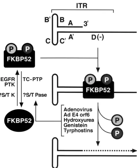
lines in vitro. In view of these data, we have revised our
pre-viously published model of AAV DNA second-strand synthesis
and transgene expression (25, 26), as shown in Fig. 9. In the
revised model, cellular FKBP52, phosphorylated either at
ty-rosine residues by EGFR-PTK or at serine or threonine
resi-dues (previously assumed to be the unphosphorylated form of
ssD-BP [27]) by an unknown cellular serine or threonine
pro-tein tyrosine kinase, interacts with the D(
⫺
) sequence in the
AAV ITR and inhibits viral second-strand DNA synthesis.
Coinfection with adenovirus, expression of adenovirus E4orf6
protein, or treatment with inhibitors of tyrosine and serine or
threonine kinase inhibitors leads to dephosphorylation of
FKBP52, which can no longer bind to the D(
⫺
) sequence,
thereby allowing viral second-strand DNA synthesis and,
con-sequently, efficient transgene expression.
In our previously published studies with HeLa cells (26), we
did not see any effect of staurosporine, a serine or threonine
kinase inhibitor, on AAV-mediated transgene expression.
Since FKBP52 present in HeLa cells is phosphorylated
pre-dominantly at tyrosine residues, that was not unexpected.
However, since KB cells contain FKBP52 phosphorylated at
both tyrosine and serine or threonine residues (40), we have
examined the effects of tyrphostin 1 and staurosporine on
AAV-mediated
lacZ transgene expression in KB cells. These
results document that treatment with tyrphostin 1 or
stauro-sporine leads to a significant increase in AAV-mediated
trans-gene expression in KB cells (data not shown). Thus, the
iden-tification of the putative cellular serine or threonine kinase
which catalyzes phosphorylation of FKBP52 remains a high
priority.
[image:7.612.69.541.86.226.2]In preliminary experiments, we have tentatively identified a
cellular tyrosine phosphatase, designated T-cell protein
ty-rosine phosphatase (TC-PTP) (24, 56), which catalyzes
de-phosphorylation of FKBP52, since stable transfection of a
TC-PTP expression plasmid into HeLa cells led to a significant
[image:7.612.316.553.392.682.2]FIG. 9. Revised model for the role of the cellular FKBP52 protein
in AAV second-strand DNA synthesis. See the text for details. Shaded
circles represent phosphorylated serine (or threonine) and tyrosine
residues. The broken-lined arrow indicates the viral second-strand
DNA synthesis.
FIG. 8. Comparative analyses of AAV-mediated transduction efficiency in cells overexpressing the FKBP52 protein. Mock-transfected or
FKBP52 expression plasmid-transfected HeLa (A), 293 (B), and NIH 3T3 (C) cells were either mock infected or infected with a recombinant
AAV-
lacZ
vector under identical conditions. Transgene expression was evaluated 48 h postinfection as described in Materials and Methods. These
data represent results from experiments performed in triplicate with the standard error of the mean. Statistical differences were determined by
using an unpaired Student
t
test.
⫹
, present;
⫺
, absent.
on November 9, 2019 by guest
http://jvi.asm.org/
increase in AAV-mediated transgene expression (K. Qing, W.
Li, M. Tan, M. C. Yoder, and A. Srivastava, unpublished data).
It is obvious, therefore, that further characterization of the
cellular serine or threonine and tyrosine phosphatases would
be instrumental in achieving optimal transduction by AAV
vectors.
It is also obvious that FKBP52 plays an important role in the
host cell. Indeed, a number of cellular processes have been
identified in which FKBP52 is involved. For example, FKBP52
has been shown to be a chaperone protein, since it can bind to
heat shock protein, HSP90, and form a complex, suggesting a
role of FKBP52 in protein folding and delivery (5).
Interest-ingly, however, FKBP52 has been documented to interact with
HSP90 only when dephosphorylated at serine or threonine
residues, and this complex has been shown to mediate
cyto-plasmic transport of a number of cellular and viral proteins to
the nucleus (37–39). Whether FKBP52 phosphorylated at
ty-rosine residues forms a complex with HSP90 remains
un-known, and whether the FKBP52-HSP90 complex is also
in-volved in AAV trafficking to the nucleus remains to be
determined. Our ongoing studies of site-directed mutagenesis
of individual serine, threonine, and tyrosine residues in
FKBP52 and the development of FKBP52 knockout cell lines
and mice will allow us to gain further knowledge of the role of
FKBP52, not only in the host cell, but also in the AAV life
cycle in general and AAV-mediated gene transfer in particular.
Thus, the elucidation of these relationships will likely have
important implications for the successful use of AAV vectors
in human gene therapy.
ACKNOWLEDGMENTS
We thank Etienne-Emile Baulieu for his kind gift of the rabbit
FKBP52 cDNA expression plasmid.
This research was supported in part by Public Health Service grants
(HL-53586, HL-58881, and DK-49218; Centers of Excellence in
Mo-lecular Hematology) from the National Institutes of Health and a
grant from the Phi Beta Psi sorority.
REFERENCES
1.Berns, K. I., and C. Giraud.1996. Biology of adeno-associated virus. Curr. Top. Microbiol. Immunol.218:1–23.
2.Carter, B. J., and T. R. Flotte.1996. Development of adeno-associated virus vectors for gene therapy of cystic fibrosis. Curr. Top. Microbiol. Immunol.
218:119–144.
3.Chatterjee, S., D. Lu, G. Podsakoff, and K. K. Wong, Jr.1995. Strategies for efficient gene transfer into hematopoietic cells: the use of adeno-associated virus vectors in gene therapy. Ann. N. Y. Acad. Sci.770:79–90.
4.Cybulsky, A. V., P. R. Goodyer, and A. J. McTavish.1994. Epidermal growth factor receptor activation in developing rat kidney. Am. J. Physiol.267:F428– F436.
5.Czar, M. J., R. H. Lyons, M. J. Welsh, J.-M. Renoir, and W. B. Pratt.1995. Evidence that the FK506-binding protein 56 is required for trafficking of the glucocorticoid receptor from the cytoplasm to the nucleus. Mol. Endocrinol.
9:1549–1560.
6.Douar, A.-M., K. Poulard, D. Stockholm, and O. Danos.2001. Intracellular trafficking of adeno-associated virus vectors: routing to the late endosomal compartment and proteasome degradation. J. Virol.75:1824–1833. 7.Duan, D., Z. Yan, J. Yang, and J. F. Engelhardt.2000. Endosomal processing
limits gene transfer to polarized airway epithelia by adeno-associated virus. J. Clin. Investig.105:1573–1587.
8.Ferrari, F. K., T. Samulski, T. Shenk, and R. J. Samulski.1996. Second-strand synthesis is a rate-limiting step for efficient transduction by recombi-nant adeno-associated virus vectors. J. Virol.70:3227–3234.
9.Fisher, K. J., G.-P. Gao, M. D. Weitzman, R. DeMatteo, J. F. Burda, and J. M. Wilson.1996. Transduction with recombinant adeno-associated virus for gene therapy is limited by leading-strand synthesis. J. Virol.70:520–532. 10. Flores, E. R., and P. F. Lambert.1997. Evidence for a switch in the mode of human papillomavirus type 16 DNA replication during the viral life cycle. J. Virol.71:7167–7179.
11. Flotte, T. R., B. J. Carter, C. K. Conrad, W. B. Guggino, T. C. Reynolds, B. Rosenstein, G. Taylor, S. Walden, and R. Wetzel.1996. A phase I study of an adeno-associated virus-CFTR gene vector in adult CF patients with mild lung disease. Hum. Gene Ther.7:1145–1159.
12. Flotte, T. R., S. A. Afione, C. Conrad, S. A. McGrath, R. Solow, H. Oka, P. L. Zeitlin, B. Guggino, and B. J. Carter.1993. Stable in vivo expression of the cystic fibrosis transmembrane conductance regulator with an adeno-associ-ated virus vector. Proc. Natl. Acad. Sci. USA90:10613–10617.
13. Hansen, J., K. Y. Qing, H.-J. Kwon, C. Mah, and A. Srivastava.2000. Impaired intracellular trafficking of adeno-associated virus type 2 vectors limits efficient transduction of murine fibroblasts. J. Virol.74:992–997. 14. Hansen, J., K. Y. Qing, and A. Srivastava.2001. Adeno-associated virus type
2-mediated gene transfer: altered endocytic processing enhances transduc-tion efficiency in murine fibroblasts. J. Virol.75:4080–4090.
15. Johansson, E., K. Hjortsberg, and L. Thelander.1998. Two YY-1-binding proximal elements regulate the promoter strength of the TATA-less mouse ribonucleotide reductase gene. J. Biol. Chem.273:29816–29821.
16. Kaplitt, M. G., P. Leone, R. J. Samulski, X. Xiao, D. W. Pfaff, K. L. O’Malley, and M. J. During.1994. Long-term gene expression and phenotypic correc-tion using adeno-associated virus vectors in the mammalian brain. Nat. Genet.8:148–153.
17. Kaplitt, M. G., X. Xiao, R. J. Samulski, J. Li, K. Ojamaa, I. L. Klein, H. Makimura, M. J. Kaplitt, R. K. Strumpf, and E. B. Diethrich.1996. Long-term gene transfer in porcine myocardium after coronary infusion of an adeno-associated virus vector. Ann. Thorac. Surg.62:1669–1676. 18. Kay, M. A., C. S. Manno, M. V. Ragni, P. J. Larson, L. B. Couto, A.
McClelland, B. Glader, A. J. Chew, S. J. Tai, R. W. Herzog, V. Arruda, F. Johnson, C. Scallan, E. Skarsgard, A. W. Flake, and K. A. High.2000. Evidence for gene transfer and expression of factor IX in hemophilia B patients treated with an AAV vector. Nat. Genet.28:257–261.
19. Kessler, P. D., G. M. Podsakoff, X. Chen, S. A. McQuiston, P. C. Colosi, L. A. Matelis, G. J. Kurtzman, and B. J. Byrne.1996. Gene delivery to skeletal muscle results in sustained expression and systemic delivery of a therapeutic protein. Proc. Natl. Acad. Sci. USA93:14082–14087.
20. Kno¨ssl, M., R. Lo¨wer, and J. Lo¨wer.1999. Expression of the human endog-enous retrovirus HTDV/HERV-K is enhanced by cellular transcription fac-tor YY1. J. Virol.73:1254–1261.
21. Koeberl, D. D., I. E. Alexander, C. L. Halbert, D. W. Russell, and A. D. Miller.1997. Persistent expression of human clotting factor IX from mouse liver after intravenous injection of adeno-associated virus vectors. Proc. Natl. Acad. Sci. USA94:1426–1431.
22. Kotin, R. M., J. C. Menninger, D. C. Ward, and K. I. Berns.1991. Mapping and direct visualization of a region-specific viral DNA integration site on chromosome 19q13-qter. Genomics10:831–834.
23. Kotin, R. M., M. Siniscalco, R. J. Samulski, X. D. Zhu, L. A. Hunter, C. A. Laughlin, S. K. McLaughlin, N. Muzyczka, M. Rocchi, and K. I. Berns.1990. Site-specific integration by adeno-associated virus. Proc. Natl. Acad. Sci. USA87:2211–2215.
24. Lorenzen, J. A., C. Y. Dadabay, and E. H. Fischer.1995. COOH-terminal sequence motifs target the T cell protein tyrosine phosphatase to the ER and nucleus. J. Biol. Chem.131:631–643.
25. Mah, C., K. Y. Qing, J. Hansen, B. Khuntrirat, M. C. Yoder, and A. Sriv-astava.1999. Gene transfer with adeno-associated virus 2 vectors: the growth factor receptor connection. Gene Ther. Mol. Biol.3:57–65.
26. Mah, C., K. Y. Qing, B. Khuntrirat, S. Ponnazhagan, X.-S. Wang, D. M. Kube, M. C. Yoder, and A. Srivastava.1998. Adeno-associated virus 2-me-diated gene transfer: role of epidermal growth factor receptor protein ty-rosine kinase in transgene expression. J. Virol.72:9835–9841.
27. McCown, T. J., X. Xiao, J. Li, G. R. Breese, and R. J. Samulski.1996. Differential and persistent expression patterns of CNS gene transfer by an adeno-associated virus (AAV) vector. Brain Res.713:99–107.
28. McElhinny, J. A., S. A. Trushin, G. D. Bren, N. Chester, and C. V. Paya.
1996. Casein kinase II phosphorylates IB␣at S-283, S-289, and T-291 and is required for its degradation. Mol. Cell. Biol.16:899–906.
29. Miyata, Y., B. Chambraud, C. Radanyi, J. Leclerc, M.-C. Lebeau, K.-M. Renoir, R. Shirai, M.-G. Catelli, I. Yahara, and E.-E. Baulieu.1997. Phos-phorylation of the immunosuppressant FK506-binding protein FKBP52 by casein kinase II: regulation of HSP90-binding activity of FKBP52. Proc. Natl. Acad. Sci. USA94:14500–14505.
30. Muller, M. T.1987. Binding of herpes simplex virus immediate-early gene product ICP4 to its own transcription start site. J. Virol.61:858–865. 31. Muzyczka, N.1992. Use of adeno-associated virus as a general transduction
vector for mammalian cells. Curr. Top. Microbiol. Immunol.158:97–129. 32. Nathwani, A. C., H. Hanawa, J. Vandergriff, P. Kelly, E. F. Vanin, and A. W.
Nienhuis.2000. Efficient gene transfer into human cord blood CD34⫹cells
and the CD34⫹CD38⫺subset using highly purified recombinant
adeno-associated viral vector preparations that are free of helper virus and wild-type AAV. Gene Ther.7:183–195.
33. Ping, P., Q. Yang, and H. K. Hammond.1996. Altered beta-adrenergic receptor signaling in heart failure, in vivo gene transfer via adeno and adeno-associated virus. Microcirculation3:225–228.
34. Ponnazhagan, S., P. Mukherjee, X.-S. Wang, K. Y. Qing, D. M. Kube, C.
on November 9, 2019 by guest
http://jvi.asm.org/
Mah, C. Kurpad, M. C. Yoder, E. F. Srour, and A. Srivastava.1997. Adeno-associated virus type 2-mediated transduction of primary human bone mar-row-derived CD34⫹ hematopoietic progenitor cells: donor variation and
correlation of transgene expression with cellular differentiation. J. Virol.
71:8262–8267.
35. Ponnazhagan, S., P. Mukherjee, M. C. Yoder, X.-S. Wang, S. Z. Zhou, J. Kaplan, S. Wadsworth, and A. Srivastava.1997. Adeno-associated virus 2-mediated gene transfer in vivo: organ-tropism and expression of trans-duced sequences in mice. Gene190:203–210.
36. Ponnazhagan, S., M. C. Yoder, and A. Srivastava.1997. Adeno-associated virus type 2-mediated transduction of murine hematopoietic cells with long-term repopulating ability and sustained expression of a human globin gene in vivo. J. Virol.71:3098–3104.
37. Pratt, W. B.1998. The hsp90-based chaperone system: involvement in signal transduction from a variety of hormone and growth factor receptors. Proc. Soc. Exp. Biol. Med.217:420–434.
38. Pratt, W. B., and D. O. Toft.1997. Steroid receptor interactions with heat shock protein and immunophilin chaperones. Endocr. Rev.18:306–360. 39. Pratt, W. B., A. M. Silverstein, and M. D. Galigniana.1999. A model for the
cytoplasmic trafficking of signaling proteins involving the hsp90-binding im-munophilins and p50cdc37. Cell. Signal.11:839–851.
40. Qing, K. Y., B. Khuntrirat, C. Mah, D. M. Kube, X.-S. Wang, S. Ponnazha-gan, S. Z. Zhou, V. J. Dwarki, M. C. Yoder, and A. Srivastava. 1998. Adeno-associated virus type 2-mediated gene transfer: correlation of ty-rosine phosphorylation of the cellular single-stranded D sequence-binding protein with transgene expression in human cells in vitro and murine tissues in vivo. J. Virol.72:1593–1599.
41. Qing, K. Y., C. Mah, J. Hansen, S. Z. Zhou, V. J. Dwarki, and A. Srivastava.
1999. Human fibroblast growth factor receptor 1 is a co-receptor for infec-tion by adeno-associated virus 2. Nat. Med.5:71–77.
42. Qing, K. Y., X.-S. Wang, D. M. Kube, S. Ponnazhagan, A. Bajpai, and A. Srivastava.1997. Role of tyrosine phosphorylation of a cellular protein in adeno-associated virus 2-mediated transgene expression. Proc. Natl. Acad. Sci. USA94:10879–10884.
43. Russo, G. L., M. T. Vandenberg, I. J. Yu, Y. S. Bae, B. R. Franza, Jr., and D. R. Marshak.1992. Casein kinase II phosphorylates p34cdc2 kinase in G1 phase of the HeLa cell division cycle. J. Biol. Chem.267:20317–20325. 44. Sambrook, J., E. F. Fritsch, and T. Maniatis.1989. Molecular cloning: a
laboratory manual, 2nd ed. Cold Spring Harbor Laboratory Press, Cold Spring Harbor, N.Y.
45. Samulski, R. J., X. Zhu, X. Xiao, J. D. Brook, D. E. Houseman, N. Epstein, and L. A. Hunter.1991. Targeted integration of adeno-associated virus
(AAV) into human chromosome 19. EMBO J.10:3941–3950.
46. Schreiber, S. L.1991. Chemistry and biology of the immunophilins and their immuno-suppressive ligands. Science251:283–287.
47. Snyder, R. O., C. H. Miao, G. A. Patijn, S. K. Spratt, O. Danos, D. Nagy, A. M. Gown, B. Winther, L. Meuse, L. K. Cohen, A. R. Thompson, and M. A. Kay.1997. Persistent and therapeutic concentrations of human factor IX in mice after hepatic gene transfer of recombinant AAV vectors. Nat. Genet.
16:270–276.
48. Srivastava, A., X.-S. Wang, S. Ponnazhagan, S. Z. Zhou, and M. C. Yoder.
1996. Adeno-associated virus 2-mediated transduction and erythroid lineage-specific expression in human hematopoietic progenitor cells. Curr. Top. Microbiol. Immunol.218:93–117.
49. Summerford, C., J. S. Bartlett, and R. J. Samulski.1999.␣V5 integrin: a co-receptor for adeno-associated virus 2 infection. Nat. Med.5:78–82. 50. Summerford, C., and R. J. Samulski.1998. Membrane-associated heparan
sulfate proteoglycan is a receptor for adeno-associated virus type 2 virions. J. Virol.72:1438–1445.
51. Walsh, C. E., J. M. Liu, J. L. Miller, A. W. Nienhuis, and R. J. Samulski.
1993. Gene therapy for human hemoglobinopathies. Proc. Soc. Exp. Biol. Med.204:289–300.
52. Wang, X.-S., S. Ponnazhagan, and A. Srivastava.1996. Rescue and replica-tion of adeno-associated virus type 2 as well as vector DNA sequences from recombinant plasmids containing deletions in the viral inverted terminal repeats: selective encapsidation of viral genomes in progeny virions. J. Virol.
70:1668–1677.
53. Wang, X.-S., K. Y. Qing, S. Ponnazhagan, and A. Srivastava.1997. Adeno-associated virus type 2 DNA replication in vivo: mutation analyses of the D sequence in viral inverted terminal repeats. J. Virol.71:3077–3082. 54. Weber, W., P. J. Bertics, and G. N. Gill.1984. Immunoaffinity purification of
the epidermal growth factor receptor. Stoichiometry of binding and kinetics of self-phosphorylation. J. Biol. Chem.259:14631–14636.
55. Xiao, X., J. Li, and R. J. Samulski.1996. Efficient long-term gene transfer into muscle tissue of immunocompetent mice by adeno-associated virus vector. J. Virol.70:8098–8108.
56. You-Ten, K. E., E. Muise, A. Itie, E. Michaliszyn, J. Wagner, S. Jothy, W. S. Lapp, and M. L. Tremblay.1997. Impaired bone marrow microenvironment and immune function in T cell protein tyrosine phosphatase-deficient mice. J. Exp. Med.186:683–693.
57. Zhou, S. Z., Q. Li, G. Stamatoyannopoulos, and A. Srivastava.1996. Adeno-associated virus 2-mediated transduction and erythroid cell-specific
expres-sion of a human-globin gene. Gene Ther.3:223–229.
on November 9, 2019 by guest
http://jvi.asm.org/