VISUAL FUNCTION FOLLOWING OPTIC
NEURITIS TREATMENT
Dissertation submitted for
M.S Degree (Branch III) Ophthalmology
April 2015
THE TAMIL NADU DR. M.G.R MEDICAL
UNIVERSITY
CERTIFICATE
This is to certify that this dissertation entitled “VISUAL FUNCTION FOLLOWING OPTIC NEURITIS TREATMENT” is a bonafide work done by Dr.D.ABIRAMASUNDARI under our guidance and supervision in the Neuro-ophthalmology Department of Aravind Eye Hospital and Post Graduate Institute of Ophthalmology, Madurai during the period of her post graduate training in Ophthalmology for May 2012 - April 2015.
DR.KOWSALYA.A DR.S.ARAVIND
Guide Head of the Department,
Consultant Aravind Eye Hospital,
Neuro-ophthalmology Madurai. Aravind Eye Hospital,
Madurai.
Dr. M.SRINIVASAN Director,
DECLARATION
I, Dr.D.ABIRAMASUNDARI, hereby declare that this dissertation entitled, “VISUAL FUNCTION FOLLOWING OPTIC NEURITIS TREATMENT”.
Is being submitted in partial fulfilment for the award of M.S.in Ophthalmology Degree by the Tamil Nadu DR.MGR Medical University in the examination to be held in April 2015.
I declare that this dissertation is my original work and has not formed the basis for the award of any other degree or diploma awarded to me previously.
Dr. D.ABIRAMASUNDARI Aravind Eye Hospital,
ACKNOWLEDGEMENT
“
Live as if you were to die tomorrow and learn as if you were to live forever” “Feeling gratitude and not expressing it is like wrapping a present and not giving it”On that note I would like to take this opportunity to acknowledge all the people who have made this course of M.S Ophthalmology a joyous adventure and a pleasurable experience. All their support rendered has aided in the compilation of this study and culmination of this book.
I thank God Almighty for bringing me into this world and showering all his graces upon me till date. It’s with his grace that I have been able to choose this path and achieved what little I have today.
First and foremost I take this opportunity to pay my respect and homage to Dr. G.Venkatasamy our founder and visionary, who dedicated his life to eliminating needless blindness through patient outreach and affordable care.
I am grateful to Dr.Mahesh kumar, Head of the Department who has rendered his constant encouragement towards all the work done for the thesis.
My heartful gratitude goes to Dr. N.V.Prajna, Director of Academics, Aravind Eye Care System for being a wonderful teacher, an inspirational leader, a catalyst of positive change throughout my residency.
A word of gratitude to Dr. R.D.Ravindran , Chairman of Aravind Eye Care System; Dr. P. Namperumalsamy, Chairman Emeritus and Director of Research; Dr.G. Natchiar, Director Emeritus (Human Resource Department); Dr. M.Srinivasan Director Emeritus and other scholars of Ophthalmology at Aravind Eye Care system.
I am thankful to the statistician Mr. VijayaKumar who has helped with the task of compiling the statistics and enabled in the preparation of results. My sincere thanks to Mrs. Kumaragurubari, librarian for her prompt and innumerable request for articles and information.
CONTENTS
PART-I
S.NO TITLE PAGE NO
1 Introduction 1
2 Review of Literature 4
3 Anatomy of Optic Nerve 6
4 Demography 14
5 Etiology 17
6 Multiple Sclerosis 19
7
Pathophysiology
20
8 Clinical assessment 26
9 Differential diagnosis 61
PART-II
S.NO TITLE PAGE NO
8 Aims & Objective 71
9 Materials & Methods 72
10 Results 78
11 Discussion 94
12 Limitations 100
13 Conclusions 101
Bibliography
Proforma
ABSTRACT
VISUAL FUNCTION FOLLOWING OPTIC NEURITIS TREATMENT
AIM:
To evaluate visual function following optic neuritis treatment in South India.
MATERIALS AND METHODS:
This is a prospective study carried out over a period of 13 months , 98 eyes
were examined and followed up for 3 months. Visual function following treatment
was assessed.
RESULTS:
Mean age was 40.0 years. Male preponderance was seen (53.4%). Papillitis
(65.3%) was more common than retrobulbar neuritis (34.7%). Bilateral
presentation was seen in 20.3%. Baseline median logMAR visual acuity was 1.48
which improved to 0.3 a t 1 month follow up. Approximately 78% of eyes showed
significant improvement in visual acuity. Other visual function including colour
vision, central fields, Brightness sensitivity and red desaturation showed
statistically significant improvement following treatment. Only 3 patients in our
CONCLUSION:
Visual function following treatment showed a good recovery. There was
less incidence of associated demyelinating disease in our study.
KEYWORDS:
Optic neuritis, visual acuity, central fields, dyschromatopsia, brightness
INTRODUCTION
Optic neuritis is an infective, inflammatory or demyelinating disease affecting the optic nerve. It is characterised by sudden loss of vision often accompanied by pain which can be lasting for several hours or days followed by gradual recovery. Women are more commonly affected than men than men. Most of the cases are idiopathic and it can also be associated with multiple sclerosis. It is the most common demyelinating disease causing optic neuritis. Other common causes include infectious, parainfectious , inflammatory, paravaccination and immunological responses.
An episode of optic neuritis increases the risk of developing multiple sclerosis. Optic neuritis is referred to as Mono symptomatic or Idiopathic or as a Clinically Isolated Syndrome in the absence of signs of multiple sclerosis.
The clinical triad of optic neuritis consist of 1. Visual loss
2. Periocular pain 3. Dyschromatopsia.
Decreased vision associated with pain on eye movements is most common in retrobulbar optic neuritis. Pain may occur due to traction of origins of the medial and superior recti on optic nerve sheath at the orbital apex. (Whitnall’s hypothesis) 1.
Rapid recovery of vision occurs with the use of corticosteroids and it is the main stay of treatment. There are reports that even after six to twelve months there is not much difference in visual outcome. A pulse therapy of intravenous methyl prednisolone in a dose of 500mg twice a day was given for 3 consecutive days followed by oral steroids of 1mg / kilogram body weight was given for 11 days is given.
REVIEW OF LITERATURE
HISTORY:
Optic neuritis was first described by Nettleship in 1884. Gunn in 1891 defined optic neuritis as rapid failure of visual acuity usually more marked in one eye. Buning et al indicated that there is no correlation between initial visual acuity or treatment and residual visual function disturbances. Uthoff 1904 described a component of interstitial inflammation in the connective tissue septa along with demyelination of optic nerve. 2,3,4
Morphologically it is classified into
1. Papillitis – associated with swollen optic disc . 2. Retrobulbar neuritis - disc appears normal.
3. Neuroretinitis – inflammation of nerve fibre layer and macular star in late stages.
Severity of vision loss can vary from slight deficit to complete loss of light perception. Even if the visual recovery is complete few may have abnormalities found in contrast sensitivity, colour vision, stereopsis and optic disc appearance.1
ON can occur in isolation or in association with multiple sclerosis.5 Women are most commonly affected than men.
The gold standard treatment for optic neuritis is based on the Optic Neuritis Treatment Trial which was to determine the efficacy of corticosteroids and to permit long term analysis.
ANATOMY OF OPTIC NERVE
It is a second cranial nerve. It extends from optic disc and upto optic chiasma. It is the backward continuation of the nerve fibre layer of the retina which consist of axons originating from ganglion cells. It also contains light reflex fibres and some centrifugal fibres. It is surrounded by meninges.
The optic nerve fibres are about million, 2-10 microns in diameter. It is about fifty millimeters in length.
INTRAOCULAR PART
Optic nerve pass through sclera, choroid and finally appears in the eye as optic disc. The average diameter of this part is 1.5mm, it expands 3mm behind the sclera where the neurons acquire a myelin sheath. It is divided into four layers from anterior to posterior.
1. Surface nerve fibre layer:
2. Prelaminar region:
The predominant structure at this level are neurons and significantly increased quantity seperates of astroglial tissue. The border tissue of Jcoby seperates the nerve from choroid.
3. Lamina cribrosa:
It is a fibrillar sieve like structure made up of fenestrated sheets of scleral connective tissue lined by glial tissue. It bridges the scleral canal. The bundle of optic nerve fibres leave the through these fenestrations. The border tissue of Elschnig intervenes between the choroid and sclera and optic nerve fibres. It gets its rich blood supply from Circle of Zinn.
4. Retrolaminar region:
INTRAORBITAL PART:
It starts from eyeball to optic foramen. It is covered by dura, arachnoid, pia. The pial sheath contains capillaries and sends septae to divide the nerve into fasciculi. The subarachnoid space contains cerebrospinal fluid. The central retinal artery along with its vein crosses the subarachnoid space to enter the nerve on its inferomedial part. Anteriorly nerve is separated from extraocular muscles by the orbital fat. Posteriorly optic nerve is surrounded by annulus of Zinn and orgin of four rectus muscles. Some fibres of superior rectus muscle and medial rectus muscle are adhered to optic nerve sheath and accounts for pain on ocular movements seen in retrobulbar neuritis. The long and short ciliary nerves and arteries surround the optic nerve before these enter the eyeball.
INTRACANALICULAR PART:
INTRACRANIAL PART:
In this part it is about 1 cm in length. It lies above the cavernous sinus where the two nerves meet to form the chiasma. It is ensheathed in piamater but receives arachnoid and dural sheaths at the point of its entry into optic canal.
ARRANGEMENT OF NERVE FIBRES:
1. In the optic nerve head: Peripheral retinal fibres lie deep in retina. It occupies the superficial part of optic disc. Fibres which are present close to the ON head lie superficially in the retina and occupies more deep portion of the disc.
THICKNESS OF NFL AT THE DISC:
It progressively increases around different quadrants of optic disc margin
Most lateral quadrant (thinnest)
Upper temporal and lower temporal quadrant
Most medial quadrant
2. In the distal region: The upper temporal and lower temporal fibres are
situated on the temporal half of the optic nerve and are separated from each other by a wedge shaped area occupied by the papillomacular bundle.
3. In the proximal region:
The macular fibres are centrally placed.
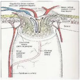
VASCULAR SUPPLY OF OPTIC NERVE:
1. Optic nerve head:
a. Surface nerve fibre layer: Its mainly supplied by capillaries derived from retinal arterioles. It anastomose with vessels of prelaminar region. Occasionally ciliary derived vessel from prelaminar region may enlarge to form cilioretinal artery.
b. The prelaminar region: It is primarly derived from peripapillary choroidal system or separate branches of short posterior ciliary arteries.
d. The retrolaminar region: It is supplied by both ciliary and retinal circulation. The central retinal artery provides centripetal branches where it arisesfrom pial plexus and also centrifugal branches.
2. The intraorbitalpart : Its supplied by two system of vessels.
a. Periaxial : It is derived from six branches of internal carotid artery.
1. Ophthalmic artery
[image:22.612.199.471.200.469.2]2. Long posterior ciliary arteries 3. Short posterior ciliary arteries
4. Lacrimal artery
5. Central artery of retina
b. Axial system: It is derived from
1. Intra neural branches of central retinal artery.
2. Central collateral arteries which comes from central retinal artery.
3. Central artery of optic nerve.
3. The inracanalicular part:
It is supplied only by periaxial system of vessels. The pial plexus in this part is mainly fed from branches of ophthalmic artery.
4. The intracranial part:
It is exclusively supplied by periaxial system of vessels. The pial plexus is contributed by 4 sources
1. Branches from internal carotid artery directly or through recurrent branch of anterior hypophsealartey
2. Branches from anterior cerebral artery
DEMOGRAPHY:
Most of the following information of visual outcome in optic neuritis comes from ONTT/LONS. The annual incidence of optic neuritis has been estimated in population based studies to be 1-5 per 1000,000.6 Most patients are between ages of 20-50 years. Females are commonly affected than males. In ONTT, 77% were female, 85% were Caucasian and the mean age was 32±7 years.5 . Optic neuritis can occur at any age. In Asia, ON is more common in relation to the incidence of MS in the United States of America or Western Europe.8
CLASSIFICATION:
Optic neuritis is classified into 4 types based on site of involvement
1. Retrobulbar neuritis – normal appearance of disc. 2. Papillitis – swollen optic disc
3. Perineuritis- its involvement of optic nerve sheath.
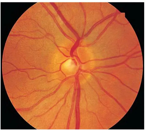
Fig 1. Papillitis – marked inflammation of the optic disc
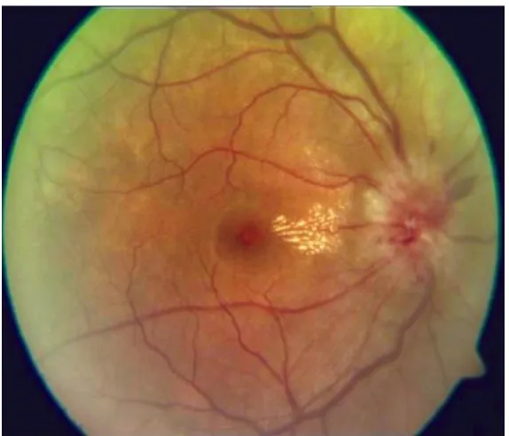
[image:25.612.186.434.371.591.2]Fig 3 Neuroretinitis- marked inflammation around the optic disc with involvement of adjoining retina
Based on topography, three principle type of neuritis is classified into
1) Perineuritis or periaxial neuritis 2) Axial neuritis
ETIOLOGY
It is idiopathic in majority of cases. It could be associated with demyelinating lesion, of which most common cause is multiple sclerosis.
A)Viral infections (e.g. chicken pox, measles, mumps etc.)
B) Granulomatous infections such as tuberculosis, cat scratch disease,
syphilis, Lyme disease or Cryptococcal meningitis.
C) Autoimmune diseases like SLE, Wegner ’s Granulamatosis or
sarcoidosis
D)Contagious inflammation from orbit, meninges and sinuses
MULTIPLE SCLEROSIS AND OPTIC NEURITIS
Optic neuritis is the most common initial manifestation of multiple sclerosis. Most common cause of visual disability among young and middle aged adults. MS is an acquired, chronic inflammatory disease of central nervous system. It is characterised by focal demyelinating lesions of varying age. It is reported to be most common aetiology in Western literature.8,9 Incidence of MS in India and other Asian countries is low.10,11,12
After an attack of optic neuritis, possibility of developing MS reaches 38% in following 10 years and the percentage rises to 56% if there are brain abnormalities in MRI.13.
PATHOPHYSIOLOGY
Inflammatory demyelination in central nervous system including optic nerves and visual pathway. It was previously thought as disease of myelin with sparing of nerve axons however neuronal and axonal loss are known to occur in MS, leading to permanent neurologic and visual impairment.
Activated peripheral T cells migrate across the blood brain barrier and release cytokines and other inflammatory mediators leading to neuronal cell death and axonal degeneration. After an acute event axons get damaged which leads to loss of axons which cause severe and irreversible neurological impairment.
Diagnostic criteria for MS was based on Poser clinical criteria
1. Clinically definite MS
2. Two attacks (relapses) of > 24 hours duration and more than one month apart together with clinical evidence of lesion in two places within the central nervous system.
2. Clinically propable MS above combination of clinical and paraclinical evidence but no oligoclonal bands.
3. Laboratory supported propable MS oligoclonal banding without clinical or paraclinical evidence of lesions.15
Retrobulbar neuritis and papillitis are mainly associated with MS.
Perineuritis and neuroretinitis are most often associated with infectious or inflammatory pathologies. 16, 17,18, 19,13. Neuroimaging (MRI) demonstrates white matter T2 signal abnormalities consistent with demyelination.20
Optic neuritis can be typical or atypical.2,3,4,14,21 It is classified based on their clinical features and course of disease.
NEUROMYELITIS OPTICA:
Features of NMO are visual loss caused by damage to the anterior visual sensory pathways and paraplegia caused by damage to the spinal cord. Pain is not a definite feature. No definite pattern of visual field loss is noticed. Fundus shows a mild disc swelling associated with venous dilatation and extensive peripapillary exudation. Optic atrophy sets as a final sequela. CSF examination reveals lymphocytic pleocytosis. MRI shows abnormal T2 weighed signals and enhancement with gadolinium in the optic nerves, chiasm and spinal cord.
INFECTIOUS/ PARAINFECTIOUS ON:
CSF may show pleomorphic lymphocytosis and an elevated protein concentration. Visual recovery is excellent without treatment. Corticosteroids hastens recovery.2,3,4
POST VACCINATION OPTIC NEURITIS :
ON can occur after vaccination against both bacterial and viral infections. It may develop after vaccination with BCG, Hepatitis B virus, Rabies virus, Tetanus Toxoid, DPT and MMR. Influenza vaccine is most commonly associated with such presentation. Onset usually within 1 to 3 weeks. Most commonly bilateral. Visual recovery occurs few weeks to months.22,23,24
Optic neuritis in multiple sclerosis is characterised by subacute / acute unilateral visual loss which stabilizes by 1 to 2 weeks in patients between age of 20 to 50 years with clinical and neurological evidence of multiple sclerosis and no evidence of other disease process.2,3,4,21,9 . Features of typical optic neuritis:
Females are predominantly affected.
Age 15 to 45 years
Unilateral
Acute and often painful loss of vision over hours to days.
Pain on eye movements.
Peak visual loss within 2 weeks.
Visual loss is subtle to complete which starts improving thereafter.
Relative afferent papillary defect often present.
Poor colour vision and contrast sensitivity.
Disc edema, vitreous cells, haemorrhages and cotton wool spots rarely present.
Fundus appears normal in retrobulbar neuritis.
Any type of visual field defect may be seen but generalised depression of visual field is the most common presentation.
VER (Visually evoked response) shows prolonged latency with normal or depressed amplitude.
Patients may complain of residual deficits in contrast sensitivity, colour vision, stereopsis, light brightness, visual acuity or visual field.
Age less than 20 or greater than 50 years
Painless loss of vision
Persistent pain lasting for more than 7 days.
Older patients are most commonly affected.
Bilateral presentation
Disc haemorrhages and cotton wool spots can occur.
Lack of significant improvement of visual function or worsening within the first three weeks after onset of symptoms.
Progression of visual field loss beyond 2 weeks.
Diagnosis or evidence of other systemic conditions ( inflammatory or infectious diseases including HIV infection) other than multiple sclerosis might cause optic neuropathy.
CLINICAL ASSESMENT
1. History 2. Visual acuity 3. Colour vision 4. Pupil reflexes
5. Biomicroscopy and Fundoscopy. 6. Visual fields
Associated symptoms might be movement phosphenes, sound induced phosphenes, visual obscuration of bright light and Uhthoff ‘s phenomenon.
1. Loss of vision
Loss of central visual acuity is the major symptom in most cases of acute optic neuritis being reported by over 90% of patients.9 Loss of vision is abrupt occurring several hours to several days. The degree of visual loss varies widely. In some cases visual acuity is minimally reduced in others there is complete blindness with no perception of light. Visual loss is monocular in most cases, it can also be binocular. Best corrected visual acuity was measured with a wall mounted Snellens Visual acuity chart at 6 metres distance.
2. Loss of visual field
Few patients complains of loss of central field of vision, some complain of loss of peripheral vision usually in a particular area of visual field such as superior or inferior region. Such patients deny loss of central acuity and may found to have 6/6 vision in the affected eye.25
3. Ocular or orbital pain
In ONTT pain was reported by 92% of patients of whom 87% indicated that it was worsened by eye movement. Rose theorized that the pain is caused by inflammation or swelling in the optic nerve sheath that are innervated by small branches of the trigeminal nerve.26 Lepore reported that among 101 eyes with optic neuritis, pain was more commonly present with retrobulbar neuritis than with papillitis.1 In ONTT pain was present in 93% of 295 eyes with retrobulbar neuritis and in 90% of 162 eyes with papillitis. Majid et al reported painful eye movements in 12 patients in his study.51
4. Positive visual phenomena
OTHER SYMPTOMS:
1) Movement phosphenes:
It can occur before an attack of optic neuritis or may accompany visual loss during the attack or may occur 6 months after full recovery. It especially occurs in horizontal movements in dim lit room; very brief flashes of light lasting only for 1 or 2 seconds occurring unilaterally and in ipsilateral affected eye even when it is maintained in lateral gaze. It is due to demyelination and demyelinated nerve fibres may discharge spontaneously when subjected to minimal mechanical stress.
2) Sound induced phosphenes:
Phosphenes precipitated by sudden noise.
3) Visual obscuration in bright light:
4) Uhthoff’s symptom:
It is an episodic transient obscuration of vision with exertion, hot baths, and showers. Patients have blurring of vision 5 to 20 minutes after exposure to provoking factor.
SIGNS:
1) Visual acuity:
It can be variable from 6/9 to loss of perception of light. In most of the cases visual acuity is reduced . Contrast sensitivity and colour vision is defective in most of the cases.
2) Pupillary reflexes:
RAPD is a office room test. It determines decreased vision in patient in optic nerve problems.
Light reflex pathway has two pathways
1. Afferent pathway extends from the retina along the optic nerve to the mid brain when a light is shone in that eye.(red)
2. Efferent pathway consist of parasympathetic fibres from mid
A swinging flash light test is used to detect RAPD. It is the paradoxical response of a pupil to light.
PROCEDURE:
3) Colour vision
Colour vision is the ability of the eye to distinguish between colours excited by light of different wavelength. Dyschromatopsia can be congenital or acquired. Acquired loss of colour vision can be due to many causes. 88% of colour vision abnormalities was seen in optic neuritis. Mixed defects like red- green and blue- yellow were reported in ONTT trial. Blue yellow defects were more common in acute phase of disease. Red-Green defects were more seen at 6 months.
Colour is purely a subjective sensation.29 It is mainly dependent on the wavelength composition of light entering the eye and on the structure of eye.31
There are three theories of colour vision 1. Opponent theory
COLOUR VISION TESTS:
The main objective is to 31
1. Screens the presence or absence of congenital or acquire colour deficiency
2. Diagnose the type and severity of colour deficiency.
3. Asses its significance in a particular vocation, employement or vocation.
4. They are designed to perform various functions
a) Screening test- identifies subjects with normal and abnormal colour vision.
b) Grading test- estimates severity of colour vision
c) Vocational tests- identifies colour matching ability,hue discrimination and colour recognition.
PSEUDOISOCHROMATIC PLATE
1. Ishihara plates
2. American Optical Hardy- Rand –Rittler Plates 3. Standard Pseudoisochromatic Plates
Ishihara charts are based on the principle of confusion of the pigment colour in red –green colour defectives. It is easy and rapid test most commonly used for screening purposes. Charts are available in 14,24 or 38 plates.
INTERPRETATION:
American Optical Hardy- Rand –Ritter (HRR) is the test for choice for quantitative diagnosis.
Standard Pseudoisochromatic Plates Part 2 for acquired colour deficiency.30,33
City University Colour Assesment
It is based on spatiotemporal luminance masking technique. In this, part of uniform background it is formed by spatially discrete elements that are equal in time averaged luminance with respect to the background, ensuring detection of the stimulus is based purely on chromatic discrimination and not luminance difference. This is designed to run on any monitor balanced approximately 9000K, which is the default factory setting for most colour monitors.
GRADING COLOUR VISION DEFICIENCIES
Spectral anomaloscope
1. Nagel anomaloscope 2. Oculus HMC
3. Neitz anomaloscope
4. Pickford – Nicolson anomaloscope.
Arrangement tests ( Hue Discrimination Tests)
1. Farnsworth Muller 100 Hue test
2. Farnsworth Muller dichotomous D 15 or Panel D 15 test 3. Lanthony desaturated D 15
4. Adams desturated D 15 test
Farnsworth Muller 100 Hue test
It is a very sensitive reliable and effective method of determining colour vision defect.
Farnsworth Muller dichotomous D 15
It is to distinguish observers with moderate or severe colour defects to those with milder defects.30
Lantern test
Test is performed in a dark room at six meters distance. Set of filters showing signal red, yellow, green and blue colours.
Edridge –Green Lantern
Marta Owidzka et al 60 reported in 27 patients of 33 eyes as contrast sensitivity is significantly reduced in all spatial frequencies both in photopic and mesopic conditions.
Contrast sensitivity:
It is the ability to perceive slight changes in luminance between regions which are not separated by definite borders. Ability to perceive sharp outlines of relatively small objects. Loss of contrast sensitivity is more important and disturbing to the patient than the loss of visual acuity. Even in presence of normal visual acuity, contrast sensitivity can be impaired.
Measurement of contrast sensitivity of human visual system was reported by Schade in form of Modulation transfer function.34
TYPES:
1. Spatial contrast sensitivity 2. Temporal contrast sensitivity
Measurement:
It is measured as (Lmax – Lmin) / (Lmax + Lmin).
L - Luminance recorded by photocells scanning across the gratings.
3 variables in measurement of contrast sensitivity:
1. Amount of light reflected depends on illumination of paper and darkness of ink.
2. Degree of blackness in relation to the white background . i.e contrast
3. The distance between the grating periods or cycles per degree of visual angle.
Deficits expressed in terms of decibels. Three types of deficits were described.
1. High frequency type characterised by increasing loss at high frequency.
2. A level loss type characterised by a similar loss for all spatial frequencies.
3. A selective loss type characterised by deficits in a narrow band of spatial frequencies.
Methods to measure contrast sensitivity include simple plate 36, cathode ray tube display on a screen letter acuity charts.46 Visual field testing using low contrast rings on stimuli, pattern discrimination test, two alternative forced choice test.
RELIABLE METHODS OF MEASURING CONTRAST
SENSITIVITY
1. Arden grating
Arden gratings:
Arden in 197836 introduced a booklet containing seven plates – one screening plate(no 1) and six diagnostic plates (no 2 -7). The contrast changes from top to bottom and covers a range of approximately 1.76 log units. Plates are studied at 57 cm, with spatial frequency increasing from 0.2 cycles /degree to 6.4 cycles/degree each being double the frequency .
Cambridge low contrast gratings:
It consist of set of ten plates containing gratings in a spiral bound booklet.
To perform this, booklet is hung on wall at a 6 metres distance. The pages are presented in pairs one above the other. One page in each pair contains gratings and the other is blank. Subject is require to choose which page top or bottom contains gratings. Pages are shown in descending contrast
Pelli – Robson contrast sensitivity test:
from one triplet to next. The log sensitivity varies from 0.00 -2.25. Chart is hung on the wall, it should be at the level of subjects eye. Chart is illuminated uniformly. Luminance of white area is between the accepatable range of 60 to 120 cd/m. It is recorded at 1 metre distance. Sensitivity is indicated by finest triplet for which two of the three letters are named correctly.
RED DESATURATION:
saturated redness in either eye indicating an acquired dyschromatopsia in that eye. Its a sign of optic nerve dysfunction.
BRIGHTNESS COMPARISON:
It can be subjective or objective.
Subjective:
Quantification:
It is done by putting a sequence of neutral density filters in the better eye and shining the light until the patient states that the brightness is same in the both the eyes.
In 89% of patients with optic neuritis poor brightness perception was reported .45,37.
Visual fields:
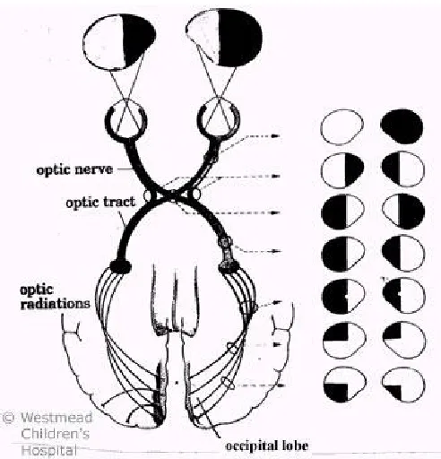
FIGURE : 1.1 Lesions along the optic pathways showing visual field defects.
Testing of central and peripheral visual fields is important:
1. To detect abnormality in vision.
2. To localize the defect along the afferent visual pathway. 3. To quantitate the defect and measure the change over time.
The following are the methods in visual field testing
a) Confrontation methods b) Amsler grid
c) Tangent screen d) Goldmann perimetry.
Confrontation methods:
This is a rapid screening method where examiner compares patients field with their own.
Finger counting:
Patients are instructed to accurately identify the number of fingers presented in each quadrant of the monocular field.
Simultaneous finger counting may bring out a subtle defect.
Test :
The patient while fixating should be asked whether he can see the examiners eyes, ears, hair, mouth etc which would reveal a large scotoma.
Hand comparison :
Simultaneous perception of hands placed on either side of vertical meridian providing a sensitive subjective comparison of the two hemifields.
Hands placed in the superior and inferior fields to determine whether there is an altitudinal defect.
Colour comparison :
Done in similar way with the patient occluding one eye. The examiner holds a red coloured object in each hand and asks whether the red objects appear the same in each hand. In a relative scotoma one object might appear less red than the other.
b) Amsler grid :
Screens central 10 degree of fixation. Patient optically corrected for near vision covers one eye and looks at fixation point in the centre of a grid. Patient is then asked to describe any scotoma.
c) Tangent screen:
Method:
The patient gazes with one eye at black screen with the other eye occluded 1- 2 metres away in good lighting and fixes on a central spot. Thin black wand with various targets on tip is used by the examiner. Initially a suprathreshold stimulus is used such as 5mm white target at 1m. stimulus is moved from non seeing area to seeing area.
The target is a flat disc white or red on one side, black on reverse so that it can be flipped over with course of the test so that black surface is not seen by the patients it blends with the screen. Patients are instructed to indicate verbally or by gesture when they first see the target only, not the wand or examiners hand.
Blind spot can be detected using larger stimulus. Central fields( fixation area) explores scotoma in the central region of fixation and the area between blind spot.
d) Goldmann perimetry:
(HVFA) sensitivity mainly requires patient’s co-operation and competence, good technician to monitor the patient.
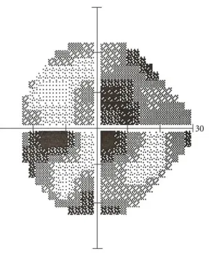
[image:64.612.222.376.443.632.2]In ONTT, patients underwent HVF testing. Perimetry revealed that 48% present with a diffuse field defect ( Figure 1.2), 20% with altitudinal or arcuate defects and 8% present with a centrocaecal defect 44. . Initially it was found that centrocaecal scotoma was common defect in optic neuritis. However it is now believed that the central scotoma found by Goldmann perimetry represents diffuse suppression of sensitivity within the central 30 degrees of vision.43 Central visual loss is common in optic neuritis patients and sparing of the periphery, it is rare for the peripheral field to be abnormal in the presence of a normal central field.
Other test are
1) Stereoacuity :
The Titmus Polaroid 3D Vectograph stereoacuity is recommended
for both children and adults with optic neuritis.
The Pulfrich effect in which patient experience a stereoillusion by
having the patient gaze at a pendulum swinging at right angles to the line
of sight and determining if the pendulum appears to the patient to be
swinging in an elliptical path is a sensitive indicator.
2) Visual Evoked Potential:
It a specific change in Electroencephalographic (EEG) recording due to stimulation of the visual pathway to either a pattern or flash stimulus. Recordings of evoked potentials can be made with the use of electrodes applied to the scalp. The main signals are detected over the occipital cortex. The VEP signal is mainly derived from the macular region as the predominant function of the occipital cortex is to subserve macular function.
It differentiates organic from functional cause of defective vision.
It tests central and perifoveal visual field and there is prolongation of P
with visual acuity and P latency is also an indicator of optic nerve
dysfunction. In pattern shift VEP it provides evidence of optic nerve
pathology in optic neuritis.
3)Pattern electroretinogram( PERG)
It monitors integrity of central ganglion cell layer. It is of value in
improved interpretation of abnormal VEP pattern when both are recorded
simultaneously to rule out if delay in pattern VEP P latency in a patient
with suspected optic nerve demyelination.
4)Pupillary light reflex latency:
Prolonged latency of papillary light reflex which is measured using
infrared reflection.
5)Foveal critical flicker frequency is impaired
Subjective brightness measured by Authorn Flicker test in relation
to flicker frequency is abnormal.
OPTIC DISC FINDINGS:
Normal in retrobulbar neuritis Swollen disc in papillitis.
MAGNETIC RESONANCE IMAGING: PRINCIPLE:
MRI shows the size ,quantity and distribution of lesions larger than 2mm, and together with supporting evidence helps in the diagnosis of MS.
Magnetic Resonance Imaging is helpful in diagnosis of clinically silent multiple sclerosis lesions.
MRI criteria for diagnosing MS
At least 3 lesions and two of the following should be present for the diagnosis of MS to be present;39
1. 1.Lesions in the lateral ventricles
2. Lesions with diameters greater than 5mm
3. Lesions present in posterior fossa (infratentorial).
MRI features in optic neuritis:
40 – 70 % of patients are reported to have periventricular white matter changes on MRI consistent with MS in cases of isolated optic neuritis. In ONTT, MRI scans were the strong predictor of developing MS performed at study entry. 10 year risk of MS ranging from 22% in patients with no MRI lesions, 56% in patients with one or more lesions.41
Optical Coherence Tomography:
SYSTEMIC INVESTIGATIONS
1. Routine hemogram
2. X-ray (Chest)
3. Mantoux
4. FTA - ABS & VDRL for syphilis
5. serology and culture for bartonella
6. Markers of viral infection
7. Serum electrolytes and fasting blood sugar
8. MRI
9. Lumbar puncture and CSF tap (IgG index and oligoclonal bands)
10. Blood culture
11. ANA, dsDNA
DIFFERENTIAL DIAGNOSIS
1. Ischemic optic neuropathy, it is characterized by lack of pain, pallid disc swelling with haemorrhages. Altitudinal visual field defect. Lack of improvement of vision with standard therapy.
2. Lebers optic neuropathy it is characterized by lack of pain, circumpapillary telengiactatic microangiopathy
3. AION
4. Toxic or nutritional deficiency amblyopia 5. Non organic visual loss
6. Other
a. posterior uveitis b. CSCR
c. Disc drusen d. Glaucoma
e. Posterior scleritis
MANAGEMENT
PREDNISOLONE:
It is a adrenocortical steroid which are naturally occurring and synthetic,readily absorbed from the gastrointestinal tract. It is a white crystalline powder very slightly soluble in water. It is designated chemically as pregna-1,4-diene-3,20-dione,11,17,21-trihydroxy-(11 β). The structural formula is represented below:
Mechanism of action:
METHYLPREDNISOLONE:
It is steroid that prevents the release of substances in the body that cause inflammation.
COMPLICATIONS OF STEROIDS:
OCULAR:
Eyelids:
a) allergic reactions b) telengiectasia c) persistent erythema
Cornea:
a) Superficial punctuate keratitis b) Delayed healing of corneal wounds
GENERAL
1. Increased IOP
2. Delayed healing of corneal wounds.
4. complicated cataract
5. Enhances lytic action of collagenase
SYSTEMIC COMPLICATIONS:
A) Dermatological :
1) Acne 2) Hirsuitism
3) Subcutaneous tissue atrophy
B) Immunological:
1) Impaired inflammatory response 2) Delayed tissue healing
C) Metabolic:
1) Secondary diabetis mellitus 2) Hyperosmotic ketoacidosis 3) Centripetal obesity
4) Hyperlipidemia
D) Cardiovascular:
1) hypertension
E) Musculoskeletal:
1) osteoporosis
2) vertebral compression fracture
F) Gastrointestinal:
1) peptic ulcer
2) gastric haemorrhage 3) intestinal perforation 4) pancreatitis
G) Endocrine:
1) Adrenal insufficiency 2) Cushing s syndrome 3) Growth failure 4) Menstrual disorders
H) Neuro Psychiatric:
1) Pseudotumour cerebi 2) insomnia
OPTIC NEURITIS TREATMENT TRIAL:
ONTT is a multicentered randomized trial involving 454 patients from 1988 to 2006. This study evaluated the efficacy of corticosteroid treatment for acute optic neuritis.
Study objective:
It evaluates the efficacy of corticosteroid treatment in acute optic neuritis relationship between optic neuritis and multiple sclerosis.
The patients in this study were randomized into 3 different groups
1. IV methyl prednisolone(250 mg 6 hrly ) for three days followed by oral prednisolone for 11 days and three day tapering of prednisolone.
2. Oral prednisolone (1mg/kg/day) for 14 days followed by 3 days in tapering doses.
3. Oral placebo for 14 days.
Results:
STUDIES INVOLVED IN OPTIC NEURITIS TREATMENT:
CHAMPS STUDY (The Controlled High Risk Avonex Multiple Sclerosis Trial)
Objective :
Avonex (interferon beta 1 a) treatment would benefit patients who had experienced a demyelinating event involving the optic nerve, brain stem/cerebellum or spinal cord. Previously,brain abnormalities in MRI predicted likelihood of future MS like events. All patients received intravenous methylprednisolone 1g per day for three days within the days of onset of neurological symptoms, followed by oral prednisolone with tapering dose of 1mg/kg for 11 days. Patients were divided into two groups
Group 1 once weekly intramuscular injection of interferon beta 1 A
Group 2 placebo injections
Results
At three years end,the probability of CDMS was 50% in the placebo treated group and 35% in the interferon 1 A treated group. There was no difference among treatment options among patients presenting with optic neuritis, brain stem/cerebellar or spinal cord events. Treatment with Avonex significantly reduces the two year likelihood of future neurological events and worsening of brain MRI in patients with a first acute CNS demyelinating event.
ETOMS (Early Treatment Of Multiple Sclerosis Study)
Objective:
To determine whether an early treatment with interferonb 1 b is effective in delaying the development of CDMS after the first attack.
CHAMPIONS STUDY (Controlled High Risk Avonex Multiple sclerosis Prevention Surveillance)
Objective:
Outcomes are compared in those who had given drug from the start of CHAMPS study (immediate treatment) versus those who had switched from placebo after about 30 months. (delayed treatment).
Results:
Immediate group had few relapses and fewer MRI lesions than the delayed group and few of its members converted to definite MS.
BENEFIT STUDY: (Betaferon in Newly Emerging MS for Initial Treatment)
Newer modalities:
It can modify the disease course
1. Copolymer 1
2. T cell receptor peptide immunisation 3. Anti CD 4 monoclonal antibody 4. Azathioprine ( Imuran)
5. Cyclophosphamide (cytoxan) 6. Oral myelin
7. Methotrexate 8. Cladribine
AIM AND OBJECTIVES
MATERIALS AND METHODS
Prospective observational case study.
To analyse visual function, response to treatment and visual outcome in patients with optic neuritis.
Study period: January 2013 to December 2013.
Study duration: 13 months (including followup)
Data source: Hospital records. (Aravind Eye Hospital)
Sample size: 88 patients /98 eyes.
Follow up period of 2 weeks, 1 month.
INCLUSION CRITERIA:
Age 16 -65 years
Loss of visual acuity or visual field, with or without pain < 1month duration.
Unilateral / bilateral
Fundus changes
EXCLUSION CRITERIA
Age < 16 years
Visual loss including toxic, metabolic, vascular, hereditary, compressive neuropathies.
Previous episode of optic neuritis in the affected eye.
CLINICAL EVALUATION:
A series of 98 eyes of 88 patients who presented to our Neuroophthalmology department were included in our study.
Optic neuritis was diagnosed based on history and clinical examination which included defective vision , dychromatopsia, pain on eye movements, presence of Relative Afferent papillary defect, normal or swollen optic disc on fundus examination of less than 1 month duration were included in our study. All these patients underwent a thorough ophthalmological and neurological evaluation.
Patient ‘s data such as name, age , sex, address were documented in a proforma.
Detailed history of each and every symptom like onset, duration, pain on eye movement , headache were documented. Patients were also enquired about history of prior visual loss.
Each one of the patient included in our study have to undergo
Visual acuity by means of Snellens’s chart
Refraction
Pupillary reaction to look for RAPD, sluggish pupil
Intraocular pressure measurement for patients above 40 years of age by Non contact tonometry method.
Fundus examination by direct ophthalmoscope and slitlamp biomicroscopy using +90 dioptre lens.
Colour vision was assessed in detail by Ishihara’s colour vision chart, red desaturation, brightness sensitivity.
Central fields tested by Bjerrums’s tangent screen.
Neuroimaging was done depending upon the need and affordability of the individual patients.
Basic blood investigations like random blood sugar, total leucocyte count, differential leucocyte count and erythrocyte sedimentation rate was done for all patients.
All patients in our study received 1 gram of intravenous methyl prednisolne for 3 days followed by oral prednisolone of 1mg/kg/body weight for 2- 4 weeks.
ANALYSIS
Analysis of collected data was done based on the following 1. Incidence
2. Age and sex distribution 3. Onset of symptoms 4. Presenting visual acuity 5. No PL/ PL / HM /1/60 6. 1/60 – 6/60
7. 6/60 – 6/6
8. Pupil - normal / RAPD.
9. Colour vision - Normal / defective / inconclusive because of poor vision.
10. Central fields
11. Fundus status – normal / abnormal.
12. Visual acuity, colour vision , red desaturation, brightness sensitivity, central fields at follow up.
STATISTICAL METHODS
RESULTS
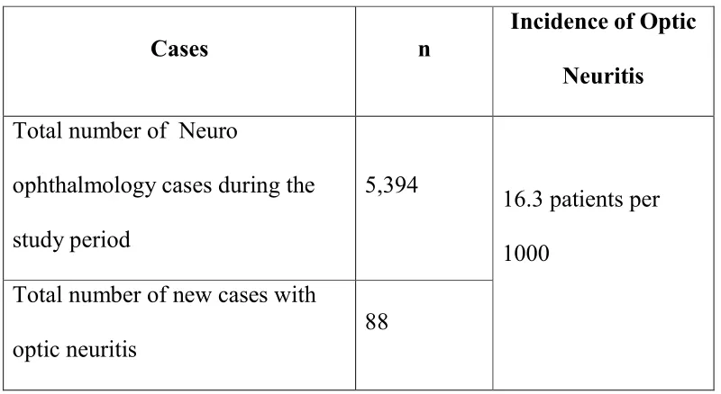
[image:88.612.107.507.266.485.2]Study included 98 eyes of 88 patients with optic neuritis. Visual function parameters showed rapid recovery following treatment with steroids compared to pretreatment levels in our patients.
TABLE 1. INCIDENCE
Cases n
Incidence of Optic Neuritis Total number of Neuro
ophthalmology cases during the study period
5,394
16.3 patients per 1000
Total number of new cases with optic neuritis
The mean age is 40.0 (12.9) years and the range is 20 to 64 years.
Majority of patients in the s years. (52.3%)
[image:89.612.117.495.162.480.2]41 - 60 40%
TABLE 2: AGE
The mean age is 40.0 (12.9) years and the range is 20 to 64 years.
Majority of patients in the study belonged to the age group 2
20 - 40 52% >60
8%
Age category
20 - 40 41 - 60 >60
The mean age is 40.0 (12.9) years and the range is 20 to 64 years.
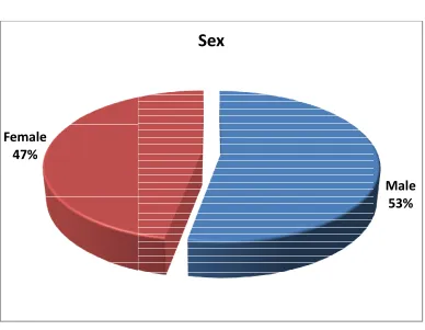
Out of 88 patients 47 were males (53.4%) and 41 were females (46.6%)
[image:90.612.115.503.102.403.2]Female 47%
TABLE 3: GENDER
Out of 88 patients 47 were males (53.4%) and 41 were females
Sex
Out of 88 patients 47 were males (53.4%) and 41 were females Male
TABLE 4:
Out of 88 patients, 78 (79.6%) of them showed unilateral involvement and 10 (20.4%) showed bilateral involvement.
Bilateral 20%
TABLE 4: LATERALITY
Out of 88 patients, 78 (79.6%) of them showed unilateral involvement and 10 (20.4%) showed bilateral involvement.
Unilateral 80% Bilateral
Laterality
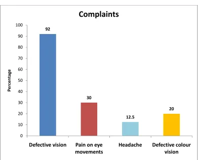
TABLE 5: COMPLAINTS
Defective vision was presented in 90 eyes (91.8%) followed by dyschromatopsia in 20 eyes(20.4%). Pain on eye movements was presented in 29 eyes (29.6%). Headache was presented in 11 patients (12.5%). 92 30 12.5 20 0 10 20 30 40 50 60 70 80 90 100
Defective vision Pain on eye
movements
Headache Defective colour
TABLE 6:VISUAL ACUITY
Visual acuity Baseline 1month
6/6 – 6/18 19(19.4) 45(58.4)
6/24 – 6/60 13(13.3) 15(19.5)
6/60 – 3/60 36(36.7) 11(14.3)
<3/60 30(30.6) 6(7.8)
Total 98 77
At baseline 32.7% of patients had V/A >6/60. 67.3% patients had V/A <6/60. About 78 % of patients had 6/60 or better vision at 1 month follow up. 19.3 58.4 13.3 19.5 36.7 14.3 30.6 7.8 0 10 20 30 40 50 60 70 Baseline 1month P e rc e n ta ge
Visual acuity
Log mar vision
N
Median
(Snellen’SEquivalent)
Mean(SD)
Min-Max
P-Value
Baseline 98 1.48(2/60) 1.54(0.94) 0 – 3.2 - 2 weeks 92 0.39(6/15) 0.74(0.84) 0 – 2.9 <0.001 1 month 77 0.3(6/12) 0.67(0.83) 0 – 2.9 <0.001
Improvement in visual acuity was statistically significant at 1 month (p = <0.001) compared to the baseline.
Baseline median LogMAR visual acuity was 1.48 which improved to 0.3 after 1 month.
PUPIL
Pupil N %
Normal 12 12.2
RAPD 86 87.8
Total 98 100.0
At baseline, in 15 eyes optic disc was normal, 44 eyes showed disc
edema, 20 eyes showed temporal pallor, hypere
month, out of 77 eyes 19
pallor, 12 eyes showed hyperemia, 16 eyes showed resolving disc edema,
3 eyes showed primary optic atrophy
0 5 10 15 20 25 30 35 40 45
Normal Disc edema 15
44
[image:95.612.116.522.106.386.2]20
TABLE 7: FUNDUS
At baseline, in 15 eyes optic disc was normal, 44 eyes showed disc
edema, 20 eyes showed temporal pallor, hyperemia in 20 eyes. At 1
month, out of 77 eyes 19 eyes was normal, 28 eyes showed temporal
pallor, 12 eyes showed hyperemia, 16 eyes showed resolving disc edema,
3 eyes showed primary optic atrophy
Disc edema Temporal pallor Hyperemia Resolving disc edema Optic atrophy 19 20 27 11 16
Fundus
Baseline 1 Month
At baseline, in 15 eyes optic disc was normal, 44 eyes showed disc
mia in 20 eyes. At 1
normal, 28 eyes showed temporal
pallor, 12 eyes showed hyperemia, 16 eyes showed resolving disc edema,
Optic atrophy
Fundus at baseline
Fundus at 1month
Total P-value Normal Abnormal
Normal 9(69.2) 4(30.8) 13 0.180
(Using McNemar’s test) Abnormal 10(15.6) 54(84.4) 64
Total 19 58 77
TABLE 8:
Colour vision at baseline
Colour vision at
Normal
Normal 7(87.5)
Dyschromatopsia 46(66.7)
Total 53(68.8)
0% 20% 40% 60% 80% 100% Baseline 12.2% P e rc e n ta ge TABLE 8:DYSCHROMATOPSIA
Colour vision at
1month Total P-value
Normal Abnormal
7(87.5) 1(12.5) 8 0.0035
(Using McNemar’s
test) 46(66.7) 23(33.3) 69
53(68.8) 24(31.2) 77
At baseline 12.2% (12) were normal 87.8% (86) were abnormal. At follow up of 1 month of 77 eyes
were abnormal . 46 eyes (66.7%) were normal at 1month follow which was abnormal in baseline. Colour vision at 1month
[image:98.612.116.532.307.646.2]statistically showed significant improvement. (P McNemar’s test) TABLE : 0 5 10 15 20 25 30 35
At baseline 12.2% (12) were normal 87.8% (86) were abnormal. At follow up of 1 month of 77 eyes 68.8% (53) were normal and 31.2% (24)
46 eyes (66.7%) were normal at 1month follow which was abnormal in baseline. Colour vision at 1month follow statistically showed significant improvement. (P-value=0.004, Using
TABLE : 9 CENTRAL FIELDS
Central Fields
At baseline 12.2% (12) were normal 87.8% (86) were abnormal. At 68.8% (53) were normal and 31.2% (24) 46 eyes (66.7%) were normal at 1month follow-up follow-up value=0.004, Using
Central fields at baseline
Central fields at
1month Total P-value
Normal Abnormal
Normal 7(100.0) - 7 <0.001
(Using McNemar’s test)
Abnormal 29(41.4) 41(58.6) 70
Total 36(46.8) 41(53.2) 77
TABLE 10: BRIGHTNESS SENSITIVITY
Brightness at baseline
Brightness at 1month Normal
Normal 8(80.0)
Reduced 35(52.2)
Total 43(55.8)
0% 20% 40% 60% 80% 100% Baseline 15.3% P e rc e n ta ge
Brightness sensitivity
TABLE 10: BRIGHTNESS SENSITIVITY
Brightness at 1month
Total P-value
Normal Abnormal
8(80.0) 2(20.0) 10 0.0001
(Using McNemar’s test) 35(52.2) 32(47.8) 67
43(55.8) 34(44.2) 77
TABLE Red Desaturation at baseline Red D Normal
Normal 6(75.0)
Reduced 37(53.6)
Total 43
[image:102.612.118.505.122.420.2]0% 20% 40% 60% 80% 100% Baseline 12.2% P e rc e n ta ge
TABLE 11: RED DESATURATION
Red Desaturation at
1month Total P-value
Normal Abnormal
6(75.0) 2(25.0) 8
0.0001 (Using McNemar’s test) 37(53.6) 32(46.4) 69
43 34 77
At baseline, 12(12.2%) eyes were normal. At follow up of 1 month, 43 (55.8%) eyes showed improvement. 37(53.6%) were normal at 1month follow-up who were abnormal at baseline. Redsaturation statistically showed significant improvement at 1month follow-up (P-value=0.0001, Using McNemar’s test).
Neuroimaging
Neuroimaging was not possible in all cases due to financial constraints. It was performed in 35 cases. MRI was done in 23 cases and CT was done in 12 cases. No lesions was seen in 7 cases, 25 had shown thickening and enhancement of optic nerve in the affected eye. Demyelinating lesions in the brain were present in 3 cases.
DISCUSSION
Optic neuritis is inflammation of optic nerve which is mostly idiopathic in nature.
In natural course of optic neuritis spontaneous recovery occurs within 1 week but it may take longer time in few cases. Typical cases of optic neuritis due to multiple sclerosis was not commonly seen in India
Many studies have been conducted which reports the association of optic neuritis with multiple sclerosis. Before ONTT, Jain et al 50 reported that clinical profile of optic neuritis in India is different from that of Western population. The aim of our study is to understand visual function following optic neuritis treatment in South India.
88 patients aged between 20 to 64 years (mean age 40.0 ) were enrolled in the study out of which 47 were male and 41 female.78 patients showed unilateral involvement, 10 patients showed bilateral involvement. A similar study was conducted by Rohit Saxena et al 48 included 83 patients between the age of 15 – 58 years ( mean age 27.6 years) of which 67 cases had unilateral involvement and 16 cases had bilateral involvement. ONTT included 457 patients with ages ranging from 18 – 46 years (mean age 31.8 years) 49.
Males were commonly affected in our study (53.4%) compared to females. A similar study conducted by Jain et al50 showed 67% of male involvement in their study. In ONTT, 77% of patients affected were females.49
neuritis and in 90% of 162 eyes with papillitis. Majid et al51 reported painful eye movements in 12 patients in their study.
In most of the eyes visual acuity was worse at baseline. At follow
up of 1 month in 15 (19.5%) eyes visual acuity was 6/24 or better. In
45(58.4%) eyes visual acuity was 6/18- 6/6. Similarly Pedro et al reported
improvement of visual acuity of 52.4% in 18 eyes out 35 eyes at follow
up.56
Out of 98 eyes 86 (87.8%) eyes showed RAPD. 12 (12.2%) eyes
were normal at baseline.
At baseline, in 15 eyes optic disc was normal, 44 eyes showed disc
edema, 20 eyes showed temporal pallor, hyperemia in 20 eyes. Other
findings were splinter haemorrhages seen in 2 eyes. ONTT reported
swollen optic disc was seen in 35%. Nikoskelainen et al reported optic
disc was normal in 46%, hyperemic/ blurred disc in 20%, disc edema in
Papillitis was seen in 65.3% and retrobulbar neuritis in 34.7 % of
patients in our study. Similarly Saxena48 et al reported 53.5% of eyes had
papillitis and retrobulbar neuritis was seen in 46.5%.
Visual fields at baseline were done only in 48 eyes, it could not be
performed in 50 eyes because of poor vision. Most common field defect
was centrocaecal scotoma seen in 22 (22.7) eyes. 7.8% in our study had
superior and inferior field defect generalised constriction of visual field
and superior field loss. In our study 1 patient had altitudinal field defect
at baseline which is common in NAION can also present in ON.
Similarly ONTT54 reported 48.2% showed diffuse loss , 8.3% central or
centrocaecal scotoma and altitudinal field defects in 23.4% eyes which
was the most common field defects at baseline. Jain et al reported
concentric contraction was seen in 25% of eyes followed by central
scotoma seen in 19.1% eyes.
Dyschromatopsia was recorded using Ishihara charts. At baseline
86 (87.8%)eyes it was abnormal 12( 12.2%) eyes it was normal. At 1
month 53(68.8%) eyes showed improvement. 24(31.2%) did not show
improvement. Jain et al reported recovery of dyschromatopsia along with
improvement in colour vision in their study. Vimala et al reported
improvement of dyschromatopsia at 1 month follow up. (Mean Log mar
at baseline was 9.14 which improved to 18.57)59
Brightness sensitivity - Out 98 eyes 83 were abnormal at baseline,
in most it could not be performed due to poor vision. At follow up of 1
month 43(55.8%) eyes out of 77 eyes showed improvement. Brightness
testing appeared to be more sensitive than other techniques in establishing
the diagnosis.37.
Red desaturation – Out of 98 eyes 86 eyes were abnormal at
baseline. At follow up of 1 month ,out of 77 eyes 43 eyes showed
improvement. In our study we have included brightness sensitivity and
Red Desaturation as one of the important parameters for evaluation of
visual function. Similarly Almong et al has shown increased desaturation
in ON in their study.55
Neuro imaging showed demyelinating lesion in the brain which
was present only in 3 cases in our study showing that incidence of MS is
consistent with MS were seen in 37.5% (8 cases out of 32) and with
contrast, it was 48.7% (203 cases out of 417). It also reported that risk
of developing MS after an attack of optic neuritis was 50% and if the
baseline MRI is negative the risk of MS was 25%. Saxena et al48 reported
12 cases showed demyelinating lesion and 4 cases showed MS in their
study. Jain et al reported that incidence of MS is low in southern part of
LIMITATIONS
1. The follow up period was very short to evaluate the long term
prognosis.
2. Favourable visual outcome of ON prohibited patients from
returning to our department for long term follow up.
3. MRI was done only in 35 patients due to financial constraints.
CONCLUSION
1. The age range is 20 to 64 years with mean age 40.0(12.9) years.
2. 79.6 % of patients had unilateral presentation.
3. 20.4% patients had bilateral presentation.
4. There is a slight male preponderance in our study.
5. Defective vision followed by pain on eye movements are the chief
complaints in our study.
6. There was a significant improvement in visual acuity.
7. The most common presentation was papillitis (65.3%) followed by
retrobulbar neuritis (34.7%)
8. Visual function like central fields, colour vision, Brightness
sensitivity, Red desaturation showed a significant improvement
following treatment.
10.3.9% of patients were considered as atypical and requires further
follow up and investigations.
11. Demyelinating disease in association with ON was less common in
our study.
Visual function following optic neuritis treatment showed good
recovery. There was less incidence of associated demyelination in our
BIBLIOGRAPHY
1. Lepore FE. The origin of pain in optic neuritis. Determinants of pain in 101 eyes with optic neuritis. Arch Neurol. 1991 Jul;48(7):748–9. 2. Glaser JS. Neuro- ophthalmology. 3rd edition. Philadelphia, Pa:
Lippincott, Williams & Willkins 1999; 118-98.
3. Miller NR, Newman NJ. Walsh and Hyot’s Clinical Neuro-opthalmology. Williams & Willkins, Vol 1; 1998 : 599-648
4. Liu GT, Volpe NJ, Galetta SL, 1st edition. Neuro-ophthalmology Philadelphia Pennsylvania: WB Saunders company 2001: 103-87. 5. Perkin GD,RoseFC.Optic neuritis and its differential diagnosis.
Oxford,UK, Oxford medical publications 1979.
6. Rodriguez M, Siva A, Cross SA, O’Brien PC, Kurland LT. Optic neuritis: a population-based study in Olmsted County, Minnesota. Neurology. 1995 Feb;45(2):244–50.
7. Wakakura M, Minei-Higa R, Oono S, Matsui Y, Tabuchi A, Kani K, et al. Baseline features of idiopathic optic neuritis as determined by a multicenter treatment trial in Japan. Optic Neuritis Treatment Trial Multicenter Cooperative Research Group (ONMRG). Jpn J Ophthalmol. 1999 Apr;43(2):127–32.
Jul;42(7):702–4.
9. The clinical profile of optic neuritis. Experience of the Optic Neuritis
Treatment Trial. Optic Neuritis Study Group. Arch Ophthalmol. 1991 Dec;109(12):1673–8.
10. Singhal BS. Multiple sclerosis--Indian experience. Ann Acad Med Singap. 1985 Jan;14(1):32–6.
11. Das A, Puvanendran K. A retrospective review of patients with clinically definite multiple sclerosis. Ann Acad Med Singap. 1998 Mar;27(2):204–9.
12. Lin Y-C, Yen M-Y, Hsu W-M, Lee H-C, Wang A-G. Low conversion rate to multiple sclerosis in idiopathic optic neuritis patients in Taiwan. Japanese journal of ophthalmology. 2006;50(2):170–5.
13. Beck RW, Trobe JD, Moke PS, Gal RL, Xing D, Bhatti MT, et al. High- and low-risk profiles for the development of multiple sclerosis within 10 years after op