The diversity and distribution of microfungi in leaf litter of an Australian wet tropics rainforest
263
0
0
Full text
(2) The Diversity and Distribution of Microfungi in Leaf Litter of an Australian Wet Tropics Rainforest. Thesis submitted by Barbara Christine PAULUS BSc, MSc NZ in March 2004. for the degree of Doctor of Philosophy in the School of Biological Sciences James Cook University.
(3) STATEMENT OF ACCESS I, the undersigned, author of this work, understand that James Cook University will make this thesis available for use within the University Library and, via the Australian Digital Theses network, for use elsewhere. I understand that, as an unpublished work, a thesis has significant protection under the Copyright Act and;. I do not wish to place any further restriction on access to this work.. The description of species in this thesis does not constitute valid form of publication.. _________________________ Signature. ______________ Date. ii.
(4) STATEMENT OF SOURCES. DECLARATION. I declare that this thesis is my own work and has not been submitted in any form for another degree or diploma at any university or other institution of tertiary education. Information derived from the published or unpublished work of others has been acknowledged in the text and a list of references is given.. ____________________________________. ____________________. Signature. Date. iii.
(5) STATEMENT ON THE CONTRIBUTION OF OTHERS In this section, a number of individuals and institutions are thanked for their direct contribution to this thesis. Many more have provided assistance in some other way and are gratefully acknowledged in the next section. Dr Paul Gadek, James Cook University, and Dr Kevin Hyde, The University of Hong Kong, supervised this project and provided academic guidance and helpful editorial comment. Assistance with identification of some microfungi was provided by the following mycologists: Ms Boonsom Bussaban, Dr Margaret Barr, Dr Pedro Crous, Dr Ewald Groenewald, Dr Wellcome Ho, Dr Kevin Hyde, Dr Peter Johnston, Dr Eric McKenzie and Dr Brian Spooner. Steve McKenna, Nigel Tucker, and Gary Werren identified the selected tree species. A number of papers have arisen from this work (Appendix M) or are in preparation. As co-author, Dr John Kanowski critically reviewed the paper that formed the basis of Chapter Five, provided additional statistical analyses for this paper and chapter, and shared information about local rainforest sites and leaf chemistry. Co-author Dr Margaret Barr confirmed two species as new to science and provided feedback on a paper that is part of Chapter Seven. Dr Roger Beaver identified the beetle associated with one species of microfungi and provided valuable feedback on the paper that formed Chapter Six. Dr Will Edwards shared his knowledge of diversity estimation and tropical rainforest ecology. Dr Elaine Harding and Dr Shannon Bros provided an excellent introduction to multivariate analysis. Microphotographs were taken at the University of Hong Kong and the University of Chiang Mai. James Cook University is gratefully acknowledged for providing a HECS exemption. Financial support was provided by the Centre for Research in Fungal Diversity, The University of Hong Kong; the Cooperative Research Centre for Rainforest Ecology and Management, Cairns; School of Tropical Biology, James Cook University and The Australian Federation of University Women – South Australia Inc. Trust Fund. All contributors are warmly thanked but although I have benefited greatly from their contributions, any errors, which may have found their way into this thesis, are mine alone.. iv.
(6) ACKNOWLEDGEMENTS I am grateful to numerous mentors, family and friends who have contributed in some way to this thesis. On a personal note, I would like to warmly thank my partner. and my family for their. ongoing encouragement, love and support. In particular, I would like to acknowledge my parents who faced some challenging times over the last years and Ian’s mother, who sadly passed away in July 2002. A big thank you to friends in Australia and New Zealand who provided timely diversion, in particular Jill, John and Coral, Gesine, Smita and Michael, Natalie, and Michaela Jane. I am deeply endebted to Dr Paul Gadek, James Cook University, and Dr Kevin Hyde, The University of Hong Kong, who supervised this project. Without their practical and academic support, feedback, and ultimately patience, this project would not have eventuated. Dr John Kanowski is gratefully acknowledged for his contribution to Chapter 5. A warm thank you is extended to Dr Will Edwards for sharing his knowledge of tropical ecosystems and for numerous valuable suggestions and for critical reviews. I would also like to thank Dr. Ceri Pearce for sharing collection trips and literature. Ceri is also thanked for proof reading Chapters One and Two and my partner Ian for proof reading the final version of the thesis. The following mycologists, biologists from a variety of disciplines, and statisticians took the time to share their knowledge, ideas, or experience to the benefit of this thesis. A warm thank you is extended to (in alphabetical order) Dr Margaret Barr, Dr Roger Beaver, Dr Shannon Bros, Dr Rafael Castañeda, Dr Pedro Crous, Mr David Goodwin, Dr Ewald Groenewald, Dr Elaine Harding, Mr Mark Harrington, Dr Wellcome Ho, Dr Peter Johnston, Dr Bryce Kendrick, Dr Ed Lieuw, Dr Eric McKenzie, Dr Michael Rosenzweig, Dr Brian Spooner, Dr Ross Storey, Dr Will Turner, Dr Larissa Vasiljeva, Dr John Walker, Mr Garry Werren, Dr Howard Wildman, and Dr Yanna. I would also like to thank Dr Barr and Dr Kanowski for contributing to papers as co-authors. The reviewers of papers that have arisen from this work are also thanked for valuable suggestions: Dr G. Bills, Dr Jean Lodge, Dr Eric McKenzie, Dr Joanne Taylor as well as anonymous reviewers. v.
(7) A warm thank you is extended to fellow postgraduate students at the Cairns campus for their good company on this journey, in particular Sandra Abel, Mary Gandini, Brett Goodman, Mark Harrington, Katie Jones, Anna Koetz, Richard Paku, Darren Peck, Matt Pye, and researcher Stuart Warboys. In addition, Steve McKenna is gratefully acknowledged for introducing me to north Queensland rainforests and Nigel Tucker for granting access to his wonderful patch of forest. Sue Kelly (faculty manager), Leanne Verrall (school office), Rod Armstrong and Steve n Stanley (computing services), Lynne Jones (molecular laboratory), Callum McCulloch and his team (technical issues), and the helpful library staff are thanked for providing valuable assistance with infrastructural matters. Petra Kieper and Henning Schreiber assisted greatly with accessing taxonomic literature, as did Helen Leung and Boonsom Bussaban. Drs Pipop and Saisamorn Lumyong, Boonsom Bussaban and all the students in the mycology lab are warmly thanked for their hospitality during my stay at Chiang Mai University.. vi.
(8) ABSTRACT This thesis examines aspects of the diversity, distribution and taxonomy of microfungi in leaf litter of several tree species in an upland tropical rainforest of Far North Queensland, Australia. The first study assessed the advantages and limitations of the particle filtration method as a potential complementary approach for estimating microfungal diversity. The observed microfungal diversity was comparable to that reported for neotropical leaf litter fungi, with a total of 253 morphotypes observed among 1365 isolates from eight samples of Neolitsea dealbata leaf litter. The isolation rate was negatively correlated with the time that leaves had been stored in a dried state while the number of observed morphotypes was similar to the control after three weeks of storage. Surface treatment with sodium hypochlorite did not affect the isolation of internal colonisers while it reduced the number of propagules on the leaf surface. The diversity of microfungi could in part be explained by the dynamic nature of tropical leaf litter where decay processes advance rapidly. In a second study that examined decaying leaves of Ficus pleurocarpa, a total of 105 taxa were recorded using a direct observational method. Applying a particle filtration method, 53 taxa were detected among 562 isolates. Distinct differences in microfungal assemblages were observed at different stages of decay, which were characterised by a rapid replacement of microfungal species at early decay and increasing similarity of collections with advancing decay. Microfungal diversity was characterised in leaf litter of six tree species belonging to four plant families common to the region, namely the Elaeocarpaceae, the Lauraceae, the Moraceae and the Proteaceae using two isolation protocols. A total of 185 taxa were observed using the direct method and 419 morphotypes were recorded in the wet season and 276 morphotypes in the dry season using a particle filtration protocol. The observed diversity of microfungi differed between some tree species and also between isolation protocols. However, both isolation methods provided congruent results in terms of microfungal distributions. Microfungal leaf litter communities were strongly shaped by host phylogeny and seasonal factors. These results indicate that microfungi in tropical leaf litter are not random assemblages but rather communities with ‘recognisable and measurable differences among repeating assemblages of fungi that occur simultaneously. vii.
(9) in similar habitats’. Species richness on leaves of different tree species was correlated with the level of total phenolics, leaf thickness and manganese. The role of chemical and physical leaf attributes in shaping overall distributional patterns as well as those of individual microfungal species requires further detailed studies. A high percentage of observed fungi were anamorphs and approximately 50 % of taxa could not be integrated into a phylogenetic scheme below the level of class. Nevertheless, families and orders previously reported from tropical habitats were also dominant among those fungi that could be integrated. While an assessment of interspecific interactions among fungi was beyond the scope of this study, interactions between a discomycete and a scolytine beetle were demonstrated and it was hypothesised that insect- fungi interactions may increase the efficiency of decomposition processes. For future studies of microfungal diversity, a centrifugal-phylogenetic approach may provide a useful strategy to extend the baseline information established in the present study. With this approach, closely related hosts are studied first and then more and more distantly related plants are included. Due to the high diversity of tree species at all taxonomic levels, the rainforests of the wet tropics of Australia would provide an ideal study site for ongoing research into the host recurrence of microfungal species.. viii.
(10) TABLE OF CONTENTS. STATEMENT OF ACCESS…………………………………………………….….ii STATEMENT OF SOURCES………………………………………………….….iii STATEMENT ON THE CONTRIBUTION OF OTHERS…………………….…iv ACKNOWLEDGEMENTS…………………………………………………………vi ABSTRACT…………………………………………………………………………vii TABLE OF CONTENTS……………………………………………………………ix LIST OF TABLES……………………………………………………………….....xiv LIST OF FIGURES……………………………………………………….…….…xvi INTRODUCTORY OVERVIEW……………………………………………….…xix CHAPTER ONE A REVIEW OF CURRENT KNOWLEDGE ................................1 1.1 Introduction............................................................................................................... 1 1.2 Taxonomy of microfungi .......................................................................................... 1 1.2.1 Fungi and microfungi defined.............................................................................. 1 1.2.2 Ascomycete taxonomy......................................................................................... 2 1.2.3 Microfungi of the wet tropics .............................................................................. 6 1.3 Diversity of Microfungi ............................................................................................ 6 1.3.1 Diversity defined.................................................................................................. 6 1.3.2 Current knowledge of microfungal diversity....................................................... 7 1.3.3 Diversity estimation............................................................................................. 9 1.3.4 Significance of assessing fungal diversity......................................................... 17 1.3.5 Global fungal species estimates ......................................................................... 18 1.4 Ecology of microfungi ............................................................................................. 19 1.4.1 Definitions and concepts .................................................................................... 19 1.4.2 Current knowledge of microfungal distributions ............................................... 21 1.4.3 Potential factors affecting diversity and distribution of microfungi .................. 23 1.4.4 Context of this study.......................................................................................... 29 1.5 Conclusion ............................................................................................................... 32. ix.
(11) CHAPTER TWO: PARTICLE FILTRATION: A TOOL FOR ESTIMATING MICROFUNGAL DIVERSITY IN LEAF LITTER? ..................................................34 2.1 Introduction............................................................................................................. 34 Aims ............................................................................................................................ 36 2.2 Materials and Methods ........................................................................................... 36 2.2.1 Collection of leaves............................................................................................ 36 2.2.2 Direct isolations ................................................................................................. 37 2.2.3 Particle filtration protocol.................................................................................. 37 2.2.4 Media ................................................................................................................. 38 2.2.5 Effect of leaf storage .......................................................................................... 38 2.2.6 Effect of surface treatments ............................................................................... 39 2.2.7 Effect of isolation media .................................................................................... 39 2.2.8 Statistical analyses ............................................................................................. 39 2.3 Results ...................................................................................................................... 40 2.3.1 Effect of leaf storage .......................................................................................... 40 2.3.2 Effect of surface treatment ................................................................................. 43 2.3.3 Effect of isolation media .................................................................................... 44 2.4 Discussion................................................................................................................. 45 2.4.1 A tool for estimating fungal diversity................................................................ 45 2.4.2 Effect of leaf storage .......................................................................................... 47 2.4.3 Effect of surface treatments ............................................................................... 49 2.4.4 Effect of isolation media .................................................................................... 50 2.5 Summary and recommendations ........................................................................... 51 CHAPTER THREE: SUCCESSIONAL PATTERNS OF MICROFUNGI IN FALLEN LEAVES OF FICUS PLEUROCARPA.....................................................53 3.1 Introduction............................................................................................................. 53 Aims ............................................................................................................................ 54 3.2 MATERIAL AND METHODS ............................................................................. 54 3.2.1 Succession study................................................................................................ 54 3.2.2 Recolonisation of leaves .................................................................................... 57 3.3 RESULTS ................................................................................................................ 58 3.3.1 Succession study................................................................................................ 58 3.3.2 Recolonisation of leaves .................................................................................... 62 3.4 DISCUSSION .......................................................................................................... 63 3.5 Summary.................................................................................................................. 68 CHAPTER FOUR: THE DIVERSITY OF MICROFUNGI IN TROPICAL LEAF LITTER..........................................................................................................................70 x.
(12) 4.1 Introduction............................................................................................................. 70 Aims ............................................................................................................................ 71 4.2 Methods .................................................................................................................... 71 4.2.1 Survey design and isolation methods................................................................. 71 4.2.2 Diversity estimation........................................................................................... 72 4.3 Results ...................................................................................................................... 75 4.3.1 Succession study................................................................................................ 75 4.3.2 Substratum study................................................................................................ 76 4.4 Discussion................................................................................................................. 87 4.5 Summary and recommendations ........................................................................... 92 CHAPTER FIVE: DISTRIBUTION OF SAPROBIC MICROFUNGI IN TROPICAL LEAF LITTER..........................................................................................95 5.1 Introduction............................................................................................................. 95 5.1.1 Potential factors affecting microfungal distributions......................................... 95 5.1.2 Definition of terms ............................................................................................. 96 5.1.3 Aims of this chapter ........................................................................................... 96 5.2 Methods .................................................................................................................... 96 5.2.1 Climatic factors at sites...................................................................................... 96 5.2.2 Direct method..................................................................................................... 97 5.2.3 Particle filtration ................................................................................................ 98 5.2.4 Definitions and statistical analyses .................................................................... 99 5.3 Results .................................................................................................................... 100 5.3.1 Climatic and microclimatic conditions ............................................................ 100 5.3.2 Direct method................................................................................................... 102 5.3.3 Particle filtration .............................................................................................. 105 5.4 Discussion............................................................................................................... 112 5.5 Summary................................................................................................................ 120 CHAPTER SIX: FUNGUS-INSECT INTERACTIONS....................................... 121 6.1 Introduction........................................................................................................... 121 Aims .......................................................................................................................... 122 6.2 Materials and Methods ......................................................................................... 122 6.2.1 Spatial and temporal distribution of Dermateaceae F472 ................................ 122 6.2.2 Recolonisation experiment in a mesocosm...................................................... 123 6.2.3 Recolonisation experiment in the field ............................................................ 124 6.3 Results .................................................................................................................... 124 6.3.1 Spatial and temporal distribution of Dermateaceae F472 and Coccotrypes aff. vulgaris...................................................................................................................... 124. xi.
(13) 6.3.2 Recolo nisation experiment in a mesocosm...................................................... 127 6.3.3 Recolonisation experiment in the field ............................................................ 129 6.4 Discussion............................................................................................................... 129 6.5 Summary................................................................................................................ 132 CHAPTER SEVEN: TAXONOMY OF MICROFUNGI ........................................ 133 7.1 Introduction........................................................................................................... 133 Aims .......................................................................................................................... 134 7.2 Description of selected taxa.................................................................................. 134 7.2.1 Methods............................................................................................................ 134 7.2.2 Results and Notes............................................................................................. 135 7.3 Taxonomic diversity of microfungi ..................................................................... 158 7.3.1 Methods............................................................................................................ 158 7.3.2 Results .............................................................................................................. 158 7.3.3 Discussion........................................................................................................ 167 CHAPTER EIGHT: CONCLUSIONS AND RECOM MENDATIONS ................ 169 8.1 Overview................................................................................................................ 169 8.2 Conclusions ............................................................................................................ 169 8.3 Recommendations ................................................................................................. 171 8.3.1 Methodological considerations ........................................................................ 171 8.3.2 Future directions .............................................................................................. 175 8.3.3 Microfungal communities as model systems ................................................... 176 REFERENCES ......................................................................................................... 178 APPENDICES........................................................................................................... 215 Appendix A. Leaf characteristics .............................................................................. 215 Appendix B. List of taxa isolated from Neolitsea dealbata leaf litter in the assessment of surface treatments ............................................................................... 219 Appendix C. Percent abundance of fungi observed on decaying leaves of Ficus pleurocarpa using a direct observational method in a succession study ................ 221 Appendix D. Percent abundance of microfungi in fallen leaves of Ficus pleurocarpa, observed during a succession study ..................................................... 225 Appendix E. Comparison of species estimates ......................................................... 227 Appendix F. Percent abundance of fungi observed in fallen leaves of Cryptocarya mackinnoniana, Elaeocarpus angustifolius, Ficus pleurocarpa, Opisthiolepis xii.
(14) heterophylla, Darlingia ferruginea and Ficus destruens using a direct obs ervational method.......................................................................................................................... 228 Appendix G. Correlation between the number of samples, number of occurrences and number of species observed during the substratum study summed for six tree species........................................................................................................................... 232 The six tree species included Cryptocarya mackinnoniana, Elaeocarpus angustifolius, Ficus pleurocarpa, Opisthiolepis heterophylla, Darlingia ferruginea and Ficus destruens ..................................................................................................... 232 Appendix H. Number of species, occurrences, Fisher’s alpha, estimated species numbers and sampling completeness for direct observations and particle filtration data ............................................................................................................................... 233 Appendix I. Abundance curves of microfungi observed by A. a particle filtration method and B. the direct method in leaf litter of six tree species........................... 234 Appendix J. Mean temperature and relative humidity measured on collection days measured over a period of two years ......................................................................... 236 Appendix K. Leaf attributes and chemistry for living leaves of Cryptocarya mackinnoniana, Elaeocarpus angustifolius, Ficus pleurocarpa, F. destruens and Darlingia ferruginea...................................................................................................... 34 Appendix L. Correlation between leaf attributes and chemistry of living leaves and number of species in decaying leaves of six tree species isolated by the direct method.......................................................................................................................... 238 Appendix M. Publications ......................................................................................... 239. xiii.
(15) LIST OF TABLES Table 1.1 Levels and types of species diversity ……………………………..…..8 Table 1.2 Econutritional groups of fungi …………………………………………28 Table 2.1 Numbers of isolates and actual versus expected numbers of morphotypes derived from Neolitsea dealbata leaf litter ……………………….41 Table 2.2 Jaccard Index of similarity calculated pair-wise for each of four cohorts of isolates derived from Neolitsea dealbata leaf litter after storage from 1 to 28 days ………………………………... ……………………………………...41 Table 3.1 Number of species, total occurrence and Shannon’s diversity indices for direct isolations and number of morphotypes, number of isolates and Shannon’s diversity index for indirect isolations ……………………………….. 59 Table 3.2 Percent abundance of microfungal species on sterilised and control leaves of Ficus pleurocarpa after 14 days of incubation on the forest floor .. 64 Table 4.1 Total number of microfungal species, occurrences, number of leaves examined, Shannon’s diversity index and evenness, Fisher’s alpha, estimated species numbers and sampling completeness for direct observations ……..77 Table 4.2 Number of morphotypes, estimated species numbers, number of isolates, number of leaves examined, Shannon’s diversity index and evenness, Fisher’s alpha, estimated species numbers and sampling completeness for the particle filtration data ……………………………………………………………..83 Table 5.1 Percent complementarity in pair-wise comparisons of microfungal assemblages in decaying leaves of six tree species ………………………. 103 Table 5.2 Number of shared microfungal species detected in decaying leaves of one to six tree species …………………………………………………………..103 Table 5.3 Summary of Motyka similarities for pair-wise comparisons of microfungi in decaying leaves of six tree species isolated by the direct method ………………………….. …………………………………………………………104 Table 5.6 Summary of Motyka similarities for pair-wise comparisons of microfungi in decaying leaves of six tree species isolated by particle filtration …………………………………………………………………………………….. 108 Table 5.4 Percent complementarity and overlap in pair-wise comparisons of microfungal assemblages in decaying leaves of six tree species isolated by particle filtration ………………………………………………………………… 108. xiv.
(16) Table 5.5 Overlap of microfungi in decaying leaves isolated by particle filtration …………………………………………………………………………. …………109 Table 6.1. Occurrence of ‘Dermateaceae F472’ during the wet and dry season 2002 on decaying leaves of Ficus pleurocarpa, F. destruens and other tree species …………………………………………………………………………..126 Table 6.2 Effects of factors and interactions in beetle and ‘Dermateaceae F472’ colonisation of Ficus pleurocarpa leaves in a laboratory experiment ……..127 Table 7.1 Conidial septation in specimens isolated from natural substrata and from culture …………………………………………………………………….139 Table 7.2 Number of taxa within taxonomic hierarchy among microfungi from leaf litter of six tree species observed during the ‘substratum’ study …… 161 Table 7.3. Microfungal genera observed in leaf litter of six tree species during the ‘substratum’ study ………………………………………………………….162 Table 7.4 Number of taxa within taxonomic hierarchy among microfungi from leaf litter of Ficus pleurocarpa observed during a succession study ……...167 Table 7.5. Microfungal genera recorded in ascomycete orders and families in leaf litter of Ficus pleurocarpa during a succession study ………………….168. xv.
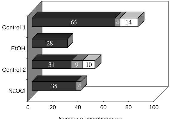
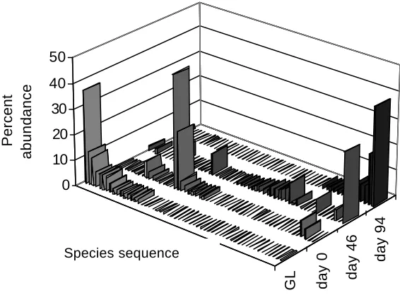
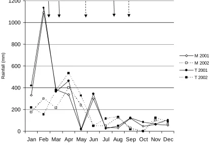
(17) LIST OF FIGURES Figure 1.1 Map of Australia and the Cairns region with study sites……………30 Figure 2.1 Frequency of isolates per morphotype derived from four Neolitsea dealbata leaf litter samples………………………………………………………….42 Figure 2.2 Scatterplot of isolation rate of leaf particles versus storage time.….42 Figure 2.3 Cumulative number of all morphotypes and common morphotypes 43 Figure 2.4 Morphotypes derived from Neolitsea dealbata leaf litter, wash water from treatment and control groups…………………………………………………44 Figure 3.1 Percent abundance of sporulating microfungi observed in Ficus pleurocarpa leaf litter………………………………………………………………..58 Figure 3.2 Percent abundance and diistribution of dominant species in green leaves and freshly fallen leaves of Ficus pleurocarpa …………………………..60 Figure 3.3 Percent abundance and diistribution of dominant species in Ficus pleurocarpa leaf baits, whch had been on the ground for 7 to 30 days………..60 Figure 3.4 Percent abundance and diistribution of domi nant species in Ficus pleurocarpa leaf baits, whch had been on the ground for 46 to 94 days………61 Figure 3.5 Shannon’s diversity indices for microfungal assemblages on fallen leaves of Ficus pleurocarpa for direct and indirect isolation methods………….62 Figure 3.6 Ordination of Bray-Curtis distances between microfungal assemblages in fallen leaves of Ficus pleurocarpa collected at different stages of decay………………………………………………………………………………..63 Figure 4.1 Observed and estimated species richness of microfungi in Ficus pleurocarpa leaves as assessed by the direct method…………………………..76 Figure 4.2 Chao2 estimates of species richness of microfungi in eight collections of Ficus pleurocarpa leaves at similar stages of decay based on direct observations……………………………………………………………….…..78 Figure 4.3 Abundance distribution for the complete dataset of microfungi in leaves of Ficus pleurocarpa obtained by the direct method……………………..78. xvi.
(18) Figure 4.4 Occurrences versus number of species in decaying leaves of Ficus pleurocarpa…………………………………………………………………………..79 Figure 4.5 Accumulation curves of observed and estimated total numbers of fungal species isolated from leaf litter of six tree species by the direct method……………………………………………………………………………..…81 Figure 4.6 Accumulation curves of observed and estimated numbers of fungal species in leaf litter isolated from six individual tree species…………………...82 Figure 4.7 Abundance distribution for the complete dataset of microfungi in decaying leaves of six tree species obtained by the direct method……………83 Figure 4.8 Accumulation curves of observed morphotypes among microfungi isolated by particle filtration…………………………………………………………84 Figure 4.9 Accumulation curves of Chao1 estimates for microfungal morphotypes isolated by the particle filtration method…………………………..85 Figure 4.10 Abundance distribution for the complete dataset of microfungi isolated from decaying leaves of four tree species by particle filtration…….. ..86 Figure 5.1 Monthly rainfall for Topaz Towalla Road and Millaa Millaa for the years 2001 and 2002………………………………………………………………101 Figure 5.2 Complementarity of microfungi in decaying leaves of six tree species in a comparison of site and season……………………………………………..105 Figure 5.3 Ordination of relative distance between microfungal assemblages from decaying leaves of six tree species using Nonmetric Multidimensional Scaling ……………………………………………………………………………..106 Figure 5.4 Dendrogram of microfungal assemblages in decaying leaves of six tree species isolated by the direct method……………………………………..107 Figure 5.5 Dendrograms of microfungal assemblages in decaying leaves of six tree species isolated by particle filtration………………………………………110 Figure 5.6 Complementarity of microfungi in decaying leaves at two sites Figure 6.1 Occurrence of ‘Dermateaceae F472’ and Coccotrypes aff. vulgaris on Ficus pleurocarpa leaves…………………………………………………....111 Figure 6.2 Abscissed leaves of Ficus pleurocarpa ……………………………. xvii.
(19) Figure 6.3 Percent occurrence of ‘Dermateaceae F472’ and Coccotrypes aff. vulgaris on sterilised and control leaves of Ficus pleurocarpa……………..…126 Figure 6.4 Interaction between ‘Dermateaceae F472’ and Coccotrypes aff. vulgaris in Ficus pleurocarpa leaf baits……………………………………….…128 Figure 7.1 Cylindrosympodium cryptocaryae showing conidia, sympodially elongating and reduced conidiophores ………………………………………….138 Figure 7.2 Number of species among fungal orders recorded from leaf litter of six tree species………………………………………………………………….….163 Figure 7.3 Number of species among fungal families recorded from leaf litter of six tree species…………………………………………………………………….164 Figure 7.4 Number of microfungal species among orders recorded in leaf litter of Ficus pleurocarpa during a succession study………………………..……...166 Figure 7.5 Number of microfungal species among families recorded in leaf litter of Ficus pleurocarpa during a succession study………………………………167. xviii.
(20) INTRODUCTORY OVERVIEW. Background “One of the most striking and perhaps characteristic features of life on Earth is its rich variety.” E. O. Wilson (1992) Fungi are among the most diverse organisms on Earth (Hammond, 1995) but the magnitude of their diversity is still unknown. They are vital contributors to ecosystems, for example through their roles in nutrient cycling (Jordan, 1985; Lodge, 1992), their mycorrhizal and endophytic associations with plants (Allen, 1991; Rodrigues and Peterini, 1997; Kumaresan and Suryanarayanan, 2002), and their interactions with insects (Wilding et al., 1989; Cafaro, 2002). Fungi also hold a vast unknown genetic potential for human endeavours, including pharmaceutical research (e.g. Bills, 1995; Wildman, 2003) and other biotechnological applications (e.g. Hyde, 1995; Vandamme, 2003). Despite the important services fungi provide to ecosystems and humans alike, fungi are an understudied element particularly of tropical regions and are rarely considered in conservation plans (Hyde, 2003). This is especially true for those fungi that cannot be observed by the unaided human eye, commonly referred to as ‘microfungi’. Among this taxonomically and functionally diverse group, those microfungi involved in the decay of leaf litter in an Australian tropical rainforest will be the focus of this project.. Research strategy General approach This study provides a rare opportunity to assess aspects of microfungal taxo nomy, diversity and ecology in a tropical ecosystem. Since information about these aspects is limited both on a regional and global scale, this project intends to be an explorative baseline survey rather than a solely experimentally based study. Understand ing the. xix.
(21) diversity and distributions of microfungi is an important first step towards understanding fungal ecology in general and any information will assist in the design of future studies to more fully elucidate the role of fungi in ecosystem processes (Cooke and Rayner, 1984). This study therefore had the following aims: •. To assess and make recommendations with respect to sampling and isolation methods for microfungi. •. To characterise the diversity and structure of microfungal assemblages from the rainforests of north Queensland. •. To assess the distribution of microfungi in leaf litter and to generate hypotheses regarding their ecology. •. To assess the taxonomy of selected microfungal taxa and to provide a reference collection of observed microfungi for future studies.. Geographical context The wet tropics of Australia (15° to 19° South, 145° to 146° East; Tracey, 1982) contain the most extensive continuous area of rainforest in Australia (Winter et al., 1991) and were declared a world heritage area in 1988. This region is characterised by an extraordinary diversity and a high degree of endemism among plants and animals (Wet Tropics Management Authority, 2004). This project was undertaken in upland rainforest on the Atherton Tablelands, north Queensland. The two study sites are part of an area of continuous forest, which also includes Bellenden Ker National Park (79,500 ha). Both sites were selected on the basis of the high diversity among tree species and were approximately matched for rainfall and rainforest type.. Choice of host species Four common plant families of this region provide a framework for this study. These include the Lauraceae, the Proteaceae, the Moraceae and the Elaeocarpaceae (Chapter 1). Microfungi were assessed on leaf litter of one or two representative species of each family, namely Cryptocarya mackinnoniana F. Muell. (Lauraceae), Elaeocarpus. xx.
(22) angustifolius Blume (Elaeocarpaceae), Ficus pleurocarpa F. Muell. (Moraceae), Ficus destruens F. Muell. ex C.T. White (Moraceae), Neolitsea dealbata (R. Br.) Merr. (Lauraceae), Opisthiolepis heterophylla L.S. Smith (Proteaceae), and Darlingia ferruginea J.F. Bailey (Proteaceae). Plant families are discussed in Chapter 1 and photos of leaves and a description of each species are provided in Appendix A.. Choice of collection methods All methods of studying microfungi impose some filter on the observed diversity. To overcome this filtering effect to some extent, I elected to use a combination of two methods. These included direct observation of fungal fruiting bodies following humid chamber incubation and the particle filtration method (Chapter 1 and 2).. Time allocation A maximum of two years could be allocated for field and laboratory work as part of this PhD project. To examine an adequate number of sampling units within each study year, I needed to weigh up whether to replicate the study over two years using the same method or whether to cross-check results with a second method in two separate years. My rationale for selecting the latter option was that if different isolation methods provided congruent results over two years with respect to the central questions, the conclusions of this study would be strengthened.. Limitations A number of limitations were encountered during this project. The amount of work that can be achieved by a single researcher using a replicated sampling strategy is a prime limitation in working with microfungi due to the labour- intensive nature of isolating and identifying these organisms. As a result, the replication within studies was low compared to some ecological studies of macro-organisms. Athough it was adequate to detect meaningful patterns in multivariate analyses, it is necessary to exercise caution when attempting to generalise these results to other forest types, ecosystems and time frames. In addition, a limitation outside my control was that one of the study years. xxi.
(23) (2002) was the driest year on record and it is not clear whether and how this has influenced microfungal diversity estimates. Another limitation was that few taxonomic resources are available for microfungi of north Queensland and testing species relationships and delimitations for more than some selected taxa was beyond the scope of this study. To circumvent this limitation to some extent, I contacted mycologists experienced in the taxonomy of tropical microfungi to assist in identifications or to confirm my preliminary identifications in some instances. These mycologists are gratefully acknowledged earlier in this thesis. Nevertheless, this limitation resulted in a conservative approach in identifying specimens to species levels.. Relevance of research Advances in the study of fungal diversity and ecology occur in small increments. In the short-term, this project adds to this incremental advance by confirming and extending the results of previous studies and by providing new information on isolating methods, sampling protocols and estimation procedures. This project also adds to the knowledge base of microfungal diversity and distributions in tropical rainforests, and generated hypotheses, which can form the basis for further synecological and autecological studies. In the medium term, the development of appropriate sampling and estimation strategies depends on an understanding of the factors, which shape fungal distributions (Lodge and Cantrell, 1995). More efficient and reliable sampling strategies for estimating microfungal diversity will benefit diverse areas of scientific research, such as conservation biology and biotechnology (Rossman, 1994; Cannon, 1997b; Hyde et al., 1997b; Hawksworth, 1998b). Despite the vital roles that microfungi play in ecosystems, a major gap exists in our understanding of the relationship between fungal diversity and ecosystem function (die Castri and Younes, 1990). Reliable methods for estimating fungal diversity are required to even begin unravelling this question. Together with advances in the taxonomic knowledge of tropical microfungi, it can also progress the utilisation of fungal genetic resources and novel compounds for biotechnology (Bills, 1995).. xxii.
(24) Thesis outline This thesis is divided into eight chapters, each dealing with a separate aspect of this project. The current state of knowledge with respect to microfungal taxonomy, diversity and distributions is reviewed in Chapter One . This is also where the reader will find definitions of terms and descriptions of relevant concepts. In Chapter Two, I will explore aspects of one isolation method for microfungi, i.e. particle filtration, and its usefulness for estimating microfungal diversity. The results of this preliminary study will be compared to those of previous studies. Successional patterns of microfungi in leaf litter of one tree species are reported in Chapter Three. In Chapter Four, I will discuss microfungal diversity and the patterns observed within microfungal assemblages. Aspects that may influence diversity estimates are also considered. An examination of the distribution of microfungi in leaf litter of six tree species is provided in Chapter Five. The distribution of fungi is discussed in relation to a number of factors such as host phylogeny, season, and site and I propose a number of hypotheses about the ecology of microfungi. In Chapter Six, I explore an association between a fungus and a beetle in decaying fig leaves. The spatial and temporal distribution of the fungus is also described and this information is integrated to generate a number of hypotheses about the nutritional modes of both organisms and their effect on decomposition processes. In Chapter Seven, I describe selected taxa, which are new to science, and provide a summary of the observed taxonomic diversity. Finally, I integrate the information gained from these separate studies and make recommendations with respect to future studies of microfungal diversity in Chapter Eight.. xxiii.
(25) CHAPTER ONE A REVIEW OF CURRENT KNOWLEDGE 1.1 Introduction The object of this chapter is to review current knowledge with respect to three different areas of research, namely the taxonomy, the diversity and the ecology of microfungi. This division into three sections serves as a frame work for discussion but is somewhat arbitrary as all three areas of research are interlinked and inform each other. In Section One, I provide a definition of fungi and their basic divisions. I discuss aspects of ascomycete taxonomy and describe available taxonomic resources in general and of the wet tropics in specific. In Section Two , I define the term diversity and summarise what is known about microfungal diversity, particularly in tropical regions. Factors that may affect diversity estimates are also reviewed; these include isolation methods for microfungi and statistical tools for estimating diversity. The significance of fungal biodiversity is considered and finally, a brief discussion is devoted to estimates of global fungal species numbers. Section Three is dedicated to a review of fungal ecology. In particular, I discuss some basic concepts and challenges of myco-ecology and review distributional patterns among microfungi. Potential factors underlying microfungal diversity and distributions, and the role of saprotrophic microfungi are considered. Finally, this studyis put into its geographic context of the region and the environmental context of leaf litter. While this chapter is focussed on microfungi, some examples are drawn from other fungal groups; this was necessary due to the paucity of data that exists for some areas of microfungal research. Where I considered it appropriate, I have also borrowed concepts from microbiology, plant ecology and general ecology.. 1.2 Taxonomy of microfungi 1.2.1 Fungi and microfungi defined The term ‘fungi’ refers to a diverse assemblage of eukaryotic organisms. The majority of these organisms are ‘true fungi’ and are placed in the kingdom Eumycota, but some. 1.
(26) belong in the kingdoms Chromista and Protoctista (Walker, 1996; Kirk et al., 2001). There is now increasing consensus based on chemical, ultrastructural and molecular data. that. Eumycota. encompass. four. phyla:. Ascomycota,. Basidiomycota,. Chytridiomycota and Zygomycota (Bruns et al., 1991; Walker, 1996; Kirk et al., 2001). These organisms are united by a lack of plastids (chloroplasts and amyloblasts). They are heterotrophs with an absorptive mode of nutrition Kendrick, 1992; Kirk et al., 2001). Their growth form is usually filamentous and consists of multicellular, coenocytic haploid hyphae although some fungi, such as yeasts, may be unicellular. Their diploid phase, if present, is usually short- lived. In contrast to plants, their cell walls contain chitin and ß-glucans (Kirk et al., 2001). Unlike many organisms grouped in Protoctista and Chromista, they are non- flagellate, or if flagella are present as in chytrids, they are lacking mastigonemes (Kendrick, 1992; Kirk et al., 2001). Fungi also have in common a number of other characters at the intracellular level (Kendrick, 1992; Kirk et al., 2001). Fungi can reproduce either sexually or asexually and are referred to as pleomorphic fungi, if more than one form or spore state is present. The whole fungus with both sexual and asexual states is referred to as ‘holomorph’ with the ‘teleomorph’ being the sexual morph and the ‘anamorph’ the asexual morph (Kendrick, 1979a; Kendrick and DiCosmo, 1979; Seifert and Samuels, 2000). The term ‘microfungi’ describes those fungi that cannot be detected with the naked human eye. It is a term of convenience and thus it may include a varied assemblage of organisms from all of the above phyla and kingdoms. In the context of this study, the majority of ‘microfungi’ detected were ascomycetes and their anamorphs, and these are discussed in more detail. The reader is referred to other texts for information on chytrids, zygomycetes and basidiomycetes (e.g. Kendrick, 1992; Alexopoulos et al., 1996).. 1.2.2 Ascomycete taxonomy 1.2.2.1 Teleomorphic ascomycetes The largest phylum of the Eumycota is the Ascomycota. The teleomorphs of these fungi are characterised by carrying spores in sac- like structures called asci (singular: ascus). Ascospores are formed following the fusion of nuc lei (karyogamy) and meiosis. Each ascus usually contains eight spores, although variable numbers have been observed in many taxa (Kendrick, 1992). A further possible uniting character among ascomycetes is 2.
(27) their lamellate hyphal wall, by which anamorphic ascomycetes can also be recognised using ultrastructural studies (Kirk et al., 2001). Asci and spores are contained within protective structures called ascomata, which can have several basic forms (apothecial, perithecial, pseudothecial and cleistothecial; Kendrick 1992). While the early taxonomy of teleomorphic ascomycetes relied mostly on fruit body shape and ascus arrangement (e.g. Engler and Prantl, 1897; Saccardo, 1882-1928; von Höhnel, 1907), in later decades increasing importance was placed on the development of ascomata (Nannfeldt, 1932) as well as the structure of asci (e.g. Luttrell, 1951; Eriksson, 1981) and their mode of discharge (Minter, 1988; Kirk et al., 2001). A comprehensive review of ascomycete taxonomy has been published in Volume 1A of the Fungi of Australia series (Walker, 1996) and an excellent review of taxonomic characters for teleomorphic ascomycetes is provided in Fröhlich & Hyde (2000); therefore these aspects will not be further discussed here. In recent years, the classification of ascomycetes has been shaped by the use of molecular sequence data and is updated continuously (Kirk et al., 2001). The most recent edition of the Dictionary of Fungi recognises 6 classes, 56 orders, 226 families, 3409 genera and 32,739 species (Kirk et al., 2001). It must be stressed that ascomycete taxonomy, in particular at higher taxonomic ranks, is still in a state of flux (Walker, 1996). For example, 41 accepted families could not be placed in orders and over 680 genera could not be assigned to families (Kirk et al., 2001). An outline of the current classification and the most recent changes are available on- line in Outlines of Ascomycetes (Eriksson, 2004).. 1.2.2.2 Anamorphic fungi Anamorphic fungi have been variously called ‘Deuteromycotina’, ‘deuteromycetes’, ‘mitotic fungi’, ‘Fungi Imperfecti’, ‘conidial fungi’ and ‘asexual fungi’ (Carmichael et al., 1980; Kirk et al., 2001). These fungi produce propagules from cells, which do not undergo meiosis (Kendrick, 1992). Most propagules are conidia but some may be derived from unspecialised mycelium (Kirk et al., 2001). Sterile mycelia or those fungi that form sclerotia, bulbils, or chlamydospores are also included in this group (Carmichael et al., 1980, Subramanian, 1983). Although many of these fungi have been linked to a sexual state classified among ascomycetes or basidiomycetes (Anateleo,. 3.
(28) 2004), a considerable number of anamorphs are still unconnected or, in some instances, appear to have lost their sexuality (Kirk et al., 2001). Reported anamorph-teleomorph connections have been established with varying degrees of rigour. These range from the least reliable, a single report of the physical proximity of an anamorphic and teleomorphic fungus on a natural substratum, to a connection proven by repeated cultural experiments (Kendrick, 1979a). The only way to unequivocally ascertain an anamorph-teleomorph relationship was to establish an axenic culture and observe the transition from one state to the other (Kendrick, 1979a). In recent years, molecular techniques have revolutionised the integration of anamorphs into a phylogenetic system. However, establishing fungal holomorphs using molecular methods remains a challenge due to inconsistent intraspecific sequence variation among genera (Egger and Sigler, 1993). In present-day taxonomy, the integration of anamorphic fungi in the teleomorph based system is considered an absolute necessity by many mycologists (Kendrick, 1979b; Seifert and Samuels, 2000; Gams et al., 2003). In fact, the latest edition of the ‘Dictionary of Fungi’ already assigns anamorphic taxa to the appropriate known level of the teleomorphic hierarchy (Kirk et al., 2001). Ultimately, the complete integration of anamorphs into a phylogenetic system is likely to be a complex task and may require numerous name changes (Hennebert and Gams, 2002). Concerns have been raised that name changes have the potential to disrupt the work of mycological practitioners and even destabilise taxonomy. These fears may be the reason why a vote taken at the Seventh International Mycological Congress 2001 indicated support for no or slow changes in funga l nomenclature (Gams et al., 2003). Currently, a dual naming system remains in use (Reynolds and Taylor, 1993; Sutton, 1993; Walker, 1996; Greuter et al., 2000; Kirk et al., 2001). In this system, the use of the suffix ‘form’ for genera and species of asexual fungi is a recognition of the artificial nature of their classification (Sutton, 1993). Formal taxonomic ranks above genera have been rejected by many (e.g. Sutton, 1993) but not all authors (e.g. Walker, 1996). In many instances, artificial classes continue to be used in an informal sense and these include •. hyphomycetes, which are mycelial in form and bear conidia on separate hyphae or aggregations of hyphae. 4.
(29) •. coelomycetes which produce conidia in pycnidial, pycnothyrial, acervular, cupulate or stromatic conidiomata and. •. aganomycetes which are sterile mycelial forms but which may produce vegetative structures such as chlamydospores or sclerotia (Kirk et al., 2001).. 1.2.2.3 Taxonomic tools The rapid advance in ascomycete systematics ha s not been matched by the availability of up to date identification tools. This may be in part due to the fact that the sheer size of the group makes it difficult to determine, which morphological features should be stressed in their delimitation (Kirk et al. 2001). This problem is compounded in those situations where molecular and morphological data do not agree (Kirk et al. 2001). “The Fungi. Volume IV A.” (Ainsworth et al. 1973) is a valuable resource and includes keys to orders, families and genera of ascomycetes; unfortunately, it is lacking recent changes and additions. A more recent key to orders of Ascomycota is that by Walker (1996). In addition to identification tools that target specific ecological groups such as palm ascomycetes (e.g. Fröhlich and Hyde, 2000) or geographical regions (Dennis, 1978), other important sources of information are treatments of orders of Ascomycota listed in Walker (1996), treatments of families, for example Rhytismataceae (Johnston, 1986, Johnston, 1989, Johnston, 1990, Johnston, 1991, Johnston, 1992), Gnomoniaceae (Monod, 1983), families of Hypocreales (Rossman et al. 1999), and other more broadly based texts (e.g.Sivanesan, 1984; Hanlin, 1998). Although an entirely artificial group, numerous identification schemes exist for anamorphic fungi; this may be due their perceived economic importance (Subramanian, 1983). For example, taxonomic treatments are available for coelomycetes (Sutton, 1980), for appendaged coelomycetes (Nag Raj, 1993), and for hyphomycetes (von Arx 1970; Ellis 1971; Matsushima 1971, 1980, 1981, 1983, 1985, 1987, 1989, 1993, 1995, 1996; Ainsworth et al. 1973, 1975; Ellis 1976; Carmichael et al. 1980; Domsch et al. 1980; Castañeda Ruiz 1985a, 1985b, 1987; Castañeda Ruiz and Arnold 1985; Castañeda Ruiz and Kendrick 1990a, 1990b, 1991; Barnett and Hunter 1998; Kiffer and Morelelt 2000; Kirk et al. 2001). Improved accessibility of mycological taxonomic resources and improved efficiency in documenting new taxa are vitally important for the progress of mycology (May, 1999).. 5.
(30) The development of databases based on coded taxonomic characters (e.g. B. Kendrick pers. comm., W. Ho pers. comm.) is one step in this direction. In addition, taxonomic resources are increasingly becoming available on-line and these include databases (Index Nominum Genericorum, 2003; The CABI Bioscience and CBS Database of Fungal Names, 2003; The NZFungi databases, 2004), keys (e.g. Rossman et al., 2003; The NZFungi databases, 2004) and digital exsiccate (e.g. Langer, 1997).. 1.2.3 Microfungi of the wet tropics Hawksworth (1993b) pointed out that our knowledge of the tropical mycobiota remains in the ‘pioneer phase’ or the first portion of alpha taxonomy (Davis and Heywood, 1963). This is certainly true for the wet tropics of Australia. While plant pathogens have received more attention than saprotrophic leaf litter fungi (e.g. Fröhlich, 1992; Hyde and Alcorn, 1993; Shivas and Alcorn, 1996; Fröhlich et al., 1997; Pearce, 1999), it is estimated that even among those only a fraction has been discovered to date (Shivas and Hyde, 1997). In the early days of European settlement, some microfungal collections, mainly of those causing leaf spots, were made by a network of collectors and these were sent to overseas mycologists for identification and description (May and Pascoe, 1996). More recently, visiting mycologists have further contributed to the knowledge of microfungi of Queensland, including leaf litter fungi (Matsushima, 1971; Taylor, 1997; Fröhlich, 1997; Hyde and Goh, 1998; Whitton, 1999; Fröhlich and Hyde, 2000; Guo et al., 2000; Johnston, 2000; Yanna et al., 2001a, 2001b; Parungao et al., 2002). A significant contribution to the taxonomy of saprotrophic microfungi was also made by K.D. Hyde during his period with the Northern Australian Quarantine Strategy at Mareeba (May and Pascoe, 1996), and W. Shipton of James Cook University published on species delimitation in Cunninghamella (Lunn and Shipton, 1983). Despite these efforts, the taxonomic diversity of leaf litter microfungi has been relatively underexplored in this region.. 1.3 Diversity of Microfungi 1.3.1 Diversity defined The term ‘biodiversity’ was coined by Wilson (1988) as a contraction of biological diversity and describes the diversity of life at all levels including genetic, species and. 6.
(31) ecosystem diversity (Hawksworth, 1991). In recent years, this term has been increasingly used in arenas outside of science and has become charged with political and social meaning (e.g. Commonwealth of Australia, 1996; Takacs, 1996; ten Kate and Laird, 1999). For this thesis, it is preferable to apply the term diversity in the sense of the richness and abundance of species (Magurran, 1988, Hubbell, 2001). Diversity has been described at different levels. For example, Whittaker (1960, 1977) defined seven levels of diversity in two main categories, namely inventory and differentiation diversities (Table 1). The former describes the diversity in one habitat at different scales, while the latter is a measure of how different (or similar) a range of habitats or samples are in terms of species richness and in some instances species abundances (Magurran, 1988). In this thesis, I assess the alpha diversity of microfungi in a particular habitat, i.e. leaf litter (Chapter 4). I also consider the pattern diversity between microfungi on different leaf species (Chapter 5) and on the same leaf species at different stages of decay (Chapter 3).. 1.3.2 Current knowledge of microfungal diversity “The knowledge gap between the known and unknown fungi is enormous.” (Hawksworth, 1998b) This statement describes the difficulty of reviewing current knowledge of microfungal diversity beyond a mere listing of studies. The diversity of soil microfungi in temperate regions has been considered ‘indeterminable’ (Christensen, 1989), tropical endophytic assemblages have been described as ‘hyperdiverse’ (Arnold et al., 2001) and a very high species richness was observed among saprotrophic leaf litter fungi in a neotropical rainforest where, for example, between 134 and 228 species were reported per five grams of leaf litter (Polishook et al., 1996). In all these instances, there was an indication that greater sampling effort would have yielded a greater number of species than was actually observed. Although there is a general agreement that microfungi are very diverse and that only a fraction of their diversity is known (e.g. Hyde, 1997a), drawing any conclusions from the body of diversity literature is difficult. This is because diversity studies are almost impossible to compare across space and time as standardised sampling and estimation protocols are lacking (Hyde, 1995; Cannon, 1997b; Cannon, 1999). Sampling approaches and hypotheses about fungal diversity. 7.
(32) could be tested if a 'known universe' of fungal species was available. For this reason, a number of mycologists have proposed an all taxa biodiversity for fungi (e.g. Rossman, 1994, 1997). A complete inventory for fungi will be costly in terms of human and material resources (Rossman, 1994) and to date this has not eventuated.. Table 1.1 Levels and types of species diversity (adapted from Whittaker, 1960, 1977). THIS TABLE HAS BEEN REMOVED DUE TO COPYRIGHT RESTRICTIONS. The taxonomic diversity of tropical microfungi has recently been reviewed (Hyde, 1997a; Hyde et al., 2004) with a focus either on specific microfungal taxonomic groups (e.g. Johnston, 1997), on microfungi on particular substrata, hosts or habitats (e.g. Hyde et al. 1997c; McKenzie, 1997; Hyde et al., 2004) or on modes of life (Shivas and Hyde, 1997; Rodriguez and Petrini, 1997). For many groups of organisms a latitudinal diversity gradient has been reported, with maximum species richness reached in the tropics (e.g. Ehrlich and Wilson, 1991). Whether this trend exists for fungi is still unclear. Some fungal groups such as the Phyllachoraceae (Cannon, 1997a), Hypocreales (Samuels, 1997) and the Xylariaceae (Whalley, 1997) appear to have a strong tropical representation. For other groups, for example the Rhytismatales (Johnston, 1997), such.
(33) a generalisation is not possible and some authors have noted the cosmopolitan nature of many microfungal genera (e.g. Johnston, 1997; McKenzie, 1997). There have also been reports that teleomorphs of some species appear restricted to the tropics while their anamorphic states were observed to be cosmopolitan (Samuels and Rossman, 1992). Similarly, the overlap between the majority of freshwater ascomycetes from tropical and temperate regions was noted to be very small (Hyde et al., 1997c) while many freshwater hyphomycetes are apparently cosmopolitan (Kuthubutheen, 1993; Goh, 1997). A greater number of plant pathogenic fungi are likely to occur in the tropics as many plant pathogenic fungi are host-specific and plants are known to be more diverse in the tropics (Shivas and Hyde, 1997). Similarly, pathogenic microfungi associated with invertebrates in the tropics appear to harbour many new taxa (Hywel-Jones, 1997) while non-pathogenic Laboulbeniales appear to have similar levels of diversity in both temperate and tropical regions (Weir and Hammond, 1997). Some substrata seem to harbour a particularly rich diversity of microfungi, for example palms (Fröhlich, 1997; Taylor, 1997), bamboo (Zhou and Hyde, 2002), Pandanaceae (McKenzie, 1997; Whitton, 1999), endemic plants of Mauritius (Dulymamode et al., 2001) and particular plant species in Great Britain (J. Cooper, pers. comm.). Most authors stressed that sampling remains incomplete in terms of the geographical regions, host taxa or niches examined and that any conclusions with respect to patterns of diversity need to be viewed in the light of this limitation.. 1.3.3 Diversity estimation 1.3.3.1 Overview An assessment of diversity is at the heart of many questions in theoretical and applied ecology. Although the concept of diversity can be grasped intuitively, it is “rather like an optical illusion” (Magurran, 1988). Our perception of diversity is shaped by numerous factors, among them our definition of diversity, the filters we inadvertently apply when we collect diversity data, and the choice of mathematical tools to measure and estimate diversity. These problems are compounded in species-rich groups, in groups that require special methods to detect ‘individuals’ and even more so in groups, such as microfungi, where ‘individuals’ are ill-defined (Seifert, 1981). For this reason, I will review methods of detecting microfungi and provide a broad overview of mathematical tools that are applied in diversity estimation.. 9.
(34) 1.3.3.2 Methods of detecting microfungi The systematic study of microfungi became possible only with the advent of microscopes and its history stretches back to the end of the 19th century (e.g. Seifert, 1981). While the observation and collection of fungal fruiting bodies remains a central focus for fungal taxonomy, in the 20th century mycologists increasingly recognised the value of “small scale gardening”, as a pioneer of this method, Dr Johanna Westerdijk, called the cultivation of microfungi (Commonwealth Agricultural Bureau, 1960). Numerous methods of isolating fungal cultures from various substrata have since been developed, each beset with their own limitations for the estimation of microfungal diversity (e.g. Booth, 1971). To overcome these limitations, it has been suggested that complementary protocols should be utilised if a more complete understanding of microfungal diversity is required (Frankland et al., 1990; Grigorova and Norris, 1990). Two main approaches for isolating fungi, the direct and indirect approach, provide such a complementary approach (Booth, 1971).. Direct approach The observation and isolation of fungal fruiting bodies from a substratum is termed the ‘direct method’ (Booth, 1971). This can either be undertaken on dried herbarium specimens, freshly collected material or following incubation of substrata in moist chambers. If desired, stains can be applied to fungal material and slides are then prepared either from squash mounts or microtome or handsections in a suitable mounting medium (Booth, 1971; Hawksworth, 1974; Gams et al., 1998). Sections are particularly useful in the study of coelomycetes and ascomycetes (Michaelides et al., 1979). Single spore cultures can be isolated from fresh specimens by plating dilutions of a spore suspension onto a suitable antibiotic medium and by transferring single spores to a fresh plate once germination has been observed (e.g. Booth, 1971). As this approach provides specimens from natural substrata and in many instances also from cultures, it is the most useful approach for taxonomic studies. In addition, single spore cultures. of. known. genetic. make- up. are. required. for. the. elucidation. of. anamorph/teleomorph connections (Kendrick and Nag Raj, 1979). On the other hand, microfungal fruit bodies are ephemeral structures and their appearance is easily missed. 10.
Figure


+7


Related documents
The sterile urine cup is also an acceptable container for collection and transfer of urine specimens provided that there is a daily pickup by ACM couriers for transport to ACM
When the key policy tools are controlled for, centralisation of the benefit and support assessment service and timely vocational rehabilitation were found to be strongly
Data sources used in the model, including electronic retrieval locations; comparison of posterior and prior distributions for the washing model, rinsing model, and microbial
A new customer web- portal will be implemented and this will be a major education tool to help our customers become familiar with online information as it
Finally, the Commission invited input from a panel of noted former public officials who were able to contribute practical insight into the intersection between ethics
The Summer Nutrition Programs The two federal Summer Nutrition Programs — the National School Lunch Program (NSLP) Seamless Summer Option and the Summer Food Service Program
where i indexes individual, j municipality of residence, and s upper secondary school starting year; Early childbearingijs is an indicator equal to one if individual i
Included in the review because the study was conducted in acute settings involving patients with older people Ericksson and Saveman 2002 Nurse’s experiences of
