STUDY OF POST OPERATIVE OUTCOME OF
VARICOSE VEIN SURGERY
Dissertation submitted in partial fulfillment of regulation for the award of
M.S. DEGREE
in
GENERAL SURGERY (BRANCH I)
THE TAMILNADU DR. M.G.R. MEDICAL UNIVERSITY
CHENNAI
APRIL 2015
CERTIFICATE
This is to certify that this dissertation titled “STUDY OF POST
OPERATIVE OUTCOME OF VARICOSE VEIN SURGERY”
submitted to the TamilNadu Dr. M.G.R. Medical University, Chennai in partial fulfillment of the requirement for the award of M.S Degree Branch - I (General Surgery) is a bonafide work done by Dr. S. Veeranan, post graduate student in General Surgery under my direct supervision and guidance during the period of September 2013 to August 2014.
Prof. D.N. RENGANATHAN, M.S., Prof. V. ELANGO, M.S.,
Professor Professor and H.O.D
Department of General Surgery Department of General Surgery Coimbatore Medical College Hospital Coimbatore Medical College Hospital
Dr. REVWATHY, M.D., Dean
DECLARATION
I solemnly declare that the dissertation titled “STUDY OF POST
OPERATIVE OUTCOME OF VARICOSE VEIN SURGERY” at
Coimbatore Medical College Hospital was done by me from September 2013 to August 2014 under the guidance and supervision of Professor Dr. D.N. RENGANATHAN, M.S. This dissertation is submitted to the Tamilnadu Dr. M.G.R. Medical University towards the partial fulfillment of the requirement for the award of M.S Degree in General Surgery (Branch I).
Place: Coimbatore
ACKNOWLEDGEMENT
I express my gratitude Dr. REVWATHY, M.D., Dean, Coimbatore Medical College Hospital for permitting me to use the clinical material for the study.
It gives me immense pleasure to express my deep sense of gratitude to my unit chief Prof. Dr. D.N. RENGANATHAN, M.S., for his excellent guidance and valuable suggestions during the course of study and in preparation of this dissertation.
I am grateful to Prof. Dr. V. ELANGO, M.S., Professor and HOD of General Surgery for allowing me to carry out this dissertation work in this department.
I express my heartfelt thanks to the unit chiefs,
Dr. P. Swaminathan,MS.D.O, Dr S. Saradha M.S., Dr. S. Natarajan,
MS., Dr. G. Ravindran, MS., Dr. Balasubramanian, MS., and
Dr. P.V. Vasanthakumar, MS, for their suggestions and support.
I am thankful to Prof. Dr. Murali, MD, Radiology,
I am greatly thankful to my Assistant Professors,
Dr. V.S. Venkadesan, MS., DA., Dr. R. Radhika, DGO, M.S., for their help and guidance.
CONTENTS
Page No.
I REVIEW OF LITERATURE
HISTORY 4
EPIDEMIOLOGY 6
ANATOMY 7
PHYSIOLOGY 23
AETIOLOGY 27
PATHOGENESIS 33
CLINICAL FEATURES 43
CLINICAL EXAMINATION 47
INVESTIGATIONS 55
TREATMENT 65
II METHODOLOGY 94
DISCUSSION 105
CONCLUSION 107
BIBLIOGRAPHY
ANNEXURES
PROFORMA CONSENT FORM QUESTIONNAIRE PHOTOS
ABBREVIATIONS
GSV GREAT SAPHENOUS VEIN
SSV SHORT SAPHENOUS VEIN
CVD CHRONIC VENOUS DISEASE
CVI CHRONIC VENOUS INSUFFICIENCY
DVT DEEP VEIN THROMBOSIS
CVU CHRONIC VENOUS ULCER
USG ULTRASONOGRAPHY
IL-1 INTER LEUKIN -1
CEAP CLINICAL ETIOLOGY ANATOMY
PATHOLOGY
SFJ SAPHENO FEMORAL JUNCTION
SPJ SAPHENO POPLITEAL JUNCTION
PL PERFORATOR LIGATION
SFJFL SAPHENO FEMORAL JUNCTION FLUSH
LIGATION
PV PERFORATOR VEIN
EVLT ENDOVENOUS LASER THERAPY
EVS EDINBURG VENOUS STUDY
SEPS SUBFACIAL ENDOSCOPIC PERFORATOR
SURGERY
LIST OF FIGURES
S. No Figure Title Page No.
1 Anatomy of Lower Limb Venous System 9
2 Venous Drainage of Foot 10
3 Venous Drainage (Posterior Aspect) 10 4 Great Saphenous Vein and Short Saphenous Vein 11
5 Deep Venous System 16
6 Lower Limb Perforators 18
7 Direct Perforator Vein 19
8 Indirect Perforator Vein 20
9 Venous valve structure 21
10 Muscle Pump 25
AIM
• To study the post operative outcome of patients who have
undergone varicose vein surgery – towards symptoms, signs of
patients after a prescribed period.
ABSTRACT
Varicose veins are dilated and tortuous veins, which affect a
significant proportion of adults. They cause physical and emotional
symptoms, and affect quality of life in sufferers. More than 5% of the
population have varicose vein, and 1% have or had venous ulceration.
Varicose vein causes substantial morbidity, cosmetic, monetary loss and
psychological effect.
OBJECTVE:
• To study the post operative outcome of patients who undergone
varicose vein surgery-towards symptom, signs of patients after
a prescribed period.
• To evaluate quality of life of patients undergone varicose vein
surgery
• To evaluate incidence of post operative complications of
varicose vein surgery and recurrence
METHOD:
The study carried out with the follow-up information relating to 53
patients who were undergone varicose vein surgery. The patients were
followed in the post operative period for the expected complications. All
the patients were subjected to duplex scan in the follow-up period ranging
A questionnaire was prepared to collect precise information
regarding the life style and symptomatic improvement of the patients.
The post operative follow-up and questionnaire data was analysed.
RESULTS:
It is observed that, out of 53 patients, 6 patients (11.25%) had mild
wound infection which did not require any additional treatment. There
was no incidence of deep vein thrombosis, pulmonary embolism. No
patients experienced neurological deficit or paraesthesia in the post
operative period. No patients experienced bleeding from wound. After
surgery 34 patients felt leg pain cured, 24 patients felt cosmetic
improvement and 7 patients had ulcer completely cured. Overall quality
of life was improved in 42 patients after surgery.
CONCLUSION:
Patients with complications of varicose vein combined with
valvular incompetence underwent surgery like saphenofemoral junction
flush ligation, with/ without stripping; with/without perforator ligation
have had improvement in their cosmetic appearance ,cure in leg pain, leg
ulcer cured, improvement in life style , improvement in ability to do
work as well as recurrence of varicose vein was not seen. The study
period was short, so the patients need long term follow-up to find out
HISTORICAL ASPECTS
Galen in second century used silk ligature to tie off blood vessels
and suggested varicose veins should be treated by incision, avulsion with
use of hooks1.
14th to 20th century:
In 14th century Maitre Henre de Mondeville successfully used
bandage on limbs with ulceration to drive back evil humours.
In 16th century anatomy of venous system was presented in great
details in work of Andeas Vesalius. Presence of venous valves was
probably first mentioned by JB Canano in 1547.
Hieronymius Fabricius de Aquapendente wrote on surgical
treatment of varicose vein in his book Opera Chirurgica published in
1593.
William Harvey described blood circulation. In 1810, Ferriar
described a patient with phlegmasia alba dolens .
Davis in 1822 recognised relation between venous thrombosis and
child birth. Brodie in 1846 described his test of venous valvular
incompetence and used compression bandages to treat venous ulcer.
Unna developed unna boot in 1854 –an elastic plaster dressing with
glycerine gelatine mixtue. Virchow described his revolutionary discovery
venous wall, stasis of venous blood and changes in blood coagulationin
his book Die cellular pathologie.
In 1864 Pravez initiated sclerotherapy and injected perchloride of
iron to sclerose varicose veins using hypodermic needles.
20th century land marks:
Proximal ligation of great saphenous vein was described by
Trendelenburg in 1891. Kellar described internal stripper in 1905.
Charles H Mayo used an external ringed stripper in 1906. Bobcock’s
contribution was development of flexible internal saphenous stripper.
Reconstructive surgery – Eck was the 1st to perform anastamosis
between portal vein and inferior vena cava. John Homan in 1917
introduced the term venous stasis and post thrombotic syndrome.
First attempt at phlebography by injection of strontium bromide
were made by Berberick & Hirsch in1923. Fibrinolytic effect of
streptococcus was 1st described in 1993 by Tillet and Garner. Bauer
showed beneficial effect of heparin in DVT in1941.
Linton described subfascial ligation of incompetent communicating
vein in 1938. Franck Cockett suggested the term blow out syndrome.
COCKETT’s book co-written by Doddwas, is the 1st standard text
EPIDEMIOLOGY
Varicose vein is a common surgical problem present in 10% of
population. Incidence is less in India compared to developed countries.
Prevalence is 20% to 25% in female, 10% to 15% in men in western
countries (Callam M J 1994 Epidemiology of varicose vein, Br. J. Surg,
81 167-173)
Edinburg venous study (EVS) published in 1998 shows prevalence
of varicose vein 39.7% in men, 32.2% in women. Prevalence of varicose
vein increases with age in published studies. Prevalence is 11.5% in 18 to
25 years of age group, 55.7% in 55-64 years age group. EVS 2004 shows
ANATOMY
NORMAL VENOUS HISTOLOGY AND FUNCTION5
HISTOLOGY OF VEIN
The vein wall contains three concentric layers6
• Tunica intima (inner layer)
• Tunica media (middle layer)
• Tunica adventitia (outer layer)
INNER INTIMAL LAYER
Inner layer is known as intima. Its main component endothelium
lines entire vascular tree including heart. This layer is absent in venules.
An internal elastic lamina is present in the sub endothelial layer of the
larger veins. Special endothelial cells produce plasminogen activator.
MIDDLE LAYER
The middle media is composed of elastin connective tissue and
smooth muscle cells. It varies in size between veins of different calibre. It
is thin in venules. This layer is thickest in arteries.
OUTER LAYER
The outer layer is known as adventitia, it consists of collagen
fibres, largely longitudinal. It contains vasa vasorium and sympathetic
VENOUS WALL FUNCTION
Large venules and veins form an extensive but variable large
volume low pressure system of vessels conveying blood back to the heart.
The outer adventitial layer of venous wall contains adrenergic fibres
mainly in cutaneous veins. Venous wall tone can be altered by central
sympathetic discharge and brainstem thermoregulatory centres and also
other stimuli like change in body temperature change in blood volume
and pain. Vein histology varies depending on calibre of vein. Smallest
veins are venules size range from 0.1mm to 1mm. The large veins present
in lower limb have less smooth muscle.
Veins are thin walled, distensible and collapsible structures. The
two important functions of vein are transportation of blood back to heart
and storage of blood. Blood flow in veins is depends on following factors
such as gravitation, competent valves, volume of blood, cardio repiratory
cycles and muscle pump. Any alteration in equilibrium of these factors
may leads to pathology of vein.
Blood flow in veins is unidirectional and this is maintained by
presence of valves in the wall of veins. The valves are more in distal part
of vein and become progressively few in proximal part of vein.
Venous valves have two leaflets. They allow unidirectional flow of
blood. The venous valves become closed if blood flow is craniocaudal
vena cava, portal vein and venous sinuses in soleus and gastracnemius are
valve less.
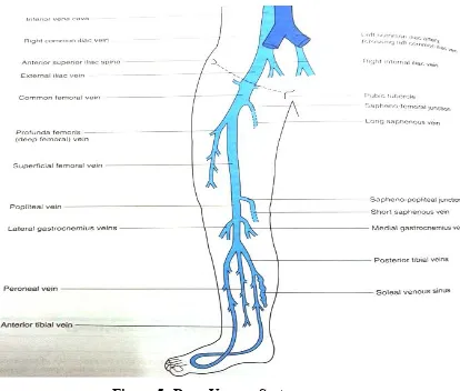
VEINS OF LOWER LIMB7
There are three systems of veins, which are
• Superficial venous system
• Deep venous system
• Communicating system
• Perforator
[image:20.612.214.433.365.680.2]• Intersaphenous
Figure 2: Venous Drainage of Foot
[image:21.612.188.441.381.679.2]SUPERFICIAL VENOUS SYSTEM OF LOWER LIMB
Superficial veins form a network that connects superficial veins to
deep veins. Superficial veins lie above deep fascia. The dorsal venous
arch lies on dorsum of foot. It receives four dorsal meta tarsal veins, each
of which is formed by union of two dorsal digital vein.
GREAT SAPHENOUS VEIN (GSV)7
This is longest vein in lower limb. It is formed by union of medial
end of dorsal venous arch with medial marginal vein. It run upwards
anterior to medial malleolus, crosses the lower one third of medial surface
of tibia obliquely and run along its medial border to back of knee. The
[image:22.612.169.506.443.682.2]saphenous nerve runs in front of GSV
In the thigh, it inclines forwards to reach the saphenous opening
where it pierces the crebriform fascia and opens into the femoral vein.
Before piercing the cribriform fascia, it receives three named tributaries
corresponding to the cutaneous arteries and also many unnamed
tributaries. It contains about 10 to 15 valves which prevent back flow of
the venous blood, which tends to occur because of the gravity. One valve
is always present at the saphenofemoral junction. The saphenofemoral
junction is present 2.5 to 3.5 cm below and lateral to pubic tubercle.
Incompetence of these valves makes the vein dilated and tortuous
leading to varicose veins. The vein is also connected to the deep veins of
the limb by perforating veins. The perforating veins are also containing
valves. These valves permit flow of blood from GSV to deep veins. If
those valves incompetent, will gives rise to varicose veins.
The saphenous nerve in leg and foot is anterior to GSV. The GSV
is accompanied by the lymphatic trunk draining the dorsum of foot and
anterior and medial aspect of the leg and thigh draining to superficial
group of inguinal lymph nodes.
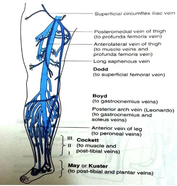
TRIBUTARIES
• At the commencement – medial marginal vein from the sole.
• Just below knee – the anterior vein of the leg runs upwards,
forwards and medially, from the lateral side of the ankle.
The posterior arch vein of Dodd and Cockett (Leonardo’s Vein)
is large and constant. It begins from series of small venous arches which
connect medial ankle perforators, runs upwards to communicate with
GSV just below the knee. In the thigh – it receives two large tributaries
which join it close to its termination. They are postero medial and antero
lateral veins. Postero medial vein drains the postero medial side of the
thigh. It may communicate with small saphenous vein. It receives
numerous small tributaries from skin and subcutaneous tissue of popliteal
fossa and upper half of the inner thigh. The anterior cutaneous vein of the
thigh drains the lower part of front of thigh.
TERMINAL TRIBUTARIES
• Just before piercing the cribriform fascia superficial epigastric,
superficial circumflex iliac and superficial external pudendal.
• Just before termination – deep external pudendal vein.
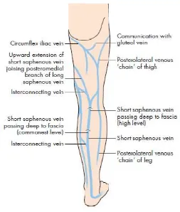
SHORT SAPHENOUS VEIN
The vein is formed on the dorsum of the foot by the union of lateral
marginal vein with dorsal venous arch. It enters back of leg by passing
behind the lateral malleolus. In the leg it ascends lateral to the
part of the popliteal fossa. It penetrate deep fascia to join the popliteal
vein.
It drains lateral border of the foot, the heel and the back of the leg.
It connected with great saphenous vein and with deep veins and is
accompanied by sural nerve. The anatomical position of saphenopopliteal
junction is variable significantly.
Tributaries – Several small vessels communicate with the short
saphenous vein to venous arches on inner side of the leg. Small
saphenous vein communicate to peroneal vein by a large constant lateral
ankle perforator vein. It communicates with soleus sinusoids and
therefore indirectly with posterior tibial and peroneal vein by an
inconstant mid calf perforator. Small saphenous vein in its upper part
communicates with GSV via posteromedial vein of thigh – vein of
giocomini.
DEEP VEINS OF LOWER LIMB
The popliteal vein and femoral vein are major veins of lower limb
and usually single conduits. They receive many tributaries from the
surrounding muscles, corresponding with arteries. The veins draining
muscles are valved with exception of those in soleus. The Soleus contains
venous sinuses, they are non-valved and empty segmentally into posterior
resting stage of muscles there is sluggish flow of blood in the soleal
sinuses.
The plantar digital veins in foot are drain into metatarsal veins
which composes deep plantar venous arch. This continues into the medial
and lateral plantar veins that then drain into posterior tibial veins. The
dorsalis pedis veins on the dorsum of the foot form the paired anterior
tibial veins at the ankle. The posterior tibial vein accompany posterior
tibial artery, receiving veins from sural muscles, especially the venous
plexus in the soleus, connections from superficial veins and peroneal
veins. Veins from soleus and superficial veins drain into the peroneal
vein.
Posterior tibial vein goes under fascia of deep posterior
compartment. Then they enter soleus and link the popliteal vein, after
joining with the paired peroneal and anterior tibial veins. There are large
venous sinuses within the soleus muscle—the soleal sinuses—that empty
into the posterior tibial and peroneal veins. There are bilateral
gastrocnemius veins that empty into the popliteal vein distal to the point
Figure
The popliteal vein enters a window in the adductor magnus, at
which point it is termed the
lies medial to femoral artery, between the heads of gastrocnemius it is
dorsal to it, proximal to the knee joint it is post
valves in popliteal vein.
• Small saphenous vein
• Veins corresponding to bran
• Muscular veins
Figure 5: Deep Venous System
The popliteal vein enters a window in the adductor magnus, at
which point it is termed the femoral vein. Popliteal vein in its distal part
dial to femoral artery, between the heads of gastrocnemius it is
dorsal to it, proximal to the knee joint it is postero lateral. There are
valves in popliteal vein. Popliteal vein tributeries are
Small saphenous vein
Veins corresponding to branches of popliteal artery
Muscular veins
The popliteal vein enters a window in the adductor magnus, at
Popliteal vein in its distal part
dial to femoral artery, between the heads of gastrocnemius it is
THE FEMORAL VEIN
The femoral vein goes together with femoral artery. It begins at the
adductor opening as the continuation of popliteal and ending posterior to
the inguinal ligament as external iliac vein. The femoral sheath middle
compartment contains femoral vein.
The femoral vein ascends and receives venous drainage from the
profunda femoris vein, or the deep femoral vein, and after this
confluence, it is called the common femoral vein. As the common
femoral vein crosses the inguinal ligament, it is called the external iliac
vein. Lateral and medial circumflex femoral vein are tributaries of
femoral vein.
INTERSAPHENOUS SYSTEM
It connects great saphenous vein and short saphenous vein.
Previously it was called as vein of GioComini.
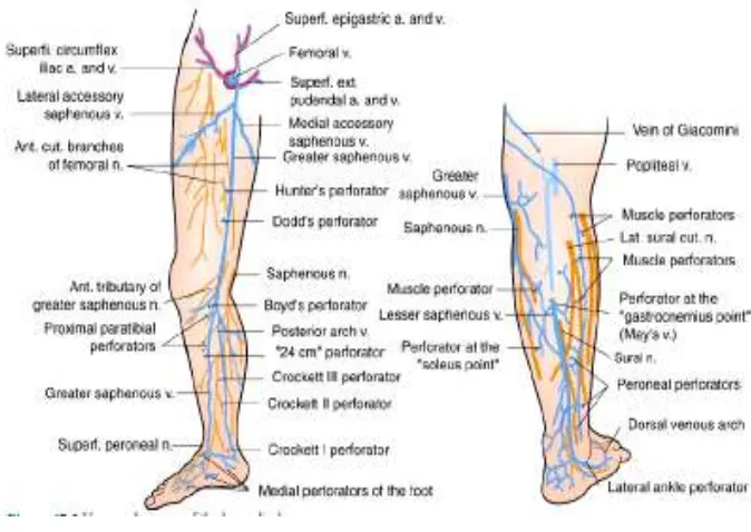
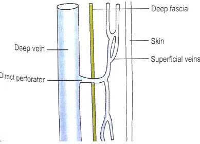
PERFORATOR SYSTEM8
The perforator veins normally allow blood to flow from superficial
to deep venous system and in healthy subjects they are valved that they
only permit blood to flow in one direction.
Two types of perforator are direct perforators and indirect
perforators. The direct perforators are the one which passes straight from
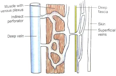
Perforators are called indirect when they connect superficial veins
with deep veins through the muscular veins.
Direct perforator
The great saphenous vein and short saphenous vein are the large
direct perforators. The small direct perforating veins are summarized
[image:29.612.133.497.312.695.2]below.
Figure
Perforators are called indirect when they connect superficial veins
with deep veins through the muscular veins.
The great saphenous vein and short saphenous vein are the large
direct perforators. The small direct perforating veins are summarized
Figure 6: Lower Limb Perforators
Perforators are called indirect when they connect superficial veins
The great saphenous vein and short saphenous vein are the large
In the thigh – There is a constant perforator vein which begin
great saphenous vein
third of thigh and end in segment of femoral vein below the deep femoral
valve in hunters canal
In the leg
BOYD Gastrocnemius perforator
vein usually runs close to the posterior border of tibia from the long
saphenous vein or its large tributaries (Posterior Arch) to the posterior
[image:30.612.134.523.332.612.2]tibial vein which is constant.
Figure
There is a constant perforator vein which begin
saphenous vein or one of its tributaries about the middle to lower
third of thigh and end in segment of femoral vein below the deep femoral
valve in hunters canal – mid thigh perforator of DODD.
BOYD Gastrocnemius perforator – just below the knee l
vein usually runs close to the posterior border of tibia from the long
saphenous vein or its large tributaries (Posterior Arch) to the posterior
tibial vein which is constant.
Figure 7: Direct Perforator Vein
There is a constant perforator vein which begins in the
one of its tributaries about the middle to lower
third of thigh and end in segment of femoral vein below the deep femoral
ust below the knee level, a
vein usually runs close to the posterior border of tibia from the long
Figure
COCKETT lower leg perforator
direct perforators of clinical importance, which are known as internal and
external ankle perforating veins. They are not isolated veins but vena
communicates accompa
MAY or KUSTER ankle perforators
These are usually present approximately 2, 4 and 6 cm from tip of
medial malleolus.
Perforators related to SSV
Figure 8: Indirect Perforator Vein
COCKETT lower leg perforator – in the distal half of leg there are
direct perforators of clinical importance, which are known as internal and
external ankle perforating veins. They are not isolated veins but vena
communicates accompanying a perforating artery to the skin.
or KUSTER ankle perforators 9
are usually present approximately 2, 4 and 6 cm from tip of
Perforators related to SSV
in the distal half of leg there are
direct perforators of clinical importance, which are known as internal and
external ankle perforating veins. They are not isolated veins but vena
Lateral or external ankle perforating vein is a constant large
perforator which connects SSV to posterior tibial vein. This perforator
plays a role in ulceration of lower third of leg.
THE VENOUS VALVES11
Valves in the superficial veins of the lower extremity are usually
located near to the termination of major tributaries. The GSV contains
about 10-15 valves, with more valves located below the knee. Valves in
the SSV are closer to each other than in the GSV. Valves in the
communicating branches between the SSV and GSV are oriented to direct
blood from the Small to the Great saphenous vein. Like superficial veins,
[image:32.612.234.411.418.635.2]deep veins have more valves in the calf than in the thigh.
Tibial veins are densely packed with valves, whereas there are only
1 or 2 valves in the popliteal vein. In the Femoral vein there are 3-5
valves, with one of them located just distal to junction of deep femoral
vein.
TYPES OF VALVES
1. Parietal valves (pocket valves) - These valves are tricuspid,
formed along the course of the large venous trunk.
2. Ostial valves- These are present at the entry point of a small vein
into the larger vein. There may be only one cusp.
STRUCTURE OF VALVE
It consists of the following parts,
• Cusp (leaf let)
• Agger (point of attachment of cusp to venous wall)
• Corne (projection of leaflet)
• Commisure (joining points of two cusps)
PHYSIOLOGY OF VENOUS RETURN
The important functions of the lower limb veins are
• Transport of venous blood to heart form lower limb
• Reservoir for storage of blood
Factors responsible for venous return to heart are,
• The muscle pump
• Capillary pressure
• Muscle tone
• Negative pressure in thorax
• The competent valves
• Vis-a-tergo
Blood enters the limb through the femoral artery before passing
through arterioles into capillaries, which have pressure of about 32 mm
Hg at their arterial ends. This pressure is reduced along the course of
capillaries and it is approximately 12mmHg at the venular end capillary.
The pressure continues to fall as it approaches the heart and it is about
-5mm Hg at the end of inferior vena cava where it enters into right atrium.
The pressure of a vein in foot on standing position is related to blood
The return of blood to heart from legs against gravity in the
standing position is facilitated by muscle pumps. There are two muscle
pumps in leg, most important one is calf muscle pump and other one is
foot pump.
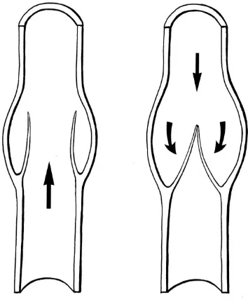
THE MUSCLE PUMPS
The calf muscle pump is the main pump which helps the blood to
be returned to the heart from legs. The soleus and gastracnemius are
importatant componants of calf pump. The calf muscles contraction
causes forcible compression of soleus and gastrocnemius sinuses and
blood propels towards heart. The pressure within calf compartment
reaches 200-300 mm Hg when muscle contract.
When muscle in relaxed position, pressure in calf compartment
falls and blood enters into deep veins from superficial veins through
sapheno femoral junction, sapheno popliteal junction and through
perforator veins. This causes fall in pressure in superficial venous
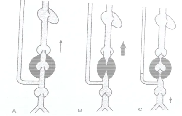
A)Rest B) Muscle Contraction
This reduction
perforators, patent deep veins and superficial veins, which must contains
competent valves. The normally functioning vein valves prevent
retrograde blood flow and maintain unidirectional blood flow towards
heart. When there is failure of
[image:36.612.165.472.71.267.2]disease and its symptoms.
[image:36.612.207.411.516.673.2]Figure 11 : Effect of Exercise on superficial venous system pressure Figure 10: Muscle Pump
Rest B) Muscle Contraction C) Muscle Relaxation
This reduction of pressure is dependent on presence of patent
orators, patent deep veins and superficial veins, which must contains
competent valves. The normally functioning vein valves prevent
retrograde blood flow and maintain unidirectional blood flow towards
heart. When there is failure of venous valves, patient develops
and its symptoms.
Effect of Exercise on superficial venous system pressure
C) Muscle Relaxation
on presence of patent
orators, patent deep veins and superficial veins, which must contains
competent valves. The normally functioning vein valves prevent
retrograde blood flow and maintain unidirectional blood flow towards
t develops venous
Figure 12 : Pressure Tracing of Calf Muscle Pump
The blood flow into deep veins occurs during the diastole of pump
only. Negative intra thoracic pressure, - speed of venous blood flow
return is increased by movements of respiration especially from upper
limb. In man quite breathing has little effect on venous pressure in leg but
deep inspiration lowers pressure.
THE RESERVOIR FUNCTION
It has been estimated that nearly one third of volume blood in the
body is found in lower limb and this blood will be available for body in
response to stimuli. This reservoir action will lost in varicose vein, even
though more amount of blood is stored in dilated elongated vein as most
stagnant and fails to react to many of stimuli. This stagnation of blood
VARICOSE VEIN AETIOLOGY
Varicose veins may be classified as
• Primary
• Secondary
• Recurrent
PRIMARY VARICOSE VEIN
Varicose vein that develop spontaneously in the absence of
identifiable cause are known as primary varicose vein. The great
saphenous vein is involved more frequently. About 12% of primary
varicose vein are associated with small saphenous system. Factors
associated with etiology of primary varicose vein are
• Valvular incompetence
• Weakness of vein wall
The incompetence of valve may be localised are segmental.
Varicosity of great saphenous vein is usually associated with
saphenofemoral junction incompetence. Varicosity of small saphenous
vein is usually associated with saphenopopliteal junction incompetence.
Valvular incompetence leading to varicose vein may be confined to short
segment of saphenous vein mainly GSV.
SECONDARY VARICOSE VEIN
Varicose veins that develop following deep vein thrombosis are
part of vein wall and any contained valve are involved in a process of
occlusion by thrombosis, fibrosis and recanalisation, subsequently results
venous dilatation in all but the smallest veins. When the perforating veins
are involved in this process and its protecting valve is destroyed a
superficial varicosity may develop.
Causes of varicose vein are
• Heriditary 12
• Race
• Sex
• Pregnancy
• Hormonal influence
• Posture
• Obesity
• Gravitational back pressure
• Primary valvular incompetence
• Incompetent perforating vein
• Weakness of vein wall
• Abnormality of vein wall
• Av fistula
Sex
Some studies show that varicose vein appear to be more common
in women. The Edinburgh vein study 13 (Evans, Fowkes,et al. 1999) and
Bulgarian cross sectional survey (Zaharivev, Anastassov et al. 2009)
showed higher prevalence of telangiectasia in women, but higher
prevelance of trunk varicosities in men.
Race 14
Varicose veins are more common in developed countries compared
to developing countries. (Beebe-Dimmer, Pfeifer et al. 2005). The
underlying reason for the racial and geographical variation is yet to be
known, but may relate to genetic factors affecting vein wall.
Age 14
With increase in age, there is increased incidence of venous disease
(Beebe-Dimmer, Pfeifer et al. 2005). The increasing prevalence with
increasing age is due to increased superficial venous pressure, due to calf
muscle weakness and slow reduction of vessel wall strength. Chronic
venous insufficiency prevalence increases with age mainly in men.
Varicose veins among people aged below 30 years are below 1% for men
and lower than 10% for female. From age 70 years and above, prevalence
Heridity 4
Patients with FOXC2 gene inheritance are associated with
development of this disease in 70 to 85% of first degree relatives,
compared to 10% of those with no family history. Klippel-trenaunay
syndrome, a valvulia, parkes-weber syndrome – here varicose veins are
atypical in distribution.
Pregnancy
The correlation between varicose vein and pregnancy is strong with
10 to 20% of pregnant females developing varicose vein and upto 70%
develop telangiectasias. Multiparous women are commonly developed
varicosity. The harmone progesterone causes dilatation and relaxation of
veins of the lower limb. This may make the valves more prone to
incompetence. The hormonal effect is more in first trimester.
As the pregnancy advances uterus enlarge in size and it may cause
compression on inferior vena cava and this may lead to secondary
varicose vein. The spontaneous resolution is seen in most patients within
3 to 6 weeks after delivery. The hormones relaxin, oestrogen and
progesterone are the principal players in this respect, having effects on
venodilatation, venous stasis, valvular dysfunction, and possibly
weakening the integrity of venous wall. (Vin, Allaert et al. 1992,
Posture
In prolonged standing (more than 6 hours per day), the column of
blood along with gravity put pressure on the vein valves, which leads to
varicosity. The qualitative dysfunction of calf muscle pump mechanism
also occurs due to prolonged standing. This causes failure of valves
giving rise to varicosities.
Obesity 16
The obesity is a risk factor for development of varicose veins, the
effect appears to be more so in women (Brand, Dannenberg et al. 1988,
Kontosic, Vukelic et al. 2000, Lee, Evans et al. 2003). Some studies
prove that women with body mass index more than 30 Kg/m2 are 3 times
more prone to develop varicose veins. The effect may be due to the
increased intra abdominal pressure in the obese (Noblett, Jensen et al.
1997), which results in decreased blood flow in pelvic veins, and
therefore increased lower limb venous pressures (Fowkes, Lee et al.
2001).
Varicose vein recurrence 17
Major cause of recurrence is faulty surgical technique. Failure to
do adequate sapheno femoral junction flush ligation and failure to ligate
Table : Causes of Varicose Vein Recurrence
Cause Preventable
Inadequate
Assessment
Deep vein Incompetence Deep vein occlusion Missed points of Incompetence
Anatomical abnormality
Yes
Inadequate Surgery Inadequate GSV Ligation Missed tributaries
Thigh GSV not stripped Inadequate SSV Ligation
Yes
Progress of disease New valvular incompetence No
Other risk factors:
Sedentary life style and prolonged standing leads to varicose vein.
Low fibre diet resulting in constipation, straining at stool may associate
with varicose veins. Edinburg vein study found that long intestinal transit
time low fibre diet and straining at stool are associated with an increased
incidence of varicose veins. Height is a positive risk factor for varicosity
and this is related to increased hydrostatic pressure. Lower limb fracture,
systemic hypertension, smoking are some other risk factors that may be
PATHOGENESIS
The three fundamental mechanisms that lead to the sustained
venous pressure are dysfunction of calf muscle pump, venous valvular
reflux, and obstruction to venous blood flow. 18
CALF MUSCLE PUMP DYSFUNCTION
Increasing age, musculo-skeletol deformity can impair the ability
of calf muscle function, so impairment of muscle pump that lead to
venous stasis. Transverse and longitudinal arch of the foot deformity can
impair the venous foot pump. Muscle tone is vital for perforator
competence. Incompetent perforators in the calf muscle leads to defect in
muscle pump function.
VENOUS VALVE REFLUX
Venous valvular reflux in the superficial and/or deep veins is
present in excess of 90% of patients with chronic venous insufficiency
(CVI). In approximately a third to a half of cases this is confined to the
[image:44.612.144.499.553.690.2]superficial venous system. In the remainder, both systems are affected.
PRIMARY VENOUS VALVULAR INCOMPETENCE
The reduction in elastin content and vein wall collagen weakening
particularly along the commissures of valves, leads to valve leaflets
separation, wall dilatation and reflux. Smooth Muscle Cell (SMC) present
in varicose vein wall are well dedifferentiated, they demonstrate
enhanced proliferative and synthetic capacity compared to SMCs present
in normal veins 19
SECONDARY VALVULAR INCOMPETENCE 4
DVT is a common clinical entity and a clear history of DVT is
found in about 20% of patients with CVU. Unknown number of people
develops asymptomatic DVT without ever knowing it. The deep vein
thrombosis leads to an inflammatory, possibly ischaemic phlebitis.
Partial or complete occlusion of the deep venous system with
thrombus leads to flow through collaterals that are: valveless, thus
permitting reflux, small diameter, so increasing the resistance to venous
outflow.
Thus blood may return from the leg by being forced out from the
deep compartment via perforators the distended so-called secondary
varicose veins. Failure of haemodynamic compensation may quickly lead
This comprises the skin changes of CVI leading to ulceration,
swelling and a bursting discomfort in the leg following exercise relieved
only by resting and elevating it (venous claudication).
CONGENITAL VARICOSE VEIN 4
Varicose veins are associated with several congenital syndromes
such as Klippel Trenaunay syndrome and Parkes Weber syndrome.
Klippel - Trenaunay syndrome is a non-familial mesodermal anamoly,
characterized by soft tissue and bone hypertrophy, varicose veins, and
capillary port-wine stains; this syndrome appears to be the most common
of the congenital abnormalities associated with varicose veins. Parkes
Weber syndrome present with varicose vein, multiple AV fistulas,
chronic venous hypertension, high output cardiac failure and ulceration.
In these syndromes, absent or hypoplastic deep veins may present, such
that malformations acts as the primary venous outflow from the leg. The
management of these conditions is mainly conservative1 with
compression hosiery, limited stab avulsion of symptomatic varices after
thorough Duplex USG scanning.
VENOUS ULCER MECHANISM
Venous disease is responsible for 60 to 70 percent of ulcers in the
lower limb. There are other causes for leg ulcers like aterial ulcers,
There are two theories explain venous ulceration that are
• White cell trapping theory
• Fibrin cuff theory
WHITE CELL TRAPPING THEORY20
This theory was proposed by Coleridge Smith and his colleagues.
White cells are bigger and less deformable than red blood cells. The
haemodynamic effect of white cell has greater effect on blood flow
through a narrow channel such as capillary. If the perfusion pressure
across the capillary bed is reduced due to an increase in venous pressure,
white cells plug the capillaries and red cells build up behind. On reaching
the post capillary venule, the white cells are forced to marginate by the
red cells. Adherence of white cells to the endothelium is then stimulated
by
• Decreased shear forces.
• Up-regulation of adhesion molecules by endothelium as a response
to venous hypertension.
The inappropriate activation of trapped white cells release
proteolytic enzymes and oxygen free radicals, causing endothelial and
tissue damage. Tumour necrosis factor α and interleukine 1 (IL-1) are
cell activation to permit the passage of much larger than normal
molecules. The patients with venous disease shows reduced fibrinolytic
activity due to IL-1 effect. The endothelial cells are stimulated to produce
plasminogen activator inhibitor I. This activation of endothelial cells is by
IL-1. IL-1 also reduces the plasminogen activator production and thereby
cause reduced fibrinolysis.
As a consequence there is release of vascular endothelial growth
factor. This cause increased microvascular permeability, which may
explain the presence of a fibrin cuff, and also produces excessive amounts
of nitric oxide. Fibrin deposition, tissue death, scarring occurs together
called as lipodermatosclerosis. Recently, McCollum demonstrated that in
patients with chronic venous insufficiency and increased venous pressure
in leg veins, have up regulation in oxygen free radicals production by
neutrophils and also enhanced production of thromboxane A2.
FIBRIN CUFF THEORY 21
In 1982, Browse and Burnand proposed that oxygen diffusion into
cutaneous tissues was restricted by a pericapillary fibrin cuff. They
suggested rise in venous pressure is directly transmitted to the capillary
bed. This results in an increase in the endothelial surface area available
for exchange and allows the passage of larger molecules i.e. fibrinogen
Fibrinogen then polymerizes to produce the `fibrin cuff’ that
surrounds cutaneous capillary walls. Measurements of capillary protein
loss by Browse and Burnand showed that fibrinogen was generally
important plasma protein leaking into tissues in patients with venous
disease. Successive measurements of fibrinolysis have shown that
patients with venous disease have decreased venous fibrinolytic activity,
which might explain why the fibrin cuff persists.
The fibrin cuff theory suggests venous ulceration is related to
deprivation of oxygen in tissues. The possible mechanism for frank
ulceration of lipodermatosclerosis lies with the enzymes matrix
metalloproteinases. These enzymes help remodel the extracellular matrix
by protein degradation, and enhanced activity of these enzymes has been
demonstrated in lipodermatosclerosis. The ambulatory venous
hypertension is the only accepted cause of ulceration. The venous
hypertension may be the result of primary valve incompetence of
saphenous veins, incompetence of perforating veins or incompetence or
obstruction of the deep veins.
INCOMPETENCE OF VENOUS VALVES
• Stasis of blood
• Chronic ambulatory venous hypertension
• RBC diffuses into tissue plane
• Lysis of RBC
• Release of haemosiderin
• Pigmentation
• Dermatitis
• Capillary endothelial damage
• Prevention of diffusion and exchange of nutrients
• Severe anoxia
• Chronic venus ulceration
CHRONIC VENOUS INSUFFICIENCY (CVI)
It is a syndrome resulting from continuous chronic venous
hypertension/ambulatory venous hypertension (more than 80mm Hg
venous pressure at ankle) in the erect posture either on standing or
exercise (in normal person venous pressure in the superficial system falls
during calf muscle contraction). CVI consists of postural discomfort,
varicose vein, oedema, pigmentation, induration, dermatitis,
lipodermatoclerosis and ulceration.
Classification of varicose vein22
Classification I
Based on system involvement,
• Great saphenous vein varicosity
• Small saphenous vein varicosity
Classification II
Based on size,
• Thread veins [dermal flare/telengiectasis/spider veins] size
0.5-1mm. These are small varices in the skin usually around the ankle
which look like dilated red or purple network of veins thread veins
are common in females.
• Reticular veins 1-4mm in size .they are slightly larger than thread
veins located in subcutaeneous regions
• Varicose veins –dilated, palpable, subcutaneous veins more than
4mm in size.
• Combination of any of these
• Corona phlebectatica are blue telengictasia on medial aspect of foot
below the malleolus around ankle level .more than five such
lesions are independant predictor of skin changes.
The CEAP (Clinical-aEtiology-Anatomy-Pathophysiology)
classification was formed in 1994 by an international ad hoc committee
of the American Venous Forum, and authorized by the Society for
Vascular Surgery. It was included into the Reporting Standards in Venous
Disease in 1995. The aim was to achieve uniformity in reports of
management of venous diseases, as well as correctly diagnose, and
appropriate management of patients with venous disorders (Beebe,
Bergan et al. 1996).
The basic CEAP classification was refined in 2004 to include the
division of C4 into 2 subclasses to reflect the severity of disease and risk
for ulcer development; the introduction of a descriptor “n” for E, A, and P
classification, where no venous abnormality is identified (Eklof,
Rutherford et al. 2004). The CEAP classification is summarised below:
Clinical classification (C0-6)10
C0 No visible or palpable signs of venous disease
C1 Telangiectasias or reticular veins
C2 Varicose veins
C3 Edema without skin changes
C4 Skin and subcutaneous tissue changes ascribed to venous disease
C4a Pigmentation or eczema
C4b Lipodermatosclerosis or atrophie blanche
C5 Skin changes as above, with healed ulceration
C6 Skin changes as above, with active ulceration
Each limb is further characterised as asymptomatic (A), or
symptomatic (S). The higher clinical classes have more severe signs, and
Etiologic classification
Ec Congenital
Ep Primary
Es Secondary
En No venous cause identified
Venous dysfunction may be congenital (c), primary (p), or
secondary. Congenital disorders are present at birth, but may not be
diagnosed until later. Primary venous dysfunction is of unidentified cause
(not congenital).
Anatomic classification
As Superficial veins
Ap Perforator veins
Ad Deep veins
An No venous location identified
Multiple venous systems might be involved, in any combination.
Pathophysiologic classification
Pr Reflux
Po Obstruction
Pr, o Reflux and obstruction
CLINICAL FEATURES
The clinical presentation of varicose vein varies among the
patients. Patients may present with asymptomatically, symptomatically
and with complications.
Asymptomatic patients presents with cosmetic problem. Edinburgh
venous study has revealed most common symptom was aching in women
53% and cramps in men 34%.17
Asymptomatic patient present with
• Venous telangiectasia
• Visible varicose veins
Symptomatic patient present with
• Aching on standing
• Heaviness in the leg
• Itching
• Cramps
• Swelling
• Restless legs
• Tenderness
Patients may present with complications like
• Bleeding
• Superficial phlebitits
• Ankle venous flare
• Atrophie blanche
• Venous eczema
• Pigmentation
• Periostitis
• Lipodermatosclerosis
• Venous ulcers
• Equinus deformity
• Calcification of vein
PIGMENTATION23
It is commonly seen in the gaiter area which is brownish to black in
colour. The red blood cells extravasation into dermis leads to lysis of red
cells and haemodiderin deposition in dermis causes pigmentation of skin.
ECZEMA-CHRONIC DERMATITIS
It may present as an acute exudative diffuse or discoid eczema to a
most chronic lichenified form.
LIPODERMATOSCLEROSIS1
Browse and Burnand coined the term, Lipodermatosclerosis to
describe progressive induration, inflammation and pigmentation,
corresponding to excessive fibrosis of the skin and subcutaneous tissues
which are induced by chronic venous hypertension.23 The skin is
thickened and tight with fixed to hard, indurated fibrosing subcutaneous
tissues. Progressive subcutaneous fibrosis gives the leg an inverted
champagne bottle appearance.
VENOUS ULCERATION
Venous ulceration of lower leg is the consequence of steadily
elevated venous pressure and its secondary effects on the microvascular
system.
HAEMORRHAGE
Trivial injury to dilated veins may lead on to profuse bleeding The
bleeding may be profuse due to high pressure within varicose vein.
Simple elevation of the leg and the application of a firm pad and bandage
PHLEBITIS
This may occur spontaneously or secondary to minor trauma. In
this condition, varicose vein become extremely tender and firm. The
overlying skin becomes red and oedematous. It may be associated with
pyrexia and malaise.
PERIOSTITIS
Periostitis causes thickening of periosteum. It delays the healing of
ulcer due to poor perfusion of ulcer bed. It is often a coincidental finding
observed in plain radiography.24
EQUINUS DEFORMITY
This only result from long standing varicosity. When the patient
finds that walking on the toes relieves pain, he continues to do so and
ultimately the Achilles tendon becomes shorter to cause this defect. 24
CALCIFICATION OF VEIN
This is occasionally seen in the walls of the veins, which are
CLINICAL EXAMINATION
INSPECTION
Patient examined in standing position. Inspection should be
performed in planned way, usually starting from distal to proximal and
from front to back. The perineal region, pubic region, and abdominal wall
also have to be inspected. The varicose veins become prominent when the
patient stands up.
In inspection look for whether varicosity has affected the great
saphenous vein or small saphenous vein or only localized. Varicosity of
GSV system is seen on the medial side of the leg and thigh while that of
short saphenous system is seen on postero lateral aspect of the leg. In a
long-standing case however, because of liberal communication between
two systems, varicosity in one may spread to the other.26
On inspection look for cutaneous ulceration, telangiectasias,
acrocyanosis, eczema, brown spots, acro dermatitis, flat angiomata,
prominent varicose veins, scars from a prior surgical operation, or
evidence of previous sclerosant injections. All visible lesions should be
measured and photographed. Healthy veins typically are visible distended
only at the foot and ankle. Visible distension of superficial veins in other
often a sign of chronic venous stasis. Ulcer over gaiter area usually
implies saphenofemoral junction incompetence.
PALPATION
The whole surface of the skin is palpated lightly with fingertips
since dilated veins are palpable even when they are not visible. Palpation
facilitates to locate both normal and abnormal veins. After light palpation
to identify superficial vascular abnormalities, deeper palpation aids to
explain the causes and sources of the superficial problems. Palpation
begins over the anteromedial surface of the lower limb by the side of the
territory of the great saphenous vein and then proceeds to the lateral
surface, where collateral varicose veins of the saphenous trunk possibly
found along the non- saphenous varicose veins.
At last, the posterior surface is palpated in the territory of SSV.
The location, size shape, and course of all varicosities are noted, and the
diameter of largest vessel is measured precisely. Distal and proximal
arterial pulses are too palpated. The saphenofemoral junction might be
palpable in patients with truncal reflux at the SFJ. It is best-palpated two
fingerbreadths below the inguinal ligament and just medial to the femoral
If reflux is present, on cough an expansile impulse may be felt at
saphenofemoral junction. The small saphenous vein might be palpable in
popliteal fossa in some slender patients. On palpation of leg, may disclose
a firm, thickened, thrombosed vein. These palpable thrombosed vessels
are superficial veins but relating DVT may exist in 40% of patients with
superficial phlebitis.
Look for pitting edema or thickening, redness or tenderness at the
lower part of the leg. These changes are due to chronic venous
hypertension following deep vein thrombosis. Sometimes a progressive
sclerosis of the skin and subcutaneous tissue may occur due to fibrin
deposition, tissue death and scarring. This is known as
lipodermatosclerosis and is also due to chronic venous hypertension. This
may follow formation of venous ulcer.
PERCUSSION
The venous percussion is helpful to identify whether the venous
segment is patent or not. The spread of a palpable pulse wave exhibits a
patent superficial venous segment with incompetent valves connecting
Brodie -Trendelenburg test
This test has two parts; Trendelenburg test 1 and
Brodie-Trendelenburg test 2. This test is done to decide the incompetency of the
saphenofemoral junction and other communicating systems.
In both tests, the patient is first placed in the supine position and
his legs are raised to empty the veins. The sapheno-femoral junction is
now compressed with the thumb of the clinician or a tourniquet is applied
just below the sapheno-femoral junction and the patient is asked to stand
up quickly. In Brodie-Trendelenburg test 1, the pressure is released. If the
veins fill quickly by a column of the blood from above, it indicates
incompetency of the saphenofemoral junction. This is called a positive
Trendelenburg test.
In Brodie-Trendelenburg test 2 - to test the communicating system,
the pressure is not released but maintained for about one minute. Gradual
filling of the veins during the period indicates incompetency of the
communicating veins, mostly situated on the medial side of the lower half
of the leg allowing the blood to flow from the deep to the superficial
veins. This is also considered as a positive Trendelenburg test. These
positive tests are indication for surgery.
In case of small saphenous system, the small saphenous vein enters
roof of the popliteal fossa. The popliteal vein has several valves. The
small saphenous vein always refills slowly even if it is varicose.
Multiple Tourniquet test
It can be called a variant of Trendelenburg test. In this test the
tourniquet is tied round the thigh or leg at different levels after the
superficial veins have been made empty by raising the leg in supine
position. The patient is now asked to stand up. If the veins above the
tourniquet fill up and those below it remain collapsed, it indicates
presence of incompetent perforator veins above the tourniquet. Similarly
if the veins below the tourniquet fill rapidly whereas veins above the
tourniquet remain empty, the incompetent perforator vein must be below
the tourniquet. Thus by applying the tourniquet at different levels one can
determine the position of the incompetent perforator vein.
Small saphenous incompetence - Application of the venous
tourniquet to the upper thigh has the paradoxical effect of increasing the
strength of the reflux, as shown by faster filling time. The sign, which has
not been described before, is pathognomic of varices of the short
Perthes’ test
The affected lower extremity is wrapped with elastic bandage.
With the elastic bandage on, the patient is instructed to move around and
exercise. Development of severe cramp like pain in calf muscle indicates
deep vein thrombosis.
Modified Perthes’ test
A tourniquet is tied just below the saphenofemoral junction without
emptying the vein. The patient is asked to walk quickly with the
tourniquet in place which precipitates bursting pain in the calf also makes
superficial veins more prominent. It indicates deep vein thrombosis.
Schwartz test
In standing position, when lower part of the long saphenous vein in
leg tapped, impulse is felt at the sapheous junction or at the upper end of
the visible part of the vein. It signifies continuous column of blood due to
valvular incompetence.
Pratt’s test
This test is done to locate the positions of the leg perforators.
Initially, an esmarch elastic bandage is applied from toes to the groin. A
tourniquet is then applied at the saphenofemoral junction. This causes
elastic bandage is taken off. The same elastic bandage is now applied
from the groin downwards. At the positions of the perforators ‘blow outs’
or visible varices can be seen. These are marked with skin pencil.26
Morrissey’s Cough Impulse Test
The limb is elevated to empty the varicose veins. The limb is then
put to bed and the patient is asked to cough forcibly. An expansile
impulse is felt at saphenofemoral junction if saphenofemoral valve is
incompetent.
Fegan’s test
On standing, the site where the perforators enter the deep fascia
bulges and this is marked. Then on lying down, button like depression in
the deep fascia is felt at the marked out points which confirms the
perforator site.
Homan’s sign
If deep vein thrombosis is suspected, dorsiflexion of the foot
causes pain. If the pain is present the sign is positive. It may also be
positive in conditions other than deep vein thrombosis.
Moses’s sign
Gentle squeezing of the relaxed calf muscles from side to side is
AUSCULTATION
Auscultation over the course of the distended veins may reveal
A-V fistulas.
REGIONAL LYMPH NODES (inguinal)
Inguinal lymph nodes are enlarged if there is infected venous ulcer.
OTHER LIMB
Other limb should be examined for the presence of varicose veins
and different tests to exclude deep vein thrombosis, incompetent
perforators and venous ulcer to plan treatment.
EXAMINATION OF THE ABDOMEN
It is the most important part of the general examination. Sometimes
a pregnant uterus or intrapelvic tumor (fibroid, ovarian cyst, cancer of
cervix or rectum or abdominal lymphadenopathy) may cause pressure on
the external iliac vein and becomes responsible for secondary varicosity.
PERIPHERAL ARTERIAL PULSES
Pulses should be examined to exclude presence of arterial
insufficiency. Ulcers in the lower limb with presence of varicose veins
may not necessarily be the venous ulcers and such ulcers may occur due
to ischemia from the arterial insufficiency and are known as arterial
INVESTIGATIONS
Various investigations can be carried out to know the condition of
deep vein and superficial venous system.
SPECIFIC NON INVASIVE INVESTIGATIONS
DOPPLER USG
It is the minimum required investigation. A number of Doppler
flow meters are commercially available. The purpose of continuous -
wave Doppler27 examination is to know the relationship of existing
varicose veins to the saphenous system, to demonstrate the presence and
duration of reflux in the SFJ incompetence28 and to know whether deep
venous system is obstructed.
For optimal results, the patient should be in a standing position as it
allows filling of incompetent venous reservoirs so that when compression
of these reservoirs is released, flow can be detected with the Doppler
probe.
DEMONSTRATION OF VALVULAR INCOMPETENCE
There are two methods of demonstrating valvular incompetence.
• By Valsalva maneuver
VALSALVA MANEUVER
Valsalva maneuver will cause the blood to flow retrograde in the
veins to the site of the probe, provided the intervening valves are
incompetent. It is most easily demonstrated in the more proximal veins.
BY APPLICATION OF COMPRESSION
This method involves compression of leg proximal to the site of
probe. In the presence incompetent valves between the compression site
and the probe, a retrograde surge of blood will be detected during
compression. Upon release of compression blood will again flow, thus
producing to and fro signal. A similar to and fro signal can be produced
by compression performed below the probe site.
In this case, blood forced cephalad during compression returns to
fill the void in the empty veins when pressure is released. The prolonged
retrograde signal > 0.5 second is abnormal and considered positive for
valve incompetence. A Doppler can be used to assess the patency and
competence of deep venous system and also to exclude arterial disease.
The Advantages are
• Non invasive
• No radiation
• Easy to handle
• Safe to use in all patients
PLETHYSMOGRAPHY
Plethysmography29 is the term given to the recording of changes in
the size of a limb. There are two main factors causing significant
variation in the volume of a limb within a short space of time:
• Changes in the tissue fluid, for example, the accumulation or loss
of edema.
• Changes due to the volume of blood pooled within the veins.
Under normal conditions, venous refilling occurs through arterial
inflow alone, a slow process taking 20 or 30 seconds when the limb is at
rest. In patients with venous incompetence, the veins also fill via venous
reflux, which speeds the refilling process. Fast refilling time means that
one or more veins in the leg are incompetent.
Plethysmography29,18
TYPES
• Photo plethysmograpy
• Fluid plethysmography
• Air Plethysmography.