JOURNAL OFVIROLOGY, Sept. 2003, p. 9156–9172 Vol. 77, No. 17
0022-538X/03/$08.00⫹0 DOI: 10.1128/JVI.77.17.9156–9172.2003
Respiratory Syncytial Virus Infection Sensitizes Cells to Apoptosis
Mediated by Tumor Necrosis Factor-Related
Apoptosis-Inducing Ligand
Alexander Kotelkin,
1Elena A. Prikhod’ko,
2Jeffrey I. Cohen,
2Peter L. Collins,
1and Alexander Bukreyev
1*
Respiratory Viruses Section, Laboratory of Infectious Diseases,1and Laboratory of Clinical Investigation,2
National Institute of Allergy and Infectious Diseases, National Institutes of Health, Bethesda, Maryland 20892
Received 19 March 2003/Accepted 3 June 2003
Respiratory syncytial virus (RSV) is an important cause of respiratory tract disease worldwide, especially in the pediatric population. For viruses in general, apoptotic death of infected cells is a mechanism for reducing virus replication. Apoptosis can also be an important factor in augmenting antigen presentation and the host immune response. We examined apoptosis in response to RSV infection of primary small airway cells, primary tracheal-bronchial cells, and A549 and HEp-2 cell lines. The primary cells and the A549 cell line gave generally similar responses, indicating their appropriateness as models in contrast to HEp-2 cells. With the use of RNase protection assays with probes representing 33 common apoptosis factors, we found strong transcriptional ac-tivation of both pro- and antiapoptotic factors in response to RSV infection, which were further studied at the protein level and by functional assays. In particular, RSV infection strongly up-regulated the expression of tu-mor necrosis factor-related apoptosis-inducing ligand (TRAIL) and its functional receptors death receptor 4 (DR4) and DR5. Furthermore, RSV-infected cells became highly sensitive to apoptosis induced by exogenous TRAIL. These findings suggest that RSV-infected cells in vivo are susceptible to killing through the TRAIL pathway by immune cells such as natural killer and CD4ⴙ cells that bear membrane-bound TRAIL. RSV
infection also induced several proapoptotic factors of the Bcl-2 family and caspases 3, 6, 7, 8, 9, and 10, repre-senting both the death receptor- and mitochondrion-dependent apoptotic pathways. RSV also mediated the strong induction of antiapoptotic factors of the Bcl-2 family, especially Mcl-1, which might account for the delayed induction of apoptosis in RSV-infected cells in the absence of exogenous induction of the TRAIL pathway.
Human respiratory syncytial virus (RSV) is the most com-mon cause of mortality associated with serious viral bronchi-olitis and pneumonia in infants throughout the world (re-viewed in reference 12). RSV also is increasingly recognized as an important cause of respiratory tract disease in adolescents and adults and as a leading cause of mortality associated with viral pulmonary infections in the elderly (67). Currently, an RSV vaccine is not yet available, although several live attenu-ated vaccine candidates are under clinical trials (74).
Apoptosis, or programmed cell death, is a common response of cells to infection with viruses (54). Apoptosis can be induced when specific transmembrane death receptors are engaged by exogenous Fas ligand, tumor necrosis factor (TNF), or TNF-related apoptosis-inducing ligand (TRAIL) borne on the mem-brane of effector immune cells or, in the case of TRAIL, also expressed as a secreted trimer. Apoptosis also can occur when intracellular changes trigger a mitochondrion-mediated pathway. Each pathway includes formation of intracellular complexes for the activation of initiator caspases (e.g., caspases 8, 9, and 10), which in turn activate effector caspases (e.g., caspases 3, 6, and 7), which in turn cleave cellular protein substrates and trigger the classic degenerative changes specific for apoptosis.
In some situations apoptosis can contribute to pathogenesis,
but more typically it is an important factor in host defense that hastens the death of infected cells and thereby limits the rep-lication and spread of the virus (see reference 4 for a review). As a consequence, many viruses have evolved various mecha-nisms to inhibit or evade apoptosis. In addition, death by apo-ptosis instead of necrosis can significantly affect the efficiency of capture of viral antigens by antigen-presenting cells and presentation to T cells. The most-potent antigen-presenting cells, dendritic cells, were shown to internalize apoptotic but not necrotic cells and process them for presentation by both major histocompatibility complex class I and class II molecules; some studies also demonstrated subsequent efficient induction of T-cell responses (1, 2, 25, 53, 55). For example, loading of dendritic cells with human immunodeficiency virus-infected apoptotic, but not necrotic, cells induced proliferation of hu-man immunodeficiency virus-specific CD4⫹ and CD8⫹ cells
(79). In the context of virus infection in vivo, a recombinant rabies virus that was engineered to express cytochromec, an effective apoptosis-inducing protein, was associated with atten-uated pathogenicity and an increased induction of virus-neu-tralizing antibodies (51). The concept that apoptosis can play a significant role in pathogenesis and the host immune response is of direct relevance for vaccine development.
Information concerning apoptosis in RSV-infected cells has been inconsistent and conflicting. Two early studies identified potential mediators of apoptosis induced in response to RSV infection, namely, interleukin-1 converting enzyme (63) and
* Corresponding author. Mailing address: LID/NIAID, 50 South Dr., Rm. 6505, Bethesda, MD. Phone: (301) 594-1854. Fax: (301) 496-8312. E-mail: ab176v@nih.gov.
9156
on November 8, 2019 by guest
http://jvi.asm.org/
Fas (46), but only in the latter study was apoptosis detected, and then only at a low level. A third study also documented elevated apoptosis in response to RSV infection (5) but pro-vided evidence that the Fas pathway was not an important mediator. Instead, the authors proposed that RSV-induced apoptosis is initiated as part of an endoplasmic reticulum-associated stress response involving caspase 12 (5). However, this hypothesis was placed in doubt by the subsequent finding that the human caspase 12 gene contains a frameshift and additional loss-of-function mutations that should preclude the expression of functional caspase 12 (20). Other recent studies implicated RSV in inducing antiapoptotic pathways. In one case, RSV infection of HEp-2 cells induced the antiapoptotic factor IEX-1L (17). In a second case, treatment of RSV-in-fected cells with an inhibitor of phosphatidylinositol 3-kinase (PI-3K) resulted in more-rapid apoptosis, implying that under normal conditions signaling through the PI-3K pathway medi-ates an inhibition of RSV-induced apoptosis (65). The idea that apoptosis is inhibited or delayed in RSV-infected cells is consistent with the minimal cytopathic effect observed in re-sponse to RSV infection of primary human airway cells in a reconstituted differentiated pseudostratified mucociliary epi-thelium in vitro (76).
In the present study, we investigated apoptosis during RSV infection, in particular in primary cell cultures as well as in transformed cell lines. RNase protection assays (RPAs) using a broad panel of probes identified up-regulation of both pro-and antiapoptotic factors, representing both the death receptor and mitochondrial pathways. In particular, RSV strongly up-regulated expression of both TRAIL and its functional recep-tors and strongly sensitized cells to apoptosis induced by exogenous TRAIL. In the absence of exogenous TRAIL, apo-ptosis occurred very late in RSV infection, a delay that might reflect the strong up-regulation of antiapoptotic factors, in-cluding Mcl-1.
MATERIALS AND METHODS
Cells and viruses.Primary normal human bronchial-tracheal epithelial cells (NHBE) (with retinoic acid to inhibit cell differentiation) and small airway epithelial cells (SAEC) from a single donor were obtained from Biowhittaker Inc. (Walkersville, Md.). The cells were passaged up to three times according to the manufacturer’s recommendations using the supplied trypsin and medium. The A549 type II alveolar adenocarcinoma cell line was obtained from the American Type Culture Collection (Manassas, Va.) and used up to passage 100. The HEp-2 line (CCL-23) was obtained from the American Type Culture Col-lection and used up to passage 400. Recombinant RSV strain A2 (13) and influenza A virus purified strain H1N1 (Advanced Biotechnologies Inc., Colum-bia, Md.) were used at a multiplicity of infection (MOI) of 2 PFU per cell. Quantitation of RSV was performed by plaque assay using monoclonal antibody (MAb) staining of plaques (45). For quantitation of intracellular RSV, cells were collected with a cell scraper, washed, resuspended in a small volume of cell culture medium (1 ml of medium per 106cells), subjected to freezing and thawing, and centrifuged to generate a clarified supernatant that was quantitated by plaque assay. For some experiments, RSV was inactivated by UV light and confirmed to lack residual infectivity by plaque assay.
Analysis of mRNA.RPA was done using a RiboQuant Multi-Probe RPA system with three different mixtures of probe templates, namely, hAPO-1c, hAPO-2b, and hAPO-3d (BD PharMingen, San Diego, Calif.), according to the manufacturer’s recommendations. The data were quantitated using a PhosphorImager 445-SI (Mo-lecular Dynamics, Sunnyvale, Calif.). For RT-PCR of TRAIL mRNA, total RNA was subjected to reverse transcription using a random primer mix and SuperScriptII RNase H⫺reverse transcriptase (Invitrogen, Carlsbad, Calif.) followed by PCR with
human-mouse TRAIL PCR Primer Pair (R&D Systems, Minneapolis, Minn.) and Vent DNA polymerase (New England Biolabs, Beverly, Mass.) using the following
parameters of PCR: melting at 94°C for 35 s; annealing at 55°C for 35 s; synthesis of DNA at 72°C for 45 s (30 cycles were performed).
Flow cytometry analyses.Annexin V staining was performed using fluorescein isothiocyanate (FITC)-labeled annexin V (BD Biosciences, San Diego, Calif.) ac-cording to the manufacturer’s recommendations. Terminal deoxynucleotidyltrans-ferase dUTP nick end labeling (TUNEL) assay was performed using FlowTACS In Situ TUNEL-based apoptosis detection kit (R&D Systems) according to the man-ufacturer’s recommendations. For flow cytometry analysis of cell surface TRAIL, cells were stained with primary anti-human TRAIL MAb 687 or mouse MAb 002 (isotype control) (both from R&D Systems), followed by secondary antibody, R-phycoerythrin (R-PE)-labeled goat anti-mouse immunoglobulin G (IgG) (Caltag Laboratories, Burlingame, Calif.). Death receptor 4 (DR4) expression was analyzed using goat polyclonal IgG specific to extracellular domain of human DR4 or normal goat IgG (isotype control) (both from R&D Systems), followed by R-PE-labeled swine anti-goat IgG (Caltag Laboratories). DR5 was analyzed using goat polyclonal IgG specific to extracellular domain of human DR5 (R&D Systems). For quantita-tion of intracellular Mcl-1 by flow cytometry, cells were fixed and permeabilized using a DAKO IntraStain fixation and permeabilization kit (DAKO Corporation, Carpinteria, Calif.) and stained with human Mcl-1-specific purified rabbit polyclonal IgG or, as a control, normal rabbit IgG (both from Santa Cruz Biotechnology, Inc., Santa Cruz, Calif.), and this was followed by staining with FITC-labeled goat anti-rabbit IgG (Caltag Laboratories).
Analysis of enzymatic activity of caspases.Analysis of caspase activity was performed using the pan-caspase inhibitor valyl-alanyl-aspartyl fluoromethyl ketone (VAD-FMK) conjugated with FITC (Promega, Madison, Wis.). A total of 500,000 cells were labeled with 10l of a 1:50 dilution of FITC-VAD-FMK for 20 min at 37°C, followed by flow cytometry. For analysis of enzymatic activities of individual caspases, cell lysates were prepared and normalized according to protein concen-tration determined using the BCA Protein Assay reagent kit (Pierce, Rockford, Ill.). Analysis was performed using caspases 8, 3, 6, and 9 (Colorimetric activity assays; R&D Systems) according to the manufacturer’s recommendations. Briefly, cell ly-sate samples were diluted with incubation buffer in 96-well plates,p -nitroaniline-labeled peptide substrates specific for the individual caspases were added, and the plates were incubated at 37°C for 2 h and read at 405 nm.
Western blot analysis.Lysates of RSV-infected or mock-infected cells were normalized for protein concentration, subjected to electrophoresis on a sodium dodecyl sulfate–4 to 20% polyacrylamide gel, transferred to a polyvinyl difluoride membrane (Invitrogen), and analyzed by incubation with rabbit anti-Mcl-1 an-tibodies (Santa Cruz Biotechnology) at a 1:200 dilution followed by peroxidase-labeled goat anti-rabbit IgG (WesternBreeze; Invitrogen) according to the man-ufacturer’s recommendations.
Immunohistochemical studies.A549 cells were seeded on glass coverslips in six-well plates and infected with RSV. At different times postinfection, cells were rinsed with phosphate-buffered saline and fixed with 2% formaldehyde at room temperature for 10 min, which was followed by two washes with phosphate-buffered saline. To detect Mcl-1 protein, cells were incubated with a 1:50 dilution of human Mcl-1-specific rabbit antibody (Santa Cruz Biotechnology), and this was followed by staining with a 1:400 dilution of FITC-conjugated donkey anti-rabbit antibody (Jackson Immunoresearch Laboratories, West Grove, Pa.). To visualize nuclei, cells were stained with the fluorescent DNA-intercalating dye 4,6-diamidino-2-phenylindole (DAPI) (1g/ml) for 30 min at room temperature. Analysis of soluble TRAIL in cell media.The level of TRAIL in medium was determined using TRAIL ActivELISA (Imgenex, San Diego, Calif.) according to the manufacturer’s recommendations. Briefly, a 96-well plate was covered with anti-TRAIL polyclonal antibodies, blocked with bovine serum albumin, and incubated with cell media or standard dilutions of recombinant soluble TRAIL. Soluble TRAIL captured on the plate was then detected by TRAIL-specific de-tecting antibody followed by incubation with alkaline phosphatase-conjugated secondary antibody and color reaction withp-nitrophenyl phosphate. Optical den-sity was measured at 405 nm and compared with those for the standard dilutions.
Sensitization of RSV-infected cells to apoptosis induced by TRAIL.A549 cells were infected with RSV. After incubation at 37°C for the various times indicated in Results, synthetic TRAIL was added to a final concentration of 500 or 250 ng/ml with or without a TRAIL-specific antibody (2g/ml) that increased the ability of TRAIL to induce apoptosis (both reagents from Alexis Corporation, San Diego, Calif.). The level of apoptosis was quantitated by annexin V staining followed by flow cytometry analysis.
RESULTS
RSV replication in primary lung epithelial cells.One pos-sible factor in the inconsistency in results from previous studies
VOL. 77, 2003 RSV INFECTION AND TRAIL-MEDIATED APOPTOSIS 9157
on November 8, 2019 by guest
http://jvi.asm.org/
of RSV-induced apoptosis was the use of established cell lines for most of the published experiments. We therefore evaluated RSV infection of primary NHBE and primary human SAEC in addition to two transformed human cell lines, A549 and HEp-2.
The A549 line originated from type II alveolar adenocarcinoma cells, and the HEp-2 line originally consisted of laryngeal epider-mal carcinoma cells but now is available commercially as a line that is mixed with HeLa cervical carcinoma cells due to
cross-FIG. 1. RSV replication in normal NHBE, SAEC, and the A549 cell line following infection with an input MOI of 2. (A) Kinetics of accumulation of cell-associated RSV and free virus released into the medium. The data represent the average titers from three wells of six-well plates per time point for each type of cell. (B) Photomicrographs illustrating cytopathological changes in NHBE, SAEC, and A549 cells at various time points following RSV infection at an MOI of 2. The 0-h time point represents the cells immediately before the infection. Mock-infected cells incubated and photographed in parallel at all time points looked similar to that at 0 h and are not shown.
9158 KOTELKIN ET AL. J. VIROL.
on November 8, 2019 by guest
http://jvi.asm.org/
[image:3.603.53.531.71.588.2]FIG. 2. Kinetics of apoptosis induced by RSV. (A) Quantitation of annexin V-positive cells by flow cytometry in the primary NHBE and SAEC cells and A549 cells. The percentages of annexin V-positive cells infected with RSV, UV-RSV, or influenza virus or mock infected are shown as the mean of two samples per group for the primary cells and four samples per group for A549 cells. (B) TUNEL assay for apoptosis of RSV-infected or mock-infected A549 cells. The results are expressed as the ratio of the median fluorescence for RSV-infected cells to that for mock-infected cells (fluorescent infected cells/fluorescent uninfected cells). The experiment was performed three times, and representative data are shown. A positive control for DNA fragmentation obtained by treatment of uninfected cells with DNase I (not shown) gave results similar to the RSV-infected culture at 72 h; a negative control using unlabeled RSV-infected cells (not shown) gave results similar to the mock-infected control at 24 h. (C) Fluorescence microscopy showing chromatin condensation (arrow) in A549 cells at 48 h postinfection. Only a minority (⬍10%) of the RSV-infected culture showed chromatin condensation; similar cells were not observed in the uninfected control.
VOL. 77, 2003 RSV INFECTION AND TRAIL-MEDIATED APOPTOSIS 9159
on November 8, 2019 by guest
http://jvi.asm.org/
contamination (10). As will be seen later, results obtained with the A549 cells generally resembled those of the primary cells, whereas this often was not the case with HEp-2 cells.
The NHBE and SAEC cells were compared to A549 cells for the ability to support the replication of RSV (Fig. 1). With regard to release of the virus into the medium, the A549 cells appeared to lag behind the primary cells at the 12- and 24-h points, but by 48 h the titer of virus released from A549 cells was similar to that for the primary cells. With regard to cell-associated virus, the A549 cells lagged behind the primary cells at 12 h, and at 24 and 48 h there was a consistent range of titers, with that of the NHBE cells being the highest, followed by those of the SAEC and A549 cells. Thus, each of the cul-tures supported efficient RSV replication, although there were cell-specific differences in kinetics and efficiency of virus re-lease.
Following RSV infection of NHBE cells, cell rounding was observed in a small fraction of cells at 12 h postinfection, followed by more extensive cell rounding and some syncytium formation at the later time points (Fig. 1B). In SAEC, exten-sive cell rounding and some syncytium formation started at 12 h. In A549 cells, significant changes were not observed at 12 and 24 h postinfection, but extensive syncytium formation and cells rounding started at 48 h. Generally, the appearance of morphological changes in monolayers of the three cell types correlated well with accumulation of the infectious virus in cells and media (Fig. 1A), with SAEC being the most sensitive and A549 the least sensitive to RSV infection. Also, A549 cells exhibited greater syncytium formation and less cell rounding than the primary cells. These cytopathic effects likely reflect a combination of direct virus-mediated damage and cell-medi-ated apoptosis. Cell rounding is associcell-medi-ated with, but is not specific for, apoptosis, while syncytium formation is mediated by the viral F protein but can amplify apoptotic signals (56).
RSV induces apoptosis in A549 and primary lung epithelial cells.To monitor apoptosis more directly, RSV-infected NHBE, SAEC and A549 cells were analyzed by staining with annexin
V (Fig. 2A). Annexin V has a high affinity for membrane phospholipid phosphatidylserine (reviewed in reference 52), which is translocated from the inner to the outer leaflet of the plasma membrane at an early stage of apoptosis (41). In A549 cells, there was little staining above that of the control cells at 24 h, but by 48 h there was extensive staining (Fig. 2A). In NHBE cells, there was a moderate increase in the percentage of apoptotic cells at 24 h, and there was a significant increase (to 55%) at 48 h. In SAEC cells, a more rapid increase in the percentage of apoptotic cells was observed, with approximately 70% of the cells staining at 24 h. The percentages of annexin V-positive mock-infected NHBE and SAEC were higher than that of A549 cells, likely due to a lower viability of the primary cells in vitro. Thus, apoptosis measured by annexin V staining generally paralleled or followed the production of infectious virus and the appearance of gross morphological changes noted in Fig. 1. Annexin V staining of cells infected with UV-inactivated RSV was essentially equivalent to that of the un-infected control, indicating that RSV replication is necessary for the induction of apoptosis (Fig. 2A). As a comparison, cells infected in parallel with influenza A virus exhibited a some-what lower level of apoptosis than those infected with RSV (Fig. 2A).
Apoptosis in A549 cells also was monitored by the TUNEL assay, which detects DNA fragmentation and thus measures a downstream step in apoptosis (Fig. 2B). Positive TUNEL staining was observed at 48 and 72 h postinfection, but not at 24 h, which correlates with the results of annexin V staining. In situ staining of RSV-infected A549 cells with the fluorescent DNA-intercalating dye DAPI demonstrated strong condensa-tion of chromatin, a characteristic apoptotic change, in a small fraction (⬍10%) of RSV-infected cells at 48 h postinfection (Fig. 2C) but not at the earlier time points (not shown).
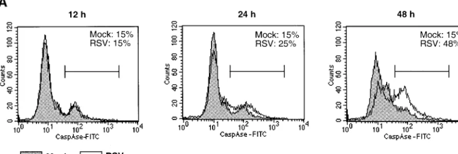
[image:5.603.71.518.76.227.2]RSV induces activation of initiator caspases 8, 9, and 10 and effector caspases 3, 6, and 7.Total caspase activation in RSV-infected cells was studied with FITC-conjugated VAD-FMK, which binds irreversibly to activated caspases. A549 cells were
FIG. 3. Induction and activation of intracellular caspases in response to RSV infection. (A) Flow cytometry analysis of activated caspases in RSV-infected A549 cells harvested at the indicated times postinfection. The cells were labeled with FITC-VAD-FMK, and the percentage of positive cells is indicated in each panel. The experiment was performed three times, and representative data are shown. (B) Kinetics of accumulation of mRNA for caspases 8, 10, 3, 6, and 7 in NHBE, SAEC, and A549 and HEp-2 cells (caspases 8 and 10 are initiator caspases; caspases 3, 6, and 7 are effector caspases). RPA was performed, and the level of each caspase mRNA is expressed as a percentage of the GAPDH housekeeping mRNA. Note the differences in the scale of theyaxis. (C) Kinetics of enzymatic activity for caspases 3, 6, 8, and 9 in RSV-infected A549 cells. Each experiment was performed twice, and representative data are shown.
9160 KOTELKIN ET AL. J. VIROL.
on November 8, 2019 by guest
http://jvi.asm.org/
FIG. 3—Continued.
VOL. 77, 2003 RSV INFECTION AND TRAIL-MEDIATED APOPTOSIS 9161
on November 8, 2019 by guest
http://jvi.asm.org/
FIG. 4. RSV induces expression of pro- and antiapoptotic members of the Bcl-2-family. (A and B) Kinetics of accumulation of mRNA for (A) proapoptotic proteins Bid, Bax, and Bak and (B) antiapoptotic proteins Mcl-1, Bcl-W, and Bcl-xLin NHBE, SAEC, A549, and HEp-2 cells that were mock-infected or infected with RSV or UV-inactivated RSV. RPAs were performed and the level of each Bcl-2 family member mRNA is shown relative to that of GADPH mRNA measured in the same gel lane. (C) Expression of Mcl-1 protein in RSV-infected A549 cells is shown at various times postinfection by intracellular immunostaining and flow cytometry. The median fluorescence⫾standard error was derived from three samples in each group. (D) Western blot analysis of Mcl-1 protein in RSV-infected or mock-infected A549 cells harvested 12 or 24 h postinfection; Mcl-1 is 37 kDa (34). (E) Immunohistochemical staining of Mcl-1 in RSV-infected A549 cells. A549 cells were infected with RSV or mock infected, fixed 24 h postinfection, and stained with anti-Mcl-1 antibody followed by an FITC-conjugated secondary antibody.
9162 KOTELKIN ET AL. J. VIROL.
on November 8, 2019 by guest
http://jvi.asm.org/
mock infected or infected with RSV for 12, 24, or 48 h; placed in suspension by treatment with trypsin; washed; stained with FITC-VAD-FMK; and assayed by flow cytometry (Fig. 3A). At 12 h postinfection, the number of FITC-VAD-FMK-positive cells was the same in RSV- and mock-infected cells. At 24 h a small increase in the number of the positive cells was observed in RSV-infected but not mock-infected cells; at 48 h approxi-mately 50% of RSV-infected cells were positive, whereas un-infected control cells did not show an increase in staining.
RPAs were used to monitor the levels of caspase mRNAs in RSV-infected NHBE, SAEC, and A549 and HEp-2 cells (Fig. 3B). Marked increases in the levels of mRNAs encoding initi-ator caspases 8 and 10 and effector caspases 3, 6, and 7 were detected in the primary NHBE and SAEC at the earliest time point analyzed (12 h). Marked increases also were observed for each of these caspase mRNAs in A549 cells by 24 h and, especially, 48 h except for caspase 6 mRNA, which was signif-icantly increased only at 72 h. In contrast, in HEp-2 cells there was only a modest, twofold increase in the levels of mRNA for caspases 10, 3, and 6, and there was no change for the mRNAs for caspase 8 and 7 (Fig. 3B). The increases in caspases 3, 8, and 6 in A549 cells were accompanied by increase in the corresponding enzymatic activities (Fig. 3C); caspases 7 and 10 were not assayed.
Caspase 9 mRNA was not detectable in NHBE and SAEC cells, while RSV infection of A549 and HEp-2 cells induced a small (up to twofold) increase in caspase 9 mRNA (data not shown). The lack of caspase 9 mRNA detectable by RPA in the primary cells might be due to a low level of transcription of this gene. However, caspase 9 enzymatic activity was detected in A549 cells beginning 36 h postinfection (Fig. 3C). No increase in caspase 2 and 5 mRNA was detected with RSV infection (not shown). Thus, RSV infection resulted in up-regulation of mRNA for caspases 8 and 10, which are initiator caspases associated with the death receptor pathway; weak up-regula-tion of caspase 9, an initiator caspase associated with the mi-tochondrial pathway; and up-regulation of caspases 3, 6, and 7, which are effector caspases utilized by either pathway.
RSV induces expression of pro- and antiapoptotic proteins of the Bcl-2 family.The Bcl-2 family contains a number of pro-and antiapoptotic factors whose balance can play a role in the decision to initiate apoptosis. RNase protection demonstrated up-regulation of a number of pro- and antiapoptotic Bcl-2 family members in response to RSV infection. mRNA encod-ing three proapoptotic members, Bid, Bax and Bak, was up-regulated two- to sevenfold in RSV-infected NHBE, SAEC, and A549 cells, whereas only Bak was up-regulated in HEp-2 cells (Fig. 4A).
Up-regulation also was observed for antiapoptotic members of the Bcl-2 family. mRNA for Mcl-1 was strongly up-regulated in RSV-infected NHBE, SAEC, and A549 and HEp-2 cells (Fig. 4B). The up-regulation of Mcl-1 mRNA was detected at 12 h for NHBE and SAEC cells and 24 h for A549 cells, which were the earliest times tested, and by 16 to 24 h for HEp-2 cells. In RSV-infected primary cells, Mcl-1 mRNA reached levels that were 200 to 400% of the amount of the abundant GAPDH (glyceraldehyde-3-phosphate dehydrogenase) mRNA, representing an increase of up to 10-fold. An increase in mRNA for two other antiapoptotic Bcl-2 family members, Bcl-W and Bcl-xL, also was observed in all four cell cultures in response to RSV infection, although the increase was less marked in HEp-2 cells (Fig. 4B). Also, the level of these mRNAs relative to GADPH mRNA was less than for Mcl-1 (Fig. 4B). Up-regulation of mRNA for Bfl-1, another antiapoptotic member of Bcl-2 family was detected in RSV-infected A549 cells but not in the other cell lines (not shown).
[image:8.603.136.451.72.272.2]The accumulation of Mcl-1 protein in RSV-infected A549 cells was analyzed at 12 and 24 h postinfection by intracellular staining and flow cytometry (Fig. 4C) and Western blotting (Fig. 4D). Flow cytometry analysis demonstrated a slight in-crease in the median expression of Mcl-1 protein at 12 h and significant (P ⬍ 0.001) increase in Mcl-1 at 24 h. Western blotting showed elevated levels of the Mcl-1 protein at 12 and 24 h post-RSV infection. The increase in Mcl-1 protein was also detected by immunohistochemistry at 12 and 24 h postin-fection (Fig. 4E).
FIG. 4—Continued.
VOL. 77, 2003 RSV INFECTION AND TRAIL-MEDIATED APOPTOSIS 9163
on November 8, 2019 by guest
http://jvi.asm.org/
FIG. 4—Continued. 9164
on November 8, 2019 by guest
http://jvi.asm.org/
RSV induces production of TRAIL in infected cells.RPAs with NHBE and SAEC primary cells and the A549 cell line showed a strong up-regulation of TRAIL mRNA (Fig. 5A), whereas no increase was observed in HEp-2 cells (not shown). TRAIL is a member of the TNF family (49, 73) and, together with its functional receptors DR4 (48) and DR5 (47, 57, 59, 71, 75), comprises one of the death receptor pathways for apoptosis. The up-regulation of TRAIL mRNA was particularly strong in pri-mary NHBE and SAEC, where it increased from the low level seen in mock- or UV-RSV-infected cell controls to a level that was within 55 to 75% of GAPDH mRNA (Fig. 5A). TRAIL mRNA also was induced in A549 cells, but its abundance relative to GADPH mRNA was nearly 10-fold lower than in primary cells. In each of the three cell types, up-regulation of TRAIL mRNA
was detected at the earliest time point tested (12 h for primary cells and 24 h for A549 cells).
[image:10.603.108.479.64.473.2]The expression of transmembrane TRAIL protein on the cell surface was quantitated in mock-infected and RSV-in-fected A549 cells by flow cytometry (Fig. 5B). An increase in the number of TRAIL-positive cells was observed at 48 and 72 h after RSV infection but not at 24 h postinfection (Fig. 5B). TRAIL also can be expressed in a soluble, truncated form that results from proteolytic cleavage of the ectodomain from the transmembrane anchor (73). However, soluble TRAIL in the medium of RSV-infected or mock-infected cells was not de-tected by an antigen-capture enzyme-linked immunosorbent assay that had a lower limit of detection of 40 pg/ml (data not shown).
FIG. 5. Induction of TRAIL mRNA and protein in response to RSV infection. (A) Production of TRAIL mRNA in RSV-, UV-RSV-, or mock-infected A549 cells, NHBE, and SAEC. Total intracellular RNA was harvested at the indicated times postinfection, and the amount of TRAIL mRNA measured by an RPA is shown as a percentage of the GAPDH mRNA measured in the same gel lane. (B) Expression of transmembrane TRAIL protein at the surface of RSV- or mock-infected A549 cells measured by flow cytometry at the indicated times postinfection. The percentage of TRAIL-positive cells is expressed as the mean⫾standard error based on three samples per group. Results obtained with an isotype control antibody were similar to those obtained with mock-infected control cells (not shown).
VOL. 77, 2003 RSV INFECTION AND TRAIL-MEDIATED APOPTOSIS 9165
on November 8, 2019 by guest
http://jvi.asm.org/
FIG. 6. Induction of the DR4 and DR5 TRAIL receptors in RSV-infected NHBE, SAEC, and A549 and HEp-2 cells. (A) Kinetics of DR4 and DR5 mRNA accumulation in the four different cell cultures. Intracellular RNA was isolated at the indicated times and the amount of DR4 and DR5 mRNA was determined by an RPA and is represented here as a percentage of the GAPDH mRNA. (B) Expression of DR4 and DR5 protein at the surface of RSV-infected A549 cells. The numbers in each box are the ratio of the median fluorescence for RSV-infected cells to that for mock-infected cells (fluorescence infected cells/fluorescence uninfected cells), based on three samples per group. The isotype control consisted of cells processed with a normal goat IgG and was similar to mock-infected cells (not shown).
9166 KOTELKIN ET AL. J. VIROL.
on November 8, 2019 by guest
http://jvi.asm.org/
RSV induces expression of TRAIL receptors DR4 and DR5.
mRNA encoding the two principal functional receptors for TRAIL, DR4 and DR5, was quantified by RPA. In NHBE, both DR4 and DR5 mRNAs were strongly up-regulated by 12 h postinfection and remained elevated at 24 and 48 h, with peak levels of 35% (for DR4) and 95% (for DR5) of that of GAPDH mRNA (Fig. 6A). In SAEC, DR4 and DR5 mRNAs were markedly elevated at 12 h but diminished thereafter, with the peak levels approximately 50 and 130%, respectively, that of GAPDH mRNA. In A549 cells, the levels of DR4 and DR5 mRNA increased steadily at 24, 48, and 72 h, but the levels relative to GAPDH mRNA were substantially lower than for primary cells. In HEp-2 cells, the up-regulation of DR4 or DR5 mRNA in response to RSV infection was twofold or less, and the level of expression was much lower than for primary or A549 cells. We also quantified the levels of TRAIL decoy receptor 1 (DcR1) (15, 57) and DcR2) (14) mRNAs, which can bind TRAIL and block its effects; no detectable increase in these mRNAs was detected after RSV infection in any of the cells (data not shown).
Expression of DR4 and DR5 proteins on the surface of RSV-infected A549 cells was assayed by flow cytometry. Up-regulation of DR4 was detected at 48 h and increased at 72 h postinfection (Fig. 6B). Expression of DR5 in RSV-infected cells was elevated at 24, 48, and 72 h. Thus, RSV infection up-regulates the expression of DR4 and DR5 receptors for TRAIL on the surface of cells.
We also observed an increase in the expression of Fas mRNA after RSV infection (data not shown), consistent with the results of O’Donnell et al. (46). Up-regulation of Fas was observed in NHBE, SAEC, and A549 cells, but not in HEp-2 cells. Infection of primary cells with RSV resulted in a 2- to 6-fold increase in Fas mRNA, while infection of A549 cells resulted in a 2- to 12-fold increase; these values were about 2 to 20% that of GAPDH mRNA (data not shown).
RSV sensitizes cells to apoptosis induced by TRAIL. In-creased surface expression of the DR4 and DR5 TRAIL re-ceptors can sensitize a cell to apoptotic killing by exogenous TRAIL, which is present on the surface of a variety of immune cells, including natural killer cells, CD4⫹T lymphocytes,
mac-rophages, and dendritic cells (27, 29–31, 40, 66), and which also can be released as a secreted soluble protein. To determine whether RSV infection resulted in an increased sensitivity to TRAIL-mediated apoptosis, A549 cells were infected with RSV, and 24 h later, exogenous TRAIL was added to the medium to a final concentration of 500 ng/ml together with a TRAIL-specific antibody that enhances TRAIL activity by cross-linking and increasing its local concentration. Before TRAIL and anti-TRAIL antibody were added, there was no difference in cell morphology between RSV-infected and non-infected cells; however, within 3 h after the addition of TRAIL plus anti-TRAIL antibody, the RSV-infected cells exhibited dramatic cytopathic effects. RSV-infected cells treated with TRAIL plus anti-TRAIL antibody rounded up, and within 5 h after the addition, most of the cells were floating in the me-dium (Fig. 7, left panels). No significant changes were observed in uninfected cells treated in parallel with TRAIL plus anti-TRAIL antibody, nor in untreated RSV-infected cells (Fig. 7). Also, addition of anti-TRAIL antibody alone to uninfected or RSV-infected cells had no effect (not shown). Addition of
TRAIL to RSV-infected cells in the absence of the TRAIL-specific antibody induced very little cytopathic effect after 5 h of incubation; following a further 3 h of incubation, the amount of cytopathic effect was increased but remained considerably less than that observed after 5 h of treatment with TRAIL plus TRAIL-specific antibody (not shown). To quantify the per-centage of apoptotic cells, the cell monolayers were harvested together with floating cells, stained with annexin V-FITC, and analyzed by flow cytometry (Fig. 7, right panels). The percent-age of annexin V-positive cells in the three negative controls, namely, mock-infected cells, mock-infected cells treated with TRAIL plus anti-TRAIL antibody, and RSV-infected cells without TRAIL or anti-TRAIL antibody, was 24 to 26%. In contrast, 42% of RSV-infected cells treated with TRAIL plus anti-TRAIL antibody were annexin V positive. Thus, RSV strongly sensitizes cells to rapid TRAIL-induced apoptosis.
The time course of acquisition of sensitivity to TRAIL-in-duced apoptosis during the first 25 h of RSV infection was studied in A549 cells (Fig. 8). Cells were infected with RSV and at various times postinfection were exposed to TRAIL (250 ng/ml) plus TRAIL-specific antibody and incubated for an additional 5 h before being collected and analyzed for annexin V staining. There was a steady increase in sensitivity to exog-enous TRAIL with increasing time after infection, with a marked increase in sensitivity evident by 11 h postinfection (Fig. 8), long before the production of infectious progeny virus (Fig. 1A).
DISCUSSION
We examined the induction of apoptosis following RSV infection of two types of human primary pulmonary epithelial cells, namely, NHBE and SAEC, and two cell lines commonly used for RSV studies, namely, A549 cells and HEp-2 cells. RPAs were used to quantify the mRNAs for 33 common apo-ptosis factors, which identified a number of pro- and antiapo-ptotic factors that were up-regulated in response to RSV in-fection. In general, results obtained with the two primary cells were in close agreement. Results with A549 cells also were generally in good agreement with those of the primary cells, although sometimes the magnitude of the response was re-duced; in contrast, the transcriptional profile of pro- and anti-apoptotic response in HEp-2 cells often was distinct, suggest-ing that HEp-2 cells are not a reliable substrate for such studies.
The results with the primary and A549 cells showed that RSV induces apoptosis in a large fraction of infected cells, although the programmed degenerative changes usually oc-curred late in infection compared to virus production. RSV infection was shown to strongly up-regulate the components of the TRAIL death receptor pathway and strongly sensitized the cells to apoptosis mediated by exogenous TRAIL. Evidence was found of activation of components of both the death re-ceptor and mitochondrial apoptotic pathways. In addition, there was strong up-regulation of antiapoptotic factors, notably Mcl-1, which might account for the apparent slow onset in the execution of apoptosis.
TRAIL is an apoptosis-inducing ligand that belongs to the TNF superfamily, together with TNF alpha and FAS ligand (FasL or CD95) (49, 73). TRAIL is a type II transmembrane
VOL. 77, 2003 RSV INFECTION AND TRAIL-MEDIATED APOPTOSIS 9167
on November 8, 2019 by guest
http://jvi.asm.org/
9168
on November 8, 2019 by guest
http://jvi.asm.org/
protein that can undergo cleavage to release a C-terminal soluble domain that can form trimers. Both the soluble and membrane-associated forms can induce apoptosis (49, 73), al-though in the present study we were unable to detect soluble TRAIL in medium from RSV-infected cells. Expression of TRAIL has been detected in a number of human tissues, most predominantly in spleen, lungs, and prostate (73); inducers include alpha/beta and gamma interferons (29, 58).
TRAIL, like TNF alpha and FasL, is an inducer of the death receptor (external) pathway of apoptosis (73) and is involved in apoptosis induced by reovirus (11), adenovirus (68), and cyto-megalovirus (58); in addition, measles virus induces TRAIL production by dendritic cells (69). TRAIL can bind to five different receptors: DR4 (48) and DR5 (47, 59) are functional receptors that mediate apoptotic responses and were strongly up-regulated by RSV infection, and DcR1 (15, 57), DcR2 (14), and osteoprotegrin (18) are decoy receptors that bind TRAIL without inducing apoptosis and were not detected in the present study. Binding of TRAIL to DR1 or DR2 leads to formation of death-inducing signaling complex consisting of (i) DR4 or DR5 trimers, (ii) an adaptor molecule such as FAS-associating protein with death domain (FADD), and (iii) pro-caspase 8, which is activated by self-cleavage upon binding the complex. Caspase 8 activates effector caspases 3 and 7, which in turn cleave cellular substrates, resulting in irreversible apo-ptosis (3, 60). Alternatively, formation of death-inducing sig-naling complex can involve caspase 10 instead of caspase 8 (32). TRAIL also can induce apoptosis through mitochondri-on-dependent pathway involving the cleavage of Bid protein by activated caspase 8, the loss of mitochondrial potential, and the release of cytochromecfrom mitochondria, resulting in acti-vation of caspase 3 (61). Transcripts for Bid, Bax, and Bak, three proapoptotic members of the Bcl-2 family associated with the mitochondrion-dependent pathway, were found to be up-regulated in the present study.
Infection with RSV also induced production of DR4 and DR5, with elevation of DR5 on the cell surface occurring earlier than that of DR4. Importantly, RSV infection efficiently sensitized cells to rapid apoptotic death induced by exogenous TRAIL added at 24 h postinfection, a time when no apoptotic changes could be detected by observation. This presumably represents a massive induction of apoptosis mediated by the RSV-induced TRAIL surface receptors, consistent with previ-ous studies demonstrating a correlation between the surface expression of TRAIL receptors and sensitivity to TRAIL-in-duced apoptosis (77, 78). It seems likely that RSV-mediated sensitization of infected cell monolayers to TRAIL-induced apoptosis in vitro reproduces a mechanism of host defense in vivo whereby infected cells render themselves susceptible to killing mediated by (i) soluble TRAIL or cell surface TRAIL expressed by neighboring RSV-infected cells or (ii) immune cells bearing cell surface TRAIL. TRAIL has been shown to mediate the cytotoxic effect of a variety of immune cells,
in-cluding NK cells, CD4⫹ T lymphocytes, macrophages, and
dendritic cells and in some studies was up-regulated on the surface of CD8⫹T lymphocytes (27, 29–31, 40, 66).
Interest-ingly, CD4⫹cell-mediated cytotoxicity was involved in
protec-tion of the upper respiratory tract from RSV infecprotec-tion in a study of an experimental RSV vaccine in mice (50).
It is noteworthy that the infected cell monolayers exhibited increased sensitivity to TRAIL-mediated apoptosis as early as 6 h postinfection, the earliest time examined, and were ex-tremely sensitive by 16 to 25 h. Thus, the up-regulation of the TRAIL receptors in response to RSV infection provides a death ligand-dependent pathway for the rapid destruction of infected cells early in the viral replicative cycle. In the absence of deliberate activation of this pathway by added TRAIL, apo-ptosis eventually was induced in a large fraction of RSV-in-fected cells, but this happened late in infection, after extensive release of progeny virus. The pathway for this late apoptosis is unclear and might involve soluble TRAIL that was below the level of detection or might involve internal activation of the mitochondrion-dependent pathway. Increased activity was ob-served for factors associated with each pathway.
[image:14.603.312.527.68.206.2]RSV also induced the early production of both pro- and antiapoptotic proteins of the Bcl-2 family: the proapoptotic members were Bid, Bax, and Bak, and the antiapoptotic mem-bers were Mcl-1, Bcl-W, and Bcl-xL. In particular, mRNA for Mcl-1 (34) was strongly up-regulated to a level that was two- to fourfold higher than that of the abundant GAPDH mRNA marker. The level of Mcl-1 protein was shown by Western blot, flow cytometry, and immunohistochemistry to be increased. All members of the Bcl-2 family have at least one of the four Bcl-2 homology (BH) functional domains, BH1 to BH4 (reviewed in
FIG. 8. Kinetics of acquisition of sensitivity to TRAIL-induced apo-ptosis during RSV infection. RSV-infected A549 cells were infected, and replicate cultures were treated with TRAIL (250 ng/ml) in the presence of TRAIL-specific antibody for 5 h beginning at 6, 11, 16, 19, 22, or 25 h postinfection. A control culture (M) was mock infected and processed in parallel with the 6-h sample. The cells were collected and stained with annexin V, and the percentage of apoptotic cells was determined.
FIG. 7. Infection with RSV sensitizes cells to TRAIL-induced apoptosis. RSV-infected or mock-infected A549 cells were treated for 5 h, beginning 24 h postinfection, with added TRAIL (500 ng/ml) in the presence of TRAIL-specific antibody or were mock treated. The cells were photographed to visualize TRAIL-induced apoptosis (panels at left) and collected and analyzed for apoptosis by annexin V staining (panels at right); the percentage of annexin V-positive-cells (bar) is indicated for each histogram. The experiment was performed four times, and repre-sentative data are shown.
VOL. 77, 2003 RSV INFECTION AND TRAIL-MEDIATED APOPTOSIS 9169
on November 8, 2019 by guest
http://jvi.asm.org/
reference 43); for example, Mcl-1 and Bcl-2 each have BH1, BH2, and BH3 domains (34). The ratio between the levels of the proapoptotic and antiapoptotic members of the family plays a role in the decision between apoptosis versus cell sur-vival (33). Bcl-2, the prototype member of the family, is local-ized in the outer mitochondrial membrane, the nuclear enve-lope, and the endoplasmic reticulum (35). Apoptosis induced by TRAIL can be delayed or inhibited by overexpression of the Bcl-2 or Bcl-xLantiapoptotic protein, an effect that has been demonstrated in various cells (21, 37), including lung cancer cells (62). It also has been suggested that Mcl-1 might be involved in the inhibition of the mitochondrial pathway of TRAIL-induced apoptosis (24). In differentiating human my-eloblastic leukemia U937 cells, antisense depletion of Mcl-1 resulted in rapid induction of apoptosis, although in that study the involvement of TRAIL was not studied (44). Up-regulation of Mcl-1 and possibly other antiapoptotic factors of the Bcl-2 family (Fig. 4B) could account for the delayed onset of apo-ptosis in RSV-infected cells. In an in vitro model of well-differentiated pseudostratified mucociliary epithelium, RSV infection resulted in minimal cytopathic changes, suggesting that apoptosis was inhibited (76). It might be that antiapoptotic proteins also are induced in that experimental system.
The up-regulation by RSV of both pro- and antiapoptotic members of the Bcl-2 family might be mediated by NF-B. Transcriptional activation by NF-B has been demonstrated for at least two members of the Bcl-2 family, namely, Bfl-1 (38, 72, 80) and Bcl-xL(9, 28, 38), both of which are up-regulated in RSV-infected cells in the present study. Activation of NF-B by RSV infection has been reported by several groups (6, 19, 22, 42). This is mediated by interaction of RSV with toll-like receptor 4 (23, 36) and possibly by the production of double-stranded RNA during the infection. In addition, engagement of receptors DR4 and DR5 by TRAIL activates the NF-B pathway (8), which might represent another mechanism of NF-B activation during RSV infection. In contrast, the up-regulation of Mcl-2 is not mediated by NF-B, but instead by the PI-3K pathway, which involves activation of the serine/ threonine kinase Akt-1 (39). Infection of cells with RSV acti-vates the PI-3K pathway (65), which might explain the stronger up-regulation of Mcl-1 than of the other members of the Bcl-2 family here (Fig. 4).
It will be important to directly demonstrate whether one or more antiapoptotic factors indeed inhibit apoptosis in RSV-infected cells. Since apoptosis limits virus replication and spread and enhances antigen presentation, inhibition of apo-ptosis could be a significant factor in viral pathogenesis and the host immune response. Suppression of Mcl-1 synthesis in vivo, for example, might result in the rapid induction of apoptosis in RSV-infected cells, thereby blocking spread of the virus to adjacent cells. Antisense oligonucleotides against Bcl-2, an-other antiapoptotic member of the family, have been demon-strated to be effective against various cancers by promoting apoptotic death of tumor cells and are currently in clinical trials for using alone or in combination with chemotherapeutic drugs (26, 70; reviewed in reference 16). It also will be inter-esting to see if individual RSV proteins have any role in pro- or antiapoptotic responses. For some viral genes, this can be directly investigated using available gene-deletion viruses (7, 64). Modification of an RSV vaccine virus to induce rather
than inhibit apoptosis might increase its attenuation and im-munogenicity.
ACKNOWLEDGMENTS
We thank Myron Hill, Kim-Chi Tran, and Elaine Lamirande for technical assistance; Brian Murphy for critical reading of the manu-script; and Stanislav Sosnovtsev for useful advice.
REFERENCES
1. Albert, M. L., S. F. Pearce, L. M. Francisco, B. Sauter, P. Roy, R. L. Silverstein, and N. Bhardwaj.1998. Immature dendritic cells phagocytose apoptotic cells via␣v5 and CD36, and cross-present antigens to cytotoxic T lymphocytes. J. Exp. Med.188:1359–1368.
2. Albert, M. L., B. Sauter, and N. Bhardwaj.1998. Dendritic cells acquire antigen from apoptotic cells and induce class I-restricted CTLs. Nature 392:86–89.
3. Ashkenazi, A., and V. M. Dixit.1998. Death receptors: signaling and mod-ulation. Science281:1305–1308.
4. Barber, G. N.2001. Host defense, viruses and apoptosis. Cell Death Differ. 8:113–126.
5. Bitko, V., and S. Barik.2001. An endoplasmic reticulum-specific stress-activated caspase (caspase-12) is implicated in the apoptosis of A549 epi-thelial cells by respiratory syncytial virus. J. Cell Biochem.80:441–454. 6. Bitko, V., A. Velazquez, L. Yang, Y. C. Yang, and S. Barik.1997.
Transcrip-tional induction of multiple cytokines by human respiratory syncytial virus requires activation of NF-kappa B and is inhibited by sodium salicylate and aspirin. Virology232:369–378.
7. Bukreyev, A., S. S. Whitehead, B. R. Murphy, and P. L. Collins.1997. Recombinant respiratory syncytial virus from which the entire SH gene has been deleted grows efficiently in cell culture and exhibits site-specific atten-uation in the respiratory tract of the mouse. J. Virol.71:8973–8982. 8. Chaudhary, P. M., M. Eby, A. Jasmin, A. Bookwalter, J. Murray, and L.
Hood.1997. Death receptor 5, a new member of the TNFR family, and DR4 induce FADD-dependent apoptosis and activate the NF-B pathway. Im-munity7:821–830.
9. Chen, C., L. C. Edelstein, and C. Gelinas.2000. The Rel/NF-B family directly activates expression of the apoptosis inhibitor Bcl-xL. Mol. Cell. Biol.
20:2687–2695.
10. Chen, T. R.1988. Re-evaluation of HeLa, HeLa S3, and HEp-2 karyotypes. Cytogenet. Cell Genet.48:19–24.
11. Clarke, P., S. M. Meintzer, S. Gibson, C. Widmann, T. P. Garrington, G. L. Johnson, and K. L. Tyler.2000. Reovirus-induced apoptosis is mediated by TRAIL. J. Virol.74:8135–8139.
12. Collins, P. L., R. M. Chanock, and B. R. Murphy.2001. Respiratory syncytial virus, p. 1443–1485.InD. M. Knipe, P. M. Howley, D. E. Griffin, R. A. Lamb, M. A. Martin, B. Roizman, and S. E. Straus (ed.), Fields virology, 4th ed., vol. 1. Lippincott-Raven Publishers, Philadelphia, Pa.
13. Collins, P. L., M. G. Hill, E. Camargo, H. Grosfeld, R. M. Chanock, and B. R. Murphy.1995. Production of infectious human respiratory syncytial virus from cloned cDNA confirms an essential role for the transcription elongation factor from the 5⬘proximal open reading frame of the M2 mRNA in gene expression and provides a capability for vaccine development. Proc. Natl. Acad. Sci. USA92:11563–11567.
14. Degli-Esposti, M. A., W. C. Dougall, P. J. Smolak, J. Y. Waugh, C. A. Smith, and R. G. Goodwin.1997. The novel receptor TRAIL-R4 induces NF-kappaB and protects against TRAIL-mediated apoptosis, yet retains an incomplete death domain. Immunity7:813–820.
15. Degli-Esposti, M. A., P. J. Smolak, H. Walczak, J. Waugh, C. P. Huang, R. F. DuBose, R. G. Goodwin, and C. A. Smith.1997. Cloning and characterization of TRAIL-R3, a novel member of the emerging TRAIL receptor family. J. Exp. Med.186:1165–1170.
16. Dias, N., and C. A. Stein.2002. Potential roles of antisense oligonucleotides in cancer therapy. The example of Bcl-2 antisense oligonucleotides. Eur. J. Pharm. Biopharm.54:263–269.
17. Domachowske, J. B., C. A. Bonville, A. J. Mortelliti, C. B. Colella, U. Kim, and H. F. Rosenberg.2000. Respiratory syncytial virus infection induces expression of the anti-apoptosis gene IEX-1L in human respiratory epithelial cells. J. Infect. Dis.181:824–830.
18. Emery, J. G., P. McDonnell, M. B. Burke, K. C. Deen, S. Lyn, C. Silverman, E. Dul, E. R. Appelbaum, C. Eichman, R. DiPrinzio, R. A. Dodds, I. E. James, M. Rosenberg, J. C. Lee, and P. R. Young.1998. Osteoprotegerin is a receptor for the cytotoxic ligand TRAIL. J. Biol. Chem.273:14363–14367. 19. Fiedler, M. A., K. Wernke-Dollries, and J. M. Stark.1996. Inhibition of viral replication reverses respiratory syncytial virus-induced NF-B activation and interleukin-8 gene expression in A549 cells. J. Virol.70:9079–9082. 20. Fischer, H., U. Koenig, L. Eckhart, and E. Tschachler.2002. Hum. caspase
12 has acquired deleterious mutations. Biochem. Biophys. Res. Commun. 293:722–726.
21. Fulda, S., E. Meyer, and K. M. Debatin.2002. Inhibition of TRAIL-induced apoptosis by Bcl-2 overexpression. Oncogene21:2283–2294.
9170 KOTELKIN ET AL. J. VIROL.
on November 8, 2019 by guest
http://jvi.asm.org/
22. Garofalo, R., M. Sabry, M. Jamaluddin, R. K. Yu, A. Casola, P. L. Ogra, and A. R. Brasier.1996. Transcriptional activation of the interleukin-8 gene by respiratory syncytial virus infection in alveolar epithelial cells: nuclear trans-location of the RelA transcription factor as a mechanism producing airway mucosal inflammation. J. Virol.70:8773–8781.
23. Haeberle, H. A., R. Takizawa, A. Casola, A. R. Brasier, H. J. Dieterich, N. Van Rooijen, Z. Gatalica, and R. P. Garofalo.2002. Respiratory syncytial virus-induced activation of nuclear factor-kappaB in the lung involves alve-olar macrophages and toll-like receptor 4-dependent pathways. J. Infect. Dis. 186:1199–1206.
24. Hersey, P., and X. D. Zhang.2001. How melanoma cells evade trail-induced apoptosis. Nat. Rev. Cancer1:142–150.
25. Hoffmann, T. K., N. Meidenbauer, G. Dworacki, H. Kanaya, and T. L. Whiteside.2000. Generation of tumor-specific T-lymphocytes by cross-prim-ing with human dendritic cells cross-prim-ingestcross-prim-ing apoptotic tumor cells. Cancer Res. 60:3542–3549.
26. Jansen, B., H. Schlagbauer-Wadl, B. D. Brown, R. N. Bryan, A. van Elsas, M. Muller, K. Wolff, H. G. Eichler, and H. Pehamberger.1998. bcl-2 antisense therapy chemosensitizes human melanoma in SCID mice. Nat. Med.4:232– 234.
27. Johnsen, A. C., J. Haux, B. Steinkjer, U. Nonstad, K. Egeberg, A. Sundan, A. Ashkenazi, and T. Espevik.1999. Regulation of APO-2 ligand/trail expres-sion in NK cells-involvement in NK cell-mediated cytotoxicity. Cytokine 11:664–672.
28. Jones, R. G., M. Parsons, M. Bonnard, V. S. Chan, W. C. Yeh, J. R. Woodgett, and P. S. Ohashi.2000. Protein kinase B regulates T lymphocyte survival, nuclear factor kappaB activation, and Bcl-X(L) levels in vivo. J. Exp. Med.191:1721–1734.
29. Kayagaki, N., N. Yamaguchi, M. Nakayama, H. Eto, K. Okumura, and H. Yagita.1999. Type I interferons (IFNs) regulate tumor necrosis factor-related apoptosis-inducing ligand (TRAIL) expression on human T cells: a novel mechanism for the antitumor effects of type I IFNs. J. Exp. Med. 189:1451–1460.
30. Kayagaki, N., N. Yamaguchi, M. Nakayama, A. Kawasaki, H. Akiba, K. Okumura, and H. Yagita.1999. Involvement of TNF-related apoptosis-inducing ligand in human CD4⫹T cell-mediated cytotoxicity. J. Immunol. 162:2639–2647.
31. Kayagaki, N., N. Yamaguchi, M. Nakayama, K. Takeda, H. Akiba, H. Tsut-sui, H. Okamura, K. Nakanishi, K. Okumura, and H. Yagita.1999. Expres-sion and function of TNF-related apoptosis-inducing ligand on murine acti-vated NK cells. J. Immunol.163:1906–1913.
32. Kischkel, F. C., D. A. Lawrence, A. Tinel, H. LeBlanc, A. Virmani, P. Schow, A. Gazdar, J. Blenis, D. Arnott, and A. Ashkenazi.2001. Death receptor recruitment of endogenous caspase-10 and apoptosis initiation in the ab-sence of caspase-8. J. Biol. Chem.276:46639–46646.
33. Korsmeyer, S. J.1999. BCL-2 gene family and the regulation of programmed cell death. Cancer Res.59(Suppl. 7):1693s–1700s.
34. Kozopas, K. M., T. Yang, H. L. Buchan, P. Zhou, and R. W. Craig.1993. MCL1, a gene expressed in programmed myeloid cell differentiation, has sequence similarity to BCL2. Proc. Natl. Acad. Sci. USA90:3516–3520. 35. Krajewski, S., S. Tanaka, S. Takayama, M. J. Schibler, W. Fenton, and J. C.
Reed.1993. Investigation of the subcellular distribution of the bcl-2 onco-protein: residence in the nuclear envelope, endoplasmic reticulum, and outer mitochondrial membranes. Cancer Res.53:4701–4714.
36. Kurt-Jones, E. A., L. Popova, L. Kwinn, L. M. Haynes, L. P. Jones, R. A. Tripp, E. E. Walsh, M. W. Freeman, D. T. Golenbock, L. J. Anderson, and R. W. Finberg.2000. Pattern recognition receptors TLR4 and CD14 mediate response to respiratory syncytial virus. Nat. Immunol.1:398–401. 37. Lamothe, B., and B. B. Aggarwal.2002. Ectopic expression of Bcl-2 and
Bcl-xL inhibits apoptosis induced by TNF-related apoptosis-inducing ligand (TRAIL) through suppression of caspases-8, 7, and 3 and BID cleavage in human acute myelogenous leukemia cell line HL-60. J. Interferon Cytokine Res.22:269–279.
38. Lee, H. H., H. Dadgostar, Q. Cheng, J. Shu, and G. Cheng.1999. NF-kappaB-mediated up-regulation of Bcl-x and Bfl-1/A1 is required for CD40 survival signaling in B lymphocytes. Proc. Natl. Acad. Sci. USA96:9136– 9141.
39. Liu, H., H. Perlman, L. J. Pagliari, and R. M. Pope.2001. Constitutively activated Akt-1 is vital for the survival of human monocyte-differentiated macrophages. Role of Mcl-1, independent of nuclear factor (NF)-kappaB, Bad, or caspase activation. J. Exp. Med.194:113–126.
40. Liu, S., Y. Yu, M. Zhang, W. Wang, and X. Cao.2001. The involvement of TNF-alpha-related apoptosis-inducing ligand in the enhanced cytotoxicity of IFN-beta-stimulated human dendritic cells to tumor cells. J. Immunol.166: 5407–5415.
41. Martin, S. J., C. P. Reutelingsperger, A. J. McGahon, J. A. Rader, R. C. van Schie, D. M. LaFace, and D. R. Green.1995. Early redistribution of plasma membrane phosphatidylserine is a general feature of apoptosis regardless of the initiating stimulus: inhibition by overexpression of Bcl-2 and Abl. J. Exp. Med.182:1545–1556.
42. Mastronarde, J. G., B. He, M. M. Monick, N. Mukaida, K. Matsushima, and G. W. Hunninghake.1996. Induction of interleukin (IL)-8 gene expression
by respiratory syncytial virus involves activation of nuclear factor (NF)-kappa B and NF-IL-6. J. Infect. Dis.174:262–267.
43. Moriishi, K., M. Koura, and Y. Matsuura.2002. Induction of Bad-mediated apoptosis by Sindbis virus infection: involvement of pro-survival members of the Bcl-2 family. Virology292:258–271.
44. Moulding, D. A., R. V. Giles, D. G. Spiller, M. R. White, D. M. Tidd, and S. W. Edwards.2000. Apoptosis is rapidly triggered by antisense depletion of MCL-1 in differentiating U937 cells. Blood96:1756–1763.
45. Murphy, B. R., A. V. Sotnikov, L. A. Lawrence, S. M. Banks, and G. A. Prince.1990. Enhanced pulmonary histopathology is observed in cotton rats immunized with formalin-inactivated respiratory syncytial virus (RSV) or purified F glycoprotein and challenged with RSV 3–6 months after immu-nization. Vaccine8:497–502.
46. O’Donnell, D. R., L. Milligan, and J. M. Stark.1999. Induction of CD95 (Fas) and apoptosis in respiratory epithelial cell cultures following respira-tory syncytial virus infection. Virology257:198–207.
47. Pan, G., J. Ni, Y. F. Wei, G. Yu, R. Gentz, and V. M. Dixit.1997. An antagonist decoy receptor and a death domain-containing receptor for TRAIL. Science277:815–818.
48. Pan, G., K. O’Rourke, A. M. Chinnaiyan, R. Gentz, R. Ebner, J. Ni, and V. M. Dixit.1997. The receptor for the cytotoxic ligand TRAIL. Science 276:111–113.
49. Pitti, R. M., S. A. Marsters, S. Ruppert, C. J. Donahue, A. Moore, and A. Ashkenazi.1996. Induction of apoptosis by Apo-2 ligand, a new member of the tumor necrosis factor cytokine family. J. Biol. Chem.271:12687–12690. 50. Plotnicky-Gilquin, H., A. Robert, L. Chevalet, J. F. Haeuw, A. Beck, J. Y. Bonnefoy, C. Brandt, C. A. Siegrist, T. N. Nguyen, and U. F. Power.2000. CD4⫹T-cell-mediated antiviral protection of the upper respiratory tract in
BALB/c mice following parenteral immunization with a recombinant respi-ratory syncytial virus G protein fragment. J. Virol.74:3455–3463. 51. Pulmanausahakul, R., M. Faber, K. Morimoto, S. Spitsin, E. Weihe, C.
Hooper, M. J. Schnell, and B. Dietzschold.2001. Overexpression of cyto-chromecby a recombinant rabies virus attenuates pathogenicity and en-hances antiviral immunity J. Virol.75:10800–10807.
52. Raynal, P., and H. B. Pollard.1994. Annexins: the problem of assessing the biological role for a gene family of multifunctional calcium- and phospho-lipid-binding proteins. Biochim. Biophys. Acta1197:63–93.
53. Ronchetti, A., P. Rovere, G. Iezzi, G. Galati, S. Heltai, M. P. Protti, M. P. Garancini, A. A. Manfredi, C. Rugarli, and M. Bellone.1999. Immunoge-nicity of apoptotic cells in vivo: role of antigen load, antigen-presenting cells, and cytokines. J. Immunol.163:130–136.
54. Roulston, A., R. C. Marcellus, and P. E. Branton.1999. Viruses and apo-ptosis. Annu. Rev. Microbiol.53:577–628.
55. Rovere, P., C. Vallinoto, A. Bondanza, M. C. Crosti, M. Rescigno, P. Ric-ciardi-Castagnoli, C. Rugarli, and A. A. Manfredi.1998. Bystander apoptosis triggers dendritic cell maturation and antigen-presenting function. J. Immu-nol.161:4467–4471.
56. Scheller, C., and C. Jassoy.2001. Syncytium formation amplifies apoptotic signals: a new view on apoptosis in HIV infection in vitro. Virology282:48– 55.
57. Schneider, P., J. L. Bodmer, M. Thome, K. Hofmann, N. Holler, and J. Tschopp.1997. Characterization of two receptors for TRAIL. FEBS Lett. 416:329–334.
58. Sedger, L. M., D. M. Shows, R. A. Blanton, J. J. Peschon, R. G. Goodwin, D. Cosman, and S. R. Wiley.1999. IFN-gamma mediates a novel antiviral activity through dynamic modulation of TRAIL and TRAIL receptor ex-pression. J. Immunol.163:920–926.
59. Sheridan, J. P., S. A. Marsters, R. M. Pitti, A. Gurney, M. Skubatch, D. Baldwin, L. Ramakrishnan, C. L. Gray, K. Baker, W. I. Wood, A. D. God-dard, P. Godowski, and A. Ashkenazi.1997. Control of TRAIL-induced apoptosis by a family of signaling and decoy receptors. Science277:818–821. 60. Sprick, M. R., M. A. Weigand, E. Rieser, C. T. Rauch, P. Juo, J. Blenis, P. H. Krammer, and H. Walczak.2000. FADD/MORT1 and caspase-8 are re-cruited to TRAIL receptors 1 and 2 and are essential for apoptosis mediated by TRAIL receptor 2. Immunity12:599–609.
61. Suliman, A., A. Lam, R. Datta, and R. K. Srivastava.2001. Intracellular mechanisms of TRAIL: apoptosis through mitochondrial-dependent and -independent pathways. Oncogene20:2122–2133.
62. Sun, S. Y., P. Yue, J. Y. Zhou, Y. Wang, H. R. Choi Kim, R. Lotan, and G. S. Wu.2001. Overexpression of BCL2 blocks TNF-related apoptosis-inducing ligand (TRAIL)-induced apoptosis in human lung cancer cells. Biochem. Biophys. Res. Commun.280:788–797.
63. Takeuchi, R., H. Tsutsumi, M. Osaki, K. Haseyama, N. Mizue, and S. Chiba. 1998. Respiratory syncytial virus infection of human alveolar epithelial cells enhances interferon regulatory factor 1 and interleukin-1-converting en-zyme gene expression but does not cause apoptosis. J. Virol.72:4498–4502. 64. Teng, M. N., S. S. Whitehead, A. Bermingham, M. St Claire, W. R. Elkins, B. R. Murphy, and P. L. Collins.2000. Recombinant respiratory syncytial virus that does not express the NS1 or M2-2 protein is highly attenuated and immunogenic in chimpanzees. J. Virol.74:9317–9321.
65. Thomas, K. W., M. M. Monick, J. M. Staber, T. Yarovinsky, A. B. Carter, and G. W. Hunninghake.2002. Respiratory syncytial virus inhibits apoptosis
VOL. 77, 2003 RSV INFECTION AND TRAIL-MEDIATED APOPTOSIS 9171
on November 8, 2019 by guest
http://jvi.asm.org/
and induces NF-kappa B activity through a phosphatidylinositol 3-kinase-dependent pathway. J. Biol. Chem.277:492–501.
66. Thomas, W. D., and P. Hersey.1998. TNF-related apoptosis-inducing ligand (TRAIL) induces apoptosis in Fas ligand-resistant melanoma cells and me-diates CD4 T cell killing of target cells. J. Immunol.161:2195–2200. 67. Thompson, W. W., D. K. Shay, E. Weintraub, L. Brammer, N. Cox, L. J.
Anderson, and K. Fukuda.2003. Mortality associated with influenza and respiratory syncytial virus in the United States. JAMA289:179–186. 68. Tollefson, A. E., K. Toth, K. Doronin, M. Kuppuswamy, O. A. Doronina,
D. L. Lichtenstein, T. W. Hermiston, C. A. Smith, and W. S. Wold.2001. Inhibition of TRAIL-induced apoptosis and forced internalization of TRAIL receptor 1 by adenovirus proteins. J. Virol.75:8875–8887.
69. Vidalain, P. O., O. Azocar, B. Lamouille, A. Astier, C. Rabourdin-Combe, and C. Servet-Delprat.2000. Measles virus induces functional TRAIL pro-duction by human dendritic cells. J. Virol.74:556–559.
70. Wacheck, V., E. Heere-Ress, J. Halaschek-Wiener, T. Lucas, H. Meyer, H. G. Eichler, and B. Jansen.2001. Bcl-2 antisense oligonucleotides chemosensi-tize human gastric cancer in a SCID mouse xenotransplantation model. J. Mol. Med.79:587–593.
71. Walczak, H., M. A. Degli-Esposti, R. S. Johnson, P. J. Smolak, J. Y. Waugh, N. Boiani, M. S. Timour, M. J. Gerhart, K. A. Schooley, C. A. Smith, R. G. Goodwin, and C. T. Rauch.1997. TRAIL-R2: a novel apoptosis-mediating receptor for TRAIL. EMBO J.16:5386–5397.
72. Wang, C. Y., D. C. Guttridge, M. W. Mayo, and A. S. Baldwin, Jr.1999. NF-B induces expression of the Bcl-2 homologue A1/Bfl-1 to preferentially suppress chemotherapy-induced apoptosis. Mol. Cell. Biol.19:5923–5929. 73. Wiley, S. R., K. Schooley, P. J. Smolak, W. S. Din, C. P. Huang, J. K. Nicholl,
G. R. Sutherland, T. D. Smith, C. Rauch, C. A. Smith, and R. G. Godwin. 1995. Identification and characterization of a new member of the TNF family that induces apoptosis. Immunity3:673–682.
74. Wright, P. F., R. A. Karron, R. B. Belshe, J. Thompson, J. E. Crowe, Jr., T. G.
Boyce, L. L. Halburnt, G. W. Reed, S. S. Whitehead, E. L. Anderson, A. E. Wittek, R. Casey, M. Eichelberger, B. Thumar, V. B. Randolph, S. A. Udem, R. M. Chanock, and B. R. Murphy.2000. Evaluation of a live, cold-passaged, temperature-sensitive, respiratory syncytial virus vaccine candidate in in-fancy. J. Infect. Dis.182:1331–1342.
75. Wu, G. S., T. F. Burns, E. R. R. McDonald, W. Jiang, R. Meng, I. D. Krantz, G. Kao, D. D. Gan, J. Y. Zhou, M. R., S. R. Hamilton, N. B. Spinner, S. Markowitz, G. Wu, and W. S. el-Deiry.1997. KILLER/DR5 is a DNA damage-inducible p53-regulated death receptor gene. Nat. Genet.17:141– 143.
76. Zhang, L., M. E. Peeples, R. C. Boucher, P. L. Collins, and R. J. Pickles. 2002. Respiratory syncytial virus infection of human airway epithelial cells is polarized, specific to ciliated cells, and without obvious cytopathology. J. Vi-rol.76:5654–5666.
77. Zhang, X. D., A. Franco, K. Myers, C. Gray, T. Nguyen, and P. Hersey.1999. Relation of TNF-related apoptosis-inducing ligand (TRAIL) receptor and FLICE-inhibitory protein expression to TRAIL-induced apoptosis of mela-noma. Cancer Res.59:2747–2753.
78. Zhang, X. D., X. Y. Zhang, C. P. Gray, T. Nguyen, and P. Hersey.2001. Tumor necrosis factor-related apoptosis-inducing ligand-induced apoptosis of human melanoma is regulated by smac/DIABLO release from mitochon-dria. Cancer Res.61:7339–7348.
79. Zhao, X. Q., X. L. Huang, P. Gupta, L. Borowski, Z. Fan, S. C. Watkins, E. K. Thomas, and C. R. Rinaldo, Jr.2002. Induction of anti-human immu-nodeficiency virus type 1 (HIV-1) CD8⫹and CD4⫹ T-cell reactivity by
dendritic cells loaded with HIV-1 X4-infected apoptotic cells. J. Virol.76: 3007–3014.
80. Zong, W. X., L. C. Edelstein, C. Chen, J. Bash, and C. Gelinas.1999. The prosurvival Bcl-2 homolog Bfl-1/A1 is a direct transcriptional target of NF-B that blocks TNF␣-induced apoptosis. Genes Dev.13:382–387.
9172 KOTELKIN ET AL. J. VIROL.