Outcome of women 40 years and
younger with Ovarian Cancer
CERTIFICATE
This is a certify that the dissertation entitled ―Outcome of women 40 years and younger‖, is the original work of Dr. Eileen Lalrinpuii done towards the M.S. Branch II (Obstetrics and Gynaecology ) Degree Examination of Tamil Nadu Dr. M.G.R Medical University , Chennai to be held in April 2015.
The Principal Professor and Head
Christian Medical College , Department of Obstetrics and Gynecology
ACKNOWLEDGEMENT
I would like to express my sincere gratitude to my guide, Dr.Abraham Peedicayil, Professor and Head , Department of Obstetrics and Gynecology for his guidance, supervision, and valuable suggestions while guiding me through this research work .
I am grateful to Dr. Rachel Chandy Professor, Departmentof Obstetrics and Gynecology, for her encouragement and practical suggestions. I am thankful to Dr. Anitha Thomas, Associate Professor also for her assistance. I am also thankful to Dr. Vinotha Thomas and Dr. Ajit Sebastian for their ideas and suggestions. I want to thank Dr. Ramani Manoj Kumar, Associate Professor and Dr. Mandeep Bindra, Department of Pathology for helping me sort out the histopathologial diagnosis of some of our patients .
I want to thank Dr. L Jeyaseelan, Professor of Biostatistics, for his help with statistical analysis. A special thanks also to Mr. John Michael, final year MSC student, Department of Biostatistics for all his help with statistical work.
I am thankful to the Department of Medical Records for granting me access to the patient charts and going out of their way to make even the old charts available online. I would like to thank staff in CHIPS for their help in identifying patients with ovarian cancer.
I am thankful to all my patients for the co-operation extended for this study. I am thankful to the Research Committee for their suggestions and financial support . I would like to thank the junior staff and students in our Department for helping me communicate with our patients through phone calls and letters.
I want to thank my family and friends for their constant love and suppor.
TABLE OF CONTENTS
S.No CONTENTS PAGE No.
1. Introduction
2. Aims and objectives
3. Review of Literature
4. Methods
5. Results
6. Limitations & Strengths
7. Implications
8. Conclusions
9. Bibliography
INTRODUCTION
AIMS and OBJECTIVES
1) To look at the clinico-pathologic patterns, in women 40 years and below, with ovarian cancer.
2) To compare the survival outcome of epithelial and non-epithelial cancer in young women.
REVIEW OF LITERATURE
Epidemiology
:
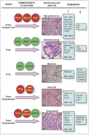
Cancer Ovary is the 7th most common cancer among women ,in fact in the west the sixth most common cause of death among women (5).According to WHO –International agency for Research on cancer 2012 report there were 238719 case of ovarian cancer detected forming age standardized incidence rate of 6.1% with 151905 death forming age standardized mortality rate of 3.8%.As things stands global result shows wide variations in the incidence rate of ovarian cancer ,the lowest rate are seen in china and the highest rates are seen in Russian federation an United Kingdom(6).growing evidence to suggest that high grade serous cancers arise from the fimbrial end of the fallopian tube.The type 1 low grade serous tumor are genetically stable and are characterized by mutations in KRAS and BRAF . The high grade type 11 serous tumor are rapidly growing , highly aggressive tumor that lack well defined precursor lesions; most are advanced stage at , or soon after ,their inception and are genetically unstable and they harbor p53 mutation(tumor protein p53 , a cellular tumor antigen encoded by TP53 gene, a tumor suppressor gene ) .It has often been assumed that that many ovarian cancers are detected late due to its vague presentation, but this could be far from the truth as the cell of origin and the behavior and the type 11 ovarian cancer are indeed different and the starting of the disease and its clinical manifestation could be very short .(8)It is also assumed from findings of previous studies that the stage matched survival rate in the younger age group as compared with those in the older age is better for Epithelial ovarian cancer .(9)
Pathology:
There are four histogenetic categories of ovarian tumours: 1. Surface epithelial stromal tumour (65-70%)
2. Sex cord stromal tumours (15-20%) 3. Germ cell tumours( 5-10%)
Table: 1 Classification of ovarian tumors
EPITHELIAL OVARIAN TUMOURS
HISTOLOGIC TYPES CELLULAR TYPE
1. SEROUS Endosalpingeal
A. Benign B. Borderline C. :Malignant
2. MUCINOUS Intestinal,Endocervical
A. Benign. B. Borderline C. Malignant
3. ENDOMETRIOID Endometrial
A. Benign B. Borderline C. Malignant
4. CLEAR CELL Mullerian
A. Benign B. Borderline C. Malignant
5. BRENNER Transitional
A. Benign
B. Borderline proliferating C. Malignant
6. Mixed epithelial type Mixed
A. Benign B. Borderline C. Malignant
7. UNDIFFERENTIATED May be anaplastic
8. UNCLASSIFIED
Benign serous cystadenoma constitute about half of all of all ovarian serous tumour Serous tumour of Low malignant potential makes up about 15% of all serous ovarian neoplasm-micropapillary pattern , stromal microinvasion and , extraovarian disease are features of borderline serous tumour , with extra ovarian disease the non invasive implants can be divided into the epithelial and desmoplastic types and the invasive form although uncommon can progress and form metastastatic implants which may progress as a proliferative disease leading to intestinal obstruction and death (10)(11)
Serous carcinoma accounts for 35 to % of all serous ovarian neoplasm , and
approximately 75% of ovarian surface epithelial –stromal carcinomas .The high grade (grade 2 to 3 ) serous carcinomas are the most common surface epithelial carcinomas and are associated with p53 mutations and somatic or germ-line abnormalities of BRCA1 and BRCA2 .
Mucinous tumors accounts for about 15% of surface epithelial neoplasm of the ovary, 80% of all are benign unilocular cystadenomas , they occur in all age group , but more freguently so in the reproductive age group .
Borderline Mucinous tumour account for 15% of all mucinous ovarian tumour of
intestinal type, the endocervival type of mucinous borderline tumor are bilateral 40% of the time .
Mucinous Carcinoma, intestinal type account for less than 10% of all mucinous ovarian neoplasms.
Endometrioid tumour, and low malignant potential endometriod tumours are entities . Endometrioid carcinoma of the ovary is comparable to grade 1 or 2 endometrioid adenocarcinoma of the uterus.associated with endometriosis 40% of the time and with primary endometrioid cancer of the uterus 20% of the time
Clear cell Carcinomas – occur in the 5th and 7th decade, it could be associated with endometriosis, and may be associated with paraneoplastic hypercalcemia or pelvic venous thromboses.
Transitional cell tumour – they are uncommon, forming about 3% of all epithelial stromal tumors , the benign, borderline and carcinomas can be seen.
Undifferentiated Epithelial tumors lacks histologic features of a specific mullerian cell type.
Mixed Surface epithelial tumors have two or more different histologic cell types.
Sex cord stromal cell tumor demonstrate ovarian, testicular, or a mixture of ovarian and testicular cell differentiation ,manyof this subtype express Inhibin , which could act as a tumor marker. Granulosa cell tumour is the commonest sex cord cell tumor ,it can occur in any age group but more commonly at the mean age of 52.call-exner bodies are characteristic of granulosa cell tumor,juvenile granulosa tumour in 90% of the time occur in females younger than 30 years
Sertoli leydic cell tumour commonly occur in women in their mid -20s . it can occur at a very young age as well as even in the seventh decade of a women’s life .one third of sertoli –leydig cell present as virilization .Sex cord tumor with annular tubules ,
stromal cell tumors . steroid cell tumours not otherwise specified , leydig cell tumours and stromal luteomas also come under sex cord stromal cell tumors.
Sex Cord—stromal Tumors
1.
Granulosa-stromal cell tumors
A. Granulosa cell tumor
B. Tumors in thecoma-fibroma group 1. Thecoma
2. Fibroma 3. Unclassified
2.
Androblastomas; Sertoli-Leydig cell tumors
A. Well-differentiated 1. Sertoli cell tumor
2. Sertoli-Leydig cell tumor
3. Leydig cell tumor; hilus cell tumor
B. Moderately differentiated
C. Poorly differentiated (sarcomatoid)
D. With heterologous elements
3.
Gynandroblastoma
4. Sex cord tumour with annular tubules
5. Sex cord stromal tumors, unclassified
A
.Stromal luteomaB.
cell tumor LeydicC.
Steroid cell tumor, not otherwise classifiedSource: Berek & Novak’s gynaecology, fifteenth edition
Germ Cell tumors
-
arise from the primordial germ cells of the ovary ,Dysgerminomas, immature teratomas, and endodermal sinus tumour are common type of germ cell tumour, Gonadoblastoma , embryonal carcinoma are other type of GCT, mixed germ cell tumors are quite common too.
Histologic Typing of Ovarian Germ Cell Tumors
1. Primitive germ cell tumors
A. Dysgerminoma B. Yolk sac tumor
C. Embryonal carcinoma
D. Polyembryoma
E. Non-gestational choriocarcinoma
F. Mixed germ cell tumor
2. Biphasic or triphasic teratoma
A. Immature teratoma
a. Dermoid cyst b. Fetiform teratoma (homunculus)
D. Carcinoma
E. Melanocytic
F. Sarcoma
G. Sebaceous tumor
H. Pituitary-type tumor
I. Others
3. Monodermal teratoma and somatic-type associated with dermoid cysts
A. Thyroid tumor 1. Struma ovarii a. Benign b. Malignant
B. Carcinoid
Table No: 2 Germ cell tumor characteristics
Source: atlas geneticsoncology.org
Subtype Frequency of OGCT
Benign/ Malignant
Uni-or Bilateral
Tumour markers expressed Metastases Route
Dysgerminoma 35-59% Malignant 10-15% are bilateral Serum lactic dehydrogenase Serum hCG Lymphatic System Endodermal Sinus tumour (EST)
20% Malignant Usually
Unilateral AFP (commonly), alpha 1- Antitripsin Intraperit oneally and hematog enously Embryonal carcinoma
Rare Malignant Usually Unilateral
AFB and hCG Intraperitoneally
Polyembryoma Rare AFB and hCG
Choriocarc inoma
Very rare Malignant Usually Unilateral
hCG
Teratoma Immature account for 20% % GCT Benign or Malignant 12-15% Are bilateral Immature tera Toma some Times secrete AFP,LDHand And CA 125. Mixed GCT 10-15% Depends on
the cell type present
Miscellanous ovarian tumors are paraganglioma:myxoma:small cell carcinoma, hypercalcemic type:small cell carcinoma,pulmonary and large neuroendocrine carcinomas .
Secondary tumors of the ovary (Metastases)include, carcinoma,lymphomaor leukemia, melanoma , and sarcoma , the colon and breast are other common sites to metastasize to the ovary.
Fallopian tube carcinomas and peritoneal cancers are treated the same as ovarian cancer and evidence suggests that these disease have common pathogenesis and maybe initiated in the fallopian tube(8)
Results of studies of incidence, histologic, patterns, prognosis and outcome of Epithelial ovarian tumor in the last 3 to 4 decades. A study done in France 1979:showed that fertility sparing surgery was feasible for young reproductive women (12)
Outcome of epithelial ovarian cancer in women under 40 years of age treated with platinum-based chemotherapy.1999(13)
Results:These results suggest that there are biological differences in the behaviour of serous carcinoma of the ovary in women of reproductive age compared with older women
Clinical characteristics and prognosis of epithelial ovarian cancer in young women a study done in china over a period of 13 years(1980-2003) the result of their study showed that serous adenocarcinoma was the commonest cancer (56%) followed by mucinous adenocarcinoma (30.9%)followed by well differentiated tumor(25.4%) and moderately differentiated (15.5%) and poorly differentiated tumor , mostly with unilateral involvement of the ovary, carrying a good prognosis with fertility preserving surgery found to be safe for stage 1a and grade 1 disease , and they found out that pathological grade and residual tumor size were independent prognostic factor
Demographic clinical and prognostic characteristics of primary ovarian, peritoneal and tubal adenocarcinomas of serous histology — A prospective comparative study(14)
Result: Primary serous peritoneal cancer has the shortest overall survival among the three,
This study was the largest prospective study comparing primary peritoneal, ovarian and fallopian tube carcinomas of serous histology
There is an increased risk of developing serous primary peritoneal cancer depending on the parity and obesity of the individual .
Two-thirds of the studied patients presented at an early stage. The common histology in descending order of frequency were: 1. immature teratoma (34.2%) 2. endodermal sinus tumor (28.9%),3. dysgerminoma (25%),4. mixed type (10.5%) and 5.choriocarcinoma (1.3%). Treatment with fertility sparing operations and adjuvant chemotherapy with a BEP regimen showed a good outcome. An advanced stage was found to be prognostic factor for recurrence.
Histologic pattern, bilaterality and clinical evaluation of 957 ovarian neoplasms: A 10-year study in a tertiary hospital of eastern India(16)
The aim of this study was to find morphologic pattern of ovarian in different age group along with bilaterality. They found out that benign disease was common in the 20-40 age group, with earlier presentation of malignant tumour at at 40-50 age group. The histological types seen idecreasing order was 1.serous cystadenoma (29.9%),2. mature teratoma (15.9%) 3. mucinous cystadenoma (11.1%).60.9% of the malignant tumour were the epithelial type serous cystadenocarcinoma forming 11.3% of all the epithelial malignant tumor.
To summarize the various studies it appears that for low stage disease conservative surgery yields good result and as far as the common histology in the young women results are concern results are conflicting as in some studies mucinous tumor seems to be more and other studies showing serous histology as the common pathology. The prognosis for stage and histology matched disease could be better in the young and there could be a difference in the biological behaviour of serous carcinoma in younger women. A study of the clinicol-patological pattern and outcome of primary peritoneal, ovarian and fallopian tube serious carcinoma showed worse prognosis for primary peritoneal cancer. A number of studies of the borderline tumor underscore the importance of proper pathological staging of each tumor and the importance of sub-classification of the extra-ovarian disease to invasive or non invasive type as the invasive type has the propensity for progress forming peritoneal implants and progressing to widespread intra-peritoneal disease which may lead on to bowel obstruction and death.
Clinical features:
More than 80% of of ovarian cancer are seen in postmenopausal women, 56 to 60 years is the peak age for invasive EOC. About 30% of ovarian tumour in postmenopausal women are malignant, whereas only about 7% of malignant ovarian tumor are malignant in premenopausal women.Etiology
: Broadly speaking ovarian cancer genesis is associated with low parity and infertility. Endometriosis,polycystic ovarian syndrome, use of an intrauterine devise ,and cigarette smoking(for mucinous carcinomas) are also known risk factors too (17) (18),)factors that are associated with decrease risk include :previous pregnancy ,history of breast feeding ,oral contraceptives use, an tubal ligation . Although a variety of epidemiologic variables like increased talc use and milk consumption are linked with ovarian cancer, and decreased risk associated with tubal ligation ,but none is as strongly correlated as prior reproductive history . As far as prevention is concern oral contraceptive pills usage is the only chemo-preventive measure identified (17)Prevention:
Ovarian cancer is seen frequently among women with infertility or low parity. Having one child reduces the risk of ovarian cancer by 30 to 40 %. Oral contraceptive pills usage for 5 years or above confers a relative risk reduction of 50 %. Bilateral salpingo-oophorectomy significantly reduce the risk of ovarian cancer in the high risk individuals, but does not totally eliminate it, as the entire peritoneum lining is still at risk.The multimodal approach for screening using imaging and a tumor marker is promising. Tumor markers are molecules or substances produced by or in response to neoplastic proliferation,that enter the circulation in detectable amounts. They indicate the likely presence of cancer and provide information about its behavior. For screening protocols, the value of the marker depends on its sensitivity (proportion of cancer detected by a positive test) and specificity (proportion of those without cancer identified by negative test). The most limiting factor is a lack of specificity for most tumor markers and a low positive predictive value. However, tumor markers have an important role to play for differential diagnosis, screening, determining therapeutic efficiency, detecting recurrences and predicting prognosis.
A variety of imaging modalities are in use to identify the morphological characteristics of cancer. These features may not be highly specific but may serve as a sensitive marker. Detailed tumor morphology can be quantified for use in a variety of scoring systems. The ultrasound features which are used for scoring are: 1. Multilocularity, 2 .Bilaterality 3. Solid areas 4. Ascites and 5. Intra-abdominal metastasis.
Different screening strategies may incorporate 1. CA 125 and Ultrasonography, 2. Ultrasonography and OVA1, 3. Risk of Malignancy Index (RMI).
Table 3. Risk of malignancy Index (RMI)
VARIABLES
FINDINGS
PROCEDURE
Menopausal status , has the women had no period for a year or more?
Is she 50 or older with a previous hysterectomy
PremenopausaI Assign 1 point
Postmenopause Assign 4 points
1.Ultrasound finding are
there cyst with more
than 1 chamber,
2.are there cyst with
solid areas inside them
3.Is there evidence of
metastases
4.Ascites
5.are there lesions on
both ovaries
0-1 findings suggestive of
malignancy
Assign 1 point
2-5 findings suggestive of
malignancy
ASSIGN 4 POINT
Serum Ca-125 Level in U/Ml FORMULA 1
Source: SU10_ALGO_TABLE_DIOGNOSIS –gif http://protomeg.ticsnetwork.c
Doppler pulsatility index of <1 and Resistance index of <0.4 points to malignancy. There is no diagnostic benefit of doppler over sonograpic tumour malignancy index alone.(19)(20)
Some of the new biomarkers which could aid in screening are the use of proteomic patterns( which are proteins and protein fragments that circulate in the blood indicating early changes caused by genetic mutation) to identify cancer using enhance laser desorption time of flight (SELDI-TOF) technology.
There is a suggestion in the literature that total and ionized calcium could help in detection of ovarian cancer. Leukocytosis detected preoperatively along with thrombocytosis could be of prognostic significance as they are found to be elevated in advance cancer (21)
Biomarkers:
Although a vast number of serum markers have been investigated few have been validated for clinical use :1. CA 125 (cancer antigen 125) sensitivity is 50% for early stage disease, sensitivity is more in postmenopausal women .
2. OVA 1 -5 proteins , CA 124 -11, Beta -2 microglobin, Transferrin, Transthyretin, Alfa fetoprotein A1.
Premenopuasal cut-off: There is a low probability of malignancy if OVA 1 < 5, high if >5 Postmenopausal cut-off: There is low probability if OVA1 <4.4, high if >/=4.4.
Sensitivity is 99% for epithelial ovarian cancer.
Activin , CA 19.9 for detecting CA 125 negative mucinous ovarian tumor , and VEGF , vascular endothelial growth factor a promoter of angiogenesis ,important in tumor growth and metastasis .
Source:
[image:26.612.137.450.185.653.2]openi.nlm.nih.gov
Genetic Risk for Ovarian Cancer:
The highest risk of ovarian cancer is associated with family history of cancer ,the BRCAI(chromosome 17) and BRCA2(chromosome 13) cancers occur in women approximately 10 years younger than those with sporadic types therefore it has been suggested that screening may be initiated at the age of 35 years or 5 to 10 years earlier than the sign of first diagnosis of ovarian cancer in the family (22) (23)(24)(25). Lynch syndrome or hereditary nonpolyposis colon cancer include multiple adenocarcinomas , involves colon, endometrial and ovarian cancer and other malignancies of the gastrointestinal and genitourinary systems (26) , mutations that are associated with this syndrome are MSH2/MLH1, PMS, PMS2 .Some of the management protocol in place for women at high risk for ovarian cancer of genetic origins are genetic counselling and testing for BRCAI and BRCA2 and 6 monthly ultrosonography, and OC Pills usage and prophylactic bilateral salpingo-opherectomy and even Hysterectomy(27)(28)(29)The. Founder effect certain ethnic group like the Ashkenazi Jews, Icelandic women have higher carrier rate of BRCA1 and BRCA2 mutated genes.The high risk individuals are now recommended to have ultrasound screening 3 monthly from the annual screening they use to follow.
symptoms. Meta analysis of 21 mostly retrospective studies shows that most women had pelvic and abdominal symptoms with epithelial ovarian cancer(30)(31). The pattern and quality of symptoms appear to differ among patients with cancer in terms of frequency / regularity or intensity of the symptoms, one study showed the symptom recurring 20-30 times versus the 2-3 times per month seen with the primary care patient((31).With the great need to detect ovarian cancer early so as to improve survival a symptom index(SI) has been developed to aid clinicians in evaluating women for early symptoms of epithelial ovarian cancer. Further validation studies are needed for this index and it is not yet recommended for routine clinical use.
Signs :
The most important sign is the presence of a pelvic mass on examination. A solid irregular fixed mass is highly suggestive of ovarian malignancy .If and upper abdominal mass or ascites is present it is almost certain it could be a malignancy.Diagnosis:
Ovarian cancer survival rate will improve through early detection of the disease, but effective screening method has not been established and to this end research is continuing to find out the diagnostic method with high sensitivity , specificity and positive predictive value, as benign neoplasm , functional cysts of the ovary should be discriminated from a malignant tumor..,ultrasound features and menopausal state to predict risk of malignancy is established. The sensitivity of CA125 that in 80-85 % of women with advance stage of epithelial cancer it is found to be high , and only 50% of the time in early disease it is elevated and it is found to be elevated in many benign conditions as uterine myomas,cystadenoma of ovary,and pelvic inflammatory diseases the specificity of the test is reduced to 75%,(33).Coming back to RMI the ultrasound score in RMI scoring is calculated as follows one point is given for the presence of ; multilocularity ,ascites and the presences of metastasis in the abdomen ,a score of 0 which indicate absences of any ultrasound features or a score of 0 to 1 or, a score 2 or more will give a U value of 1 or 3 , and the menopausal status represented by the letter M is allocated a score of 1 for premenopausal state and 3 for post menopause, the menopause score, CA 125 values at the time of evaluation and the ultrasound score were used to complete RMI work analysis. Studies have shown of late that HE4 can emerge as a promising tumor marker for the diagnosis of ovarian cancer ,not many study have been done to test if ROMA or RMI has a better diognostic performance, a multicenter study shows that ROMA could be a better system with a a better predictive value as compared to RMI for diagnosis of malignant ovarian cancer (34).
Differential diagnosis
: Pelvic inflammatory disease, endometriosis,pedunculated uterine myomas .Prognostic factors :
Can be grouped into Pathologic, Biologic and Clinical factors . Important prognostic variables among the Pathologic factors are the morphology, histologic pattern and architecture and grade of the disease, grade of the lesion is not taken as an independent prognostic factor .Clear cell carcinomas have the worse prognosis (35)(36).Clinical factors in addition to stage , the residual disease after primary cytoreduction, the volume of ascites , patients age and performance status are all independent prognostic variables(37) , intraoperative rupture or spillage does not worsen prognosis. For early stage disease poor prognostic variable are tumour grade (38), capsular penetration, surface excrescences and malignant ascites and not iotrogenic rupture (39) Epithelial carcinoma of the ovaries, fallopian tubes, and peritoneum are clinically similar.Preoperative evaluation:
Preoperative testing include: imaging and other testing for metastases, tumor markers for diagnosis and as a baseline for post treatment surveillance,and testing for hereditary cancer syndrome.2014 FIGO ovarian, fallopian tube, and peritoneal cancer staging system and corresponding TNM.
I Tumor confined to ovaries or fallopian tube(s) T1
IA Tumor limited to one ovary (capsule intact) or fallopian tube, No tumor on ovarian or fallopian tube surface No malignant cells in the ascites or peritoneal washings
T1a
IB Tumor limited to both ovaries (capsules intact) or fallopian tubes No tumor on ovarian or fallopian tube surface
No malignant cells in the ascites or peritoneal washings T1b
IC Tumor limited to one or both ovaries or fallopian tubes, with any of the following: IC1 Surgical spill intraoperatively
IC2 Capsule ruptured before surgery or tumor on ovarian or fallopian tube surface IC3 Malignant cells present in the ascites or peritoneal washings
T1c
II Tumor involves one or both ovaries or fallopian tubes with pelvic extension (below pelvic brim) or peritoneal cancer (Tp) T2
IIA Extension and/or implants on the uterus and/or fallopian tubes/and/or ovaries T2a IIB Extension to other pelvic intraperitoneal tissues T2b
III Tumor involves one or both ovaries, or fallopian tubes, or primary peritoneal cancer, with cytologically or histologically confirmed spread to the
IIIA Metastasis to the retroperitoneal lymph nodes with or without microscopic peritoneal involvement beyond the pelvis T1,T2,T3aN1
IIIA1 Positive retroperitoneal lymph nodes only (cytologically or histologically proven) IIIA1(i) Metastasis ≤ 10 mm in greatest dimension (note this is tumor dimension and not lymph node dimension) T3a/T3aN1
IIIA1(ii) Metastasis N 10 mm in greatest dimension
IIIA 2 Microscopic extrapelvic (above the pelvic brim) peritoneal involvement with or without positive retroperitoneal lymph nodes T3a/T3aN1
IIIB Macroscopic peritoneal metastases beyond the pelvic brim ≤ 2 cm in greatest dimension, with or without metastasis to the retroperitoneal lymph nodes T3b/T3bN1 III C Macroscopic peritoneal metastases beyond the pelvic brim N 2 cm in greatest dimension, with or without metastases to the retroperitoneal nodes (Note 1) T3c/T3cN1 IV Distant metastasis excluding peritoneal metastases
Stage IV A: Pleural effusion with positive cytology
Stage IV B: Metastases to extra-abdominal organs (including inguinal lymph nodes and lymph nodes outside of abdominal cavity) (Note 2)
Any T, Any N, M1
(Note 1: includes extension of tumor to capsule of liver and spleen without parenchymal involvement of either organ)
Notes: 1. Includes extension of tumor to capsule of liver and spleen without parenchymal involvement of either organ.
2. Parenchymal metastases are Stage IV B. Technigue for surgical staging
A. Staging procedure includes Mid-line incision on the abdomen.
B.Collection of ascitic fluid / peritoneal washing (50-100 ml saline instilled and recovered) C.Systematic exploration of all the intra-abdomianl surfaces and viscera.
D.Biopsy of all the suspicious areas or adhesions of the peritoneal surfaces, E.Sampling of the diaphragm by biopsy or scraping ,
F.Omentectomy and
G. Retroperitoneal space exploration .
Results- Metastases in apparent stage 1 and 11 epithelial ovarian cancer can occur in as many as 3 in 10 patients.
Early stage ovarian cancer – The primary treatment for early stage 1 Epithelial ovarian cancer is surgical –that is total abdominal hysterectomy ,bilateral salpingo-oopherectomy , and surgical staging.
Borderline tumors –the principal treatment is the surgical resection of the primary tumor. Fertility Preservation in Early stage ovarian cancer can be advised for stage 1A-1C with grade 1 to 2 disease , followed by post treatment surveillance with CA125 level determination and routine transvaginal ultrasonography , then ovary and uterus may be removed at the completion of child bearing.
possible along with the metastatic disease this procedure is referred to as debulking or cytoreductive surger (40) (41)(42). The pre-operative assessment of respectability is limited ,CA 125 cut –off of 500 U/mL level has been suggested as a means of predicting the probability of optimal resection . The rationale of cytoreductive surgery is 1. The physiologic benefit of tumor excision and 2. The improved tumor perfusion and increased growth fraction.both of which increase the likelihood of response to chemotherapy or radiation therapy. Large tumor mass has higher chance of having phenotypically resistant clones of cells.
Cytoreductive surgery include –Incision of the abdomen by vertical midline incision , ascitic fluid / peritoneal washing collection , exploration and biopsies in a systematic manner assessing the status of the pelvic pelvic organ ,small and large intestine ,mesentery, appendix, stomach, liver gall bladder, spleen , omentum ,both diaphragms dome, entire peritoneum, retroperitoneal structure such as kidneys,pancreas, and lymph nodes palpation along with palpation of the umbilicus is also for any resectable tumor .
or bowel from lateral aspect beneath the bladder and posterior cul de sac reflection, biopsy is taken from all suspicious sites.Pseudomyxoma peritonei can be associated with benign, malignant and borderline tumor , Frozen section maybe sent,omentectomy and salpingo-oopherectomy , lymph node sampling and appendectomy may follow.
Factors which may limit achievement of optimal cytoreduction maybe technical or related to poor performance status. Factors that preclude optimal cytoreduction include 1.Presence of extraperitoneal or retroperitoneal disease or large tumor bulk
2. Extensive bowel involvement
3. Parenchymal liver and lung involvement 4. Presence of massive ascites
5. Ability of patient to tolerate cytoreduction, based on age, performance status, medical co-morbidities and preoperative nutritional state.
Cytoreduction is associated with increased survival. Splenectomy maybe done if tumor nodules extend into the splenic hilum; partial hepatic resection too is an option if a lobe is involved, in case of diaphragmatic disease stripping or resection can be done; bowel resection could be an important part of cytoreduction; there may be a need to do bladder and ureteral resection, placement of a port for intraperitoneal chemotherapy may be done simultaneously.
Optimal cytoreduction maybe considered if there is no residual tumor at the end of the surgery (R0) although many would consider residual nodules < 1 cm as adequate.
Laparoscopy. In early stages, it may be done if patients are selected carefully, but many surgeons think it is not prudent to do laparoscopic surgery in ovarian cancer .
Adjuvant treatment:
Early stage (IA) low risk ovarian cancers do not need further adjuvant treatment.
Early stage (IA and IB) high-risk ovarian cancers are treated with three cycles of adjuvant carboplatin and paclitaxel chemotherapy.
The current GOG trial includes patients with high risk stage I and II disease and offers three cycles of carboplatin and paclitaxel followed by randomizing them either to observation versus 26 weeks of weekly low dose paclitaxel (40mg/m2). High risk stage I is defined as stage IA or IB grade 3, stage IC or clear cell carcinomas.
Table: 4 Chemotherapy drug protocol for Advanced stage ovarian cancer
Combination chemotherapy for advanced ovarian cancer :Recommended Regimen
Drugs Dose Administration (hr) interval No.of treatment Standard Regimen
Paclitaxel 175 mg/m2 3 Every 3 weeks 6-8 cycles Carboplatin AUC =5-6
Paclitaxel 135 mg/m2 3 Every 3 weeks 6-8 cycles Alternative drugsa (can be given with platinum)
Topotecan 1.0-1.25 mg/m2 Daily x 3-5 days
4.0 mg/m2 Every 3 weeks or weekly Gemcitabine 800-1,000 mg/m2 Every 3 weeks
Doxorubicin, liposomal 40-50 mg/m2 Every 4 weeks
AUC , area under curve dose by Calvert formula;
Drugs that can be substituted for paclitaxel if hypersensitivity to that drug occurs.
Patients who cannot tolerate IV chemotherapy can be given an oral alkylating agent or oral etoposide.
Consolidation and maintenance of clinical response to first line chemotherapy- the benefit of consolidation and maintenance chemotherapy is doubtful, patient and physicians may consider prolonged single agent paclitaxel an option, but it should not be considered as standard care.
Radiation therapy is an alternative to first line combination therapy for selected patients with metastatic disease.
Hormonal therapy as a first line drug is not practiced but may be used to suppress recurrent disease
Immunotherapy is on trial.
Treatment Assesment may be done using tumor markers ,rising level during treatment indicate treatment failure,radiologic assessment with Computed tomography or PET scan or fluorodeoxyglucose FDG-PET can be done .
Second look laparotomy is not recommended, but second look laparoscopy could have a role.
Secondary treatment . Secondary cytoreduction or second line chemotherapy can be administered.
Table 5: Second line chemotherapy in recurrent or persistent Epithelial ovarian cancer
Drugs used in Platinum and Taxane sensitive Disease Response Rate 20-40%
Cisplatin Carboplatin Paclitaxel (Taxol) Docetaxel (Taxotere)
Drugs used in Platinum and Taxane resistant and Refractory disease Response Rate 10-25%
Topotecan (Hycamtin) Etoposide (oral) (VP-16) Liposomal doxorubicin (Doxil) Gemsitabine (Gemsar)
Hexamethylmelamine (Altretamine)
Platinum sensitive disease Platinum and paclitaxel regimen in patient who have not received paclitaxel in their primary chemothereupeutic regimen may provide slight advantage .
Hormonal therapy as second line therapy – 15 to20% response rate with tamoxifen is seen in well differentiated carcinomas of the ovary, Aromatse inhibitors e.g letrozole, anatrozole, and exemestane are also being used.
Targetted therapies Incorporation of angiogenesis inhibitors (bevacizumab) and other VEGF inhibitors for first line treatment is not recommended as of now, but may be used as a maintenance therapy.
Dose intense second line chemotherapy, or
High dose chemotherapy and Autologous bone marrow transplantation may not show significant difference in progression free survival (PFS) or overall survival.
Whole abdominal radiation may be effective in a small subset of patients, but is associated with relatively high morbidity.
Intestinal obstruction maybe related to mechanical obstruction or to carcinomatous ileus – parental alimentation may be the best option for this group.
Genome wide tumor analysis if the molecular tumor profile of each individual patient can be maintained ,this will help in selecting molecular targeted treatment just right for the patient. (43)
Survival :
There is a trend toward improved survival for ovarian cancer(44). Survival rate for all stages from 1976 to 1978 was 29.8%,, for the interval of 1999 to 2001 the survival rate was 49.7%The survival of patients with borderline tumor is excellent , with stage 1 lesion having a 98% 15 year survival (47) . Patients who relapsed within 6 months of completing first line chemotherapy are classified as platinum resistant and have a median survival of 6-9 months and a 10% likelihood of responding to chemotherapy, patients who progress while on treatment are classified as platinum refractory disease.
Non epithelial tumours account for about 10% of all ovarian cancers,in the first two decades of life , almost 70% of ovarian tumour are of germ cell origin and 1/3 of these are malignant , in contrast to the relatively slow growing EO tumour the Germ cell malignancies grow rapidly, The most common type of GCT are dysgerminomas, immature teratomas and Endodermal sinus tumour , although 20-25% of all benign and malignant ovarian neoplasm are are of germ cell origin only about 3% are malignant,it account for 5 % of all ovarian cancer according to western figure where as the observation in Asia and Africa shows that 15 % of ovarian cancer are of Germ cell type and epithelial ovarian cancer is relatively less. The survival rate for all stages, ages and types are as follows:
Table 6. Survival of ovarian cancer.
Epithelial ovarian cancer Stage
5 year survival
Stage I
94 %
Stage II
73 %
Stage III
28 %
Non Epithelial Ovarian
cancer Histologic type
Stage
5
years
survival
10
years
survival
Germ
cell
tumour
(Dysgerminoma)
I A
Advanced
95%
68-83%
Immature Teratoma
I
All stages
90-95%
70-80 %
Yolk sac tumour
All stages 60-70 %
Granulosa cell tumour
>95 %
90%
Sertoli Leydig tumour
70-90%
METHODS
This study was approved by the Research Committee and Insitutional Review Board (Ethics Committee) of Christian Medical College, Vellore.
obtained through telephonic interview with patient or relatives after getting their consent. Data was analyzed using SPSS software.
Study Design
: This was a hybrid of a retrospective and prospective design. Survival analysis was done for the primary outcomes and a cohort design was used for assessing risk factors.FIGURE :2 Flow chart of patient recruitment
4 non cancers
8 recurrent cancers
10 borderline tumors
115 women
with ovarian tumors
ovarian
ovarian tumor
111 women with
Ovarian tumors
103 women <40 years with primary ovarian tumors
t
93 women < 40 years with
Inclusion Criteria:
Patients with all of the features below were selected. 1. Patients ≤ 40 years with histologically proven ovarian cancer2. Operated in CMC Vellore between January 2008 and December 2012. 3. Primary ovarian cancers
Exclusion Criteria:
Patients with any of the features below were excluded.1. Patients above age 40 years
2. Those who did not have cancer ovary on final histopathology 3. Patients who were treated with chemotherapy only and not operated. 4. Those who were operated elsewhere
5. Those who had ovarian tumors secondary to cancers from other organs
Primary outcomes :
Survival and recurrence pattern and of women 40 years and below with ovarian cancer treated in our institution from January 2008 to December 20121. Death and time to death
2. Recurrence of cancer and time to recurrence
Secondary outcomes :
1. Complications of treatment 2. Pregnancy
Sample size:
With 90 % expected survival probability, 95% confidence interval and 7.5 % precision, the calculated sample was 72. The hazard ratio was calculated based on the difference in survival probabilities in the two group, the power of the study was kept at 80%, with alpha error kept at 5% (2 sided alpha).Statistical Analysis:
Descriptive statistics were obtained for all variables. Continuous variables which were normally distributed were expressed using mean and standard deviation; non-normal continuous variables were expressed using median and range. For categorical variables count and percentages were given, and for comparing two categorical variables crosstabs with chi-square statistics were obtained. Kaplan-Meir estimation method was used to get the estimates of mean survival and recurrence time. Tarone-Ware test was applied to check the significance of difference between two groups. Statistical analysis was carried out using SPSS version 20.RESULTS
115 patients were retrospectively enlisted for our study out of which 93 could fulfil our inclusion criteria.
Table 7. Year of surgery
Year Number of Patients 2008 31
2009 28 2010 12 2011 24 2012 20
Table 8. Place Distribution
Number of patients Percent (%)
Vellore 22 19.1
Rest of Tamil Nadu 10 8.7
Rest of India 75 65.2
Outside India 7 6.1
Majority of our patients were coming from outside Tamil Nadu state
.
Table 9. Histological Distribution of the study patients
Histiopathological
pathological classification (n=111)
Frequency Percent Notes
Epithelial 79/111 71
Serous 31 28 2 with sarcomatous
changes
Mucinous 23 21 2intestinal
Subtypes
Endometrioid 11 10
Cell Clear 2 1.8
Not specified 2 1.8
Non epithelial 32 29
Germ cell tumours 21 18.9
Mixed GCT 9 8.1
Immature teratoma 5 4.5
Yolk sac 4 3.6
Dysgerminoma 3 2.7
Sex cord stromal 7 6.3
Granulosa 3 2.2
sertoli-leydic 2 1.8
Gynanroblastoma 1 0.9
Androgen secreting tumor 1 0.9
Others 4 3.6
Metastatic 2 1.8
Hemangio-endothelioma 1 0.9
Granulocytic sarcoma 1 0.9
Borderline 10 9 4 serous,6mucinous
Looking at the 111 patient with ovarian tumor 79(71.1%) patients had epithelial tumour, with the following cell types: serous 31, mucinous 23, endometrioid 11, clear cell 2 and adenocarcinoma not otherwise specified 2.
Of the 111 ovarian tumours, 10 were borderline tumors (9 %); 4 of these were serous and 6 were mucinous. There were a total of 7 sex cord stromal tumors (6.3%): 3 granulosa cell tumors, 2 sertoli leydig cells, one gynandroblastoma an one androgen secreting tumor that was not otherwise specified. There were a total of 21 germ cell tumors (18.9%): 9 were mixed germ cell tumors, 5 immature teratoma , 4 yolk sac, and 3 dysgerminoma,
There were two metastatic tumors, and two other tumors namely a granulocytic sarcoma and a Hemangioendothelioma.
The median age of the patients was 32 years with a range of 13 to 40.
Table 10. BMI BMI FREG Percent
Under weight
<18.50 22 25.9
Norma
18.5-24.99
35 41.2
Obese
> 30 21 24.7
Severely obese
> 40 7 8.2
Total 85 100.0
Variable N Mean (S.D)
Perfomance status of a patient has prognostic value as with residual disease after surgery and volume of ascites. Of the 93, performance status was documented for 87 patients, 36 of them had ECOG of 0, 45 with ECOG 1 , 5 with ECOG of 2 and I with ECOG 3.
Table 11. Performance status Distribution
Performance
status
Frequency Percent
0 36 41.38
1 45 51.72
2 5 5.75
3 1 1.15
Most patients were of parity 2 or less as shown in Table 12.
Table 12. Parity of the patients
[image:51.612.223.391.163.403.2]Regarding oral contraceptive use we could get information on only 46 patients out of which 9 (19.6%) gave history of its use and 37(80.4%) did not.
Table 13. Oral contraceptive use
Parity Frequency Percent %
0 31 33.3
1 27 29.0
2 27 29.0
3 5 5.4
4 2 2.2
6 1 1.1
Total 93 100
Oral
Contraceptive use
Frequency Percent
(%)
Yes 9 19.6
No 37 80.0
Of the presenting symptoms we could get information for 91 of the 93 patients , 2(2.2%) had dyspepsia as the presenting symptom, 26(28.6%) complained of distension of abdomen , majority of the patient 44(47.2% came with complaints of pain, 20 had other symptoms like
Table 14. Clinical presentation distribution
Presentation Frequency Percent
(%)
Dyspepsia
2
2.2
Distension
26
28.6
Pain
43
47.2
Other
20
22.0
Total
91
100
Table 15. Tumour markers
Variables
n
Median(min,Max
CA 125
82 92.2(2,19100)
hCG
35 1 (0.1,6142)
AFP
36 6.3 (0.5,496274)
LDH
35 609 (67.3,17400)
Serum
Albumin
63
Mean (S.D)
Table 16. Biomarkers
CA125 Freq. Percent (%)
<200 54 65.9
>200 28 34.1
Total 82 100.0
hCG Freq. Percent (%)
<5 21 60.0
>5 14 40.0
Total 35 100.0
AFP Freq. Percent
(%)
<5.5 18 50.0
>5.5 18 50.0
Total 36 100.0
LDH Freq. Percent (%)
<460 11 31.4
>460 24 68.6
Serum
Albumin
Freq. Percent
(%)
<3.3 12 19.1
>3.4 51 80.9
Total 63 100.0
[image:55.612.216.386.71.222.2]Serum assay of CA 125 for 82 patients ,HCG for 35,AFP for 36, LDH for 35 , and serum Albumin for 63 was done , and of the 93 patients with serum CA125 28 patients had assay >200 U/Ml,d of the 35 who had HCG assay 14 had values >5 and of the 36 patients with serum AFP assay ,18 had values >5.5 and of the patient with pre-operative serum Albumin ,of the 63, 12 had serum albumin less than 3.3 mg/dl.
Table 17. Primary treatment distribution:
Primary
Treatment
Frequency Percent
Surgery
72
77.4
Chemo
21
22.6
Surgical staging was done according to FIGO (International Federation of Gynecology and Obstetrics) 1988. 47 of the 93 were staged as Early (from stage 1a to stage 2c ) and 46 patients were staged as advanced stage from stage 3a to stage 4) .
Table 18. Stage distribution
Stages Freq. Percent
Early stage 47 50.5
Advanced stage 46 49.5
Total 93 100
Table 19. Complexity of surgery distribution
Complexity
of surgery
Frequency Percent
%
Simple
21
22.6
Intermediate 69
74.2
Complex
3
3.2
Surgery for most patients was of intermediate complexity as sown in Table 19.
[image:57.612.176.424.254.588.2]Regarding optimal cytoreduction or surgical clearance , 53 of the 93 had no gross residual tumor ,15 had less than 1 cm residual (optimal cytoreduction) , 19 had > 1 cm residual and three had open laparotomy with biopsy .
Table 20. Surgical clearance
Surgical
Clearance
Frequency
Percent %
No gross residual 56
60.2
Less than 1 cm
Residual
15
16.1
More than 1 cm
Residual
19
20.4
Biopsy only
3
3.2
Fertility sparing surgery was done in 23 patients (27%). It was found out that three of our patients had become pregnant and had given birth.
Table 21 . Fertility sparing surgery distribution
Uterus
Frequency Percent %
Removed 63
73
Not
Removed
23
27
Total
86
100
Table 22. Complications of surgery
COMPLICATION
N
PERCENT
Fever
21
19
Paralytic ileus
1
0.9
Thromboembolism
0
0
Others
7
6.3
Majority of our postoperative cases did not have complications.
Table 23. Tumour Types distribution
Tumor
Frequency Percent %
Epithelial tumour
64
69.6
Non-epithelial tumour 28
30.4
Table 24. Histological type distribution
Histology
Frequency Percent
Epithelial
64
68.8
Sex cord
Stromal
4
4.3
Germ cell
21
22.6
Other
4
3.2
Total
93
100
For secondary treatment of the disease 5 had surgery,23 had chemotherapy, I had radiotherapy, 1 (androgen secreting tumor) , and 31 patients did not have secondary treatment with us.
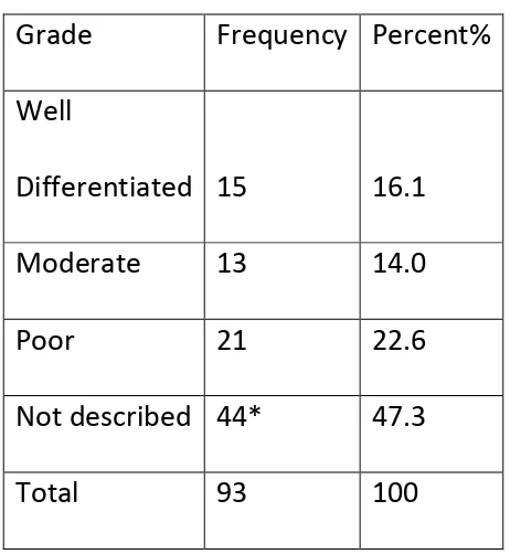
Table 25. Grade of tumour
Grade
Frequency Percent%
Well
Differentiated
15
16.1
Moderate
13
14.0
Poor
21
22.6
Not described 44*
47.3
Total
93
100
*
Many serous tumours did not have grading as they are assumed to be high grade by its very nature. Sex cord and germ cells tumours are usually not graded .Secondary
treatment
Freq.
Percent
(%)
Surgery 5 8.2
Chemotherapy 23 37.7
Radiotherapy 1 1.6
Other 1 1.6
None 31 50.8
Total 61 100
A significant number of our patients could not afford secondary treatment.
Table 27. Recurrence Distribution
Recurrence Freq.
Percent
(%)
Yes 29 37.7
No 48 62.3
Total 77 100
There were 76 patients on whom survival and progression free time was calculated, and out of the 76 patients there were total of 10 deaths during our follow period with an estimate mean survival time of 5.4 years with standard error of .239 years(95%CI 4.971-5.908).66 (87 %) patients were censored mainly for two reasons ,first reason being that quite a number of our patients(14 of them) survival was known up to certain time after the surgery followed by loss of contact , this year 2014 we were sure of 29 patients survival, there were 23 patients who were lost to follow-up after surgery.
Table 28. Survival distribution
Survival
status
Freq. Percent (%)
Alive 66 86.8
Dead 10 13.2
Total 76 100
Tumour
Survival status
Total
Recurrence
Total
Alive Dead Yes No
Epithelial tumour 42 9 51 23 29 52
Non-epithelial
tumour
23 1 24 5 19 24
Total 65 10 75 28 48 76
9 of the patients in epithelial group and 1 in the non epithelial group died during follow up
Table 30. Survival based on stage of the disease
Stages
Epithelial tumour Non-epithelial tumour
Survival status
Total
Survival status
Total
Alive Dead Alive Dead
Early stage 23 0 23 15 0 15
Advanced stage 19 9 28 8 1 9
Total 42 9 51 23 1 24
Table 31. Recurrence based on tumor types and stages
Stages
Epithelial tumor Non-Epithelial tumor
Recurrence status
Total
Recurrence status
Total
Yes No Yes No
Early stage 0 23 23 3 12 15
Advanced stage 23 6 29 2 7 9
Total 23 29 52 5 19 24
Table. 32 Survival and recurrence state based on primary treatment
Primary
treatment
Survival status
Total
Recurrence status
Total
Alive Dead Yes No
Surgery 57 3 60 16 44 60
Chemo 9 7 16 13 4 17
Total 66 10 76 29 48 77
Table 33.
Time to death
Dead Censored Mean Survival time
Total No. of Patients
No. of Events
Total No. of Patients
% Estimate Std. Error
95% Confidence Interval
Lower Bound Upper Bound
[image:66.612.68.541.118.637.2]76 10 66 86.8% 5.439 .239 4.971 5.908
Figure 3. Overall survival
Dead Censored Mean Recurrence time
Total No. of Patients
No. of Events
Total No. of Patient s
Percent (%)
Estimate Std. Error
95% Confidence Interval
Lower Bound
Upper Bound
[image:67.612.68.504.72.239.2]76 28 48 63.2% 4.261 .295 3.682 4.840
[image:67.612.75.500.156.637.2]Figure 4. Overall Progression Free Survival
Tumours
Total No of Patients
Total No of Events
Censored Mean Survival time
Siga Total
No of Patients Percent (%) Estimate Std. Error
95% Confidence Interval
Lower Bound
Upper Bound Epithelial 51 9 42 82.4% 5.000 .347 4.319 5.681
0.100 Non
Epithelial
24 1 23 95.8% 6.028 .235 5.568 6.489
Overall 75 10 65 86.7% 5.431 .241 4.958 5.903
Table 37. Time to Recurrence by histology
Tumours
Total No of Patients
Total No of Events
Censored Mean Recurrence time
Siga Total
No of Patients Percent (%) Estimate Std. Error
95% Confidence Interval
Lower Bound
Upper Bound Epithelial 51 22 29 56.9% 3.799 .378 3.058 4.539
0.022 Non
Epithelial
24 5 19 79.2% 5.315 .389 4.553 6.078
Overall 75 27 48 64.0% 4.304 .297 3.722 4.886 a. Significance is calculated using Tarone-Ware test.
Table 38.
Time to Death by primary t
reatment (Surgery Vs Chemotherapy)
Treatment Group
Total No of patients
Total No of Events
Censored Mean survival time
Siga Total
No of patients Percent (%) Estimate Std. Error
95% Confidence Interval
Lower Bound
Upper Bound
Surgery 60 3 57 95.0% 5.945 .183 5.587 6.302
<0.001
Chemo 16 7 9 56.3% 3.641 .555 2.554 4.728
Overall 76 10 66 86.8% 5.439 .239 4.971 5.908
a. Significance is calculated using Tarone-Ware test.
Table 39. Time to recurrence
Treatment Group
Total No of patients
Total No of Events
Censored Mean Recurrence time
Siga Total
No of patients Percent (%) Estimate Std. Error
95% Confidence Interval
Lower Bound
Upper Bound
Surgery 60 16 44 73.3% 4.809 .306 4.209 5.408
<0.001
Chemo 16 12 4 25.0% 2.246 .517 1.233 3.259
Overall 76 28 48 63.2% 4.261 .295 3.682 4.840
a. Significance is calculated using Tarone-Ware test.
Table 40.
Time to death by Stage of disease (Early vs Advanced)
Stages
Total No of patients
Total No of Events
Censored Mean survival time
Siga Total
No of patients Percent (%) Estimate Std. Error
95% Confidence Interval Lower Bound Upper Bound Early stage
38 0 38 100.0%
Can’t be calculated because in early stage group every patients were censored, i.e no events occurred.
- Advanced
stage
38 10 28 73.7%
Overall 76 10 66 86.8%
Figure 8. Overall survival by Stage of disease
Table 41. Time to Recurrence by Stage of disease
Stages
Total No of patients
Total No of Events
Censored Mean survival time
Siga Total
No of patients Percent (%) Estimate Std. Error
95% Confidence Interval Lower Bound Upper Bound Early stage
38 3 35 92.1% 5.877 .176 5.532 6.221
<0.001 Advanced
stage
38 25 13 34.2% 2.651 .398 1.872 3.430
Figure 9. Progression Free Survival by stage of disease
Table: 44 Cytoreduction (Optimal Vs Sub-Optimal)
Time to death
Cytoreduction Total No of patients Total No of Events
Censored Mean survival time
Siga Total
No of patients
Percent
(%) Estimate
Std. Error 95% Confidence Interval Lower Bound Upper Bound Optimal
cytoreduction 59 5 54 91.50% 5.73 0.20 5.34 6.13
0.003
Sub-optimal
cytoreduction 17 5 12 70.60% 3.67 0.72 2.25 5.09
Overall 76 10 66 86.80% 5.44 0.24 4.97 5.91
a. Significance is calculated using Tarone-Ware test.
Figure:11
[image:80.612.83.491.341.686.2]Table: 45 Time to recurrence
Cytoreduction Total No of patients Total No of EventsCensored Mean survival time
Siga Total
No of patients
Percent
(%) Estimate
Std. Error 95% Confidence Interval Lower Bound Upper Bound Optimal cytoreduction
59 18 41 69.50% 4.66 0.30 4.07 5.25
0.002
Sub-optimal cytoreduction
17 10 7 41.20% 2.43 0.64 1.18 3.68
Overall 76 28 48 63.20% 4.26 0.30 3.68 4.84
Figure:
Tables: 46Table 42: Univariate and multivariate Cox regression analyses of potential risk factors affecting survival of ovarian cancer patients after surgery
Factors N
Survival status
Unadjusted (Univariate) Adjusted (Multivariate)
Dead (n)
Alive (n)
HR (95% C.I) P-value HR (95% C.I) P-value
Histology Non-Epithelial Tumour Epithelial tumour 24 51 1 9 23 42 1 5.81 (0.73-46.22) 0.10 1 2.22 (0.23-21.34) 0.49 Treatment Surgery Chemotherapy 60 16 3 7 57 9 1 10.17 (2.62-39.4) <0.001 1 2.42 (0.54-10.64) 0.25
[image:83.612.68.586.134.557.2]Table 43: Univaritate and Multivariate Cox Regression analyses of potential risk factors for recurrence of ovarian cancer after surgery
Variables N
Recurrence status
Unadjusted (Univariate) Adjusted (Multivariate)
Yes (n) No (n)
HR (95% C.I) P-value HR (95% C.I) P-value Histology Non-Epithelial Epithelial 24 52 5 23 19 29 1
2.7 (1.02-7.30)
0.05 1 2.36 (0.74-7.51) 0.15 Treatment Surgery Chemotherapy 60 17 16 13 44 4 1
4.3 (2.01-9.31)
<0.001 1 1.44 (0.60-3.48) 0.42 Stages Early stage Advance stage 38 39 3 26 35 13 1 14.1 (4.21-47.4) <0.001 1 12.6(3.49-45.49) <0.001
DISCUSSION
Cancer of the ovary is the most fatal of all female gynecological cancers. Worldwide, it is the 7th most common cause of death in the female. In India, it is the third most common cancer affecting women, next to cancer of the breast and cervix. Although most of the literature combines clinical features and treatment of epithelial and non-epithelial tumors, these need to be studied separately. It is also not clear how often epithelial tumors effect young women and whether the bi-modal age distribution clearly separates out epithelial and non epithelial tumors, hence the aim and objective of this study was to evaluate the clinico-pathological features and survival outcome of women 40 years and younger with ovarian cancer treated at a tertiary care hospital in India.
Most of the studies from India on ovarian cancer are case reports or clinicopathological studies. There are no survival studies done in the below 40 year age group, especially for epithelial ovarian cancer.
Time to death and time to recurrence could be calculated in the 76 patients. There were a total of 10 deaths during our follow period with an estimated mean survival time of 5.4 years with a 95% confidence interval of 4.97 to 5.91. There were no peri-operative deaths. Of the 28 cases who had recurrence of disease 10 went on to die. All those who died had stage III or IV disease; 5 had serous adenocarcinoma (grade 2 or 3), 2 had endometrioid type , 1 with mucinous, and one with non specified adenocarcinoma. The only death from the non-epithelial group was a rare type of ovarian cancer, a hemangi-endothelioma. We could not show a significant difference in time to death between epithelial and non-epithelial tumours as the numbers wee small. The mean time to recurrence was 4.3 years (95% confidence interval 3.68 to 4.84). There was a statistically significant difference (p=0.022) in time to recurrence between epithelial (3.8 years) and non-epithelial tumours (5.3 years).
In our study, epithelail cancers were seen in 69%. The commonest histological type was serous cystadenocarcinoma(28%). Germ cell tumours constituted 23 % of cases.
I or II. In the study from east India, the overall survival rate was 85% for stage I tumor, 65% for stage II, 30 % for stage III, and 15.5 % for stage IV tumor. In our study, survival of the early stage cancer (stage I and II) showed 100% survival rate .whereas for advanced stage (Stage III and IV), the survival rate was 74% with overall survival rate of 87%.
A Chinese study on young women showed the commonest histologic type to be serous adenocarcinoma (56%). For stage IA disease, their 2 and 5 year survival was 86% and 82%.(55). They concluded their study by stating that women under 35 years with epithelial ovarian cancer mostly have serous adenocarcinoma with a good prognosis so such wo