A DISSERTATION ON
“THE ANALYSIS ON THE TREND OF SCREENING
POSITIVE RATE AND CORELATIVE RISK FACTORS OF
RETINOPATHY OF PREMATURITY”
THE TAMIL NADU DR. MGR. MEDICAL UNIVERSITY, CHENNAI–600032. TAMILNADU.
In partial fulfillment of the regulations
For the award of the degree of
M.D. DEGREE BRANCH-VII
PAEDIATRICS
May 2018
GOVERNMENT MOHAN KUMARAMANGALAM
ACKOWLEDGEMENT
I gratefully acknowledge and sincerely thank our beloved Dean
Dr. P. KANAGARAJ M.D., Government Mohan Kumaramangalam Medical College and Hospital, for his whole hearted co-operation and
support for the completion of this dissertation.
I am grateful to Prof. Dr.T.S.SUNDARARAJAN MD, DCH.,
Professor and Head, Department of Paediatrics, Government Mohan
Kumaramangalam Medical College and Hospital for permitting me to do
the study with constant encouragement and guidance in the preparation of
this dissertation.
I extend my sincere and heartful thanks to Assistant professor
DR.SURESH KANNAN, MD, my co guide and all Assistant professors of Paediatrics for their sincere support and valuable suggestions for my
study.
I also like to thank the Dr. KARTHIKEYAN, M.D.,(Community Medicine) for helping in carrying out the statistical analysis of the study.
I would like to express my deepest gratitude to my parents who
prepared me for life and who led me to this run on ladder of my
I would also like thank NICU assistants and staffs GMKMCH,
Salem and ROP Screening Team Aravind eye Hospital, Coimbatore for
their help and assistance. I express my sincere thanks to post graduate
colleagues and friends who have helped me in preparing this dissertation.
I am greatly indebted to all my patients and their mothers for their
co-operation in spite of pain and suffering from disease without whom
this study would have been impossible.
Date:
Place: Salem
Signature of the Candidate
TABLE OF CONTENTS
S.NO PARTICULARS PAGE NO.
1. INTRODUCTION 1
2. AIMS AND OBJECTIVES 3
3. REVIEW OF LITERATURE 6
4. METHODOLOGY 52
5. RESULTS 55
6. DISCUSSION 65
7. CONCLUSION 72
8. SUMMARY 74
9. BIBLIOGRAPHY
ANNEXURES
LIST OF TABLES
TABLE
NO. TABLES
PAGE NO.
1 CLASSIFICATION OF ROP 27
2 INCIDENCE OF ROP 55
3 STAGES OF ROP 56
4 SEX DISTRIBUTION OF ROP 57
5 BIRTH WEIGHT AND ROP 58
6 GESTATIONAL AGE AND ROP 59
7 OXYGEN AND ROP 60
8 SEPSIS AND ROP 61
9 TRANSFUSION AND ROP 62
10 APONEA OF PREMATURITY AND ROP 63
11 ANALYSIS OF RISK FACTOR 64
12 INCIDENCE OF NATIONAL AND INTERNATIONAL
STUDIES 68
13 COMPARISION OF OXYGEN AND ROP IN DIFFERENT
[image:13.596.102.533.118.595.2]S NO. GRAPHS PAGE NO.
1 INCIDENCE OF ROP 55
2 STAGES OF ROP 56
3 SEX DISTRIBUTION OF ROP 57
4 BIRTH WEIGHT AND ROP 58
5 GESTATIONAL AGE AND ROP 59
6 OXYGEN AND ROP 60
7 SEPSIS AND ROP 61
8 TRANSFUSION AND ROP 62
LIST OF PHOTOGRAPH
SR No. PHOTOGRAPH Page
No.
1 Stage 1 ROP :Demarcation line 29
2 Stage 2 ROP :Ridge 29
3 Stage 3 ROP :Mild 29
4 Stage 3 ROP :Moderate 29
5 Stage 3 ROP :Severe 29
6 Stage 4 ROP :Partial RD- Exudative and tractional 30
7 Stage 5 ROP :Open funnel RD 30
8 Plus Disease 30
9 APROP 30
LIST OF FIGURES
S. No. FIGURES Page
No.
LIST OF ABBREVIATIONS
AOP : Apnoea of prematurity
APROP : Aggressive posteriorretinopathy of prematurity
BT.WT. : Birth weight
CRYOROP : Cryotherapy for retinopathy of prematurity study 2, 3
DPG : 2, 3- Diphosphoglycerate
ELBW : Extremely Low Birth Weight (<1000 gms)
ETROP : Early treatment for ROP trial
FIO2 : Fraction of inspired concentration of oxygen
gms : grams
Hb : Haemoglobin
IGF-1 : Insulin-like growth factor-1
IVH : Intraventricular haemorrhage
ICROP : International Classification of ROP
Light-ROP : Light reduction on ROP study
NICU : Neonatal Intensive Care Unit
O2 : Oxygen
PHPV : Persistent Hyperplastic Primary Vitreous
PDA : Patent Ductus Arteriosus
PIH : Pregnancy Induced Hypertension
ROP : Retinopathy of Prematurity
RLF : Retrolental Fibroplasia
RDS : Respiratory Distress Syndrome
ABSTRACT
Background: Retinopathy of prematurity (ROP) is a common blinding disease in children in the developed world despite current treatment, and is
becoming increasingly prevalent in the developing world. Improved
survival of preterm neonates has increased the incidence of retinopathy of
prematurity (ROP) in India.
Objective: To know the incidence of ROP in preterm infants with birth weight ≤ 1500 grams to 2000gms with risk factor and/or gestational age
≤32 to 35 weeks with risk factors and to correlate between development
of ROP and the risk factors.
Methods: A longitudinal study of 100 infants weighing ≤1,500 grams to 2000grams and/or GA ≤32 to 35 weeks at birth was conducted. The main
clinical outcomes were the incidence of any stage of ROP and severe
ROP. The variables considered for the study were: birth weight,
gestational age, oxygen, occurrence of sepsis, transfusion, apnoea,.
Results: The incidence of ROP in this study was found to be 27%. 22.22% babies were in stage 1, 59.26% were in stage 2 and 3.70% was in stage3
ROP,and 14.82% developed APROP.
The mean birth weight of the ROP babies was 1280gm, Lower birth
<1500gms (p = <0.001) of ROP. The incidence of ROP was 48.15% in
babies weighing ≤ 1500gm at birth.
The mean gestational age of the ROP babies was32 weeks, The
incidence of ROP was 36.36% in babies born ≤ 32 weeks of gestational
age. Gestational age was found to be a significant risk factor for the
development of ROP (p=0.011).
CONCLUSION:
The present study reflects the problem of ROP in a tertiary care
centre. Early examination was significantly associated with chances of
early detection of ROP and hence all babies should have their first
screening within the first four weeks after birth. In our opinion, the
effective management of ROP requires a team effort of the neonatologist,
ophthalmologist and the NICU staff. Regular screening programme with a
criteria of birth weight <1500 gms and gestational age <32 weeks or both
and babies more than 1500 gms and >32 weeks with other risk factors
should be screened at the discretion of the neonatologist and
ophthalmologist. Along with regular screening, an effective control of
oxygen delivery, reduction of apneic spells and their early recognition and
effective management.
1
INTRODUCTION
Retinopathy of prematurity (ROP) is a vasoproliferative disorder
affecting premature infants. It is one of the most common causes of visual
loss in children and can lead to lifelong vision impairment and blindness.
There are approximately 45 million blind people in the world today
out of which, 30% are in Asia. Of the total blindness, childhood blindness
accounts for 4%. It is estimated that there are about 1.4 million blind
children, 1 million of whom live in Asia. India shares 20% of the world’s
childhood blindness1. ROP afflicts over 3,00,000 infants worldwide2 .In
developing countries like India, the incidence of ROP has been reported
at 24 – 47 % among high risk preterm infants 3. It is important not only in
terms of economic burden, but in its severe social implication, which is
very long in terms of blind years.
In the context of our country, we are sitting at the summit of two
volcanoes - one where all latest state of the art health care is available and
the other, where even minimal basic health care is unavailable. ROP is
known to grow in both these conditions. Among the preventable causes of
blindness in children (57%), ROP figures very high in the agenda. Low
birth weight and gestational age were found to be the most important risk
With neonatalogical units being equipped with the state-of-art
technological background and highly qualified personnel providing
optimum care of extremely immature newborns, ROP incidence is on a
rise. By early detection and timely intervention, blindness due to ROP is
preventable.
The purpose of this study is to know the incidence of ROP with
3
AIMS AND OBJECTIVES
Primary objective - To know the incidence of Retinopathy of Prematurity
in preterm infants, with birth weight upto 2000 grams and/or gestational
age 35 weeks with risk factor.
Secondary objective – To know the relationship between development of
INCLUSION CRITERIA
Selecting neonates for screening depends on incidence of ROP at
different gestation ages. Gestation age and birth weight cut-off for
screening shifts lower as smaller and sicker neonates start surviving.
Based on current incidence and risk factors reported in Indian
literature following group of neonates should be screened.
Babies with birth weight <=1500 g to upto 2000gms with risk
factor
Babies born with ≤32 to 35 weeks of gestation with risk factor
Selected preterm infants with a birth weight between 1500 and 2000
g or gestational age of more than 32 weeks with sickness like need
of cardiorespiratory support, prolonged oxygen therapy, apnea of
5 EXCLUSION CRITERIA
Term babies with exclusive breast feeding
Late preterm with risk factors
Large for gestational age babies
Term babies with IUGR & LBW
REVIEW OF LITERATURE
HISTORY
RETINOPATHY OF PREMATURITY (ROP) was originally
designated Retrolental Fibroplasia (RLF) by Dr. Theodore L. Terry who
first connected the condition with premature birth. He proposed that the
primary change was the proliferation of the embryonic hyaloid system
which incorporated the retina. He stated the unilateral pathological
specimens and provided details which that may be identical with bilateral
retrolental fibroplasia.
Heath coined the term ROP in 1951. In 1951, Dr Kate Campbell
observed that, in a smaller hospital each infant’s family was charged for
each tank of oxygen that was used and thus much less oxygen was
administered, and there was a lower incidence of RLF. She concluded
that, “normal oxygen environment required for full‐term infant is
7 INCIDENCE
The survival of ELBW-infants increased from 5-65% in the last 40
years, while in VLBW-infants, it increased from 35-90%. This increase
in survival rates has lead to increasing the number of diagnosed ROP
cases.
In the CRYO-ROP study, the incidence of the disease in a group of
premature newborns, with a birth weight <1251gms was 65.8% and it
was 81.6% for infants of less than 1000 g birth weight16. The overall
incidence of more-severe ROP (prethreshold) was 36.9% among infants
with ROP in the ETROP Study, whereas the incidence was 27.1% for
patients in the CRYO-ROP Study who developed ROP17.
Some study states that, the incidence and severity of ROP increases
with decreasing gestational age and birth weight. The incidence of ROP
in different studies done outside India was found to be 9.4%-38.9%18-21.A
study from the Indian subcontinent reveals the incidence to be
17.5%-46%22-26
The incidence of severe form of the disease known as Threshold
disease is decreasing. There is an overall decrease in incidence of disease
whenever there is an ongoing surveillance programme. Aggarwal and co-
workers found a drop from 46 to 21% in their study over a period of 7
There was a significant decrease in incidence of ROP in infants
weighing more than 1250gm20. Similar observations were made in
multicentre study in UK27. Nair22 and colleagues, Gupta28 and co-workers
9 PATHOGENESIS
NORMAL RETINAL VASCULAR DEVELOPMENT:
Ocular blood supply consists of development of angiogenesis,
arteriogenesis, and vascularization. Angiogenesis is the formation of
endothelial lined blood vessels. Arteriogenesis is the addition of smooth
muscle cells to endothelial cells, forming intact arterioles. Vascularisation
is the new arterialization of retina. The combination of these for forming
the embryonic vascular tree is termed vasculogenesis.
The posterior segment of the eye has dual blood supply:choroidal
and retinal. The choroidal nourishes the outer retina while the inner retina
is supplied by the retinal circulation. Up to 16 weeks of gestation, both
outer and inner retina is nourished by choridal blod supply, practically
inner retina remains avascular. After 16 weeks of gestational age, the first
blood supply to inner retina appears in the form of mesenchymal "spindle
cells". These spindle cells arises from the adventitia of the hyaloid
artery.The origins of the retinal circulation reside at the optic nerve head.
The vasculogenic elements begin to spread out over the and
vascularisation starts in a relatively concentric fashion out to the ora
serrata 34. Vessels reach the nasal ora first, because the fovea is the eye’s
center, thus optic nerve is nasal to the retinal center and uninterrupted
PATHOGENESIS OF ROP
At birth, fetal circulation involvesswitch from placental
oxygenation to lung oxygenation. Oxygen saturation rises from mixed
venous levels to arterial levels. Since the fetal lungs are immature,they
are not capable of fully mature oxygen transfer. Medical intervention
provides inhaled supplemental oxygen, enhancing the oxygen transfer.
Several factors lead to a potential initially hyperoxic state: mixed venous
oxygenation to arterial oxygenation; supplemental oxygen; immature but
as yet undamaged lungs; and a low retinal metabolic rate of oxygen
consumption. After birth, this relative hyperoxia begins to change. The
lungs get damaged, alveolar – blood oxygen exchange is compromised,
and retinal metabolic demand for oxygen rises according to embryologic
events. This gives rise to relative hypoxia. This transition is not a smooth,
linear one. There are undoubtedly dramatic swings during the gradual
change over from hyperoxia to hypoxia.
Retinal vascularisation is modulated by VEGF. This process is
acutely sensitive to relative states of hyperoxia and hypoxia. Hypoxia
up-regulates VEGF and hyperoxia down-up-regulates its production. Along with
VEGF, there are insulin like growth factor( IGF-1), basic fibroblast
11
RISK FACTORS FOR DEVELOPMENT OF ROP51
ROP is a multifactorial disease. Based on the clinical and
epidemiological studies, numerous risk factors have been proposed for
ROP.
Definitive and well accepted factors
Oxygen supplementation (Mechanical ventilation)
Prematurity/ Gestational age/ Birth weight
Associated factors
Apnea with bag/ mask ventilation Sepsis
Blood transfusions / Exchange transfusions
Methyl xanthine administration
Respiratory distress syndrome
Asphyxia / Hypoxia
Shock
Hypercarbia / Hypocarbia
Acidosis / Alkalosis
Vitamin E deficiency
Intraventricular hemorrhage
Light
13
PREMATURITY AND BIRTH WEIGHT
In the CRYO-ROP study, the incidence of the disease in a group of
premature newborns with a birth weight <1251gms was 65.8% and 81.6%
for infants of less than 1000 g birth weight16. In the ETROP ( multi
centric ) study done 15 years later, the overall incidence of ROP was
found to be 68% in babies with birth weight <1251 grams.. The incidence
of ROP in different studies done outside India was found to be
9.4%-38.9%18-21.A study from the Indian subcontinent reveals the incidence to
be 17.5%-46%22-26. A Danish study found a statistically significant
decrease in incidence of ROP in infants weighing more than 1250gm20.
Gupta28 and co-workers found no cases of ROP in babies weighing more
than 1250gm. A review of literature reveals that, the incidence and
severity of ROP increases with decreasing gestational age and birth
weight.
Severe ROP is often encountered in babies with birth weight more
than 1250 grams in developing countries based on the retrospective study
done by Anand Vinekar52 et al they suggested that, the western screening
guidelines may require modifications before application in developing
OXYGEN AND ROP
The excessive use of oxygen has been proved to be one of the
main risk factor for the development of ROP. During this short transition
period, the retina probably undergoes frequent and potentially wide
swings of hyperoxia /hypoxia. The sickest infants have the most volatile
transition phase of retinal oxygenation. The ultimate cause of ROP is
related to the mismatch of tissue oxygen need and tissue oxygen supply.
ROP occurs also due to Sudden discontinuation of oxygen and
duration of oxygen therapy. Gunn analyzed data from their low birth
weight survivors and found a significant association between, the more
severe grade of cicatricial disease and duration of oxygen therapy54.Their
finding concurred with the co-operative study of 1977, in which Kinsey
emphasized that the strongest association with occurrence of ROP, apart
from birth weight, was time in oxygen.
He also noted that, infants under 1200gm, concentration of
oxygen administered was significantly associated with ROP . When
comparing mean Pa02 levels in normal infants and in ROP infants, he
found differences only in babies of low birth weight and only Pa02
levels greater than 150mmHg53.
15
essential. A definite safe range for arterial Pa02 is not known. Until such
guidelines are established, keeping Pao2
<100 mm Hg is recommended preferably between 50 and 70mm Hg and
VITAMIN E DEFICIENCY:
Vitamin- E is a fat soluble antioxidant and as a result it is able to
scavenge free radicals derived from oxygen. Prophylactic vitamin E has
been suggested for the management of retinopathy of prematurity (ROP)
.this is because the premature infant and the retina are likely to be
particularly vulnerable to the deleterious effects of these oxygen derived
free radicals, as a result Three clinical trials57-59 have document. The
spindle cells, mesenchymal precursors of the inner retinal capillaries, are
the primary inducers of the neovascularisation associated with ROP.
Exposure of spindle cells to elevated oxygen tension increases their gap
junction area. This stops the normal vasoformative process and eventually
triggers the neovascularisation that is observed clinically 8–12 weeks
later. Vitamin E supplementation suppresses gap junction formation and
17 APNOEA
Apnoea independently increased the incidence of ROP. In a study
by Kim et al61, they found that frequent apnoeic attacks increased the
progression of threshold ROP to threshold ROP. In addition,
pre-existing ROP also gets worsened by apnoea. A higher incidence of
hypoxemia and apnoeic episodes requiring bagging was found among
infants with severe ROP, than in a control group26,54. The babies with
increased frequency of apnoea appeared to have longer duration of high
pC02. Similarly in a study by Chen et al62 they found that apnoea was one
SEPSIS
Sepsis is an independent risk factor for the development of
ROP. It may act through cytokines and endotoxins or by oxidative burst
in the neutrophils consequent to infection. Agarwal26 reported positive
blood cultures in 67 percent of infants who later developed ROP, and in
only 31 percent of infants with normal eyes. Liu PM et alfund that sepsis
is the most significant factor contributing to ROP. Paediatric Research
study found that, Candida sepsis is independently associated with
increased severity of ROP and the need for laser surgery in ELBW
infants.study done by Parupia H, et al73 als stressed on the same point.
Other studies also support these findings54. Thus,Its prevention and early
19 BLOOD TRANSFUSIONS
In recent years, the role of blood transfusions and iron intake as
risk factors for ROP has been strongly emphasized77,78. Some studies
suggest that anaemia per-se is a risk factor for ROP, while others show
that a high haematocrit ratio and frequent blood transfusions are
important independent risk factors79-81.
The usual explanation is that, tissue (including retinal) oxygen
levels are increased by transfusion, owing to the reduced affinity of adult
haemoglobin to oxygen, as compared to fetal haemoglobin. An
alternative hypothesis is that, damaging effects of blood transfusion on
the retina are mediated by, an increase
in free iron load which may react with various intermediates of oxygen
generating highly reactive oxygen radical. Otherwise, protection against
free iron is provided by ceruloplasmin and transferrin, but in preterm
infants with gestational age lower than 34 weeks, the levels of these
binding proteins are very low, and rapid saturation of transferrin
occurs77,78.
A study done by Akter S et al82 showed that, blood transfusion
in first week of life and repeated blood transfusion resulting in large
cumulative volume are very significantly associated with occurrence of
OTHER RISK FACTORS ASSOCIATED WITH ROP
In recent years, additional factors have been implicated in the
evolution of ROP. The task of defining any of these factors in the setting
of other major factors, such as low birth weight and early gestational age,
is again formidable. Because oxidative injury contributes to the
development of ROP, bilirubin has been suggested as a physiologically
important antioxidant. However, a recent study found no definite
association between bilirubin levels and severe ROP.
The use of dopamine in the management of hypotension in high
risk prematurely born infants (birth weight < 1,000 gm) has been
associated with increased risk for the development of threshold ROP.
Thus more vigilant screening of high-risk infants requiring dopamine
therapy, for systemic hypotension may be warranted87.
It is hypothesized that replacement of fetal blood by adult blood,
would reduce the overall oxygen affinity of haemoglobin and
consequently promote the unloading of oxygen, to the tissues at relatively
lower arterial oxygen levels. Several studies have shown that, transfusion
of adult blood either by exchange transfusion or top-up transfusion is
associated with ROP22, 88. In an effort to elucidate further the oxidative
21
the glutathione status of red blood cells in patients with ROP both in vivo
and after an in vitro oxidative challenge. After an in vitro oxidative
challenge, Infants with active ROP have the lowest levels of reduced
glutathione (GSH), the highest levels of the oxidized form (GSSG), the
highest GSSG/GSH ratios and the greatest fall in GSH. After an in vitro
oxidative stress, defective glutathione recycling was found in patients
with preceding ROP (not active ROP) It was suggested as a factor
predisposing to oxidative haemolysis. The glutathione redox ratio was
CLASSIFICATION OF ROP
INTERNATIONAL CLASSIFICATION OF ROP (ICROP)15:
A.Classification - Consists of five components.
1.Location refers to, how far the developing retinal blood vessels have
[image:41.596.126.519.434.634.2]progressed. The retina is divided into three concentric circles or zones.
23 ZONES
Zone 1 consists of an imaginary circle with the optic nerve at the center
and a radius of twice the distance from the optic nerve to the macula.
Zone 2 extends from the edge of zone 1 to the equator on the nasal side of
the eye and about half the distance to the ora-serrata on the temporal side.
Zone 3 consists of the outer crescent - shaped area extending from zone 2
STAGES OF ROP
Stage 1- A demarcation line appears as a thin white line that separates the normal retina from the undeveloped avascular retina .
Stage 2- A ridge of scar tissue with height and width replaces the line of stage 1. It extends inward from the plane of the retina.
Stage 3- The ridge has extra retinal fibro-vascular proliferation. Abnormal blood vessels and fibrous tissue develop on the edge of the
ridge and extend into the vitreous.
Stage 4- Partial retinal detachment may result when scar tissue pulls on the retina.
• Stage 4A is partial detachment outside the macula, so that the
chance for vision is good if the retina reattaches.
• Stage 4B is partial detachment that involves the macula, thus
limiting the likelihood of usable vision in that eye.
Stage 5- Complete retinal detachment occurs. The retina assumes a funnel shaped appearance and is described as open or narrow in the
25
1. Plus disease: It is an additional designation, which refers to the presence of vascular dilatation and tortuosity of the posterior
retinal vessels. This indicates a more severe degree of ROP and
may be associated with iris vascular engorgement, pupillary
rigidity, and vitreous haze. Plus disease, that is associated with
zone 1 ROP is termed rush disease; this type of ROP tends to
progress extremely rapidly.
2. Extent - refers to the circumferential location of the disease and is reported as clock hours in the appropriate zone.
3. Pre-plus disease - Vascular abnormalities of the posterior pole that are insufficient for the diagnosis of plus disease, but that
demonstrate more arterial tortuosity and more venous dilatation
than normal.
B.Definition of threshold and prethreshold ROP:
Threshold ROP is present if five or more contiguous or eight cumulative clock hours (30-degree sectors) of stage 3 with plus
disease in either zone 1 or 2 are present. This is the level of severity
at which the risk of blindness is predicted to approach 50% and thus
treatment is recommended.
zone 2 ROP with stage 3 without plus disease; or zone 2 ROP at
stage 3 with plus disease with fewer than the threshold number of
sectors of stage 3. Infants with prethreshold ROP have a 1 in 3
chance of needing surgical treatment and a 1 in 6 chance of extreme
loss of vision if treatment is not done promptly when threshold is
reached. With therapy, they have a 1 in 12 chance of extreme visual
loss35.
Aggressive posterior ROP (AP-ROP):
It is a rapidly progressing, severe form of ROP. If untreated, it
usually progresses to stage 5 ROP. The characteristic features of this type
of ROP are its posterior location, prominence of plus disease, and the
ill-defined nature of the retinopathy. This may not have classical ridge or
extraretinal fibrovascular proliferation. This rapidly progressing
retinopathy has been referred previously as "type II ROP" and "Rush
disease”. Observed most commonly in Zone I, but may also occur in
27
Table :1 Classification of Retinopathy of Prematurity
Figure 2: Classification of retinopathy of prematurity
Stages of ROP:
Photograph 1: Stage 1- Demarcation line
Photograph 2: Stage 2- Ridge
Photograph 3: Stage 3 ROP - Mild Photograph 4: Stage 3 ROP
- moderate
Stages of ROP: (Continue…)
Photograph 6: Stage 4 ROP – Partial RD- Exudative and
Tractional
Photograph 7: Stage 5 ROP – Open Funnel RD
DIAGNOSIS Screening Window:
Progression of ROP follows a distinct time-table according
to the post- menstrual age of the baby. Hardly any ROP is detected
before 32 weeks of gestation. The median age for detection of stage
1 ROP is 34 weeks. Pre-threshold ROP appears at 36 weeks of
post-menstrual age and threshold disease at 37 weeks. Vascularisation is
complete by 40 weeks of gestation. Thus the crucial period for
detection of ROP is from 32 weeks to 40 weeks of post-menstrual
period. The critical phase is from; 34-35 weeks to 37-38 weeks age
during which, the progression of the disease takes place and treatment
may have to be instituted It may also be noted that, ROP usually does
not develop before 2 weeks of postnatal age.
Babies to screen:
Selecting neonates for screening depends on incidence of ROP at
different gestation ages. Based on current incidence and risk factors
reported in Indian literature, following group of neonates should be
screened.
Babies born at ≤32 weeks of gestation Selected preterm infants
with a birth weight between, 1500 and 2000 g or gestational age
of more than 32 weeks, with sickness requiring cardiorespiratory
support, prolonged oxygen therapy, apnoea of prematurity ,
anaemia needing blood transfusion and neonatal sepsis or believed
by their attending paediatrician or neonatologist to be at high risk.
This ‘third criterion’ is important as it brings in many larger babies
into the screening guidelines, without raising the screening
parameters90.
Follow up:
Follow-up examinations are done based on the retinal findings.
1-week or less follow-up
• Stage 1 or 2 ROP: Zone I
• Stage 3 ROP: Zone II
1to 2-week follow-up
• Immature vascularisation: Zone I - no ROP
• Stage 2 ROP: Zone II
2-week follow-up
• Stage 1 ROP: Zone II
• Regressing ROP: Zone II
2- to 3-week follow-up
• Immature vascularisation: Zone II—no ROP
• Stage 1 or 2 ROP: Zone III
• Regressing ROP: Zone III
Findings that suggest further examinations are not needed include:
• Zone III retinal vascularisation attained without previous Zone I or
II ROP
• Full retinal vascularisation
• Postmenstrual age of 45 weeks and no Prethreshold disease
(defined as stage 3 ROP in Zone II, any ROP in Zone I) or worse
ROP is present
SCREENING91:
Pupils are dilated with Phenylephrine 2.5% and Tropicamide 0.5%.
One drop of Tropicamide is instilled every 10-15 minutes, for 4 times
starting 1 hour before the scheduled time for examination. This is
followed by Phenylephrine, one drop just before examination.
Phenylephrine is available in 10% concentration; it should be diluted 4
times before use in neonates. Repeated instillation of phenylephrine is
avoided for the fear of hypertension.
Screening of ROP involves, indirect ophthalmoscopy using 20 D or
28/30 D lens by an experienced ophthalmologist. After instilling a topical
anesthetic drop like proparacaine, a wire speculum is inserted to keep the
eye-lids apart. First, the anterior segment of the eye is examined to look
for tunica vasculosa-lentis, pupillary dilation, and lens / media clarity;
followed by the posterior pole, to look for plus disease; followed by,
sequential examination of all clock hours of the peripheral retina. A
scleral depressor is often used to indent the eye externally, to examine
areas of interest, rotate and stabilize the eye.
Ophthalmological notes should be made after each ROP
examination, detailing zone, stage and extent in terms of clock hours, of
any ROP and the presence of any pre-plus or plus disease. These notes
(if any) and be kept with the baby’s medical record.
ROP screening examinations can have short-term effects on blood
pressure, heart rate and respiratory function in the premature baby92.The
examinations should be kept as short as possible and precautions should
RETCAM
Photograph 10: RETICAM
A wide-field digital camera (RetCam) capable of retinal imaging in
preterm infants, has been evaluated as an alternative to indirect
ophthalmoscopy for screening. Retinal images taken by camera can be
stored, transmitted to expert, reviewed, analyzed and sequentially
compared over time and are useful for telemedicine purposes. Studies
comparing RetCam with the indirect ophthalmoscope, have reported
sensitivity, specificity, and accuracy when image quality is poor, it is not
recommended to replace bedside ophthalmoscopic examination. Digital
fundus images can be used as a useful adjunct to conventional bedside
ROP screening by indirect ophthalmoscopy.
Place of screening:
All eligible babies were screened at Neonatal Intensive Care Unit.
Preparation of the child:
The pupils were dilated with a mixture of Phenylephrine 2.5% and
Tropicamide 0.5% instilled 3 times at 10mins interval about 1 hour before
the scheduled examination. Resistance to dilation was noted. Care was
taken to wipe off any eye drops with sterile cotton that come out of eyes
to cheeks and not to feed the baby immediately before examination as the
child might vomit or aspirate.
Instruments used:
• Cordless Indirect ophthalmoscope with 20D lens.
• Pediatric wire speculum.
• Scleral indentor.
Procedure:
All preterm babies who satisfied any one of the inclusion criteria
were taken up for the study.
explained the nature of the examination and informed consent was taken.
Demographic history and risk factors like respiratory distress syndrome,
sepsis, multiple blood transfusions, multiple births, apneic episodes and
oxygen given was documented using a data collection instrument.
First examination was done at 4 weeks post natal age (age in weeks
after birth) by taking all aseptic precautions in a temperature controlled
room in the presence of a neonatologist.
Indirect ophthalmoscopic examination was done .One drop of
topical paracaine eyedrops was used to anaesthetise the cornea. A
pediatric wire speculum was used to keep the eyelids apart .After
decreasing the room illumination the anterior segment was first visualized
to look for tunica vasculosa lentis, pupillary dilatation and lens and media
clarity. Then the posterior pole was examined for any Plus disease. A
scleral indenter was used to visualize the periphery. The periphery was
examined in all clock hours to look for the extent of changes from nasal
to temporal retina. Care was taken not to put too much pressure on the
globe. During examination, untoward neonatal complications were
looked for and managed appropriately.
The changes in the retina were graded according to the
International Committee for the Classification of Retinopathy of
Follow up protocol:
If no ROP was detected at initial examination, the infants were
re-evaluated once every two weeks until vascularisation was complete. If
ROP was detected, the examinations were performed weekly for stage
1-2 disease and more frequently for stage 3 disease, till the disease started
resolving or progressed to threshold stage. Babies showing evidence of
regression were followed up till vascularisation was complete. Babies
Treatment
Early Treatment of Retinopathy of Prematurity (ETROP) trial
recruited neonates at 26 centres in the US and compared early treatment
of high-risk prethreshold disease with conventional threshold treatment94.
Four hundred and one babies meeting the criteria for ‘high-risk’ of an
unfavourable outcome with prethreshold in at least one eye were
randomized to receive either early or conventional treatment. The level of
risk was determined by a risk analysis programme which used, among
other factors, degree of ROP (stage, zone and presence of plus), rate of
ROP progression, birth weight, gestational age and ethnicity to classify
eyes as, either ‘high-risk’ (i.e. 15% chance) or ≥ ‘low-risk’ (<15%
chance) of an unfavourable outcome without treatment. The results
showed, an overall significant benefit for the early treatment of eyes with
high-risk prethreshold disease. Based on results of ETROP, two new
terminologies have been suggested:
Type 1 ROP:
• Zone I, any stage ROP with plus disease
• Zone I, stage 3 ROP with or without plus disease
Type 2 ROP:
• Zone I, stage 1 or 2 ROP without plus disease
• Zone II, stage 3 ROP without plus disease.
Peripheral retinal ablation should be carried out for all cases with type 1
ROP and continued serial examinations are advised for type 2 ROP.
Treatment modalities :
Peripheral retinal ablation of avascular retina anterior to the ridge
can be done by either cryotherapy or diode laser. Diode laser ablation has
replaced cryotherapy, due to lower rate of postoperative ocular and
systemic complications and less damage to the adjacent tissues compared
to cryotherapy. Other advantages are that, the laser spots are visible
during treatment, minimizing the risk of missing areas requiring treatment
and that, laser equipment is portable allowing use outside of the operating
theatre. The procedure can be carried out under general anesthesia or
under sedation, depending on the feasibility and expertise. Treatment for
ROP should include, the entire avascular retina anterior to the ridge, with
burn spacing between 0.5 to 1 burn-widths apart.
CRYOTHERAPY:
Cryotherapy is an ablative procedure used in severe active ROP. It
rapidly growing vessels that are presumably being driven to grow by an
angiogenic factor released by the peripheral avascular retina. The
cryotherapy for Retinopathy of Prematurity (Cryo-ROP) is a major
landmark study in the battle against ROP. The Cryo-ROP study53
recommended cryotherapy for threshold ROP, defined as 5 contiguous or
8 cumulative clock-hours of stage 3 plus ROP in zone I and zone II. The
technique of cryotherapy involves using the cryo probe to create
contiguous cryo marks on the avascular retina. Treatments are performed
under continuous monitoring of heart rate, respiratory rate, blood pressure
and oxygen saturation. General endotracheal tube anaesthesia is
preferred. Cryotherapy is administered with a cryotherapy probe, such as
a hammer head-shaped pediatric probe. Continuous cryotherapy is
performed under direct observation of the fundus, avoiding over
treatment and re treatment. After the cryo treatment, patients can be
discharged home on a topical steroid, cycloplegic and antibiotic on the
same day, if not anaesthetized95.
Laser Photocoagulation:
The use of indirect laser via an ophthalmoscope, for retinal
photocoagulation in the treatment of ROP has been established.
McNamara and coworkers95 conducted a prospective clinical trial, that
argon laser photocoagulation and showed that infants treated with laser
had less ocular inflammation, fewer systemic complications, and no
significant difference in effectiveness as compared with those treated
with cryotherapy. In addition, general anesthesia typically was not
required for infants treated with laser (27% of infants in the Cryo-ROP
study underwent general anesthesia). Some experts in ROP have also
advocated earlier intervention with laser therapy in eyes with ROP,
especially when there is plus disease in any zone (particularly in zone I)
and vitreous haze.
Trans-scleral diode laser photocoagulation has been evaluated for
the treatment of threshold ROP and results suggested that, it is as
effective in the treatment of threshold ROP as is transpupillary diode
laser photocoagulation. Trans - scleral diode laser photocoagulation
seems to be an advantageous treatment method, if trans-pupillary
treatment bears an increased risk of cataract formation57.
Pre-anesthetic medication:
Oral feeds should be discontinued 3 hours prior to the procedure.
Baby should be started on intravenous fluids, and put on
cardio-respiratory monitor. Dilatation of pupil is done by using 0.5%
Tropicamide and 2.5% Phenylephrine as described in the section on
Anesthesia/ Sedation:
Topical anaesthesia alone provides insufficient analgesia for ROP
treatment and should not be used. Babies may be treated under adequate
sedation and analgesia in an operation theatre, if this can be arranged in a
timely way. If shifting to operation theatre is not possible or is causing
delay in treatment, babies may be treated more rapidly in the neonatal
unit under adequate sedation and analgesia.
Procedure:
Both the eyes can be treated at the same sitting time, unless
contraindicated by instability of the baby. If baby is not tolerating the
procedure, consider abandoning the procedure for the time being. Vital
signs and oxygen saturation should be monitored very closely.
Monitoring after laser therapy:
After laser therapy, first examination should take place 5-7 days
after treatment and should be continued at least weekly for signs of
decreasing activity and regression. Re-treatment should be performed
usually 10-14 days after initial treatment, when there has been a failure of
the ROP to regress.
Post-operative care:
feeds can be started shortly after the procedure. Premature babies,
especially those with chronic lung disease may have an increase or
re-appearance of apneic episodes, or an increase in oxygen requirement.
Therefore they should be carefully monitored for 48-72 hours after the
procedure. Antibiotic drops (such as chloramphenicol) should be instilled
6-8 hourly for 2-3 days.
Future therapeutic targets96:
The discovery of the importance of VEGF and IGF-1 in the
development of ROP is a step forward in our understanding of the
pathogenesis of this disease. These studies suggest a number of ways to
intervene medically in the disease process. The use of anti-VEGF therapy
is the first medical treatment for neovascularisation in age-related
macular degeneration and is likely to be useful for proliferative
retinopathy. However, prevention of vessel loss will be even more
important in the treatment of ROP, since the extent of the second
destructive phase of ROP is determined by the amount of vessel loss in
the first phase. The finding that, late development of ROP is associated
with low levels of IGF-1 after premature birth suggests that,
physiological replacement of IGF-1 to levels found in-utero, might
prevent the disease by allowing normal vascular development. In
addition, the use of a specific agonist to VEGFR-1, PIGF-1, might be
used early in the disease process to prevent vessel loss without promoting
proliferative disease. The current understanding of ROP pathogenesis
the two phases of ROP require very different approaches. Inhibition of
either VEGF or IGF-1 early after birth can prevent normal blood vessel
growth while, at the second phase, might prevent pathological
neovascularisation. Similarly, providing VEGF or IGF-1 early on, might
promote vessel growth, whereas late supplementation in the neovascular
phase could exacerbate the disease. In the fragile neonate, any
intervention must be made very carefully to promote normal
PREVENTION
Prevention can be subdivided into the prevention of premature
birth, eliminating ROP at the source; optimizing neonatal care,
eliminating ROP by facilitating normal physiologic maturation; and
preventing or minimizing ROP itself. The statistics on premature birth are
not encouraging. Public health success would provide the greatest social,
economic, and medical benefit but unfortunately is often not well funded.
Maximizing neonatal care means mimicking the in-utero environment as
much as possible.
Prenatal steroids
Use of prenatal steroids is a well-known approach to prevent
respiratory distress and intraventricular hemorrhage, two important risk
reduced occurrence of ROP, perhaps because it saves smaller babies who
are at higher risk of developing ROP, but, as it reduces sickness level in
preterm infants, prenatal steroids are likely to reduce severe ROP.
Judicious oxygen therapy
Oxygen is a drug and it should be used judiciously. Each neonatal
unit should have a written policy regarding when and how to use oxygen
If a preterm neonate <32 weeks gestation needs resuscitation at
birth, inhaled oxygen concentration.
(FiO2) should be titrated to prevent hyperoxia and achieve gradual
increase in oxygen saturation.
(70% at 3 minute and 80% at 5 minute after birth).21 During acute
care of a sick preterm neonate, ROP is more likely to develop if partial
pressure of oxygen in arterial blood is more than 80 mm Hg.
Oxygen level in blood should be continuously monitored using pulse
oximetry keeping a saturation target of 90% to 93%, with limits set at
88% and 95%.
It has been observed that if oxygen saturation in a baby on oxygen
therapy is kept between 85% and 93%, in about 90% samples partial
pressure of oxygen is in desirable range (40 to 80 mm Hg).It is important
that a work culture is inculcated wherein physicians and nurses respond
Judicious use of blood transfusions
Transfusion of packed RBCs is another risk factor of ROP. Adult RBCs
are rich in 2,3 DPG and adult
What is evidence?
A large scale RCT (SUPPORT trial) indicated that maintaining low
saturations (85% to 89%) compared to high saturations (91% to 95%) in
preterm infants<28 week did not reduce composite outcome of death or
severe ROP but it resulted in lower severe ROP and higher death rates.
Therefore it is recommended that saturations in preterm neonates be
maintained between 90% and 95%. Saturations should be monitored in
preterm infants receiving oxygen therapy to prevent hyperoxia or
hypoxia.
Hb binds less firmly to oxygen, thus releasing more oxygen to the retinal
tissue. Packed cell transfusions should be given when hematocrit falls
below following ranges:
Ventilated infants: 40%
Infants with cardio-pulmonary disease but not on ventilators: 35%.
Sick infants but no cardiopulmonary instability: 30%,
Symptomatic anemia (tachycardia >180/minute or respiratory rate
> 60 for ≥ 24hour, doubling of the oxygen requirement in last 48
<7.20 or weight gain less than 10 grams/kg/day over 4 days while
receiving al/kg/day): 25%
Asymptomatic anemia:20%.
Bevacizumab
Intravitreal injection of bevacizumab, a neutralizing anti-VEGF
molecule has been demonstrated to diminish the neovascular response
significantly in animal models105. However, due to uncertainties with
respect to the dosing, frequency, timing, and adjunct therapies to be used
and potential to cause serious systemic adverse effects, use of
bevacizumab is not recommended outside the scope of clinical trial.
Other interventions
Supplementation of high doses of Vitamin E or reduced ambient
light exposure is not associated with reduced incidence of ROP. In
neonates with early stages of ROP administration of supplementation
oxygen to achieve oxygen saturation in supra-physiological range and to
AUDITING
Following outcomes should be regularly audited in units with ROP
screening and treatment programme.
Completeness of screening program: Percentage of eligible babies who receive at least one ROP eye examination.
Timing of first screen: Percentage of eligible babies receiving first ROP screening exam by 4 weeks of postnatal age.
Timing of treatment: Percentage of babies needing ROP treatment for their ROP who are treated within 48 hours of the
METHODOLOGY
Source of Data: All preterm infants born with a birth weight of ≤1500 to
2000grams with risk factor at Government Mohan Kumaramangalam
Medical College Hospital, Salem.
Method of collection of Data:
A data collection instrument was used in which data was collected
1. Hospital records
2. Examination of the infant
STUDY DESIGN: Longitudinal study
Study period:
One year -1st March 2016 to 31st March2017.
Sample size: Sample size for incidence with specified confidence level and specified relative precision
Method of Statistical Analysis:
The following methods of statistical analysis have been used in this study.
1. The results for each parameter (numbers and percentages) for
continuous data are presented in Tables and Figures. The
proportions were compared using Chi-square test of significance.
Chi-Square (χ2) test for (2 x 2 tables)
GROUP Attribute Characteristic finding
ABSENT PRESENT TOTAL
Group 1 A B a+b
Group 2 C D c+d
Total
a+c b+d N
a,b,c,d are the observed numbers.
N is the Grand Total
DF=(r-1)*(c-1), where r=rows and c=columns
DF= Degrees of Freedom (Number of observation that are free to vary
after certain restriction have been placed on the data)
2. Student “t’ test.
difference between male and female subjects in the parameters measured.
Student’s t test is as follows:
3. The association between potential related risk factors with ROP and
without ROP were studied initially through an Univariate analysis.
The categorical variables were assessed using Pearson chi-square and
Yates correction applied where needed. Odds Ratio (OR) and 95%
Confidence Interval (CI) was calculated. To estimate the independent
effect of the factors that were significantly associated with ROP and
without ROP the confounding effect they may have on each other,
logistic regression analysis was done. The variables were included if
their respective univariate analysis yielded P <0.10. A backward
stepwise elimination procedure based on the likelihood statistics
(using removal probability of 0.10 and considering the change in
classification accuracy) was also performed to identify the best subset
of variables. In the entire test, the “p” value of less than 0.05 was
RESULTS INCIDENCE
100 babies who fulfilled the inclusion criteria were screened and 27
babies were found to have ROP. The incidence of ROP in this study is
[image:74.596.108.486.462.676.2]27%.
Table 2: Incidence of ROP
ROP No. %
PRESENT 27 27%
ABSENT 73 73%
TOTAL 100 100
Graph 1:INCIDENCE of ROP
Graph 1:INCIDENCE of ROP
ROP PRESENT 27%
STAGES OF ROP:
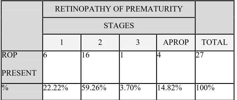
Out of 27 babies with ROP, only 6 babies(22.22%) were in stage 1, 16
babies(59.26%) were in stage 2, 1 baby(3.70%) was in stage3 and 4
[image:75.596.106.518.249.423.2]babies (14.82%) developed APROP (stage4).
Table 3: Stages of ROP
Graph 2: Stages of ROP
6 16 1 4 0 2 4 6 8 10 12 14 16 18 Stage 1 Stage 2 Stage 3 Stage 4
RETINOPATHY OF PREMATURITY
STAGES
1 2 3 APROP TOTAL
ROP
PRESENT
6 16 1 4 27
SEX DISTRIBUTION:
Out of 100 babies screened, 57 were male and 43 were female. Among 57
male babies 18 (31.58%) developed ROP and out of 43 female babies 9
(20.90%) had ROP. In this study gender did not significantly influence
[image:76.596.107.509.530.741.2]the incidence (p=0.82) of ROP.
Table 4: Sex distribution of ROP babies
SEX and ROP
ROP
PRESENT ABSENT TOTAL
SEX
MALE 18(31.58%) 39(68.42%) 57
FEMALE 9(20.90%) 34(79.10%) 43
TOTAL 27 73 100
P value =0.82
Graph 3: Sex distribution of ROP babies
0 5 10 15 20 25 30 35 40
Category 1 Category 2
BIRTH WEIGHT AND ROP
The birth weight of the ROP babies ranged from 800gm-1500 gm (mean
1280 gm).
Lower birth weight was significantly associated with birth weight <
1500gm.
This study included the birth weight upto 2000gm, p value is 0.48 which
is insignificant
[image:77.596.107.518.338.544.2]The incidence of ROP was 48.15% in babies weighing ≤ 1500gm at birth, And more than 1500gm -2000gm the incidence was 51.85%.
Table 4: Distribution as per birth weight
Graph 4: Distribution as per birth weight
0 5 10 15 20 25 30 35
<=1500 1501-1800 1801-2000
ROP present ROP absent
DISTRIBUTION AS PER BIRTH WEIGHT
ROP
PRESENT ABSENT TOTAL
BIRTH
WEIGHT
<=1500 13 26 39
1501-1800 10 32 42
1801-2000 4 15 19
GESTATIONAL AGE AND ROP:
The incidence of ROP was 36.36% in babies born ≤ 32 weeks of
gestational age. Gestational age was found to be a significant risk factor
[image:78.596.111.517.244.459.2]for the development of ROP (p=0.011).
Table 5: Distribution of gestational age and ROP
DISTRIBUTION OF GESTATIONAL AGE
ROP
PRESENT ABSENT TOTAL
GESTATIONAL AGE
<=32 12(36.36%) 21(63.64%) 33
33-35 14(33.33%) 28(66.67%) 42
>35 1(4.00%) 24(96.00%) 25
TOTAL 27 73 100
p value=0.011
Graph 5: Distribution of gestational age and ROP
0 5 10 15 20 25 30
<=32wks 33-35wks >35wks
OXYGEN AND ROP:
Out of 100 babies screened 70 were given O2 and 25 (35.71%) babies
developed ROP. Oxygen administration was a significant risk factor for
[image:79.596.109.525.544.768.2]the development of ROP (p = 0.001).
Table 6: OXYGEN AND ROP
OXYGEN AND ROP
ROP
OXYGEN
Present Absent Total
Given 25(35.71%) 45(64.29%) 70
Not Given 2(6.67%) 28(93.33%) 30
Total 27 73 100
p value = 0.001
Graph 6: OXYGEN AND ROP
SEPSIS AND ROP:
Out of 100 babies screened, 44 had sepsis and 15 babies (34.1%)
developed ROP. Sepsis was not a significant risk factor for the
[image:80.596.106.508.301.749.2]development of ROP in this study (p=0.16).
Table 7: SEPSIS AND ROP
p value = 0.16
Graph 7: SEPSIS AND ROP
0 5 10 15 20 25 30 35 40 45
Sepsis Non sepsis
ROP present ROP absent
SEPSIS AND ROP
ROP
Present Absent Total
SEPSIS
Present 15(34.1%) 29(65.9%) 44
Absent 12(21.4%) 44(78.6%) 75
TRANSFUSION
Out of 100 babies screened, 45 were given transfusion and 20(44.44%)
developed ROP. Transfusion was found to be a significant risk factor for
[image:81.596.109.514.239.439.2]the development of ROP in this study (p=0.004)
Table 8: TRANSFUSION AND ROP
TRANSFUSION AND ROP
ROP
PRESENT ABSENT TOTAL
TRANSFUSIO N
GIVEN 20(44.44%) 25(55.56%) 45
NOT GIVEN 7(12.73%) 48(87.27%) 55
TOTAL 27 73 100
p value = 0.004
Graph 8: TRANSFUSION AND ROP
0 5 10 15 20 25 30 35 40 45 50
Given Not Given
APNOEA OF PREMATURITY AND ROP:
Out of 100 babies screened, 39 babies had AOP and 11(28.21%)
developed ROP. AOP was found to be a insignificant factor for the
development of ROP in this study (p=0.8).
Table 9: APNOEA OF PREMATURITY AND ROP
APNOEA OF PREMATURITY AND ROP
ROP
PRESENT ABSENT TOTAL
AOP
PRESENT 11(28.21%) 28(71.79%) 39
ABSENT 16(26.23%) 45 (73.77%) 61
TOTAL 27 73 100
p = 0.8
Graph 9: APNOEA OF PREMATURITY AND ROP
0 5 10 15 20 25 30 35 40 45
AOP present AOP absent
Table 10: Analysis of risk factors
Gestational age p= 0.011
Birth weight p = 0.48
Oxygen p = 0.001
Sepsis p = 0.16
Transfusion p = 0.04
DISCUSSION
Retinopathy of prematurity (ROP) is a vasoproliferative disorder
affecting premature infants. It is one of the most common causes of visual
loss in children. It can lead to lifelong vision impairment and blindness.
Out of 45 million blind people in the world today, there are about
1.4 million blind children. ROP afflicts over 3,00,000 infants worldwide2
.In developing countries like India, the incidence of ROP has been
reported at 24 – 47 % among high risk preterm infants3. Among the
preventable causes of blindness in children (57%), ROP figures very high
in the agenda. Low birth weight and gestational age were found to be the
most important risk factors for the development of ROP.
We screened babies admitted to our NICU with birth weight ≤
1500g and gestation ≤ 32 weeks. Infants with birth weight >1500g and
gestation more than 32 weeks were screened only if they had additional
risk factors. In an article, Chawla et al91, have suggested the same
INCIDENCE:
With neonatalogical units equipped with the state of art
technological background and highly qualified personnel providing
optimum care of extremely immature newborns, ROP incidence is on a
rise.
The overall incidence in the present study was found to be 27%,
with only one case of severe ROP (APROP). It is of current knowledge
that, aggressive posterior ROP seems to occur especially among smaller
and more immature neonates .In our study the baby which developed
APROP, the birth weight was 1500 grams and gestational age was 33
weeks. In the CRYO-ROP study, the incidence of the disease in a group
of premature newborns with a birth weight <1251gms was 65.8% and
81.6% for infants of less than 1000 g birth weight16..
A review of literature reveals that, the incidence and severity of
ROP increases with decreasing gestational age and birth weight. The
incidence of ROP in different studies done outside India was found to be
9.4%-38.9%18-21.A study from the Indian subcontinent reveals the
incidence to be 17.5% - 46%22-26 comparable to the present study. Patil et
al25 in 1997 reported overall incidence as 17.5% and no severe ROP.
Maheshwari et al24 in 1996 reported overall incidence as 20% and
severe ROP as 7%. They studied 66 babies with <35wk or < 1500gms.
Gupta et al28 in 2003 reported overall incidence as 21.7% and severe ROP
as 5%. They studied 60 babies with ≤ 35wk or ≤1500gms. Dutta et al88
screened 108 babies of ≤32 wk or ≤1700gms and reported overall
incidence as 21%.
The incidence of severe form of the disease (Threshold disease) is
decreasing A Danish study20 found a statistically significant decrease in
incidence of ROP in infants weighing more than 1250gm. Agarwal and
co-workers11 found a drop from 46 to 21% in their study over a period of
7 years.. Similar observations were also made in a multicentre study in
UK27. Nair22 and colleagues, Gupta28 and co-workers found no cases of
ROP in babies weighing more than 1250gm.
TABLE 11: Comparison of incidence of present study with other national and international studies
INDIAN STUDIES
G AGE BT WT INCIDENCE
1.Rekha23,1996 ≤ 34 <1500 46%
2.Maheshwari24,1996 <35 <1500 20%
3.Patil25,1997 <32 <1250 17.5%
4.Agarwal26,2002 <35 <1500 20%
5.Gupta28, 2003 ≤35 ≤1500 21.7%
6.Nair22,2003 ≤32 <1500 25.4%
7.Present study ≤32-35 ≤1500-2000 27%
INTERNATIONAL STUDIES
1.Hussain29,1999 <32 <1500 21.3%
2.Fledelius20,2000 <32 <1500 9.4%
3.Blair21, 2001 <30 <1250 38%
4.Conarth30,2004 ≤32 <1750 10%
5.Shah18 ,2005 <1500 29.2%
RISK FACTORS:
In our study birth weight, gestational age, oxygen, transfusion and
apnoea of prematurity were found to be significant risk factors on Chi
Square analysis. On univariate analysis, risk factors associated with ROP
were oxygen, and transfusion. Using the forward method oxygen and
transfusion were also found to be significant.
Birth Weight and Gestational age:
In our study both low birth weight (p=<0.48) and prematurity (p=
0.011) were found to be significant risk factors for the development of
ROP.
The birth weight of the ROP babies ranged from 800-1500 gm (mean
1280gm) Lower birth weight was insignificantly associated with this
study because birth weight included upto 2000gms,whereas significant
upto 1500gms. The incidence of ROP was 48.15% in babies weighing ≤
1500gm at birth.
The incidence of ROP was 26.32% in babies born ≤ 32 weeks of
gestational age. Gestational age was found to be a significant risk factor
Oxygen:
In our study oxygen was found to be significant risk factor for the
development of ROP on Chi square analysis (p=0.005), on univariate
analysis and also on multivariate analysis. Out of 100 babies screened 59
were given O2 and 15 (25.42%) babies developed ROP. The causal link
between ROP and supplemental oxygen has been confirmed by controlled
trials and clinical studies. Gunn analyzed data from their low birth weight
survivors and found a significant association between, the more severe
grade of cicatrical disease and duration of oxygen therapy54.Kinsey noted
that, concentration of oxygen administered was significantly associated
with ROP in infants under 1200gm. When comparing mean Pa02 levels
in normal infants and in ROP infants, he found differences only in babies
of low birth weight and only Pa02 levels greater than 150mmHg.
However, a safe level of oxygen usage has not been defined, keeping
Pao2 <100 mm Hg is recommended, preferably between 50 and 70mm
Hg and saturation between 90-95%. Preliminary work has suggested that,
Table 12: Comparison of oxygen as a risk factor of ROP in different studies
Study p-value
Gupta28 et al 0.002
Rekha23et al 0.005
CONCLUSION
1. The present study reflects the problem of ROP with various risk
factors in a tertiary care centre.
2. The incidence of ROP in the present study is 27%.
3. Out of 27 babies with ROP, only 6 babies(18.75%) were in stage 1,
16 babies(68.75%) were in stage 2,1 baby(6.25%) was in stage3
and 4 baby(6.25%) developed APROP.
4. The birth weight of the ROP babies ranged from 800-1500 gm
(mean 1.32 gm) lower birth weight was significantly associated
with increased incidence (p = 0.48) of ROP. The incidence of ROP
was 48 % in babies weighing ≤ 1500gm at birth.
5. The incidence of ROP was 36.36 % in babies born ≤ 32 weeks of
gestational age. Gestational age was found to be a significant risk
factor for the development of ROP (p=0.011).
6. Oxygen, Sepsis, Transfusion and Apnoea of Prematurity were
found to be significant risk factors on Chi Square analysis.
7. The risk factors associated with ROP were Oxygen, Sepsis and
Transfusion .