0022-538X/07/$08.00
⫹
0
doi:10.1128/JVI.00648-06
Copyright © 2007, American Society for Microbiology. All Rights Reserved.
Modulation of Host Gene Expression by the K15 Protein of Kaposi’s
Sarcoma-Associated Herpesvirus
䌤
Melanie M. Brinkmann,
1† Marcel Pietrek,
1Oliver Dittrich-Breiholz,
2Michael Kracht,
2and Thomas F. Schulz
1*
Institut fu
¨r Virologie, Medizinische Hochschule Hannover, Carl-Neuberg Strasse 1, D-30625 Hannover, Germany,
1and Institut fu
¨r
Pharmakologie, Medizinische Hochschule Hannover, Carl-Neuberg Strasse 1, D-30625 Hannover, Germany
2Received 30 March 2006/Accepted 27 September 2006
Kaposi’s sarcoma-associated herpesvirus (KSHV) contains several open reading frames (ORFs) encoding
proteins capable of initiating signal transduction pathways. Among them is the K15 ORF, which consists of
eight exons encoding a protein with 12 predicted transmembrane domains and a cytoplasmic C terminus. When
transiently expressed, the 8-exon K15 transcript gives rise to a protein with an apparent molecular mass of 45
kDa. K15 interacts with cellular proteins, TRAF (tumor necrosis factor receptor-associated factor) and Src
kinases, and activates AP-1, NF-
B, and the mitogen-activated protein kinases (MAPKs) c-jun-N-terminal
kinase and extracellular signal-regulated kinase. This signaling activity of K15 is related to phosphorylation
of Y
481of the K15 SH2-B motif Y
481EEV. In this study we demonstrate the expression of an endogenous 45-kDa
K15 protein in KSHV BAC36-infected epithelial cells. This endogenous K15 protein shows the same
intracel-lular localization as transiently expressed K15, and expression kinetic studies suggest it to be a lytic gene. We
have further determined the downstream target genes of K15 signaling using DNA oligonucleotide
microar-rays. We demonstrate that K15 is capable of inducing expression of multiple cytokines and chemokines,
including interleukin-8 (IL-8), IL-6, CCL20, CCL2, CXCL3, and IL-1
␣
/

, as well as expression of Dscr1 and
Cox-2. In epithelial cells, K15-induced upregulation of most genes was dependent on phosphorylation of Y
481,
whereas in endothelial cells mutation of Y
481did not result in a complete loss of Dscr1 and Cox-2 expression
and NFAT-activity. Our study establishes K15 as one of the KSHV lytic genes that are inducing expression of
multiple cytokines, which have been shown to play an important role in KSHV-associated pathogenesis.
The eighth human herpesvirus, Kaposi’s sarcoma-associated
herpesvirus (KSHV or HHV-8), was originally identified in
Kaposi’s sarcoma (KS) tissues by representational difference
analysis (21). KSHV plays an essential role in the pathogenesis
of three human neoplastic disorders, Kaposi’s sarcoma (81,
114), a very rare form of B-cell lymphoma called body
cavity-based lymphoma (BCBL) or primary effusion lymphoma
(PEL) (19), and the plasma cell variant of multicentric
Castle-man’s disease (120).
KS is a multicentric vascular neoplasm involving the skin and
mucosal surfaces and in aggressive cases may involve visceral
organs and lymph nodes. KS lesions contain distinctive
prolif-erating spindle cells, activated endothelial cells, fibroblasts,
smooth muscle cells, and infiltrating inflammatory cells (82,
103, 104). Endothelial cell-derived spindle cells represent the
neoplastic component of the KS tumor and are infected by
KSHV (10, 32, 60, 94, 101, 115). In these cells, four latent viral
genes are expressed: open reading frame (ORF)73/Lana-1,
ORF K12/kaposin, ORF K13/vFLIP, ORF72/vCyclin. In some
spindle cells KSHV is not strictly latent but undergoes lytic
replication (60, 94, 122). Similarly, in cell lines derived from
PEL (9, 20), only latent genes are expressed in the majority of
cells, but in some PEL cell lines a minority of KSHV-infected
cells will spontaneously switch into the lytic cycle.
The lytic viral replication cycle of KSHV can be
experimen-tally induced in PEL cells by addition of the phorbol ester
12-
O
-tetradecanoylphorbol-13-acetate (TPA) or
n
-butyrate
(78, 107). Virus harvested from induced PEL cell lines can
infect the epithelial cell line 293 (39, 106), primary endothelial
cell cultures (16, 24, 38), immortalized endothelial cell lines
(64, 82), human and murine fibroblasts, HeLa cells, and the
endothelial SLK cell line (7). Infectious KSHV can also be
produced from latently infected 293 and TIME cells after
in-duction of the lytic cycle following transfection or transin-duction
of the viral lytic transactivator regulator of transcriptional
ac-tivation (RTA/ORF50) (7). Viral particles generated from 293
cells stably transfected with a recombinant KSHV genome in a
bacterial artificial chromosome (BAC) can infect 293, HeLa,
and human endothelial cells (135).
Several studies have demonstrated the importance of
in-flammatory cytokines in KSHV-associated pathogenesis (34).
KSHV encodes various genes capable of regulating cellular
cytokine expression such as the viral G-protein-coupled
recep-tor (vGPCR) (99, 117), K1 (67), kaposin B (75), and
homo-logues of human interleukin-6 (IL-6) and cellular CC
chemo-kines (87). The lytic KSHV membrane proteins vGPCR and
K1 are signaling proteins capable of activating multiple cellular
signaling cascades such as mitogen-activated protein (MAP)
kinase pathways and several transcription factors implicated in
inflammatory responses (14, 88).
Another KSHV-encoded membrane protein capable of
elic-iting cellular signal transduction pathways is K15 (14). The K15
* Corresponding author. Mailing address: Institut fu
¨r Virologie,
Medizinische Hochschule Hannover, Carl-Neuberg Str. 1, D-30625
Hannover, Germany. Phone: 49 511 532 6736. Fax: 49 511 532 8736.
E-mail: schulz.thomas@mh-hannover.de.
† Present address: Whitehead Institute for Biomedical Research, 9
Cambridge Center, Cambridge, MA 02142.
䌤
Published ahead of print on 18 October 2006.
42
on November 8, 2019 by guest
http://jvi.asm.org/
ORF is located between the viral terminal repeat region and
ORF75 of the KSHV genome (23, 44, 100). Multiple
alterna-tively spliced transcripts are generated from the K15 gene, with
the most prominent transcript encompassing eight exons. The
sequences of all K15 cDNA clones isolated so far are predicted
to contain a common C-terminal cytoplasmic region (encoded
by exon 8) linked to a variable number of transmembrane
domains, with the full-length transcript (exons 1 to 8) coding
for a protein with up to 12 transmembrane domains.
Expression of K15 transcripts was identified in unstimulated
KSHV-positive PEL cells and was shown to be upregulated
upon lytic cycle induction (23, 44, 100). Gene array studies
indicate that K15 is predominantly expressed during the lytic
cycle in PEL cell lines (57, 85, 98). In the PEL cell line
BCBL-1, ectopic RTA/ORF50 expression, and TPA induction
were shown to mediate K15 promoter transactivation (130).
Taken together, these findings suggest that K15 is expressed
after activation of the lytic cycle in PEL cells.
Cloned into mammalian expression vectors, the eight-exon
K15 isoform is translated into a protein with an apparent mass
of
⬃
45 kDa that associates with lipid rafts and forms patches
on the plasma and cytoplasmic membranes in 293 and Cos-1
cells with a mainly perinuclear localization (13, 23, 44, 116).
However, in uninduced PEL cell lines, a protein of 23 kDa was
detected with a monoclonal antibody raised to the C-terminal
domain of K15, and immunofluorescence studies showed a
cytoplasmic staining in the majority of these cells (116). Upon
induction of the lytic cycle, expression of this 23-kDa protein
decreased over time, suggesting latent kinetics for this protein.
By immunostaining with a monoclonal K15 antibody, K15
ex-pression has so far been observed in multicentric Castleman’s
disease plasmablasts but not in KS tumors (116).
The cytoplasmic domain of K15 contains several signaling
motifs: a proline-rich motif that could potentially serve as an
SH3-binding (SH3-B) motif (PP
387PLPP), a motif reminiscent
of a tumor necrosis factor receptor-associated factor (TRAF)
binding site (ATQ
475PTDD), and two potential SH2-binding
(SH2-B) sites (VFGY
431ASI and DDLY
481EEV). K15 shows
structural and functional similarities to Epstein-Barr virus
(EBV) latent membrane proteins LMP1 and LMP2A (14).
Like LMP1, K15 interacts with TRAF-1, TRAF-2, and
TRAF-3, activates the MAP kinase c-jun-N-terminal kinase
(JNK) 1, and the transcription factors NF-
B and AP-1 (13,
44). Reminiscent of LMP2A, K15 interacts with members of
the Src family of protein tyrosine kinases via its C-terminal
domain and is phosphorylated in vitro at Y
481by these
kinases (13). A chimera of the CD8 molecule fused to the
K15 cytoplasmic domain is constitutively phosphorylated at
Y
481of the Y
481EEV motif in vivo (23). The CD8-K15
chimera was shown to downregulate B-cell receptor
signal-ing in B cells, which seems to be mediated via the putative
SH3-B and the SH2-B motif Y
481EEV (23). Further, K15
can induce activation of the MAP kinase Erk2 via the
clas-sical Ras/Raf/MEK pathway (13).
Here, we show the expression of a 45-kDa K15 protein in the
epithelial cell line 293 stably transfected with the entire KSHV
genome cloned as a bacterial artificial chromosome (KSHV
BAC36). This 45-kDa K15 protein expressed in the context of
the complete KSHV genome was significantly induced upon
lytic cycle induction and shows a similar intracellular
localiza-tion as the transfected eight-exon cDNA of K15. Employing
two types of DNA oligonucleotide microarrays, we identified
several cytokines, chemokines, and cellular genes such as
Down syndrome critical region 1 (Dscr1), cyclooxygenase 2
(Cox-2), and matrix metalloproteases as downstream targets of
K15. With few exceptions, K15-induced activation of gene
ex-pression in epithelial cells was dependent on Y
481of the SH2-B
motif of K15. In contrast, in SLK endothelial cells the K15
mutant K15 F
481was still able to induce expression of
K15-downstream targets Dscr1 and Cox-2 and NFAT activity,
indi-cating host-cell-dependent functions of Y
481. These results
es-tablish K15 as one of the KSHV-encoded proteins that on their
own have the capacity to modulate the host cell response
toward an inflammatory phenotype and may therefore be
in-volved in KSHV-associated pathogenesis.
MATERIALS AND METHODS
Cell lines, transfections, and lytic cycle induction.The cell lines HEK (human embryonic kidney) 293-T, HEK 293, Cos-7, and human SLK endothelial cells were cultured in Dulbecco’s modified Eagle medium (Gibco) supplemented with 10% heat-inactivated fetal calf serum (FCS), 50 IU/ml penicillin, and 50g/ml streptomycin at 37°C in humidified air with 5% CO2. The endothelial SLK cell line was established from a biopsy of an oral KS tumor of an iatrogenically immunosuppressed human immunodeficiency virus-negative patient and has since lost the KSHV genome (49). The human cervix epithelial HeLa cell line and the epithelial kidney cell line PtK2 (isolated fromPotorous tridactylis) were cultured in minimal essential medium (Cytogen) supplemented as above. Sf9 insect cells were cultured in Grace’s insect medium (Gibco) with 10% FCS, 50 IU/ml penicillin, and 50g/ml streptomycin at 27°C. For transfections, cells were grown to subconfluence in six-well plates. 293-T cells, Cos-7, PtK2, and HeLa cells were transfected with FuGENE transfection reagent (Roche) (FuGENE: DNA ratio of 3l:1g). SLK cells were transfected with Lipofectamine 2000 according to the manufacturer’s instructions with a Lipofectamine:DNA ratio of 4l:4g (Invitrogen). For immunofluorescence studies, SLK cells were trans-fected with 2g of DNA. The KSHV BAC clone 36 (135) was kindly provided by S. J. Gao. KSHV BAC36 DNA for transfection of 293 cells was prepared from Escherichia colistrain DH10B with the Maxi-BAC Kit (Machery and Nagel). For transfection, 293 cells were plated at 3⫻105
cells per well of a six-well plate and transfected 2 days later at 70% confluence with Lipofectamine 2000, according to the manufacturer’s instructions (Invitrogen) (DNA:Lipofectamine ratio of 4 g:10l). The transfection efficiency could be monitored by green fluorescent protein expression. The medium was changed the following day, and 48 h post-transfection cells were trypsinized and split 1:2. Eight days postpost-transfection, cells were trypsinized and split 1:3 in Dulbecco’s modified Eagle medium containing HygromycinB (150g/ml). The medium was changed every 3 days and when foci developed, individual foci were picked and transferred into a 96-well plate under selection with HygromycinB.
The lytic viral life cycle was induced 1 or 2 days after seeding 293 KSHV BAC36 cells by addition of 1 mM sodium butyrate (Sigma) or infection with baculovirus coding for KSHV ORF50/RTA or both. At different time points postinduction, cells were lysed in 250l of TBS-T (20 mM Tris-HCl, pH 7.4, 150 mM NaCl, 1 mM EDTA, 1% Triton X-100, and protease inhibitors phenylmeth-ylsulfonyl fluoride [1 mM], leupeptin [50M], aprotinin [100 U/ml], benzami-dine [200M], pepstatin A [1M]) per well of a six-well plate. The generation of the recombinant baculovirus expressing KSHV ORF50/RTA was described elsewhere (128). For infection of mammalian cells, RTA baculovirus containing Sf9 insect cell culture supernatant was cleared by centrifugation (500⫻gfor 15 min), and 200l of supernatant was added per well of a six-well plate.
DNA constructs.The full-length K15 cDNA clone (K15 amino acids [aa] 1 to 489, exons 1 to 8) and the point mutant K15 Y481
F used in this study were generated as follows. The full-length K15 wild-type (WT) construct containing an artificial Kozak sequence (bold) 5⬘ of the ATG in exon 1 (underlined) was generated by PCR on clone MBK15 using the primers K15EcoRIKozakfor (5⬘-TATGAATTCGCCACCATGAAGACACTCATATTCTTCTGG-3⬘) and FlagLAMPa3⬘binding to exon 8 (5⬘-TATGAATTCCTAGTTCCTGGGAAAT AAAAC-3⬘) (underlining indicates the stop codon in exon 8). The PCR fragment was cloned EcoRI in pFJ-EA. The K15 point mutant K15 Y481F was generated as the K15 WT construct but with reverse primer LAMPaY:Frev (5⬘-TATGAA TTCCTAGTTCCTGGGAAATAAAACCTCCTCAAACAGGTC-3⬘)
on November 8, 2019 by guest
http://jvi.asm.org/
ing the amino acid substitution Y4813F481(bold). The generation of the full-length K15 P-type cDNA clone MBK15 (K15 aa 1 to 489) from the BCP-1 KSHV isolate (primary effusion cell line infected with KSHV) has already been de-scribed (13).
The reporter plasmids Dscr1 (in pGL3b) and Cox-2 (in pGL3b) contain the promoter regions of thedscr1andcox-2genes cloned upstream of the luciferase gene of the pGL3b vector, respectively, and were kindly provided by Elia J. Duh (132). The promoterless pGL3b reporter (Promega) served as negative control. The pTA-Luc vector (Clontech) contains the minimal TA promoter upstream of the luciferase reporter gene. The pNFAT-TA-Luc vector (Clontech) additionally contains three tandem copies of the NFAT consensus sequence upstream of the minimal TA promoter and the luciferase reporter gene.
siRNA design and transfection.The K15 small interfering RNA (siRNA) was targeted to exon 8 of K15 (CAACCACCUUGGCAAUAAU) and was pur-chased from Dharmacon. HeLa and 293-T cells (in six-well plates) were trans-fected with 160 pmol of K15 siRNA (diluted in 250l of Opti-MEM I medium [Gibco]) and 5l of Lipofectamine 2000 (Invitrogen) (diluted in 250l of Opti-MEM I) at 30 to 50% confluence according to the manufacturer’s instruc-tions. Six hours later, the medium was replaced and cells were transfected with 1g of K15 expression construct (FuGENE).
293 KSHV BAC36 cells were plated at a cell density of 6⫻105cells/well of a six-well plate and transfected the following day with K15 siRNA as described above. Six hours later, cells were washed, and fresh medium was added. Twenty hours after siRNA transfection, cells were either left untreated or stimulated with 1 mM butyrate or baculovirus coding for KSHV ORF50/RTA or both and lysed 24 h postinduction.
Oligonucleotide DNA microarray experiments.HeLa cells were seeded in six-well plates at a density of 8⫻104cells per well and transfected 24 h later with 1g of DNA per well using the FuGENE transfection reagent. Thirty-two hours posttransfection, cells were lysed for RNA extraction according to the manufac-turer’s instructions (RNeasy kit; QIAGEN). For controlling protein expression levels by Western blotting, cells were lysed 32 h posttransfection for 10 min on ice with 250l of radioimmunoprecipitation assay (RIPA) 100 buffer (20 mM Tris, pH 7.5, 1 mM EDTA, 100 mM NaCl, 1% Triton X-100, 0.5% sodium deoxycholate, 0.1% sodium dodecyl sulfate [SDS]) with protease inhibitors (1 mM phenylmethylsulfonyl fluoride, 50M leupeptin, 100 U/ml aprotinin, 200 M benzamidine, 1M pepstatin A) per well. Lysates were cleared by centrif-ugation (10,000⫻gfor 10 min at 4°C) and analyzed by Western blotting. To control transfection efficiency, HeLa cells were seeded onto glass coverslips and analyzed by immunofluorescence using a polyclonal antibody to K15 (13) 32 h posttransfection.
For microarray experiments the Human 40K:A Array and the Human Inflam-mation Array (MWG Biotech) were used (now distributed by Ocimum Biosolu-tions as Human 40K A OciChipTM and Human Inflammation OciChipTM, respectively). The Human Inflammation Array contains 155 validated oligonu-cleotide probes for 136 inflammatory and 19 housekeeping genes. The Human 40K:A Array contains approximately 20,000 probes covering mainly well-char-acterized human transcripts.
Total RNA from cells transfected as described above was purified with the RNeasy kit followed by on-column DNase I digestion (QIAGEN). RNA was used to prepare Cy3- or Cy5-labeled cRNA by oligo(dT)-T7-primed double-stranded cDNA synthesis (cDNA synthesis system; Roche), followed by in vitro transcription with T7-polymerase (MEGAscript T7 kit; Ambion) as directed by the manufacturers. cRNA yields were determined photometrically.
For low-density microarray experiments, 16g of Cy3-labeled cRNA derived from approximately 2.5g of total RNA was hybridized individually to microar-rays (one sample per array). For high-density microarray experiments, 40g of Cy3- and Cy5-labeled cRNA populations, derived from approximately 5g of total RNA, representing two different experimental conditions, were combined and cohybridized onto the same microarray (two-color experiment).
cRNAs were fragmented, repurified, and hybridized to microarrays in prepre-pared hybridization solution (MWG Biotech) at 42°C overnight and then washed sequentially in 2⫻SSC (1⫻SSC is 0.15 M NaCl plus 0.015 M sodium citrate), 0.1% SDS, 1⫻SSC, and 0.5⫻SSC. Hybridized arrays were scanned on an Affymetrix 428 scanner at variable photomultiplier tube voltage settings. Fluo-rescence intensity values were processed using Imagene 4.2 software (Biodiscovery). In order to obtain maximal signal intensities without saturation effects, intensity values from TIFF images were integrated into one value per probe by the MAVI software (version Pro 2.5.1; MWG Biotech). Data were filtered for flagged spots and low intensity values; remaining data were used to calculate ratios of gene expression using Excel macros. Additional information on the microarrays and methodology used can be obtained at http://www.mh-hannover.de/forschung /sfb566/microarray/index.phtml.
Real-time PCR.The same cDNAs that were used for high-density microarray experiments were used to quantify the amounts of mRNA ofactb(Hs99999903_m1), cxcl8(Hs00174103_m1), andvegf(Hs00173626_m1) by the indicated assays on de-mand (Applied Biosystems). Real-time reactions were performed in 25-l mixtures using an ABI7500 instrument, and cycle threshold (CT) values for each PCR product
were calculated by the 7500 Fast System software (version 1.3.0).CTvalues
ob-tained forcxcl8andvegfwere normalized by subtracting theCTvalues obtained
for-actin (actb) as a housekeeping reference. The resulting⌬CTvalues were
then used to calculate relative changes of mRNA expression as the ratio (R) of mRNA expression of transfected/mock-transfected cells according to the equa-tion:R⫽2⫺(⌬Ct(transfected)⫺ ⌬Ct(mock-transfected))
.
Reverse transcription-PCR (RT-PCR). SLK cells were plated at 3⫻105 cells/well of a six-well plate and transfected with Lipofectamine 2000. The me-dium was changed 6 h posttransfection. At 32 h posttransfection, cells were lysed for RNA extraction according to the manufacturer’s instructions (RNeasy kit; QIAGEN). cDNA was prepared as described for DNA microarray experiments. For the subsequent PCR, 7 ng of HeLa or SLK cDNA was used per PCR with primers spanning the intron between exons 5 and 6 of thedscr1gene (dscr1 347-365-for, 5⬘-CCTTCTCCGCAGCAGATGC-3⬘; dscr1 543-524-rev, 5⬘-GACT GGGGTCGCATCTTCCAC-3⬘). This primer combination detects the same dscr1isoforms as the inflammation DNA microarray and amplifies a spliced transcript of 196 bp and would amplify a genomic fragment of 554 bp if genomic traces of DNA were present. The PCR was performed with the Hot StarTaqkit (QIAGEN) with the following PCR conditions: 15 min at 95°C followed by 22 to 24 cycles of 30 s at 94°C, 30 s at 57°C, 20 s at 72°C, and 7-min final extension at 72°C. The housekeeping gene glyceraldehyde-3-phosphate dehydrogenase (GAPDH) was amplified with primers GAPDHfor (5⬘-ACCACAGTCCATGC CATCAC-3⬘) and GAPDHrev (5⬘-TCCACCACCCTGTTGGTGTA-3⬘) and the following PCR conditions: 15 min at 95°C followed by 22 to 24 cycles of 30 s at 94°C, 30 s at 60°C, 30 s at 72°C, and 7-min final extension at 72°C. A total of 5 pmol of each primer was used for one PCR of 25l. PCR products were separated on 1.5% agarose gels and visualized by ethidium bromide staining. To control equal expression levels of the K15 proteins, SLK cell lysates were ana-lyzed by Western blotting as described for HeLa cells (DNA microarray).
ELISA.HeLa cells were transfected exactly as described for the DNA mi-croarrays. At 32 h posttransfection, the conditioned medium was collected and centrifuged for 5 min at full speed in a table-top centrifuge, and supernatants were analyzed by enzyme-linked immunosorbent assay (ELISA) according to the manufacturer’s instructions (Immunotools). The cells were lysed in RIPA 100 buffer to control K15 protein expression by Western Blotting as described for the DNA microarrays.
Cytokine antibody array.HeLa cells were transfected exactly as described for the DNA microarrays. At 32 h posttransfection, the conditioned medium was collected and centrifuged for 5 min at full speed in a table-top centrifuge, and supernatants were analyzed with the Cytokinearray III encompassing 42 different human cytokines according to the manufacturer’s instructions (RayBiotech). The captured cytokines were detected with a mixture of biotin-labeled anticytokine antibodies, followed by streptavidin-horseradish peroxidase and chemilumines-cence. Quantification of the spots was performed with the Kodak 1D imaging software. The cells were lysed in RIPA 100 buffer to control K15 protein expres-sion by Western blotting as described for DNA microarrays.
Luciferase-based reporter assays.293-T cells were plated at a cell density of 5⫻ 105
cells per well of a six-well plate and transiently cotransfected the following day with 100 ng of the reporter plasmid Dscr1 or Cox-2 or the control reporter plasmid pGL3b and 1g of K15 expression constructs or empty vector (pFJ-EA) per well of a six-well plate. At 20 h posttransfection, the medium was replaced with medium containing 1% FCS. At 29 h posttransfection, cells were washed once with phosphate-buffered saline (PBS) and lysed in reporter lysis buffer (Promega). Luciferase activities were measured in cleared lysates using a luciferase assay system according to the manufacturer’s instructions (Promega). Luciferase activity was calculated as relative induction (n-fold) compared to mock (empty expression vector)-transfected controls. The reporter assays with the pTA (negative control) or pNFAT-TA luciferase reporter plasmids were performed as described above, with the exception that the serum was reduced to 0% FCS 20 h posttransfection. As a positive control for NFAT activation, transfected 293-T cells were stimulated with TPA (200 ng/ml; Sigma) and cal-cium ionophore (5M; Sigma) or as negative control with dimethyl sulfoxide (DMSO) 4 h before cells were lysed. SLK cells were seeded at a cell density of 3⫻105cells per well of a six-well plate and cotransfected the following day with 3g of K15 expression constructs or empty vector together with 1g of the reporter vectors pGL3b, pTA (negative controls), Dscr1, Cox-2, or pNFAT-TA using the Lipofectamine 2000 transfection reagent (Invitrogen) according to the manufacturer’s instructions. Medium was changed 6 h posttransfection. At 21 h
on November 8, 2019 by guest
http://jvi.asm.org/
posttransfection, medium was replaced with medium containing 0% FCS, and cells were lysed in reporter lysis buffer 29 h posttransfection.
Immunoblotting.For detection of proteins by Western blotting, cleared cell lysates containing K15 proteins were not boiled prior to SDS-polyacrylamide gel electrophoresis. As indicated, the following primary antibodies were used for immunostaining of immunoblots: mouse actin (Chemikon), mouse anti-vimentin (Chemikon), and rabbit anti-K15 (13) antibodies. Immunoblots were analyzed using horseradish peroxidase-coupled secondary antibodies (Dako) and a standard enhanced chemiluminescence reaction.
Immunofluorescence.HeLa, SLK, and PtK2 cells were washed once with PBS and fixed for 20 min with paraformaldehyde (3% in PBS) and then washed three times with PBS and incubated with NH4CL (50 mM) in PBS for 10 min. Cells were then permeabilized for 5 min with 0.1 to 0.2% Triton X-100 in PBS, washed three times with PBS, and blocked for 1 h in 10% FCS in PBS at room temper-ature. The first antibody (diluted in 2% FCS in PBS) was applied for 1 h at room temperature, and cells were washed three times with PBS and then incubated for 30 min at room temperature with the secondary antibody (diluted in 2% FCS in PBS). After three washes with PBS, cell nuclei were stained with Hoechst, washed with PBS, and embedded in MOWIOL with Dabco (25 mg/ml, Sigma) and analyzed by fluorescence microscopy. The primary K15 polyclonal antibody was diluted 1:200. Secondary antibodies were anti-rabbit Cy3 (1:200; Jackson ImmunoResearch) or anti-rabbit fluorescein isothiocyanate (1:40) (Dako).
293 KSHV BAC36 cells were plated on coverslips at a cell density of 4⫻105 to 5⫻105cells per well of a six-well plate coated with 0.1% gelatin in PBS. The lytic viral life cycle was induced the following day by the addition of sodium butyrate (1 mM final concentration; Sigma) or baculovirus encoding the lytic switch protein KSHV RTA/ORF50. At 23 to 38 h postinduction, cells were analyzed by fluorescence microscopy as described above.
RESULTS
A K15 protein of 45 kDa is expressed in the 293 KSHV
BAC36 cell line.
When transiently expressed in cell lines such
as 293-T or HeLa, the eight-exon K15 transcript is translated
into a membrane protein of
⬃
45 kDa (13, 23, 116). Until now,
full-length K15 transcripts (exons 1 to 8) have been detected in
KSHV-positive PEL cell lines by RT-PCR (23, 44), but a K15
protein of 45 kDa has, until now, not been observed in any
KSHV-positive cell line analyzed.
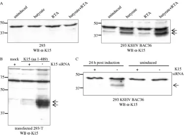
[image:4.585.106.479.66.340.2]We detected a K15 protein of 45 kDa in 293 cells that were
stably transfected with the complete KSHV genome derived
from the PEL cell line BCBL-1 and which had previously been
cloned into a bacterial artificial chromosome, KSHV BAC
clone 36 (135). As shown in Fig. 1A (right blot), this 45-kDa
K15 protein was weakly expressed in uninduced 293 KSHV
BAC36 cells, and its expression increased upon induction of
the lytic viral life cycle with sodium butyrate. Whereas
induc-tion of the lytic viral life cycle by infecinduc-tion with a baculovirus
coding for the KSHV lytic switch protein RTA/ORF50 did not
increase K15 expression, the addition of both compounds
(so-dium butyrate and baculovirus RTA/ORF50) was most
effec-tive in the upregulation of K15 expression (Fig. 1A, right blot).
FIG. 1. A K15 protein of 45 kDa is expressed in 293 cells stably transfected with KSHV BAC36. (A) In 293 cells stably transfected with KSHV
BAC36, a 45-kDa protein was detected with the K15 polyclonal antibody (right blot, indicated by arrows), which was not present in 293 cells (left
blot). The K15 polyclonal antibody detects an unspecific band of approximately 48 kDa in lysates of 293 and 293 KSHV BAC36. For induction
of the lytic viral life cycle, cells were either treated with butyrate or infected with baculovirus coding for the KSHV RTA/ORF50 (regulator of
transcriptional activation) protein, or both. At 48 h postinduction, cells were lysed and analyzed for K15 protein expression by Western blotting
with the K15 polyclonal antibody. 293 cells were treated in a similar manner. (B) 293-T cells were transiently transfected with empty expression
vector pFJ-EA (mock) or a full-length K15 cDNA construct encompassing exons 1 to 8 (aa 1 to 489). At 6 h prior to DNA transfection, cells were
transfected with a K15 siRNA directed against exon 8, which codes for the C-terminal cytoplasmic domain of K15 (
⫹
, K15 siRNA transfected;
⫺
,
not K15 siRNA transfected). At 36 h posttransfection, cells were lysed and analyzed by Western blotting with the polyclonal K15 antibody. (C) The
293 KSHV BAC36 cell line was transfected with K15 siRNA as described in Material and Methods. Twenty hours after siRNA transfection, either
cells were left untreated (uninduced) or the lytic viral life cycle was induced by addition of butyrate and baculovirus RTA/ORF50. Cells were lysed
24 h postinduction, and lysates were analyzed by Western blotting with the polyclonal K15 antibody. WB, Western blotting.
on November 8, 2019 by guest
http://jvi.asm.org/
The K15 protein expressed from the complete KSHV genome
migrates as a doublet (Fig. 1A, right blot) similar to the K15
protein expressed from a cDNA expression vector (exons 1 to
8) in transiently transfected 293-T cells (Fig. 1B), suggesting
posttranslational modifications.
To verify that the 45-kDa protein detected by our polyclonal
K15 antibody in the 293 KSHV BAC36 cell line was indeed
derived from ORF K15, an siRNA directed against exon 8 of
K15 was first validated in a cotransfection experiment in 293-T
cells with the K15 expression construct comprised of exons 1 to
8 (Fig. 1B). After transfection into the 293 KSHV BAC36 cell
line, this K15 siRNA was able to significantly reduce the
ex-pression of the 45-kDa protein detected by the polyclonal K15
antibody in sodium butyrate and RTA/ORF50-induced 293
KSHV BAC36 cells (Fig. 1C), indicating that the latter is in
fact derived from ORF K15.
Intracellular localization of endogenously and transiently
expressed K15 protein.
Different intracellular localization
pat-terns of the transiently expressed K15 protein have been
re-ported which range from endoplasmic reticulum localization in
293 and HeLa cells (116) to the formation of large patches on
external and cytoplasmic membranes in 293 cells (44), and
cytoplasmic, plasma membrane, and perinuclear localization in
Cos-1 and 293 cells (23, 116). In the epithelial cell line PtK2
and the endothelial cell line SLK, the transfected eight-exon
K15 expression construct produced a distinct punctuate
ex-pression pattern evenly distributed over the entire cell and
concentrated in the perinuclear region, which was visualized
with either a monoclonal K15 antibody directed against amino
acids encoded in exon 8 (data not shown) or the polyclonal K15
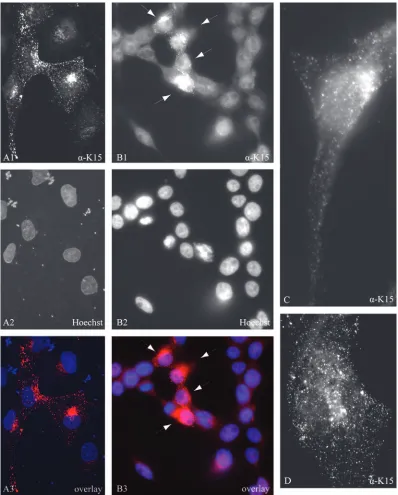
antibody (Fig. 2; PtK2 in image A1 and SLK in image D). In
K15-expressing PtK2 cells, the majority of K15 did not
colo-calize with the endoplasmic reticulum marker calnexin (data
not shown).
We next examined the 293 KSHV BAC36 cell line, after
treatment with butyrate, for the expression of endogenous K15
protein by immunofluorescence with the K15 polyclonal
anti-body. As observed for transiently expressed K15 protein in
SLK and PtK2 cells, endogenous K15 expression was
concen-trated in the perinuclear region (Fig. 2, image B1). Image C
(Fig. 2), which shows a higher magnification of a
K15-express-ing 293 KSHV BAC36 cell, illustrates that the K15 protein
expressed in the context of the entire KSHV genome shows a
punctuate localization similar to that observed in PtK2 or SLK
cells transiently transfected with K15 (Fig. 2, images A1 and
D). In uninduced 293 KSHV BAC36 cells only very few cells
were positive for the K15 protein (data not shown). We
ob-served that a small percentage of 293 KSHV BAC36 cells
expressed the late lytic glycoprotein K8.1 A/B and the early
lytic protein ORF59 without induction of the lytic cycle (data
not shown). It is therefore likely that the K15 expression
ob-served in uninduced 293 KSHV BAC36 cells occurs in a subset
of cells that have spontaneously switched on the lytic cycle.
Genome-wide investigation of K15-regulated cellular genes
reveals several genes related to inflammation.
The K15
mem-brane protein activates the MAP kinases extracellular
signal-regulated kinase (ERK) and JNK and the transcription factors
AP-1 and NF-
B (13). K15 is constitutively tyrosine
phosphor-ylated at Y
481in vivo (23), and we have shown that members of
the Src family of protein tyrosine kinases phosphorylate Y
481in
vitro (13). All signaling activities of K15 described so far are
significantly impaired when Y
481is mutated to F
481(13).
In order to explore the downstream targets of K15 signaling,
we performed high-density DNA microarrays on
K15-trans-fected cells. Since the 293 KSHV BAC36 epithelial cell line
was the first cell system in which we could detect the expression
of the 45-kDa K15 protein in the context of the complete viral
genome, we chose epithelial cells for further experiments.
HeLa cells were transiently transfected with either pFJ-EA
(empty vector control; mock) or K15 expression constructs K15
WT or the K15 F
481mutant. At 32 h posttransfection, cells
were lysed for RNA extraction, and cDNA was prepared as
described in Material and Methods and the notes to Table 1. In
these experiments, the transfection efficiency of HeLa cells was
about 50% for both K15 WT and K15 F
481, and both proteins
were expressed at equal levels as judged by
immunofluores-cence and Western blotting (data not shown).
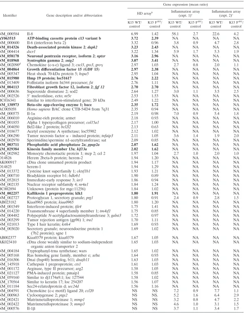
As depicted in Table 1, by the filter criteria applied, 56 genes
were consistently upregulated more than 1.5-fold by K15, and
3 genes were downregulated. Cellular genes induced by K15
included several involved in inflammation, such as the
chemo-kines interleukin-8 (
cxcl8
),
gro1
(
cxcl1
/
gro
␣
), and monocyte
chemotactic protein 1 (
mcp-1
;
ccl2
), as well as the cytokines
interleukin-6 and interleukin-1
␣
(Table 1). The K15 F
481mu-tant only marginally affected expression of these five
inflam-matory genes (Table 1).
Among the K15-induced genes were also several whose
ex-pression was upregulated to the same extent by K15 WT and
the K15 F
481mutant (depicted in bold in Table 1), e.g.,
death-associated protein kinase 2 (
dapk2
), neuronal pentraxin
recep-tor (
nptxr
), syntrophin (
sntg2
), prostate differentiation factor
(
plab
), hmp19 protein, and fibroblast growth factor 12 (
fgf12
).
We extended the analysis of K15-regulated inflammatory
genes by an additional series of experiments using a DNA
oligonucleotide microarray containing 155 validated
oligonu-cleotide probes for 136 inflammatory genes. Among the 136
inflammatory genes are chemokines, cytokines, growth factors
and the respective receptors, adhesion molecules, enzymes,
matrix proteins, signal transduction transcription factors, and
matrix metalloproteases (52, 53). Using this type of microarray,
27 genes were found to be upregulated by K15 more than
1.5-fold in two independent experiments (Table 1).
As on the high-density array, IL-8 was the cellular gene most
strongly upregulated (36-fold) by K15 WT (Table 1), whereas
the K15 F
481mutant was strongly impaired in its ability to
upregulate IL-8 (3.5-fold) (Table 1). Besides IL-8, the genes of
the chemokines CCL20, CXCL3, and CCL2 and of the
inter-leukins IL-1
␣
, IL-1

, and IL-6 were significantly upregulated
by wild-type K15 but not by mutant K15 F
481(Table 1).
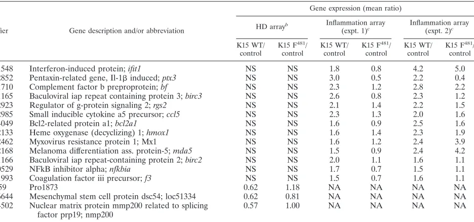
Notably, K15 induced the expression of three genes that are
downstream targets of vascular endothelial growth factor
(VEGF) (Table 1): Down syndrome critical region 1 (
dscr1
;
5.6-fold), cyclooxygenase 2 (
ptgs2
/
cox-2
; 5.8-fold), and tissue
factor (
f3
; 1.6-fold). The K15 F
481mutant slightly activated
dscr1
(1.8-fold) and
cox-2
(2.1-fold) gene expression and did not
activate tissue factor/f3 gene expression (0.9-fold) (Table 1).
In summary, we have shown that K15 has the capacity to
modulate the host cell response toward an inflammatory
phe-notype: K15 induces gene expression of chemokines (C-X-C
chemokines CXCL8/IL-8, CXCL3/Gro
␥
and CCL2; C-C
on November 8, 2019 by guest
http://jvi.asm.org/
mokines CCL5 and CCL20/MIP-3
␣
), cytokines (IL-1
␣
, IL-1

,
and IL-6), antiapoptotic genes (
tnfaip3
/
A20
,
bf
,
birc3
,
birc2
, and
bcl2a1
), genes involved in signaling (NF-
bia and rgs2),
angio-genesis (IL-8, Dscr1, Mmp1, Mmp3, and tissue factor), and
various other classical inflammatory genes such as Cox-2, heme
oxygenase 1, Ptx3/pentraxin-3, and Sod2.
[image:6.585.93.491.68.563.2]K15 induces the secretion of inflammatory cytokines.
In
order to confirm the data obtained with the DNA microarrays,
FIG. 2. Subcellular localization of K15 in 293 KSHV BAC36 cells and in transiently transfected PtK2 and SLK cells. (A) PtK2 cells were
transiently transfected with the eight-exon K15 expression construct. At 48 h posttransfection, cells were fixed and labeled with the K15 polyclonal
antibody and anti-rabbit Cy3 as secondary antibody (A1) as described in Material and Methods. Nuclei were stained with Hoechst 33258 (A2). A3
shows an overlay of images A1 and A2. (B) 293 cells stably transfected with KSHV BAC36 were treated with 1 mM butyrate for 23 h, fixed, and
labeled with the K15 polyclonal antibody (B1) as described in Material and Methods. The arrows indicate cells expressing K15 protein. Nuclei were
stained with Hoechst 33258 (B2). B3 shows an overlay of images B1 and B2. (C) Image showing a single 293 KSHV BAC36 cell expressing
endogenous K15. The lytic cycle was induced in 293 KSHV BAC36 cells by adding 1 mM butyrate and baculovirus coding for KSHV RTA/ORF50
to the cell culture medium. At 38 h postinduction, cells were fixed and labeled with the K15 polyclonal antibody as described in Material and
Methods. (D) SLK endothelial cells were transiently transfected with the eight-exon K15 cDNA expression construct, fixed, labeled with the K15
polyclonal antibody, and analyzed by fluorescence microscopy.
on November 8, 2019 by guest
http://jvi.asm.org/
TABLE 1. Identification of K15-regulated genes
aIdentifier Gene description and/or abbreviation
Gene expression (mean ratio)
HD arrayb Inflammation array
(expt. 1)c
Inflammation array (expt. 2)c
K15 WT/ control
K15 F481 / control
K15 WT/ control
K15 F481 / control
K15 WT/ control
K15 F481 / control
NM_000584
Il-8
6.99
1.42
58.1
2.7
22.6
4.2
AY063515
ATP-binding cassette protein c13 variant b
3.72
2.39
NA
NA
NA
NA
NM_000600
Il-6 (interferon beta 2)
3.32
0.96
3.8
0.9
4.7
2.8
NM_014326
Death-associated protein kinase 2
;
dapk2
3.23
2.43
NA
NA
NA
NA
NM_004414
dscr1
3.22
1.34
5.9
1.7
5.3
1.9
NM_058178
Neuronal pentraxin receptor, isoform 2
;
nptxr
3.16
2.96
NA
NA
NA
NA
NM_018968
Syntrophin gamma 2
;
sntg2
3.07
3.41
NA
NA
NA
NA
NM_002090
dChemokine (c-x-c) ligand 3;
cxcl3
,
gro3
,
gro
␥
2.97
1.03
2.7
0.8
2.0
1.1
NM_004864
Growth differentiation factor 15 (GDF 15)
2.97
2.38
NA
NA
NA
NA
NM_005347
Heat shock 70-kDa protein 5;
hspa5
2.95
1.04
NA
NA
NA
NA
NM_015980
Hmp 19 protein; loc51617
2.76
2.22
NA
NA
NA
NA
NM_013409
Follistatin isoform fst344 precursor;
fst
2.76
1.11
NA
NA
NA
NA
NM_004113
Fibroblast growth factor 12, isoform 2
;
fgf 12
2.70
2.70
NA
NA
NA
NA
NM_000636
Superoxide dismutase 2; sod2
2.64
1.27
3.0
1.1
3.5
2.5
NM_002526
5
⬘
nucleotidase, ecto;
nt5e
2.53
1.53
NA
NA
NA
NA
BC016341
Similar to interferon-stimulated gene; 20 kDa
2.49
1.22
NA
NA
NA
NA
NM_138973
Beta-site app-cleaving enzyme 1; bace
2.35
1.72
NA
NA
NA
NA
AC005058
Homo sapiens
BAC clone CTB-54D4 from 7q31
2.35
0.94
NA
NA
NA
NA
NM_000575
Il-1
␣
2.19
0.98
7.0
0.9
5.4
1.8
NM_006010
Arginine-rich protein; armet
2.18
0.93
NA
NA
NA
NA
NM_001855
Alpha 1 typexycollagen precursor;
col15a1
2.17
1.00
NA
NA
NA
NA
NM_004050
Bcl2-like 2 protein;
bcl2l2
2.13
0.63
NA
NA
NA
NA
NM_018677
Acetyl coenzyme A synthetase; loc55902
2.12
1.02
NA
NA
NA
NA
NM_006290
Tumor necrosis factor
␣
- induced protein;
tnfaip3
2.11
1.05
3.6
1.4
1.9
2.0
NM_002970
Sperimidine/spermine n1-acetyltransferase; sat
2.09
0.85
NA
NA
NA
NA
NM_003711
Phosphatidic acid phosphatase 2a
;
ppap2a
2.07
1.62
NA
NA
NA
NA
XM_029384
Kinesin family member 13a
;
kif13a
2.02
1.62
NA
NA
NA
NA
NM_002982
Monocyte chemotactic protein 1; mcp 2; ccl 2
1.97
1.04
2.7
1.0
2.0
1.0
D14826
Herem 2beta-b protein; herem-2
1.94
1.20
NA
NA
NA
NA
AK000917
cDna clone unnamed protein product
1.94
1.17
NA
NA
NA
NA
D14825
herem-1
1.94
1.29
NA
NA
NA
NA
NM_013372
Cysteine knot superfamily 1;
cktsf1b1
1.93
1.21
NA
NA
NA
NA
NM_000710
Bradykinin receptor b1;
bdkrb1
1.90
0.89
NA
NA
NA
NA
NM_052815
Immediate-early response 3;
ier3
1.86
0.94
NA
NA
NA
NA
NM_002135
Nuclear receptor subfamily 4;
nr4a1
1.84
1.24
NA
NA
NA
NA
BC002894
Unknown (protein for mgc:11256)
1.84
1.02
NA
NA
NA
NA
NM_002257
Kallikrein 1 preproprotein; klk1
1.80
1.91
NA
NA
NA
NA
NM_002727
Proteoglycan 1, secretory granule;
prg1
1.80
0.91
2.4
0.9
2.8
1.5
AB023182
Kiaa0965 protein;
kiaa0965
1.80
1.20
NA
NA
NA
NA
NM_001549
Interferon-induced protein;
ifit4
1.75
1.41
NA
NA
NA
NA
NM_014220
Transmembrane 4 superfamily member 1;
tm4sf1
1.75
0.85
NA
NA
NA
NA
NM_004482
Polypeptide
N
-acetylgalactosaminyltransferase 3;
galnt3
1.72
0.97
NA
NA
NA
NA
NM_003299
Tumor rejection antigen (gp96) 1;
tra1
1.70
1.11
NA
NA
NA
NA
NM_021013
Type I hair keratin;
krth4
1.69
0.93
NA
NA
NA
NA
NM_003020
Secretory granule; neuroendocrine protein 1
(7b2 protein); sgne 1
1.69
1.02
NA
NA
NA
NA
AB002377
Kiaa0379 protein; kiaa0379
1.67
1.05
NA
NA
NA
NA
AK023410
cDna clone weakly similar to sodium-independent
organic anion transporter 2
1.65
1.03
NA
NA
NA
NA
NM_004184
Tryptophanyl-trna synthetase; wars
1.65
1.02
NA
NA
NA
NA
NM_005168
Ras homolog gene family, member e; arhe
1.64
0.93
NA
NA
NA
NA
NM_016306
Dnai (hsp40) homolog, b11;
dnajb11
1.63
1.03
NA
NA
NA
NA
NM_145918
Cathepsin 1 preproprotein;
cts1
1.61
1.03
NA
NA
NA
NA
NM_001172
Arginase, type II precursor; arg2
1.58
1.05
NA
NA
NA
NA
NM_021127
PMA-induced protein; pmaip1
1.58
0.85
NA
NA
NA
NA
XM_059160
Similar to dj1174n9.1; loc 127544
1.58
1.02
NA
NA
NA
NA
XM_170564
Similar to keratin 17; loc 254207
1.56
1.07
NA
NA
NA
NA
XM_011184
Sec24-relatedprotein d;
sec24d
1.56
1.16
NA
NA
NA
NA
NM_004591
Chemokine (c-c motif) ligand 20;
ccl20
NS
NS
7.2
0.9
7.7
1.1
NM_000963
Cyclooxygenase 2;
ptgs2
NS
NS.
5.2
1.6
6.4
2.9
NM_002421
Matrixmetalloproteinase 1;
mmp1
NS
NS
3.2
0.8
4.7
2.2
NM_002422
Matrixmetalloproteinase 3;
mmp3
NS
NS
4.6
1.0
3.1
1.5
NM_000576
Il-1

NS
NS
3.7
1.1
3.4
1.7
Continued on facing page
on November 8, 2019 by guest
http://jvi.asm.org/
we analyzed cytokine secretion of K15-transfected HeLa cells.
We used a cytokine antibody array spotted with 42 different
human cytokine antibodies. The supernatants of HeLa cells
transiently transfected with empty vector pFJ-EA, K15 WT, or
K15 F
481were incubated with the antibody array membrane,
and binding of secreted cytokines was measured as described
in Materials and Methods. This assay showed that K15
expres-sion induced the secretion of IL-6, IL-8, and GRO
␥
/CXCL3
proteins in HeLa cells (Fig. 3A). The control spots (Fig. 3A)
are marked as well as the spots for IL-8, IL-6, and GRO
␥
.
Quantification of the spots in Fig. 3A shows that K15
upregu-lated IL-8 secretion 16-fold, IL-6 expression 4-fold, and GRO
␥
expression 2-fold (Fig. 3B), which confirms the changes seen at
the mRNA level (Table 1). The mutant K15 F
481was
signifi-cantly impaired in the induction of cytokine secretion (IL-6,
1.5-fold; IL-8, 1; GRO
␥
, 0.3-fold) (Fig. 3A and B).
To quantitatively determine the specific amounts of IL-6 and
IL-8 secreted from HeLa cells transiently transfected with K15,
an ELISA was performed. Consistent with the DNA
microar-ray and cytokine antibody armicroar-ray, K15 WT significantly induced
IL-6 and IL-8 production (Fig. 3C) (IL-6, 762 pg/ml; IL-8, 1318
pg/ml), while K15 F
481did not induce IL-6 and IL-8 production
compared to empty vector-transfected HeLa cells (Fig. 3C)
(mock IL-6, 167 pg/ml; K15 F
481IL-6, 160 pg/ml; mock IL-8, 51
pg/ml; K15 F
481IL-8, 51 pg/ml). Equal protein expression
levels for K15 WT and K15 F
481in HeLa cells used for the
cytokine antibody array and ELISA are shown in Fig. 3D.
To test whether K15 can induce gene expression of VEGF,
we performed a real-time PCR with probes for VEGF and, as
a positive control, IL-8. IL-8 was significantly upregulated by
K15 (101-fold) but only marginally by K15 F
481(3.4-fold) as
expected, whereas VEGF levels were only slightly upregulated
(1.9-fold) in K15-expressing HeLa cells (Fig. 3E).
Activation of the
dscr1
and
cox-2
promoters by K15 in 293-T
cells.
The DNA microarrays showed that expression of
dscr1
[image:8.585.72.540.80.299.2]and
cox-2
was significantly upregulated by K15.
dscr1
was only
recently described as a target for VEGF and seems to have a
role in angiogenesis (50, 54, 132). To confirm the DNA
mi-croarray data, we performed luciferase-based reporter assays
in 293-T and HeLa cells with reporter plasmids containing the
promoter region of either
dscr1
or
cox-2
cloned upstream of
the luciferase gene in the promoterless reporter vector pGL3b
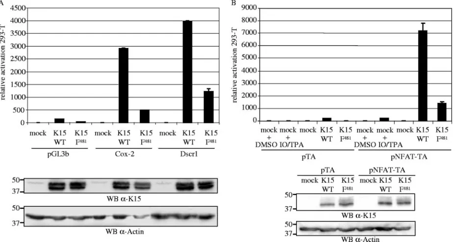
(132). Figure 4A shows the luciferase activity of 293-T cells
cotransfected with K15 WT or K15 F
481with the reporter
plasmids pGL3b, Cox-2, or Dscr1 in relation to cells
cotrans-fected with pFJ-EA (mock) and the respective reporter
plas-mid (luciferase activity of mock-transfected cells was set at 1
for each reporter plasmid). K15 WT was found to upregulate
the
cox-2
and
dscr1
promoters in 293-T cells, whereas the
mutant K15 F
481was significantly impaired in the activation of
the two promoters (Fig. 4A). Similar results were obtained in
HeLa cells (data not shown). Expression of the K15 WT and
K15 F
481proteins was comparable (Fig. 4A).
TABLE 1—
Continued
Identifier Gene description and/or abbreviation
Gene expression (mean ratio)
HD arrayb Inflammation array
(expt. 1)c
Inflammation array (expt. 2)c
K15 WT/ control
K15 F481 / control
K15 WT/ control
K15 F481 / control
K15 WT/ control
K15 F481 / control
NM_001548
Interferon-induced protein;
ifit1
NS
NS
1.8
0.8
4.2
5.0
NM_002852
Pentaxin-related gene, Il-1

induced;
ptx3
NS
NS
3.0
0.5
2.2
0.4
NM_001710
Complement factor b preproprotein;
bf
NS
NS
2.3
1.2
2.8
2.2
NM_001165
Baculoviral iap repeat containing protein 3;
birc3
NS
NS
2.6
0.8
2.3
1.2
NM_002923
Regulator of g-protein signaling 2;
rgs2
NS
NS
2.1
1.4
2.2
1.5
NM_002985
Small inducible cytokine a5 precursor;
ccl5
NS
NS
2.3
1.3
2.0
1.6
NM_004049
Bcl2-related protein a1;
bcl2a1
NS
NS
1.6
0.9
2.5
1.6
NM_002133
Heme oxygenase (decyclizing) 1;
hmox1
NS
NS
1.6
1.4
2.3
1.9
NM_002462
Myxovirus resistance protein 1; Mx1
NS
NS
1.6
1.2
2.4
3.9
NM_022168
Melanoma differentiation ass. protein-5;
mda5
NS
NS
1.5
0.9
2.4
4.2
NM_001166
Baculoviral iap repeat-containing protein 2;
birc2
NS
NS
2.0
1.1
1.6
1.1
NM_020529
NFkB inhibitor alpha;
nfkbia
NS
NS
1.7
0.7
1.5
1.1
NM_001993
Coagulation factor iii precursor;
f3
NS
NS
1.5
0.7
1.6
1.1
AF119859
Pro1873
0.62
1.18
NA
NA
NA
NA
NM_016644
Mesenchymal stem cell protein dsc54; loc51334
0.62
0.81
NA
NA
NA
NA
NM_014502
Nuclear matrix protein mmp200 related to splicing
factor prp19; nmp200
0.57
1.00
NA
NA
NA
NA
aHeLa cells were transiently transfected with the K15 expression constructs K15 WT (exons 1 to 8) or the K15 mutant K15 F481or empty vector pFJ-EA (mock, or control). At 32 h posttransfection, RNA was harvested. Additional information on the methodology of the microarrays is available at http://www.mh-hannover.de /forschung/sfb566/microarray/index.phtml. The complete data set is available on request.
bHigh density (HD) microarray experiments were performed as “two-color hybridizations”. The vector control cRNA population was cohybridized with cRNAs
generated from transient transfections with K15 WT or K15 F481expression vectors. For each of the two resulting comparisons (K15 WT/control and K15 F481/control) an additional “dye-swap hybridization” was carried out. Thus, four high-density microarray hybridizations were performed altogether. After hybridization and data extraction (as described in Material and Methods), the whole data set was filtered for genes that were regulated at least 1.5-fold by the K15 WT construct in each of the two corresponding “dye swap arrays”. Listed are the mean ratios of gene expression for 59 cellular genes represented on the HD microarray. Genes that are upregulated by both K15 WT and K15 F481are shown in bold. NS, not significant.
c For the customized inflammation array, 27 genes were found to be regulated by K15. Shown are the values comparing gene expression of K15 WT or K15 F481 with empty vector-transfected cells (control) from two independent experiments. Only genes that were regulated by at least 1.5-fold are shown. NA, not available.
dThe oligonucleotide probe detecting NM_002090gro3(cxcl3andgro␥) also detectsgro1(cxcl1andgro␣) andgro2(cxcl2andgro).
on November 8, 2019 by guest
http://jvi.asm.org/
K15 activates the NFAT transcription factor in 293-T cells.
Recently, VEGF was reported to induce
dscr1
expression via
activation of the NFAT transcription factor (50). Given the
activation of
dscr1
by K15, we investigated whether K15 was
able to induce NFAT activity. 293-T cells were transiently
cotransfected with K15 expression constructs or the empty
vector pFJ-EA together with the control reporter plasmid
pTA, containing the TA promoter, or the reporter plasmid
pNFAT-TA, which additionally contains three tandem copies
of the NFAT transcription factor consensus sequence
up-stream of the TA promoter. As a positive control for NFAT
activation, empty vector-transfected 293-T cells were
stimu-lated with TPA and calcium ionophore 4 h before cells were
lysed. In order to reduce serum-induced signaling pathways,
cells were serum starved 9 h prior to cell lysis. The luciferase
activity of mock-transfected 293-T cells was set at 1 for each
reporter plasmid. K15 significantly induced the activity of the
NFAT transcription factor in 293-T cells (Fig. 4B), and similar
FIG. 3. Induction of cytokine and chemokine secretion by K15. (A) HeLa cells were transiently transfected with K15 expression constructs (K15
WT and K15 F
481) or the empty vector pFJ-EA (mock). At 32 h posttransfection, the conditioned medium was collected and analyzed with a
cytokine antibody array encompassing 42 different human cytokines spotted in duplicate onto a nitrocellulose membrane. The cytokine antibody
array contains two different controls (c), one spotted in quadruplicate (upper left corner) and one in duplicate (lower right corner) and was
performed as described in Materials and Methods. Arrays are labeled at left. The spots that were upregulated by K15 are marked as indicated on
the figure. (B) Quantification of the cytokine antibody array shown in panel A. The intensity of the spots was measured by densitometry and
normalized to the control spots (c). The signal reached with medium of cells that were mock-transfected was set at 1. Standard deviation is shown.
(C) The ELISA was carried out according to the manufacturer’s instructions with supernatant from transiently transfected HeLa cells as described
in panel A. Shown is the mean value of two independent experiments. (D) After the conditioned medium was collected from transiently transfected
HeLa cells for the cytokine antibody array (A) and ELISA (C), cells were lysed and analyzed by Western blotting for K15 protein expression with
the K15 polyclonal antibody. Actin served as a loading control. (E) Real-time PCR analysis for IL-8 and VEGF was performed with 7 ng of cDNA
from HeLa cells transiently transfected with K15 expression constructs (K15 WT and K15 F
481) or pFJ-EA (mock). The graph shows the relative
changes of mRNA expression after normalization against

-actin (
⌬⌬
C
Tmethod; see Material and Methods).WB, Western blotting.
on November 8, 2019 by guest
http://jvi.asm.org/
results were obtained in HeLa cells (data not shown). The
mutant K15 F
481was impaired in the activation of NFAT in
293-T (Fig. 4B) and HeLa cells (data not shown). K15 WT and
mutant K15 F
481were expressed at comparable levels (Fig. 4B).
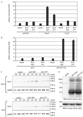
K15 activates the
dscr1
and
cox-2
promoters and the NFAT
transcription factor in SLK endothelial cells.
Since KS is a
tumor of endothelial origin, we investigated the capability of K15
to induce
dscr1
and
cox-2
gene expression and NFAT activity in
the KSHV-negative SLK endothelial cell line that was established
from a KS tumor (49). K15 expression constructs or pFJ-EA were
cotransfected together with the Dscr1, Cox-2 (Fig. 5A), or NFAT
(Fig. 5B) luciferase reporter plasmids. The luciferase activity is
shown as relative activity compared to empty-vector-transfected
cells for each reporter. As observed for epithelial cells, K15 WT
was able to activate the
dscr1
and
cox-2
promoters (Fig. 5A) and
the NFAT transcription factor (Fig. 5B) in endothelial cells. The
K15 F
481mutant was only slightly impaired in the activation of the
dscr1
and
cox-2
promoters (Fig. 5A) and as potent as K15 WT to
induce NFAT activity (Fig. 5B), unlike its effect in epithelial cells
(Fig. 4A and B).
In order to confirm the SLK luciferase reporter assay data,
we performed a semiquantitative RT-PCR analysis for the
dscr1
transcript. RNA was extracted 32 h posttransfection from
transfected HeLa and SLK cells, and cDNA was prepared as
described for the DNA microarray. The
dscr1
primers detect,
as do the probes for the inflammatory gene array, three
dif-ferent splice variants of the
dscr1
gene. As depicted in Fig. 5C,
the
dscr1
transcript appeared after fewer PCR cycles in HeLa
and SLK cells that were transfected with K15 WT compared to
cells transfected with empty vector. The K15 F
481mutant was
impaired in the upregulation of
dscr1
expression in HeLa but
not in SLK cells (Fig. 5C), which confirms the results obtained
with the luciferase reporter assays (Fig. 5A). The PCR with
primers for the housekeeping gene GAPDH shows equal
amounts of cDNA used for the PCRs (Fig. 5C), and equal
protein expression of K15 WT and K15 F
481in the SLK cells is
shown in Fig. 5D.
DISCUSSION
Previous studies with transiently transfected K15 (exons 1 to
8; aa 1 to 489) had shown that it has an apparent molecular
mass of approximately 45 kDa (13, 23, 116). However, it had
not been shown up to now whether this protein is expressed in
the context of a complete viral genome. Here we show the
existence of a 45-kDa K15-derived protein in cells stably
trans-fected with KSHV BAC36 undergoing productive replication.
Based on its molecular size and intracellular distribution, this
K15 protein appears to be similar to the transiently expressed
full-length (exons 1 to 8) K15 protein. The increased
expres-FIG. 4. K15 activates the
cox-2
and
dscr1
promoter and the NFAT transcription factor in luciferase-based reporter assays in 293-T cells. 293-T cells
were transiently transfected with 1
g of K15 WT or K15 F
481and 100 ng of reporter plasmid per well of a six-well plate. The Dscr1 and Cox-2 reporter
plasmids contain the promoter regions of the
dscr1
and
cox-2
genes, respectively, cloned in the promoterless pGL3b vector (A). The pTA-Luc reporter
plasmid contains the minimal TA promoter upstream of the luciferase reporter gene. The pNFAT-TA-Luc reporter plasmid additionally contains three
tandem copies of the NFAT transcription factor consensus sequence (B). At 20 h posttransfection, cells were serum starved by reducing the serum
concentration from 10% to 1% (for
dscr1
and
cox-2
) or 0% (for NFAT). At 29 h posttransfection, cells were lysed, and luciferase activities were
determined. As a positive control for NFAT activation, mock-transfected 293-T cells were incubated with TPA and calcium ionophore (IO; both dissolved
in DMSO) 4 h before cells were lysed, and treatment of mock-transfected cells with DMSO served as negative control (B). The luciferase activities are
depicted as relative activity compared to mock transfections (luciferase activity of mock and pGL3b, Dscr1, and Cox-2 transfections in panel A or
mock-and pTA- mock-and pNFAT-TA-cotransfected cells treated with DMSO in panel B was set at 1). The data are representative of three independent experiments,
each performed in duplicate. Standard deviations are shown. For both panels, lysates were analyzed by SDS-polyacrylamide gel electrophoresis and
Western blotting (WB) with the K15 polyclonal antibody. Membranes were stripped and probed with an
␣
-actin antibody as loading control.
on November 8, 2019 by guest
http://jvi.asm.org/
[image:10.585.62.526.70.317.2]FIG. 5. K15 activates the
cox-2
and
dscr1
promoter and the NFAT transcription factor in SLK endothelial cells. SLK cells were transiently
cotransfected with 3
g of K15 WT, K15 F
481, or pFJ-EA (empty vector, mock) and 1
g of reporter plasmids pGL3b (negative control), Dscr1,
or Cox-2 (A) or 1
g of reporter plasmids pTA (negative control) or pNFAT-TA (B). At 21 h posttransfection, cells were serum starved by reducing
the serum concentration from 10% to 0%. At 29 h posttransfection, cells were lysed, and luciferase activities were determined. The luciferase
activities are depicted as relative activity compared to mock transfections. The data are from one experiment representative of three independent
experiments, each performed in duplicate. Standard deviations are shown. Equal expression levels of K15 proteins were analyzed by Western
blotting (data not shown). (C) Dscr1 and GAPDH semiquantitative RT-PCR. cDNA of SLK or HeLa cells transiently transfected with empty
vector (mock) or K15 expression constructs K15 WT or K15 F
481was generated as described in Material and Methods. A semiquantitative PCR
with 22, 23, or 24 PCR cycles was performed with dscr1-for and dscr1-rev primers amplifying a spliced transcript of 196 bp. The GAPDH PCR
shows that equal amounts of cDNA were utilized for the PCR. (D) To control equal protein expression of K15 WT and K15 F
481in transiently
transfected SLK cells, cells were lysed for Western blotting (WB) analysis in parallel to the cells being lysed for RNA preparation and subsequent
RT-PCR analysis. K15 proteins were detected with the K15 polyclonal antibody. Blots were stripped and probed with an
␣
-actin antibody as loading
control. The transfection efficiency of SLK cells with K15 expression constructs was about 40% as judged by immunofluorescence (data not shown).
on November 8, 2019 by guest
http://jvi.asm.org/
sion of the 45-kDa K15 protein in the 293 KSHV BAC36 cell
line during the lytic replication cycle correlates well with
pub-lished RT-PCR analyses (23, 44, 100) and KSHV gene arrays
(57, 98) that describe upregulation of K15 mRNA expression
upon lytic cycle induction in PEL cell lines. However, low
expression levels of K15 transcripts (23, 44, 100) and K15
protein (this study) have been detected in uninduced
KSHV-infected cells. Since a small percentage of latently KSHV-infected cells
are known to undergo spontaneous lytic replication (46, 134,
135), we favor the interpretation that K15 expression observed
in uninduced KSHV-infected cell cultures may be attributable
to the small percentage of cells undergoing spontaneous lytic
replication.
In PEL cell lines, a 23-kDa protein recognized by a
mono-clonal antibody to the cytoplasmic domain of K15 has
previ-ously been reported (116). The polyclonal antibody to the
cytoplasmic domain of K15 used in our studies (13, 44)
de-tected a similar protein (data not shown). However, we found
this protein not to be associated with cellular membranes (data
not shown), although a K15-derived protein of similar size and
containing the cytoplasmic domain (K15 exons 6 to 8; aa 296 to
489) was found in the membrane fraction (data not shown). An
artificially initiated protein containing only the (hydrophilic)
cytoplasmic domain (aa 355 to 489) had a lower molecular
mass (15 kDa; data not shown), suggesting that the 23-kDa
protein in PEL cells is unlikely to represent a proteolytic
frag-ment of a precursor K15 protein that only contains the
cyto-plasmic domain. We cannot exclude that the 23-kDa protein in
PEL cells is derived from K15, but its nature and derivation are
not yet resolved.
Since the 293 BAC36 cell line is so far the only cell system
in which expression of the endogenous 45-kDa K15 protein
could be observed in the context of the entire viral genome, we
decided to study its function in epithelial cells. Epithelial cells
play a significant role in the establishment of KSHV infection
and viral transmission in vivo and in vitro. KSHV DNA has
been detected in scattered keratinocytes of the epidermis
over-lying cutaneous lesions and in eccrine ductular epithelial cells
and in spindle and endothelial cells lining the well-formed
blood vessels within and surrounding KS lesions (102).
Fur-ther, KSHV DNA was detected in epithelial cells from several
organs (111). Moreover, detection of infectious viral particles
in the saliva of KSHV-infected patients suggests that the
epi-thelial cells in the oral mucosa or in salivary glands are a source
for the virus (58, 97, 127). KSHV shows a broad host range in
culture in that it can efficiently establish latency after de novo
infection in many human cells such as HeLa (epithelial origin),
human foreskin fibroblasts, and SLK cells (7). Other studies
have demonstrated that the human embryonal kidney
epithe-lial cell line 293 (7, 39, 106) as well as cultured primary oral
epithelial cells (33) can support KSHV infection in vitro.
Fur-thermore, KSHV can infect and replicate in primary human
keratinocytes in vitro (18).
We have now analyzed the downstream targets of K15
sig-naling using DNA gene array analyses. Our study establishes
K15 as one of the KSHV genes that is capable of inducing the
expression of multiple cytokines, including in particular IL-8
and IL-6 (Table 1 and Fig. 3), which have been shown to play
a role in KSHV-associated pathogenesis. The IL-8 promoter
contains an NF-
B element that is required for activation in all
cell types studied, as well as a single consensus AP-1 site (22,
51). Numerous lines of evidence exist suggesting that
coordi-nated activation of the MAP kinases ERK, JNK, and p38 is
important for IL-8 regulation in response to proinflammatory
cytokines such as IL-1 (63), viruses (15), bacteria (8, 45, 80),
and various stress inducers (51). The 5
⬘
flanking region of the
IL-6 gene contains a number of
cis
-acting elements, among
them an AP-1 (126) and NF-
B (69) consensus site. The
exis-tence of these
cis
elements in the IL-8 and IL-6 gene promoter
suggests that K15-mediated activation of cellular transcription
factor activity results in the upregulation of cytokine
expres-sion. This notion is further supported by the crucial role of the
Y
481residue of the K15 SH2-B motif Y
481EEV, both in
acti-vation of MAP kinase pathways and transcription factors
NF-
B and AP-1 (13), as well as in the induction of cytokine
expression (Table 1 and Fig. 3). However, K15-induced
up-regulation of IL-8 and IL-6 may also be mediated indirectly.
Expression of IL-1
␣
and IL-1

was induced by wild-type K15
but not by the K15 mutant K15 F
481(Table 1), and IL-1 is
known to induce IL-6 and IL-8 production via the JNK MAP
kinase pathway (63). Induction of the chemokine
CCL20/MIP-3
␣
, which was significantly upregulated by K15 (7.4-fold) but
not by K15 F
481, has been attributed to tumor necrosis factor
alpha and IL-1

, probably via MAP kinases Erk1/2 and p38, in
epithelial cells (105). Whether K15 signaling directly
upregu-lates IL-6, IL-8, and/or CCL20/MIP-3
␣
expression cannot be
answered at this stage.
Notably, several other KSHV-encoded genes upregulate
ex-pression of IL-8 and IL-6, suggesting that dysregulated
expres-sion of these cytokines is important for KSHV-associated
pathogenesis. The lytic vGPCR constitutively activates NF-
B