0022-538X/07/$08.00⫹0 doi:10.1128/JVI.01422-06
Copyright © 2007, American Society for Microbiology. All Rights Reserved.
MINIREVIEW
Human Cytomegalovirus Tropism for Endothelial Cells: Not All
Endothelial Cells Are Created Equal
䌤
Michael A. Jarvis
1and Jay A. Nelson
1,2*
Vaccine and Gene Therapy Institute1and Department of Molecular Microbiology and Immunology,2
Oregon Health Science University, Portland, Oregon
Human cytomegalovirus (HCMV) is a ubiquitous herpesvi-rus that persists for the life of the host following initial infec-tion. Genome analysis indicates that mammalian CMVs have cospeciated with their respective host over the last 80 million years (39). This prolonged period of coevolution has resulted in a high level of coadaptation between virus and host. The study of CMV, a virus that is exquisitely adapted for persis-tence within the host, is beginning to reveal strategies critical for virus survival. Although all herpesviruses persist for the life span of the host, recent findings suggest that HCMV has a unique replication strategy for maintenance within the host, wherein the virus establishes sites of persistent active replica-tion even in the presence of high levels of preexisting HCMV-specific immunity. A number of cell types, including myeloid lineage cells, smooth muscle cells, and endothelial cells (ECs), appear to be critical as sites of HCMV persistent replication and latency. HCMV infections of myeloid lineage and of smooth muscle cells have been the focuses of previous reviews (see references 32 and 66). This review will focus on HCMV infection of ECs and the role of this cell type in virus persis-tence and latency. We will describe a “genomic island” of three genes that are essential for HCMV EC tropism and discuss mechanisms by which the products of these genes mediate HCMV infection in ECs.
CMV IS ADAPTED FOR PERSISTENT REPLICATION IN THE IMMUNOCOMPETENT HOST
All herpesviruses persist for the life span of the host (53). However, HCMV appears to have a unique replication strat-egy for maintenance within the host, wherein the virus estab-lishes sites of persistent active replication (or frequent virus reactivation) even in the presence of high levels of preexisting HCMV-specific immunity. Consistent with this ability of the virus to replicate irrespective of host immunity, HCMV has been shown to frequently reinfect healthy HCMV-seropositive individuals, with active replication in these individuals for months to years (1, 7). Further evidence for persistent virus replication is the observation that a surprisingly large compo-nent of an individual’s T-cell repertoire is directed against
HCMV-encoded epitopes (57, 65); in some cases, in excess of 40% of an individual’s CD4⫹ T-cell response is directed against HCMV (57). Epitopes recognized by these T cells are present in HCMV proteins expressed at all stages of the viral replication cycle (65), consistent with continual exposure of the host immune response to HCMV antigens from persistently replicating virus. Consequently, HCMV appears to be exqui-sitely adapted to maintain an active, persistent replication for the life span of the immunocompetent host.
ECs ARE A SITE OF CMV LATENCY AND PERSISTENCE
Acute HCMV disease is primarily limited to the immuno-compromised host (44). The tissue distribution of virus during acute disease can be viewed as one resulting from uncontrolled replication of a virus with an extremely wide cellular tropism. During acute disease, a diverse population of cell types are infected, including ECs, various leukocyte populations, epithe-lial cells, hepatocytes, smooth muscle cells, and fibroblasts (5, 13, 17, 29, 42, 49, 59, 69). The extent of organ involvement in many of these cases can be remarkable. For example, in one case, that of a congenitally infected neonate with CMV inclu-sion disease, HCMV was highly disseminated, with infection observed in all organs examined (lung, pancreas, kidney, spleen, adrenal, small bowel, placenta, liver, brain, bone mar-row, and heart) (5).
Analysis of tissues from healthy individuals in the absence of acute disease identifies a number of cell types that may serve as sites of HCMV persistent or latent infection in the normal individual. Early studies identified HCMV DNA, mRNA, and antigen within the vessel walls of major arteries throughout the body (19, 23–26, 40, 70). Although the infected cell types were not conclusively determined, the identification of HCMV in the walls of these vessels was taken as evidence of persistent or latent infection of smooth muscle cells and ECs. To function as a site of HCMV persistence, virus infection would be expected to be accompanied by minimal cytopathology. Consistent with this requirement, HCMV infection in these healthy individuals was frequently observed in healthy, histologically normal ar-teries. More recently, ECs were definitively shown to be one of the cell types infected by HCMV within the arterial wall on the basis of viral DNA and antigen positivity in cells staining for an EC marker (43). However, only a subset of HCMV DNA-positive ECs were observed to contain detectable levels of virus
* Corresponding author. Mailing address: Vaccine and Gene Ther-apy Institute, Oregon Health Science University, 3181 SW Sam Jack-son Park Road, Portland, OR 97239. Phone: (503) 418-2700. Fax: (503) 418-2701. E-mail: nelsonj@ohsu.edu.
䌤Published ahead of print on 6 September 2006.
2095
on November 8, 2019 by guest
http://jvi.asm.org/
antigen, identifying ECs as a potential site of latency as well as persistent viral replication in healthy individuals. Interestingly, in a recent study (51), Reeves et al. were unable to detect HCMV DNA in saphenous vein tissue samples obtained from healthy individuals. Given the consistent identification of CMV in arterial vessels in multiple studies, this finding sug-gests that ECs from different anatomic locations may differ in their susceptibility to CMV infection, a result which is not unexpected given the high level of diversity of this cell type (see below). In the closely related murine CMV (MCMV) model, ECs are also identified as a site of viral latency in multiple organs (35). Only ECs from small vessels and capillaries were shown to harbor the MCMV genome, suggesting a similar influence of EC anatomic locations on infection and the estab-lishment of latency by MCMV (35).
ECs ARE A HIGHLY DIVERSE POPULATION OF DISTINCT CELL TYPES
ECs form the inner lining of blood and lymphatic vessels throughout the body and have a number of phenotypic simi-larities that reflect their involvement in common processes, such as the regulation of coagulation, tissue homeostasis, and inflammation. However, additional requirements of ECs to perform highly specialized, tissue-specific functions result in a wide diversification of EC phenotypes according to anatomic location and tissue source. The high level of EC diversity was initially suggested by morphological differences between indi-vidual EC types (12). Analyses of antigen expression by ECs from different vascular sources, initially using antibodies (3) and more recently using in vivo screening of phage display libraries (48, 55), extend these studies to ECs throughout the systemic vasculature. These studies indicate that ECs are ex-tremely heterogeneous and express unique cell surface anti-gens that together comprise an EC “vascular address” system (55). DNA array analyses of EC gene expression profiles fur-ther emphasize the level of EC diversity, with ECs from dif-ferent sources even within the same organ differing signifi-cantly in their gene expression profiles (11, 22, 27, 30, 34). In one extensive analysis of 52 different EC types from 14 different anatomical sites, characteristic clusters of genes were expressed by ECs of different tissues, as well as by arterial compared to venous ECs and by macro- compared to micro-vascular ECs. Over 200 macro- and⬎1,000 microvasculature EC-specific genes were identified, and similar high levels of diversity in arterial, venous, and tissue-specific EC gene ex-pression were observed. ECs were also shown to differ in their expression levels of immune response molecules as well as of receptors for specific pathogens, such as CD36 (Plasmodium falciparum) and CD66a (Neisseria sp.), suggesting that ECs from different tissue locales may differ in their susceptibilities and responses to infection by various pathogens (11).
HCMV REPLICATION IS INFLUENCED BY EC ORIGIN
The effect of EC diversity on CMV replication and patho-genesis remains largely unaddressed, with most studies using EC types such as macrovascular human umbilical vein ECs (HUVECs) that are not normally infected in vivo. This restric-tion of studies to EC types of limited biological relevance has
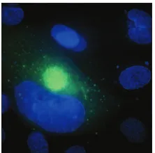
[image:2.585.366.477.68.178.2]potentially serious implications for our understanding of HCMV biology in ECs. For example, DNA array analyses show that micro- and macrovascular ECs differ considerably in expression levels of various molecules involved in CMV entry, such as integrins and epidermal growth factor receptors (11, 34). Indeed, levels of HCMV infection in HUVECs compared to intestinal microvascular ECs have been shown to be to significantly decreased (58), and virus production in HUVECs compared to other macrovasculature (aortic) and microvascu-lature (uterine) is similarly reduced (by 1 to 2 logs) (37). ECs also differ in other characteristics associated with HCMV in-fection. For example, a comparison of HCMV infection in brain microvascular (BMVECs) and aortic macrovascular ECs (AECs) shows that, although both EC types express viral pro-teins and support HCMV replication, virus fails to accumulate intracellularly in AECs, resulting in reduced levels of cell-associated virus compared to supernatant virus. This difference in the distribution of virus corresponds to a lytic infection in BMVECs, but not AECs, and suggests that efficient removal of mature intracellular virions (by either export or degradation) may prolong cell survival. Figure 1 shows HCMV-infected AECs stained for two major viral proteins, glycoprotein B and IE2. Interestingly, HCMV has also been shown to establish a persistent noncytopathic infection in a number of other EC types (52, 64). The ability of HCMV to produce a persistent long-term productive infection with minimal cytopathology may be a prerequisite for a site of persistent infection and suggests that distinct types of ECs may be more important than others as sites of persistent infection. A further level of com-plexity is revealed by the observation that distinct strains of HCMV differ in their cytopathic effects in ECs (15, 33, 37, 64). This observation indicates that genetic determinants of the virus also influence characteristics of replication in ECs and that lack of cytopathology may not be a strict requirement for a site of persistence. Alternatively, viral functions in addition to those required for replication in ECs may be necessary to modulate aspects of the virus infection process to facilitate long-term persistent, instead of acute cytolytic, replication in this cell type. For example, the deletion of US16 from HCMV increases replication in microvascular ECs, identifying US16 as a negative modulator of infection that may be required to maintain viral replication below cytopathic levels in this cell
FIG. 1. Immunofluorescence micrograph showing HCMV-infected AECs. AECs were infected with HCMV. At day 4 postinfection, cells were stained with antibodies directed against the major viral transcrip-tional activator expressed at immediate early times of infection, IE-2 (Cy5 [blue]), and the major virion glycoprotein expressed at early/late times of infection, gB (fluorescein isothiocyanate [green]).
on November 8, 2019 by guest
http://jvi.asm.org/
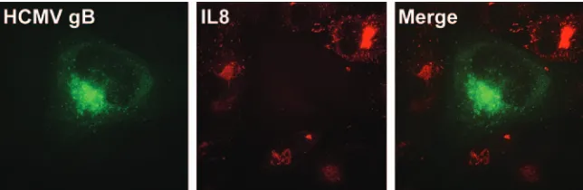
type (14). Similarly, HCMV was recently shown to induce a global inhibition of proinflammatory signaling in multiple cell types (Fig. 2), which may be a critical immune evasion mech-anism for cells persistently infected in vivo, such as ECs (31). The expansion of future studies to types of ECs relevant to a specific aspect of HCMV biology and disease using multiple strains of genetically stable virus is clearly needed before we are able to fully appreciate the characteristics of CMV repli-cation in this diverse cell type.
GENETIC DETERMINANTS OF HCMV EC TROPISM
Early studies observed that HCMV strains differed in their ability to infect ECs, suggesting that genetic determinants of the virus were required for replication in ECs (33, 36, 61). However, identification of viral genes involved in EC tropism was hindered by the lack of genetically stable viruses and a robust genetic system for the construction of viral mutants. In these early studies, comparisons of growth characteristics of EC and non-EC tropic strains suggested that viruses were comparable in their ability to enter ECs but that non-EC strains were impaired in their ability to translocate the viral genome to the nucleus (6, 60, 63). However, an interpretation of results from these studies was complicated by observed differences in the capacities of even identical strains of HCMV to replicate in ECs (6, 33), which presumably resulted from differences in virus preparation, methods of EC culture, and the derivation of specific HCMV strains.
GENETIC DETERMINANTS OF CMV TROPISM GENES IN THE BAC ERA
Many of the technical problems described above have been overcome by the recent cloning of multiple CMVs as geneti-cally stable bacterial artificial chromosomes (BACs) and the development of a suitable genetic system for mutagenesis of these BACs. The first BAC-based approach to identify a CMV-encoded determinant of EC tropism was performed using the closely related virus MCMV (8). In this study, the virally en-coded antiapoptosis gene M45 was shown to be necessary for MCMV growth in murine ECs in vitro. Since ECs represent a site of persistent virus infection, the ability to prevent the normal apoptotic death response of these cells to viral infec-tion may be crucial to maintain long-term viability of the
in-fected cells. However, the role of HCMV-encoded inhibitors of apoptosis in EC tropism and persistence is not clear. The HCMV M45 homologue (UL45) does not inhibit apoptosis and was not required for growth of a BAC-cloned recent clin-ical isolate (designated fusion-inducing factor X [FIX]) in HUVECs, indicating that the HCMV homologue does not function in a similar fashion (20). Alternatively, given the di-vergence of EC types, the function of UL45 during infection of HUVECs may not accurately reflect the role of this gene during HCMV replication in EC types normally infected in vivo. Additional HCMV proteins (IE1, IE2, pUL36/vICA, and pUL37x1/vMIA) have been shown to inhibit apoptosis follow-ing overexpression of the recombinant protein (18, 62, 71). Analysis of IE1 and IE2 is complicated by the function of these proteins as critical transcriptional regulators of the virus (38, 41). The antiapoptotic function of vMIA was recently shown to be essential for HCMV replication in fibroblasts (50), whereas vICA appears not to be required for normal virus replication (46). However, since the recent vMIA studies were performed with a vICA-defective virus background, the possibility of re-dundancy in vMIA and vICA antiapoptotic function remains. In rhesus CMV (RhCMV), the Rh10 open reading frame (ORF), which encodes a viral cyclooxygenase 2 homologue (vCOX-2), was recently shown to be required for replication in rhesus microvascular brain ECs. In this study, deletion of vCOX-2 using BAC-based mutagenesis resulted in a 4-log re-duction in the prore-duction of progeny virus in ECs without affecting replication in fibroblasts (54). Although HCMV does not encode a vCOX-2, COX-2 activity is induced by HCMV infection and is required for normal HCMV replication in fibroblasts (72). A large-scale targeted deletion mapping study has also identified UL24 as required for HCMV replication in microvascular ECs (14). The mechanisms by which vCOX-2 of RhCMV and UL24 of HCMV function as EC tropism deter-minants remain unclear.
HCMV UL128, UL130, AND UL131A: “GENOMIC ISLAND” OF CELLULAR TROPISM
[image:3.585.131.453.69.174.2]The most extensive studies of HCMV EC tropism have focused on a “genomic tropism island” comprised of three ORFs: UL128, UL130, and UL131A. An initial indication that an area containing these genes was important for EC tropism
FIG. 2. HCMV inhibits IL-1 and tumor necrosis factor alpha (TNF-␣) proinflammatory pathways. ECs (HUVECs) were infected with HCMV. At day 3 postinfection, cells were treated with proinflammatory cytokines (in this case, IL-1). The effect of HCMV infection on cellular proinflammatory signaling pathways was then determined by an immunofluorescence analysis of the IL-1/TNF-␣-induced chemokine IL-8. Antibodies used were directed against gB (fluorescein isothiocyanate [green]) and cellular IL-8 (Texas Red [red]). The absence of IL-8 in HCMV (gB-positive) cells shows the profound inhibitory effect of HCMV on proinflammatory pathways.
on November 8, 2019 by guest
http://jvi.asm.org/
was suggested by the observation that the ULb⬘ genomic re-gion (UL128 to UL151 [UL128-UL151]) was in large part absent from non-EC-tropic laboratory strains (9, 47). Subse-quently, Hahn et al. (21), using mutagenesis of the BAC-cloned EC-tropic clinical isolate FIX, identified UL128, UL130, and UL131A that together were required for replica-tion in HUVECs. In that study, delereplica-tional mutagenesis iden-tified UL128, UL130, and UL131A as each individually being required for replication in ECs (HUVECs). The inability of laboratory strains Toledo, Towne, and AD169 to replicate in ECs was also consistent with these viruses encoding inactivated forms of UL128, UL130, and UL131A, respectively. Impor-tantly, the capacity of heterologous expression of the products of each of these ORFs to recover EC tropism of viruses ex-pressing inactivated versions of the respective ORF indicated that the product of each ORF was individually required for EC tropism. Additional studies have shown UL131A to be re-quired for replication in lung microvascular ECs and a variety of epithelial cell types (67) as well as monocyte-derived den-dritic cells (granulocyte-macrophage colony-stimulating factor and interleukin 4 [IL-4] derived) (16). However, the require-ment of these ORFs for replication in other biologically rele-vant types of ECs has not been determined.
In a study by Wang and Shenk (67), repair of the non-EC-tropic AD169 with a functional UL131A recovered EC tropism but resulted in a syncytium-inducing virus with impaired rep-lication in fibroblasts (67). This observation suggests that UL128-UL131A, while required for replication in a variety of cell types, may be detrimental for replication in fibroblasts. This hypothesis is supported by the rapid selection of viruses with inactivating mutations in UL128-UL131A following pas-sage of clinical isolates in fibroblasts (2). However, the situa-tion is clearly more complex, as the EC-tropic FIX and a FIX mutant with a deletion of UL131A grow to similar levels in fibroblasts, a result which suggests the presence of additional genetic determinants that affect HCMV replication in fibro-blasts (21). The genetic stability of the UL128-UL131A region during passage in fibroblasts is also not clear. Viruses with inactivating mutations in UL128-UL131A are rapidly selected following passage of patient isolates in fibroblasts (2). How-ever, a previous study showed that a plaque-purified EC tropic virus clone, TB40/E, maintained EC tropism irrespective of multiple (⬎40) serial passage in fibroblasts (60). Since patient isolates presumably represent a genetically heterogeneous population of virus variants, these findings would indicate that enrichment for viruses with inactivated UL128-UL131A arises from the selection of preexisting viruses in contrast to de novo mutation followed by selection. In addition to increasing our understanding of the role of UL128-UL131A for HCMV rep-lication in fibroblasts, an appreciation of the stability of this region in cloned viruses is technically critical given the use of fibroblasts for reconstitution of BAC-cloned viruses.
The original annotation of the UL128-UL131 region pre-dicted four unspliced ORFs designated UL128, UL129, UL130, and UL131 (10). However, reannotation based on alignment with the closely related chimpanzee CMV identified the three ORFs now known to be present within this region: UL128, UL130, and UL131A (2). The reannotated UL128 shares protein identity with the carboxyl region of the earlier UL128 but is now comprised of three exons; UL131A occupies the same region as the earlier
UL131 but is in a different reading frame and is comprised of two exons, and UL130 remains unchanged (2). The UL128-UL131A region encodes two major mRNA transcripts (2 kb and 0.8 kb) that are transcribed with late kinetics and coterminate down-stream of the UL128 consensus polyadenylation signal sequence (2, 21). The smaller transcript is UL128 specific, with a start site located within the UL130 ORF (21, 67). The start site of the larger transcript is located upstream of UL131 and contains all three ORFs, but the gene(s) encoded by this transcript are still unclear (67). Consistent with their essential function in cellular tropism, these ORFs are observed to be highly conserved in vivo. In one study of 34 clinical isolates derived from distinct patient populations, identity conservation levels of greater than 90% were observed for all three ORFs, compared to 73% for a hyper-variable region of gB (4).
UL128-, UL130-, AND UL131A-ENCODED PROTEINS ARE REQUIRED FOR VIRUS ENTRY INTO ECs
Amino acid sequence analysis predicts that all three proteins encoded by these ORFs (pUL128, pUL130, and pUL131) are lumenal proteins of the secretory system with a consensus N-terminal signal sequence (2). A CC-() chemokine motif (2) and monocyte chemoattractant protein fold were also pre-dicted for the N-terminal regions of pUL128 and pUL130, respectively (21). pUL130 and pUL128 were recently shown to be components of the virion envelope and involved in cell entry (45, 56, 68). Although pUL130 was shown to be targeted to the lumen of the secretory pathway with cleavage of the signal peptide, pUL130 was only found associated with the virion and was not secreted from the cell, indicating a considerable level of interaction with components of the virion envelope. Consis-tent with a function of pUL130 at the level of viral entry, a UL130-deficient non-EC-tropic (Towne) strain produced in noncomplementing cells was unable to efficiently enter ECs (HUVECs), even when the EC target cells expressed UL130. However, the production of Towne in a UL130-complement-ing cell line resulted in a virus that was able to efficiently enter and complete a single cycle of replication in ECs (45).
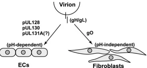
pUL130, as well as pUL128, has subsequently been shown to form a complex with two envelope glycoproteins, gH and gL, which are known to be involved in virus entry and fusion processes (68) (Fig. 3). The gH and gL glycoproteins had previously been known to complex with a third glycoprotein, gO, and to be required for replication in fibroblasts (28). In the present study, two distinct complexes were identified in EC-tropic virions comprised of gH/gL complexed with either pUL128/pUL130 or gO (68). Antibodies directed against ei-ther pUL130 or pUL128 inhibited infection of ECs (HUVECs) and epithelial cells but did not block infection of fibroblasts. Although the severe attenuation of gO-deficient virus growth has prevented a direct assessment of the requirement for gO in EC infection (28), these results support a model wherein pUL128/pUL130 and the gO-containing gH/gL complex are required for infection of ECs/epithelial cells and fibroblasts, respectively. The role of ORF UL131A in HCMV infection of ECs/epithelial cells is unclear and represents a lack of knowl-edge at this stage of research. pUL131 was not detected in the gH/gL complex with pUL128 and pUL130. However, a
on November 8, 2019 by guest
http://jvi.asm.org/
tional UL131 ORF was required for the incorporation of pUL128 and pUL130 into the gH/gL virion-associated com-plex. In an earlier study, UL131A was shown to be required for an early stage of the virus replication cycle in ECs (67). These observations suggest that UL131 is also probably involved in mediating virus entry into ECs but is required at submolar levels, functioning perhaps indirectly, or is more weakly asso-ciated with the complex.
The requirement of UL128-UL131A for HCMV entry into both ECs and epithelial cells suggests that the HCMV entry processes for these two cell types are closely related to one another. A recent finding suggests that the HCMV infection pathways for these two cell types may diverge at a step shortly after entry (56). In that study, an EC tropic BAC-cloned virus (designated TR) was shown to enter ECs and epithelial cells by endocytosis followed by low pH-de-pendent fusion. In contrast, entry into fibroblasts occurred at the cell surface and was pH independent. Consistent with the role of UL128-UL131A in viral entry, a TR virus with a deletion of the ULb⬘region (UL128 to UL150) or of AD169 (UL131A deficient) was unable to enter ECs and epithelial cells, and treatment with the fusogenic agent polyethylene glycol (PEG) overcame this block in both cell types. How-ever, viral gene expression following PEG treatment, corre-sponding to nuclear translocation of the viral genome, was observed only with epithelial cells. These results indicate that genes, presumably UL128-UL131A, are involved in the fusion process in both ECs and epithelial cells as well as in a postentry step unique to ECs. Combined with the identi-fication of unique gH/gL complexes required for entry into EC/epithelial cells compared to fibroblasts (67), these results also suggest that the association of gH/gL with pUL130/UL128 or gO alters the fusion mechanism from one that is dependent on pH to one that is independent of pH, respectively.
CONCLUDING REMARKS
HCMV utilizes a unique strategy of persistent active in-fection to maintain itself within the host, and ECs appear to play a critical role in this process. The application of BAC-based mutagenesis technology to questions of EC tropism enables the identification of determinants required for rep-lication in ECs. However, ECs are a remarkably diverse cell type, and the use of biologically relevant types of ECs is essential to ensure relevance to HCMV biology in the host. Virus persistence is also, presumably, a sum of more than merely the capacity to enter and replicate in a cell. Future studies focused on mechanisms by which HCMV modulates cellular functions to establish a long-term infection in ECs are expected to add considerably to our understanding of virus persistence.
REFERENCES
1.Adler, S. P.1988. Molecular epidemiology of cytomegalovirus: viral trans-mission among children attending a day care center, their parents, and
caretakers. J. Pediatr.112:366–372.
2.Akter, P., C. Cunningham, B. P. McSharry, A. Dolan, C. Addison, D. J. Dargan, A. F. Hassan-Walker, V. C. Emery, P. D. Griffiths, G. W. Wilkinson, and A. J. Davison.2003. Two novel spliced genes in human cytomegalovirus.
J. Gen. Virol.84:1117–1122.
3.Auerbach, R., L. Alby, L. W. Morrissey, M. Tu, and J. Joseph.1985. Expres-sion of organ-specific antigens on capillary endothelial cells. Microvasc. Res.
29:401–411.
4.Baldanti, F., S. Paolucci, G. Campanini, A. Sarasini, E. Percivalle, M. G. Revello, and G. Gerna.2006. Human cytomegalovirus UL131A, UL130 and
UL128 genes are highly conserved among field isolates. Arch. Virol.151:
1225–1233.
5.Bissinger, A. L., C. Sinzger, E. Kaiserling, and G. Jahn. 2002. Human cytomegalovirus as a direct pathogen: correlation of multiorgan involvement and cell distribution with clinical and pathological findings in a case of
congenital inclusion disease. J. Med. Virol.67:200–206.
6.Bolovan-Fritts, C., and J. A. Wiedeman. 2001. Human cytomegalovirus strain Toledo lacks a virus-encoded tropism factor required for infection of
aortic endothelial cells. J. Infect. Dis.184:1252–1261.
7.Boppana, S. B., L. B. Rivera, K. B. Fowler, M. Mach, and W. J. Britt.2001. Intrauterine transmission of cytomegalovirus to infants of women with
pre-conceptional immunity. N. Engl. J. Med.344:1366–1371.
8.Brune, W., C. Menard, J. Heesemann, and U. H. Koszinowski.2001. A ribonucleotide reductase homolog of cytomegalovirus and endothelial cell
tropism. Science291:303–305.
9.Cha, T. A., E. Tom, G. W. Kemble, G. M. Duke, E. S. Mocarski, and R. R. Spaete.1996. Human cytomegalovirus clinical isolates carry at least 19 genes
not found in laboratory strains. J. Virol.70:78–83.
10.Chee, M. S., A. T. Bankier, S. beck, R. Bohni, C. M. Browne, R. Cerny, T. Horsnell, C. A. Hutchison III, T. Kouzarides, J. A. Martignetti, E. Preddie, S. C. Satchwell, P. Tomlinson, K. M. Weston, and B. G. Barrell.1990. Analysis of the protein-coding content of the sequence of human
cytomeg-alovirus strain AD169, p. 125–171.InJ. K. McDougall (ed.),
Cytomegalovi-ruses. Springer-Verlag, Berlin, Germany.
11.Chi, J. T., H. Y. Chang, G. Haraldsen, F. L. Jahnsen, O. G. Troyanskaya, D. S. Chang, Z. Wang, S. G. Rockson, M. van de Rijn, D. Botstein, and P. O. Brown.2003. Endothelial cell diversity revealed by global expression
profil-ing. Proc. Natl. Acad. Sci. USA100:10623–10628.
12.Conway, E. M., and P. Carmeliet.2004. The diversity of endothelial cells: a
challenge for therapeutic angiogenesis. Genome Biol.5:207.
13.Dankner, W. M., J. A. McCutchan, D. D. Richman, K. Hirata, and S. A. Spector.1990. Localization of human cytomegalovirus in peripheral blood
leukocytes by in situ hybridization. J. Infect. Dis.161:31–36.
14.Dunn, W., C. Chou, H. Li, R. Hai, D. Patterson, V. Stolc, H. Zhu, and F. Liu.
2003. Functional profiling of a human cytomegalovirus genome. Proc. Natl.
Acad. Sci. USA100:14223–14228.
15.Fish, K. N., C. Soderberg-Naucler, L. K. Mills, S. Stenglein, and J. A. Nelson.1998. Human cytomegalovirus persistently infects aortic endothelial
cells. J. Virol.72:5661–5668.
16.Gerna, G., E. Percivalle, D. Lilleri, L. Lozza, C. Fornara, G. Hahn, F. Baldanti, and M. G. Revello.2005. Dendritic-cell infection by human cyto-megalovirus is restricted to strains carrying functional UL131-128 genes and
mediates efficient viral antigen presentation to CD8⫹T cells. J. Gen. Virol.
86:275–284.
17.Gnann, J. W., Jr., J. Ahlmen, C. Svalander, L. Olding, M. B. Oldstone, and J. A. Nelson.1988. Inflammatory cells in transplanted kidneys are infected by
[image:5.585.44.284.67.179.2]human cytomegalovirus. Am. J. Pathol.132:239–248.
FIG. 3. Schematic showing roles of pUL128, pUL130, and pUL131A in EC tropism. The gH/gL glycoproteins form a disulfide-linked complex that is essential for viral entry and fusion. The gH/gL exists in two distinct forms, one composed of pUL128, pUL130, and pUL131A and the other composed of gO alone. The gH/gL/pUL128/pUL130/pUL131A complex is required for pH-dependent entry into ECs, whereas the gH/gL/gO complex is required for pH-independent entry into fibroblasts. The pUL128 and pUL130 proteins have been found in association with gH/gL. Although UL131A is required at early times of infection of ECs, the presence of pUL131A in the complex has not been definitively demon-strated. The gO protein is found in association with gH/gL and is required for replication in fibroblasts. However, the requirement of gO for entry into ECs has not been determined.
on November 8, 2019 by guest
http://jvi.asm.org/
18.Goldmacher, V. S., L. M. Bartle, A. Skaletskaya, C. A. Dionne, N. L. Kedersha, C. A. Vater, J. W. Han, R. J. Lutz, S. Watanabe, E. D. Cahir McFarland, E. D. Kieff, E. S. Mocarski, and T. Chittenden.1999. A cytomegalovirus-encoded mitochondria-localized inhibitor of apoptosis structurally unrelated to Bcl-2.
Proc. Natl. Acad. Sci. USA96:12536–12541.
19.Gyorkey, F., J. L. Melnick, G. A. Guinn, P. Gyorkey, and M. E. DeBakey.
1984. Herpesviridae in the endothelial and smooth muscle cells of the
prox-imal aorta in arteriosclerotic patients. Exp. Mol. Pathol.40:328–339.
20.Hahn, G., H. Khan, F. Baldanti, U. H. Koszinowski, M. G. Revello, and G. Gerna.2002. The human cytomegalovirus ribonucleotide reductase homolog UL45 is dispensable for growth in endothelial cells, as determined by a BAC-cloned clinical isolate of human cytomegalovirus with preserved
wild-type characteristics. J. Virol.76:9551–9555.
21.Hahn, G., M. G. Revello, M. Patrone, E. Percivalle, G. Campanini, A. Sarasini, M. Wagner, A. Gallina, G. Milanesi, U. Koszinowski, F. Baldanti, and G. Gerna.2004. Human cytomegalovirus UL131-128 genes are indis-pensable for virus growth in endothelial cells and virus transfer to leukocytes.
J. Virol.78:10023–10033.
22.Hendrickx, J., K. Doggen, E. O. Weinberg, P. Van Tongelen, P. Fransen, and G. W. De Keulenaer.2004. Molecular diversity of cardiac endothelial cells in
vitro and in vivo. Physiol. Genomics19:198–206.
23.Hendrix, M., P. H. J. Dormans, P. Kitseelar, F. Bosman, and C. A. Bruggeman.
1989. The presence of CMV nucleic acids arterial walls of atherosclerotic and
non-atherosclerotic patients. Am. J. Pathol.134:1151–1157.
24.Hendrix, M. G., M. Daemen, and C. A. Bruggeman.1991. Cytomegalovirus nucleic acid distribution within the human vascular tree. Am. J. Pathol.
138:563–567.
25.Hendrix, M. G., M. M. Salimans, C. P. van Boven, and C. A. Bruggeman.
1990. High prevalence of latently present cytomegalovirus in arterial walls of
patients suffering from grade III atherosclerosis. Am. J. Pathol.136:23–28.
26.Hendrix, R. M., M. Wagenaar, R. L. Slobbe, and C. A. Bruggeman.1997. Widespread presence of cytomegalovirus DNA in tissues of healthy trauma
victims. J. Clin. Pathol.50:59–63.
27.Ho, M., E. Yang, G. Matcuk, D. Deng, N. Sampas, A. Tsalenko, R. Tabibiazar, Y. Zhang, M. Chen, S. Talbi, Y. D. Ho, J. Wang, P. S. Tsao, A. Ben-Dor, Z. Yakhini, L. Bruhn, and T. Quertermous.2003. Identification of endothelial cell genes by combined database mining and microarray analysis. Physiol. Genomics
13:249–262.
28.Hobom, U., W. Brune, M. Messerle, G. Hahn, and U. H. Koszinowski.2000. Fast screening procedures for random transposon libraries of cloned her-pesvirus genomes: mutational analysis of human cytomegalovirus envelope
glycoprotein genes. J. Virol.74:7720–7729.
29.Howell, C. L., M. J. Miller, and W. J. Martin.1979. Comparison of rates of virus isolation from leukocyte populations separated from blood by
conven-tional and Ficoll-Paque/Macrodex methods. J. Clin. Microbiol.10:533–537.
30.Huminiecki, L., and R. Bicknell.2000. In silico cloning of novel
endothelial-specific genes. Genome Res.10:1796–1806.
31.Jarvis, M. A., J. A. Borton, A. M. Keech, J. Wong, W. J. Britt, B. E. Magun, and J. A. Nelson.2006. Human cytomegalovirus attenuates interleukin-1 and tumor necrosis factor alpha proinflammatory signaling by inhibition of
NF-B activation. J. Virol.80:5588–5598.
32.Jarvis, M. A., and J. A. Nelson.2002. Human cytomegalovirus persistence and latency in endothelial cells and macrophages. Curr. Opin. Microbiol.
5:403–407.
33.Kahl, M., D. Siegel-Axel, S. Stenglein, G. Jahn, and C. Sinzger.2000. Effi-cient lytic infection of human arterial endothelial cells by human
cytomeg-alovirus strains. J. Virol.74:7628–7635.
34.Kallmann, B. A., S. Wagner, V. Hummel, M. Buttmann, A. Bayas, J. C. Tonn, and P. Rieckmann.2002. Characteristic gene expression profile of primary
human cerebral endothelial cells. FASEB J.16:589–591.
35.Koffron, A. J., M. Hummel, B. K. Patterson, S. Yan, D. B. Kaufman, J. P. Fryer, F. P. Stuart, and M. I. Abecassis.1998. Cellular localization of latent
murine cytomegalovirus. J. Virol.72:95–103.
36.MacCormac, L. P., and J. E. Grundy.1999. Two clinical isolates and the Toledo strain of cytomegalovirus contain endothelial cell tropic variants that are not present in the AD169, Towne, or Davis strains. J. Med. Virol.
57:298–307.
37.Maidji, E., E. Percivalle, G. Gerna, S. Fisher, and L. Pereira.2002. Trans-mission of human cytomegalovirus from infected uterine microvascular en-dothelial cells to differentiating/invasive placental cytotrophoblasts. Virology
304:53–69.
38.Marchini, A., H. Liu, and H. Zhu.2001. Human cytomegalovirus with IE-2
(UL122) deleted fails to express early lytic genes. J. Virol.75:1870–1878.
39.McGeoch, D. J., S. Cook, A. Dolan, F. E. Jamieson, and E. A. Telford.1995. Molecular phylogeny and evolutionary timescale for the family of
mamma-lian herpesviruses. J. Mol. Biol.247:443–458.
40.Melnick, J. L., B. L. Petrie, G. R. Dreesman, J. Burek, C. H. McCollum, and M. E. DeBakey. 1983. Cytomegalovirus antigen within human arterial
smooth muscle cells. Lancetii:644–647.
41.Mocarski, E. S., G. W. Kemble, J. M. Lyle, and R. F. Greaves.1996. A
deletion mutant in the human cytomegalovirus gene encoding IE1491aais
replication defective due to a failure in autoregulation. Proc. Natl. Acad. Sci.
USA93:11321–11326.
42.Myerson, D., R. C. Hackman, J. A. Nelson, D. C. Ward, and J. K. McDougall.
1984. Widespread presence of histologically occult cytomegalovirus. Hum.
Pathol.15:430–439.
43.Pampou, S., S. N. Gnedoy, V. B. Bystrevskaya, V. N. Smirnov, E. I. Chazov, J. L. Melnick, and M. E. DeBakey.2000. Cytomegalovirus genome and the immediate-early antigen in cells of different layers of human aorta. Virchows
Arch.436:539–552.
44.Pass, R. F.2001. Cytomegalovirus, p. 2675–2705.InD. M. Knipe, P. M. Howley, D. E. Griffin, R. A. Lamb, M. A. Martin, B. Roizman, and S. E. Straus (ed.), Fields virology, 4th ed. Lippincott Williams & Wilkins, Phila-delphia, Pa.
45.Patrone, M., M. Secchi, L. Fiorina, M. Ierardi, G. Milanesi, and A. Gallina.
2005. Human cytomegalovirus UL130 protein promotes endothelial cell
in-fection through a producer cell modification of the virion. J. Virol.79:8361–
8373.
46.Patterson, C. E., and T. Shenk.1999. Human cytomegalovirus UL36 protein
is dispensable for viral replication in cultured cells. J. Virol.73:7126–7131.
47.Prichard, M. N., M. E. Penfold, G. M. Duke, R. R. Spaete, and G. W. Kemble.2001. A review of genetic differences between limited and
exten-sively passaged human cytomegalovirus strains. Rev. Med. Virol.11:191–200.
48.Rajotte, D., W. Arap, M. Hagedorn, E. Koivunen, R. Pasqualini, and E. Ruoslahti.1998. Molecular heterogeneity of the vascular endothelium
re-vealed by in vivo phage display. J. Clin. Investig.102:430–437.
49.Read, R. W., J. A. Zhang, S. I. Ishimoto, and N. A. Rao.1999. Evaluation of the role of human retinal vascular endothelial cells in the pathogenesis of
CMV retinitis. Ocul. Immunol. Inflamm.7:139–146.
50.Reboredo, M., R. F. Greaves, and G. Hahn.2004. Human cytomegalovirus proteins encoded by UL37 exon 1 protect infected fibroblasts against virus-induced apoptosis and are required for efficient virus replication. J. Gen.
Virol.85:3555–3567.
51.Reeves, M. B., H. Coleman, J. Chadderton, M. Goddard, J. G. Sissons, and J. H. Sinclair.2004. Vascular endothelial and smooth muscle cells are un-likely to be major sites of latency of human cytomegalovirus in vivo. J. Gen.
Virol.85:3337–3341.
52.Ricotta, D., G. Alessandri, C. Pollara, S. Fiorentini, F. Favilli, M. Tosetti, A. Mantovani, M. Grassi, E. Garrafa, L. Dei Cas, C. Muneretto, and A. Caruso.
2001. Adult human heart microvascular endothelial cells are permissive for
non-lytic infection by human cytomegalovirus. Cardiovasc. Res.49:440–448.
53.Roizman, B.1996.Herpesviridae.InB. N. Fields, D. M. Knipe, and P. M. Howley (ed.), Fields virology, 3rd ed. Lippincott-Raven Publishers, Phila-delphia, Pa.
54.Rue, C. A., M. A. Jarvis, A. J. Knoche, H. L. Meyers, V. R. DeFilippis, S. G. Hansen, M. Wagner, K. Fruh, D. G. Anders, S. W. Wong, P. A. Barry, and J. A. Nelson.2004. A cyclooxygenase-2 homologue encoded by rhesus
cyto-megalovirus is a determinant for endothelial cell tropism. J. Virol.78:12529–
12536.
55.Ruoslahti, E., and D. Rajotte.2000. An address system in the vasculature of
normal tissues and tumors. Ann. Rev. Immunol.18:813–827.
56.Ryckman, B. J., M. A. Jarvis, D. D. Drummond, J. A. Nelson, and D. C. Johnson.2006. Human cytomegalovirus entry into epithelial and endothelial cells depends on genes UL128 to UL150 and occurs by endocytosis and
low-pH fusion. J. Virol.80:710–722.
57.Sester, M., U. Sester, B. Gartner, B. Kubuschok, M. Girndt, A. Meyerhans, and H. Kohler.2002. Sustained high frequencies of specific CD4 T cells
restricted to a single persistent virus. J. Virol.76:3748–3755.
58.Sindre, H., G. Haraldsen, S. Beck, K. Hestdal, D. Kvale, P. Brandtzaeg, M. Degre, and H. Rollag.2000. Human intestinal endothelium shows high sus-ceptibility to cytomegalovirus and altered expression of adhesion molecules
after infection. Scand. J. Immunol.51:354–360.
59.Sinzger, C., A. Grefte, B. Plachter, A. S. Gouw, T. H. The, and G. Jahn.1995. Fibroblasts, epithelial cells, endothelial cells and smooth muscle cells are major targets of human cytomegalovirus infection in lung and
gastrointesti-nal tissues. J. Gen. Virol.76:741–750.
60.Sinzger, C., M. Kahl, K. Laib, K. Klingel, P. Rieger, B. Plachter, and G. Jahn.2000. Tropism of human cytomegalovirus for endothelial cells is de-termined by a post-entry step dependent on efficient translocation to the
nucleus. J. Gen. Virol.81:3021–3035.
61.Sinzger, C., K. Schmidt, J. Knapp, M. Kahl, R. Beck, J. Waldman, H. Hebart, H. Einsele, and G. Jahn.1999. Modification of human cytomegalo-virus tropism through propagation in vitro is associated with changes in the
viral genome. J. Gen. Virol.80:2867–2877.
62.Skaletskaya, A., L. M. Bartle, T. Chittenden, A. L. McCormick, E. S. Mocarski, and V. S. Goldmacher.2001. A cytomegalovirus-encoded inhibi-tor of apoptosis that suppresses caspase-8 activation. Proc. Natl. Acad. Sci.
USA98:7829–7834.
63.Slobbe-van Drunen, M. E., A. T. Hendrickx, R. C. Vossen, E. J. Speel, M. C. van Dam-Mieras, and C. A. Bruggeman.1998. Nuclear import as a barrier to infection of human umbilical vein endothelial cells by human
cytomegalovi-rus strain AD169. Vicytomegalovi-rus Res.56:149–156.
64.Smiley, M. L., E. C. Mar, and E. S. Huang.1988. Cytomegalovirus infection
on November 8, 2019 by guest
http://jvi.asm.org/
and viral-induced transformation of human endothelial cells. J. Med. Virol.
25:213–226.
65.Sylwester, A. W., B. L. Mitchell, J. B. Edgar, C. Taormina, C. Pelte, F. Ruchti, P. R. Sleath, K. H. Grabstein, N. A. Hosken, F. Kern, J. A. Nelson, and L. J. Picker.2005. Broadly targeted human cytomegalovirus-specific
CD4⫹and CD8⫹T cells dominate the memory compartments of exposed
subjects. J. Exp. Med.202:673–685.
66.Vossen, R. C., M. C. van Dam-Mieras, and C. A. Bruggeman.1996.
Cyto-megalovirus infection and vessel wall pathology. Intervirology39:213–221.
67.Wang, D., and T. Shenk.2005. Human cytomegalovirus UL131 open reading
frame is required for epithelial cell tropism. J. Virol.79:10330–10338.
68.Wang, D., and T. Shenk.2005. Human cytomegalovirus virion protein
com-plex required for epithelial and endothelial cell tropism. Proc. Natl. Acad.
Sci. USA102:18153–18158.
69.Wiley, C. A., and J. A. Nelson.1988. Role of human immunodeficiency virus
and cytomegalovirus in AIDS encephalitis. Am. J. Pathol.133:73–81.
70.Yamashiroya, H. M., L. Ghosh, R. Yang, and A. L. Robertson.1988. Her-pesviridae in the coronary arteries and aorta of young trauma victims. Am. J.
Pathol.130:71–79.
71.Zhu, H., Y. Shen, and T. Shenk.1995. Human cytomegalovirus IE1 and IE2
proteins block apoptosis. J. Virol.69:7960–7970.
72.Zhu, H., J. P. Cong, D. Yu, W. A. Bresnahan, and T. E. Shenk.2002. Inhibition of cyclooxygenase 2 blocks human cytomegalovirus replication.
Proc. Natl. Acad. Sci. USA99:3932–3937.