0022-538X/05/$08.00⫹0 doi:10.1128/JVI.79.13.8480–8492.2005
Copyright © 2005, American Society for Microbiology. All Rights Reserved.
DNA Vaccines Expressing Different Forms of Simian Immunodeficiency
Virus Antigens Decrease Viremia upon SIVmac251 Challenge
Margherita Rosati,
1Agneta von Gegerfelt,
1Patricia Roth,
1Candido Alicea,
2Antonio Valentin,
1Marjorie Robert-Guroff,
3David Venzon,
4David C. Montefiori,
5Phil Markham,
6Barbara K. Felber,
2and George N. Pavlakis
1*
Human Retrovirus Section1and Human Retrovirus Pathogenesis Section,2Vaccine Branch, Center for Cancer Research,
National Cancer Institute at Frederick, Frederick, Maryland; Immune Biology of Retroviral Infection Section, Vaccine Branch, Center for Cancer Research, National Cancer Institute, Bethesda, Maryland3;
Biostatistics and Data Management Section, National Cancer Institute, Bethesda, Maryland4; Duke University Medical Center, Durham, North Carolina5;
and Advanced BioScience Laboratories, Inc., Kensington, Maryland6
Received 30 July 2004/Accepted 7 March 2005
We have tested the efficacy of DNA immunization as a single vaccination modality for rhesus macaques followed by highly pathogenic SIVmac251 challenge. To further improve immunogenicity of the native proteins, we generated expression vectors producing fusion of the proteins Gag and Env to the secreted chemokine MCP3, targeting the viral proteins to the secretory pathway and to a-catenin (CATE) peptide, targeting the viral proteins to the intracellular degradation pathway. Macaques immunized with vectors expressing the MCP3-tagged fusion proteins developed stronger antibody responses. Following mucosal challenge with patho-genic SIVmac251, the vaccinated animals showed a statistically significant decrease in viral load (Pⴝ0.010). Interestingly, macaques immunized with a combination of vectors expressing three forms of antigens (native protein and MCP3 and CATE fusion proteins) showed the strongest decrease in viral load (P ⴝ 0.0059). Postchallenge enzyme-linked immunospot values for Gag and Env as well asgag-specific T-helper responses correlated with control of viremia. Our data show that the combinations of DNA vaccines producing native and modified forms of antigens elicit more balanced immune responses able to significantly reduce viremia for a long period (8 months) following pathogenic challenge with SIVmac251.
The development of effective AIDS vaccines remains a pri-mary challenge in human immunodeficiency virus (HIV) re-search. Evidence suggests that high titers of neutralizing anti-bodies and a broad cellular immunity are inversely correlated with disease progression in humans (18, 51, 64), indicating that both humoral and cellular immune responses are important for disease containment. DNA-based vaccines have generated substantial interest due to their ability to elicit immune re-sponses against native proteins with complex structures, as well as their capacity to stimulate both humoral and cellular immu-nity (13, 40, 65, 67). DNA immunization may provide several advantages over conventional vaccines in terms of proper an-tigen processing, immunogenicity, safety, and stability (re-viewed in references 14, 19, 22, and 35). DNA vectors express-ing human immunodeficiency virus type 1 (HIV-1) and simian immunodeficiency virus (SIV) proteins have been shown to elicit specific humoral and cellular immunity in both mice (38, 53, 69, 71) and macaques (9, 15, 16, 33, 36, 37, 44, 45, 60, 62, 70, 75), although it was observed that antibody development after DNA vaccination was inefficient in macaques. It was also reported that DNA vaccination provides a level of protection in nonhuman primates. Several groups have reported evidence of protection, by measuring reduced levels of chronic viremia, using the simian-human immunodeficiency virus (SHIV)
ma-caque challenge model (reviewed in reference 54). These trials included DNA with interleukin-2 (IL-2) as an adjuvant (5) and DNA priming followed by boosting with recombinant virus, protein, or inactivated virus (2, 15, 16, 61). Other studies, using DNA vaccination followed by SIVmne challenge in cynomol-gus macaques, identified some animals capable of controlling viremia (21, 42, 43). However, in other studies, the generated immune responses failed to protect against a highly pathogenic SIV challenge (26, 37, 68). It is important to assess methods able to further improve vaccine protocols using the most ap-propriate primate models. One important model is SIVmac251 infection of Indian rhesus macaques, which closely parallels HIV-1 infection and AIDS development in humans (12). In this model, little evidence for persistent protection after DNA vaccination has been reported, especially in the absence of recombinant viral boost (17, 26, 37, 44).
Improvement of DNA vaccine vectors expressing HIV and SIV proteins is an important objective in our laboratory. We have demonstrated that the inefficient expression of HIV struc-tural proteins in absence of the essential regulatory protein Rev is due to the presence of instability sequences scattered throughout the messenger RNAs (59). In fact, removal of such sequences by point mutations enhanced the expression of viral proteins in the absence of Rev (46, 57, 58). Such constructs are highly immunogenic in mice and macaques, whereas previous vectors showed low immunogenicity. A DNA-prime/attenu-ated poxvirus-booster vaccine study with this first generation of optimized SIV constructs (RNA or codon optimized)
demon-* Corresponding author. Mailing address: Human Retrovirus Sec-tion, Vaccine Branch, Bldg. 535, Rm. 210, NCI-Frederick, Frederick, MD 21702. Phone: (301) 846-1475. Fax: (301) 846-7146. E-mail: [email protected].
8480
on November 8, 2019 by guest
http://jvi.asm.org/
strated increased cytotoxic T lymphocyte and lymphoprolifera-tive responses (LPR) as well as a reduction of viremia after mucosal challenge with highly pathogenic SIVmac251 (23, 24). The present study was performed to evaluate the ability of DNA-based vaccines expressing combinations of native and modified SIV antigens (Gag and Env) to elicit specific immune responses and to provide protection against a pathogenic SIV-mac251 challenge. Reduction in plasma levels of the challenge virus was observed in the vaccinated animals. Hence, a com-bination of DNA vectors expressing native and modified forms of viral antigens provides protection against a highly patho-genic virus challenge. These results demonstrate long-lasting protective immunity and reduced viremia after vaccination with DNA only and provide evidence for the use of such DNA vectors as part of AIDS vaccine strategies.
MATERIALS AND METHODS
DNA vectors.The plasmids used for DNA vaccination contain the cytomega-lovirus promoter, the bovine growth hormone polyadenylation site, and the
kanamycin-resistant gene. The RNA optimized expression vectors forgagand
envwere generated by removal of the inhibitory sequences by multiple silent
point mutations not affecting the sequence of the encoded proteins, as previously
described for HIV-1gagandenv(46, 57–59), using synthetic DNAs. The secreted
and intracellularly degraded variants of the SIV antigens were generated by fusion of either IP10-MCP3 (7) or of a beta-catenin (CATE)-derived peptide (amino acids [aa] 18 to 47) (1) at the N terminus of Gag and Env, replacing the myristoylation signal of Gag or the signal peptide of Env, respectively. Plasmids gagDX (1S) and p39gag (71S) are fully optimized and produce p57gag and p39gag, respectively. Gag fusion proteins contain five amino acids (ASAGA)
linking the respective signal peptide to the second amino acid ofgag, generating
pCATE-gagDX (2S), pCATE-p39gag (3S), and pMCP3-p39gag (4S). For all vaccinations except the last pCATE-p39gag (3S) was used, which was then replaced by pCATE-gagDX (2S). pEnv-CTE (56S) is partially optimized and
contains the constitutive transport element (CTE) of SRV-1 located betweenenv
and the polyadenylation signal, while pEnv (61S) is fully optimized, producing higher levels of Env. The latter variant was used in the last vaccination. The Env fusion proteins contain a 3-aa linker (ICS) between the signal and the 25th aa of Env, generating pMCP3-env (60S) and pCATE-env (59S).
Transient transfections and protein analysis.Human 293 cells were trans-fected by the calcium phosphate coprecipitation technique. The supernatants and cells were harvested after 48 h and were analyzed by Western immunoblot and SIVp27gag antigen capture assays as described (20).
Immunization, challenge, and sample collection.All animals in the study were
colony-bred Indian rhesus macaques (Macaca mulatta) and were housed and
handled in accordance with the standards of the Association for the Assessment and Accreditation of Laboratory Animal Care International. Screening for 10 major histocompatibility complex (MHC) class I alleles (A01, A02, A08, A11, B01, B03, B04, B17, w201, 0401/06) was performed by PCR (D. Watkins, Wis-consin Regional Primate Center). Highly purified, endotoxin-free DNA plasmid preparations were produced using a QIAGEN kit (Hilden, Germany) and were adjusted to 1 mg/ml in phosphate-buffered saline (PBS). The animals were immunized via the intramuscular route with a total of 6 mg of DNA (3 mg each
ofgag- andenv-expressing plasmids) (see Fig. 2A). Each DNA was injected
separately at four different sites. For the last boost, all animals received the fully
optimizedenvexpression plasmids and CATE-p57gag (2S), while for the
previ-ous vaccinationsenvwas expressed from partially optimized mRNAs containing
the RNA export signal CTE.
The SIVmac251 challenge stock (prepared by Ranajit Pal, Advanced Bio-Science Laboratory) was derived from the SIVmac251 stock originally prepared by Ronald Desrosiers. Animals were challenged intrarectally with a 1:10 dilution of the virus stock, containing approximately 10 animal-infectious units (52).
Blood samples were obtained in acid citrate dextrose tubes. After
centrifuga-tion, plasma was collected and stored at⫺80°C. Peripheral blood mononuclear
cells (PBMCs) were obtained by density gradient centrifugation over Histopaque (Sigma) and were used fresh for lymphocyte proliferation assays. Aliquots were viably frozen in fetal bovine serum and 10% dimethyl sulfoxide and used for enzyme-linked immunospot (ELISPOT) assays.
Lymphocyte proliferation assay.Fresh PBMCs (3⫻106
cells/ml) were resus-pended in RPMI 1640 medium supplemented with 5% heat-inactivated human
AB serum (Sigma-Aldrich, St. Louis, MO), seeded in 96-well plates (density, 3⫻
105
cells/well) in triplicate, and cultured for 3 days in the absence or presence of
1g/ml of native high-pressure liquid chromatography-purified SIV p27gag or
gp120env proteins (Advanced BioScience Laboratories, Rockville, MD) per well. Phytohemagglutinin was used as a positive control. The cells were then pulsed
overnight with 1Ci of3
H-labeled thymidine before harvesting. The stimulation index was calculated as the ratio between the thymidine incorporated by PBMCs in the presence of viral proteins and the thymidine incorporated by PBMCs cultured in medium only.
ELISPOT assay.ELISPOT assays were performed according to a modified version of the method of Newberg et al. (47). Ethanol-treated 96-well
Multi-screen-IP (Millipore, Bedford, MA) plates were coated overnight with 100l of
7.5g/ml anti-human gamma interferon (IFN-␥) (B27 clone; BD Biosciences,
San Diego, CA) per well, washed with PBS containing 0.01% Tween 20, blocked for 2 h with PBS containing 3% human serum at room temperature, and washed.
PBMCs at 3⫻105
/well were assayed in triplicate in RPMI 1640 containing 3%
human serum and SIV-specific peptides (1g/ml for each peptide) using pools
of 15-mer peptides with an overlap of 11 amino acids (Infinity Inc. Biotech
Research and Resource, Aston, PA) spanning the completegaggene and the
N-terminal half of theenvgene. The plates were incubated for 18 h at 37°C in 5%
CO2, treated with ice cold water, and washed. Biotinylated rabbit polyclonal
anti-IFN-␥(Biosource, Camarillo, CA.) at 0.5g/ml in PBS containing 0.5%
human sera (filtered with Steriflip-GP; Millipore, Bedford, MA) was added to the plates and incubated for 2 h at room temperature, washed, and incubated for 1 h at room temperature with streptavidin-alkaline phosphatase (Southern Bio-technology, Birmingham, AL) in PBS containing 3% human sera and 0.005% Tween. Following washes, the plates were developed with one-step Nitro Blue Tetrazolium/5-bromo-4-chloro-3-indolylphospate (Pierce, Rockford, IL). Spots were counted on a C.T.L. ELISPOT reader (Cellular Technology Ltd., BD Biosciences, San Diego, CA) and analyzed by using ImmunoSpot software ver-sion 2.06. The cutoff was defined as the average spots in the negative control plus 2 standard deviations. Cells treated with medium alone were used as the negative control. Specific spots to a given peptide pool were calculated by subtracting the cutoff value and adjusted to the number of spot-forming cells per million PB-MCs.
Antibody assays. Serial dilutions of plasma were incubated with purified p27gag or gp120 Env protein of SIVmac251 bound to microtiter plates, and optical absorbance at 450 nm was determined. The binding titers are reported as the reciprocal of the highest dilution scoring positive (having a value at least two times higher than the average values obtained with negative-control sera). Neu-tralizing antibodies against primary SIVmac251 (PBMC-grown) were measured in M7-Luc cells as described (41). The primary isolate neutralizing antibody titer is the reciprocal plasma dilution at which the relative luciferase units were reduced 80% compared to virus control cells (primary-SIVmac251).
Measurement of SIV RNA copy number.SIV RNA copy numbers were de-termined by a nucleic acid sequence-based isothermal amplification assay using SIVmac251-specific primers (55). The assay has a sensitivity threshold of 2,000
copies/input volume of 100l. Plasma samples having values below the assay
threshold were assigned a value of 10,000 copies/ml.
Statistical analysis.For statistical analysis median viremia levels during acute-phase infection (weeks 1 to 4) and chronic acute-phase infection (weeks 5 to 25) were compared using the Wei-Johnson test (72). Correlations of immune responses with levels of viremia were determined using the Spearman rank correlation
method. ThePvalues for all analyses were corrected by the method of Hochberg
(25) for multiple comparisons between pairs of vaccine groups, over phases of infection or over assays of immune response.
RESULTS
Expression vectors for SIVgagandenvantigens.High levels of SIVmac239 gag and env expression have been achieved through RNA optimization (46, 57–59) (Fig. 1B, lane 1, for Gag and Fig. 1E, lane 1, for Env). Such vectors, expressing the native forms of Gag and Env, were previously shown to induce SIV-specific immune responses in immunized macaques (24). Their use in a DNA prime/poxvirus-based NYVAC SIV boost further showed great benefit of the DNA priming resulting in control of the SIVmac251 challenge virus (23). In an effort to further improve antigen presentation and immunogenicity, two types of modifications were made. First, a signal sequence and
VOL. 79, 2005 DNA VACCINATION WITH MODIFIED ANTIGENS 8481
on November 8, 2019 by guest
http://jvi.asm.org/
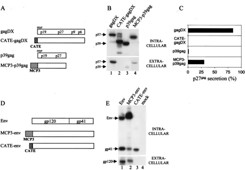
the entire chemokine MCP-3 coding region, previously shown to enhance the ability of some antigens to trigger strong im-mune response (7), were linked to the N termini of Gag and Env (Fig. 1A and D). Alternatively, a peptide derived from beta-catenin providing a ubiquitination signal was linked to the N terminus of Gag and Env. The resulting modified SIV anti-gens have different properties as shown in Fig. 1. Whereas the native p57gag protein expressed from pGagDX formed parti-cles and was secreted (Fig. 1B), CATE-gagDX was rapidly degraded in the cells and did not appear in the medium. In contrast, MCP3-p39gag fusion was stable and was secreted more efficiently than p39gag (Fig. 1B [compare lane 3 contain-ing unmodified p39gag to lane 4] and Fig. 1C). In similarity to Gag results, the MCP3-env chimera was secreted (Fig. 1E, lane 2), whereas the CATE-env fusion was unstable and did not appear extracellularly (Fig. 1E, lane 3). The gp120/gp160 form of the CATE-env was not visualized because of its instability, and only a cell-associated Env fragment of approximately 40 kDa was visible. Thus, both Gag and Env are degraded upon
fusion to the CATE signal. Env degradation appears to be more rapid, indicating that Gag may be more resistant to degradation. These results indicate that the introduced modi-fications altered significantly the stability and trafficking of the antigens, justifying the analysis of immunogenicity of such forms. Vaccination studies using mice indicated that the com-bination of three different plasmids (unmodified, MCP-3 fu-sion, and CATE fusion) showed better immunogenicity (M. Rosati, A. Von Gegerfelt, C. Alicea, B. K. Felber, and G. N. Pavlakis, unpublished data). In addition, we found that the fusions to MCP-3 provided the best antibody response. This was verified in preliminary DNA vaccination experiments using two macaques, which did not develop antibodies after repeated vaccination with an optimized expression vector producing na-tive Gag but developed a strong antibody response after vac-cination with MCP3-gag (Rosati et al., unpublished).
[image:3.585.47.537.64.403.2]DNA-based vaccination decreases viremia following muco-sal challenge. To test the ability of the antigens to protect against SIV challenge, we immunized Indian rhesus macaques
FIG. 1. SIV protein expression analysis in 293 cells. (A) Schematic representation of RNA-optimized SIVgagexpression vectors encoding the p57gag (gagDX) and p39gag proteins. Sequences fused togagare shown in boxes, striped for the catenin peptide (CATE) and grey for MCP-3 chemokine (MCP3). (B) Expression and localization of the different gag constructs. Human 293 cells were transfected with the indicated plasmids, and protein production was analyzed by Western immunoblotting using sera from SIV-infected macaques. For this analysis 1/50 of intracellular protein extract and 1/150 of the supernatant was used. (C) Quantitation of Gag levels in the intracellular and extracellular compartments of the experiment shown in Fig. 1B using p27gag antigen capture assay. The percentage of secreted Gag is shown. (D) Schematic representation of RNA-optimized SIVenvexpression vectors. Symbols are as described for Fig. 1A. (E) Expression and localization of the different Env constructs. Human 293 cells were transfected with the indicated plasmids, and protein production was analyzed by Western immunoblotting in both intra- and extracellular compartments as described for Fig. 1B.
on November 8, 2019 by guest
http://jvi.asm.org/
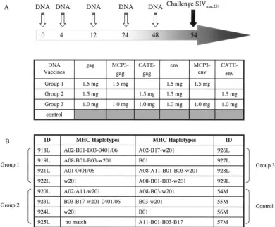
with plasmids expressing the native and modified Gag and Env proteins in different combinations (Fig. 2A) and examined virus propagation as well as cellular and humoral immune responses before and after pathogenic challenge by SIV-mac251. Prior to vaccination, all animals were analyzed for a set of 10 MHC class I alleles (Fig. 2B). The individual animals showed a diverse array of MHC class I alleles, and only animal 921L (group 1) had the MamuA*01 allele. Three groups of four animals were immunized via the intramuscular route us-ing a total of 6 mg plasmid DNA per vaccination (3 mg of gag and 3 mg of Env-expressing DNA vectors, respectively) in-jected at different sites.
On the basis of the preliminary immunogenicity analysis in mice and macaques, three combinations of antigens were tested in macaques in an effort to improve the outcome of DNA vaccination. All three groups received the vectors ex-pressing native Gag and Env (Fig. 2A). Group 1 received
native and MCP3-tagged antigens (1.5 mg each of
pSIVp57gag, pMCP3-p39, p239env, and pMCP3-239env). Group 2 received a combination of native and intracellularly
[image:4.585.97.487.71.394.2]degraded antigens (1.5 mg each of p57gag, pCATEp57 p239env, and pCATE-239env). Group 3 received a combina-tion of all six molecules (1 mg each). The vaccinacombina-tion schedule included a total of 6 mg plasmid DNA per immunization and a total of five vaccinations given at weeks 0, 4, 12, 24, and 48. Six weeks after the last vaccination the immunized and four naive macaques were intrarectally challenged with a highly pathogenic stock of SIVmac251. All animals became infected, although we observed significant differences among the vacci-nated and the control animals (Fig. 3). We also included in the analysis 10 additional control animals, which were infected with the same dilution of the virus stock in the same animal facility within a few weeks of our challenge and which were analyzed with the same plasma virus load assay method. Re-sults obtained with eight of these animals (8 M, 12 M, 13 M, 27F, 34 M, 39F, 44F, 46F) were published elsewhere (52), and two animals (538L and 539L) were from a separate study. The median peak virus load during the acute phase for the 14 control animals was 108.2RNA copies/ml, and the median virus
FIG. 2. Study design. (A) Four groups of four Indian rhesus macaques were enrolled. Group 4 remained unvaccinated and served as the control group. All other animals were vaccinated five times at 0, 4, 12, 24, and 48 weeks with a total of 6 mg of plasmid DNAs (3 mg for Gag and 3 mg for Env), followed by mucosal challenge with SIVmac251. All animals received the vectors expressing the native SIV Gag and Env proteins. In addition, all animals received vectors expressing modified Gag and Env proteins. Group 1 received p57gag (gagDX) and gp160 (env) together with MCP-3 fusion proteins pMCP3-p39gag and pMCP3-env; group 2 received the expression vectors for the native antigens together with CATE fusion proteins pCATE-p39gag and pCATE-Env; group 3 received the combination of all six plasmids. Different plasmids expressing identical Env fusions as well as pCATE-p57gag were used for the last vaccination, as detailed in Materials and Methods. (B) MHC haplotypes of the individual animals using a set of 10 MHC class I alleles. Only one animal had the MamuA*01 haplotype. Ten additional control animals were used in the comparisons (see text). Of these, two (39F, 44F) were MamuA*01 positive.
VOL. 79, 2005 DNA VACCINATION WITH MODIFIED ANTIGENS 8483
on November 8, 2019 by guest
http://jvi.asm.org/
load during the entire chronic phase was 106.7RNA copies/ml,
confirming the robustness of the viral stock.
The log-transformed median virus loads of all the vaccinated animals as a group (a combination of groups 1, 2, and 3;n⫽
12) were compared to the virus load of the 14 control animals (Fig. 3A). This comparison showed a statistically significant difference between the vaccinee (n⫽12) and the control (n⫽
14) in both the acute phase (P⫽0.010) and the chronic phase (P⫽0.010) of infection (Wei-Johnson test). Therefore, DNA vaccination alone was effective and protected the animals from development of high viremia for a long period during chronic infection.
We next compared the individual vaccinated groups to the controls, to evaluate the efficacy of the three tested vector combinations. The comparison of the log-transformed median virus loads of the individual groups (group 1, 2, and 3;n⫽4 for
each group) and the combination of all controls (n⫽ 14) is shown in Fig. 3B. Of note is group 3, which received the combination of all antigens and shows strongest control of viremia (1 to 2 logs less than control) up to the end of the study at week 33. Analysis using the Wei-Johnson test showed that group 3 differed significantly from the control group during both the acute phase (days 7 to 28,P⫽0.023) and the chronic phase (days 35 to 171,P ⫽ 0.0059). The difference between group 2 and controls was statistically significant only during the acute phase at the P ⫽ 0.024 level, whereas no significant difference was found for group 1.
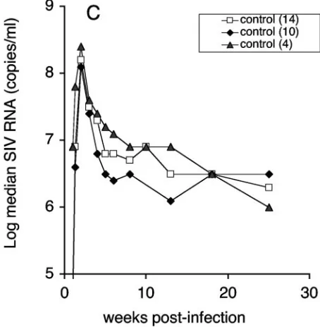
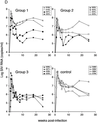
[image:5.585.312.539.71.303.2]We note that analysis including all 14 control animals leads to a conservative evaluation of differences between vaccine and control groups. Figure 3C shows the median virus loads of the 4 controls and of the additional 10 controls compared to the total control group ofn⫽14. Inclusion of the 10 additional
FIG. 3. Viral loads of DNA-immunized and control animals after mucosal challenge with SIVmac251. (A) The log median SIV RNA loads for all vaccinees (n⫽12) and controls (n⫽14) are shown. The
Pvalue (0.01) indicates significant difference during both the acute and chronic phase. Controls include 10 additional macaques challenged with the same virus stock as described in Materials and Methods. (B) The log median SIV RNA loads are shown for each individual group and the 14 control animals. ThePvalue indicates significant difference between group 3 and control (P⫽0.0059). In addition, virus loads in groups 2 and 3 were significantly lower than in the control groups during acute viremia (P⫽0.024 andP⫽0.023, respectively). (C) Log median virus loads of the two control groups used for the comparisons (n⫽4,n⫽10) and the combined control group (n⫽14). (D) Sequential viral load determinations for individual animals in the four groups, shown as log SIV RNA copies per ml of plasma. Filled symbols indicate individual animals with viral loads less than the con-trol animal loads.
on November 8, 2019 by guest
http://jvi.asm.org/
controls leads to more-conservative estimates of the difference between control and vaccinee groups, because the 10 addi-tional controls had lower viral loads than the group of 4 con-trols at almost every time point. Analysis by the Wei-Johnson method showed that the two groups of controls were not dif-ferent during the chronic period of infection. The log-trans-formed virus loads of the individual animals in each group are shown in Fig. 3D. Interestingly, all animals in group 3 showed a consistent control of viremia with little animal-to-animal variation, despite the genetic differences among the monkeys (Fig. 1B). In contrast, in groups 1 and 2 half of the animals (indicated with filled symbols) showed decreased levels of vire-mia during the chronic phase similar to those seen with group 3.
The analysis by the Wei-Johnson nonparametric test does not make assumptions about the form of the distributions in the groups. Alternative parametric analyses were also per-formed using repeated-measure analysis of variance
[image:6.585.125.463.71.489.2](ANOVA) to compare each group to the group of the four controls. The P values derived from the repeated-measure ANOVA were adjusted by Dunnett’s method in analyses where multiple groups were compared to the same controls. The difference between group 3 and the four control animals is significant also using repeated-measure ANOVA (P⫽0.035), even when the greatest estimate of variance across all four groups was applied, in agreement with the results of the non-parametric Wei-Johnson test. The difference between group 1 or 2 and the four controls did not reach statistical significance. Therefore, DNA vaccination alone elicited immune responses capable of reducing viremia during both the acute and chronic phases of SIV infection. This long-lasting effect on viremia against a highly pathogenic stock of SIVmac251 was more prominent when the different forms of DNA antigens were combined (group 3). Analysis of immune responses also indi-cated that group 3 showed strong postchallenge immune re-sponses to SIV antigens (see below). These results suggest that
FIG. 3—Continued.
VOL. 79, 2005 DNA VACCINATION WITH MODIFIED ANTIGENS 8485
on November 8, 2019 by guest
http://jvi.asm.org/
the combination of three different forms of antigens used in group 3 may have resulted in a better control of viremia.
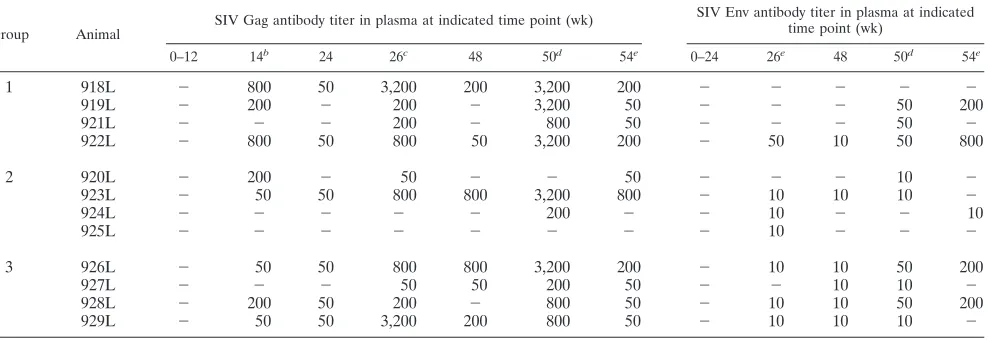
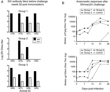
Enhancement of humoral immunity.We measured the pres-ence of binding antibodies to both p27gag and gp120 by en-zyme-linked immunosorbent assay in the vaccinated animals both before and after challenge (Table 1 and Fig. 4). Table 1 shows the development of anti-Gag and anti-Env antibodies during DNA vaccination, and Fig. 4A shows the reciprocal titers at week 50, 2 weeks after the last immunization.
After three DNA inoculations, 8 of 12 animals developed specific humoral immune responses to Gag. The number of animals responding was further increased to 10 of 12 animals after the fourth immunization. The humoral immune response to Env was in general lower and appeared later compared to Gag results. At week 50 (Fig. 4A), anti-Env antibodies were detected in 9 of 12 animals (titers of 10 to 50). Despite the differeknces in the magnitude of Gag and Env humoral im-mune responses, all the animals in groups 1 and 3, which received native and MCP-3 tagged antigens, developed anti-bodies. In contrast, only two of four animals in group 2, which received native and CATE-fusion antigens, showed persistent Gag and Env humoral immune responses (Table 1). Therefore, the MCP-3 fused forms of the antigens (common in groups 1 and 3) were associated with stronger humoral immune re-sponses. Following challenge (Fig. 4B), we observed rapid in-creases (detectable by days 3 to 14 postchallenge) of humoral immune responses, predominantly to Gag, demonstrating an anamnestic immune response in all the vaccinated groups com-pared to the challenged control animals.
Group 3 animals also developed neutralizing antibodies against SIVmac251 grown in primary cells faster than the other groups, as early as weeks 4 to 8 after challenge (Table 2). No neutralizing antibodies were detected prior to challenge by this assay. Therefore, the development of neutralizing antibodies was more rapid after challenge in group 3 compared to the other vaccinated groups and the controls. However, no
statis-tically significant differences in binding antibody titers or in neutralizing activity were observed between the DNA-immu-nized animals and the control group when comparing the mag-nitude of the response. Therefore, the contribution of humoral immune responses to the control of viremia is unclear.
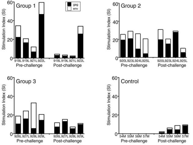
Correlation of SIV-specific postchallenge cellular immune responses to the control of viremia.We measured lymphopro-liferative responses (LPR) to Gag and Env proteins at different times both before and after challenge (Fig. 5). Before chal-lenge, all vaccinated animals developed specific LPR to Gag and Env, with peak stimulation indices ranging between 4 and 47 and between 4 and 27, respectively. Following SIV chal-lenge, we observed a decline in LPR to gp120env in all groups. LPR to p27gag were similar postchallenge except for group 1, which had reduced LPR. Control animals had low LPR after challenge.
We also used ELISPOT assays to measure the frequency of antigen-specific IFN-␥-producing PBMCs after overnight incu-bation with pools of overlapping SIV Gag or Env peptides (Fig. 6). Similar peak ELISPOT values for SIV Gag were measured in all three vaccine groups before challenge (Fig. 6A). The ELISPOT values for Env were significantly lower (Fig. 6B). Whereas all animals in group 1 showed Env-specific ELISPOT values, only 1 and 3 animals responded in groups 2 and 3, respectively. Following challenge, we observed an en-hancement in the frequencies of ELISPOTs for both viral antigens in vaccinated animals. The control animals had low ELISPOT values compared to the other groups. Animals in group 3 showed the strongest responses to Gag and Env.
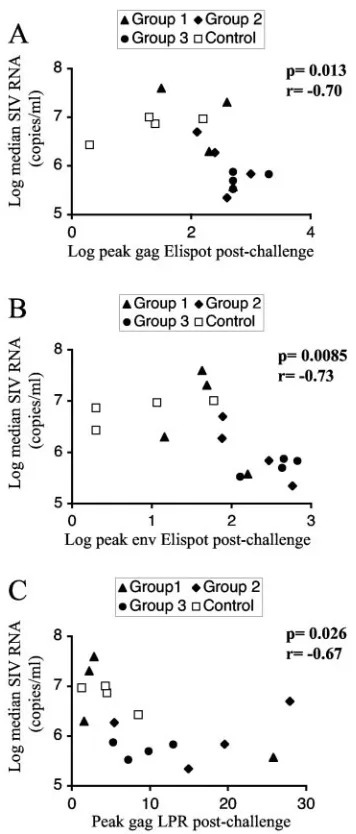
Figure 7 shows the correlation between the log-transformed median viral load measures and the postchallenge immune responses during the chronic phase using the Spearman rank correlation method. We found a strong negative correlation between chronic viremia and SIV-specific ELISPOT values for both Gag (P ⫽ 0.013, r ⫽ ⫺0.70) (Fig. 7A) and Env (P ⫽
[image:7.585.49.543.91.261.2]0.0085,r⫽ ⫺0.73) (Fig. 7B). Therefore, protection from high
TABLE 1. Binding antibodies to SIV Gag (left panel) and Env (right panel) in the plasma of the immunized animals at indicated time pointsa
Group Animal
SIV Gag antibody titer in plasma at indicated time point (wk) SIV Env antibody titer in plasma at indicated
time point (wk)
0–12 14b
24 26c
48 50d
54e
0–24 26e
48 50d
54e
1 918L ⫺ 800 50 3,200 200 3,200 200 ⫺ ⫺ ⫺ ⫺ ⫺
919L ⫺ 200 ⫺ 200 ⫺ 3,200 50 ⫺ ⫺ ⫺ 50 200
921L ⫺ ⫺ ⫺ 200 ⫺ 800 50 ⫺ ⫺ ⫺ 50 ⫺
922L ⫺ 800 50 800 50 3,200 200 ⫺ 50 10 50 800
2 920L ⫺ 200 ⫺ 50 ⫺ ⫺ 50 ⫺ ⫺ ⫺ 10 ⫺
923L ⫺ 50 50 800 800 3,200 800 ⫺ 10 10 10 ⫺
924L ⫺ ⫺ ⫺ ⫺ ⫺ 200 ⫺ ⫺ 10 ⫺ ⫺ 10
925L ⫺ ⫺ ⫺ ⫺ ⫺ ⫺ ⫺ ⫺ 10 ⫺ ⫺ ⫺
3 926L ⫺ 50 50 800 800 3,200 200 ⫺ 10 10 50 200
927L ⫺ ⫺ ⫺ 50 50 200 50 ⫺ ⫺ 10 10 ⫺
928L ⫺ 200 50 200 ⫺ 800 50 ⫺ 10 10 50 200
929L ⫺ 50 50 3,200 200 800 50 ⫺ 10 10 10 ⫺
aReciprocal titers an given.⫺, negative values.
bTwo weeks after third immunization.
cTwo weeks after fourth immunization.
dTwo weeks after fifth immunization.
eFour weeks after fifth immunization.
on November 8, 2019 by guest
http://jvi.asm.org/
viremia in this model was associated with high levels in ELISPOT assays for the structural proteins Gag and Env. A moderate negative correlation was found between T-helper responses to gag (LPR) and virus loads (P⫽0.026,r⫽ ⫺0.65) (Fig. 7C). Similar significant negative correlations were found when comparing the viral load during acute infection to the postchallenge immune responses. There were no significant correlations with prechallenge immune responses. In conclu-sion, we found a significant negative correlation between virus load and cellular immune responses, since the animals having high responses in all groups had the lowest virus loads.
DISCUSSION
In the present study we established that DNA vaccine vec-tors producing a combination of native and modified forms of SIV antigens enhanced the ability of both arms of the immune system to mount an immune response against the virus. The combination of DNA vectors led to the development of a more balanced immune response than previously observed in DNA-vaccinated macaques. We demonstrated that DNA-immunized monkeys developed Gag and Env binding antibodies (Table 1 and Fig. 4). An anamnestic antibody response was observed after challenge, and the animals in group 3 also developed neutralizing antibody earlier than all other groups and con-trols. In addition, the DNA-immunized animals showed
cellu-lar immune responses as determined by LPR and ELISPOT assays during the immunization period (Fig. 5 and 6). ELISPOT values were increased after challenge and correlated with protection from high viremia (Fig. 7).
[image:8.585.112.471.73.373.2]Although all animals were infected by the SIVmac251 chal-lenge, immunized animals were protected from high viremia compared to unvaccinated controls in both the acute and the chronic phase of infection. Animals in group 3 receiving the combination of the three different forms of antigens achieved the greatest reduction of viremia, which was statistically signif-icant. Although the results for the other two groups did not achieve statistical significance in the chronic phase, we note that 2/4 animals in each of these groups (in total 8/12 vacci-nated animals in this study) had lower level of viremia com-pared to the controls. Comparison of all 12 vaccinated animals to the controls also showed a statistically significant difference in chronic viremia, which is a predictor of disease develop-ment. The DNA-immunized monkeys showed reduced viremia for 8 months following SIVmac251 challenge. This finding suggests that the combinations of vectors used in this study are useful for the generation of protective immunity and should be also studied in prime-boost experiments. The levels of viremia in the vaccinated animals, although reduced, remain substan-tial. This may be a consequence of the highly pathogenic virus stock used, which resulted in peak viremia values higher than 108. Although DNA alone is not sufficient to provide complete
FIG. 4. Humoral immune responses. (A) Binding antibodies to SIV Gag (solid bar) and Env (open bar) in the plasma of vaccinated animals, determined at week 50 (4 weeks prior to challenge). The log reciprocal titers are shown. The control animals were negative prior to challenge (see also panel B). (B) The median reciprocal antibody titers to SIV Gag (top panel) and Env (bottom panel) in the plasma of vaccinated (groups 1 through 3) and control animals were determined at the indicated days after SIVmac251 challenge.
VOL. 79, 2005 DNA VACCINATION WITH MODIFIED ANTIGENS 8487
on November 8, 2019 by guest
http://jvi.asm.org/
virus control in this model, our results indicate the utility of DNA vaccination using multiple optimized vectors as part of vaccine strategies against HIV.
The animal model used here is considered one of the closest to HIV-1 infection in humans, since SIVmac251-infected ma-caques develop high primary viremia and a chronic active in-fection with high plasma virus loads, which leads to immuno-deficiency and death within a half year to 3 years in the
majority of the animals. As in human populations, rapid and slow disease progressors are observed. This model has not been used extensively for efficacy trials of DNA-only vaccina-tion, and some reported trials showed little or no evidence of long-lasting vaccine protection. Many studies involving DNA vaccination have been performed using the SHIV challenge model, especially challenge with the pathogenic SHIV89.6P. Infection by this virus leads to a rapid disease progression and severe depletion of CD4⫹ cells. This is in contrast to SIV-mac251 in rhesus macaques and HIV-1 in humans, where disease progression and lymphocyte depletion is a much slower process. SHIV89.6P infects different subset of cells due to its CXCR4 tropism, compared to primarily CCR5-tropic HIV-1 in humans (48). It can be argued that SIVmac251 challenge reflects more accurately the chronic active disease caused by CCR5-tropic HIV-1 in humans.
Several groups conducting DNA vaccine studies using SIV challenge stocks have concluded that there is minimal or no long-lasting benefit of DNA-only vaccination (5, 17, 26, 37, 44). In contrast, our study provides evidence for control of viremia after challenge with highly pathogenic SIVmac251. In agree-ment with other reports showing that the containagree-ment of vire-mia was associated with an increase in T-cell-specific responses (2, 5, 24), we find a strong correlation between the Gag-specific or Env-specific IFN-␥ immune responses and the ability to control viremia in the vaccinated animals.
Recently, the immunogenicity of the DNA vaccine regi-ments has been improved by different kinds of adjuvants in-cluding genetic adjuvants and different delivery methods (50, 56, 61, 63). Although the efficacy of several novel techniques in controlling a pathogenic virus challenge is yet to be proven, Barouch and coworkers reported that seven of eight animals receiving DNA together with IL-2 immunoglobulin fusion
[image:9.585.42.284.88.297.2]pro-FIG. 5. SIV-specific T-helper responses. Pre- and postchallenge peak lymphoproliferative responses to Gag and Env of the individual macaques in the four groups, after stimulation with purified proteins as detailed in Materials and Methods. Solid bars, Gag severalfold stimulation over control; open bars, Env severalfold stimulation.
TABLE 2. Postchallenge neutralizing antibody titer in plasma to primary SIVmac251(in M7-luc cells)
Group Animal
Titer at indicated time point (wk)a
0 4 8 13
1 918L ⫺ ⫺ ⫺ 20
919L ⫺ ⫺ ⫺ 73
921L ⫺ ⫺ ⫺ ⫺
922L ⫺ ⫺ ⫺ ⫺
2 920L ⫺ ⫺ ⫺ 70
923L ⫺ ⫺ ⫺ 180
924L ⫺ ⫺ ⫺ 68
925L ⫺ ⫺ ⫺ ⬎540
3 926L ⫺ 94 59 86
927L ⫺ ⫺ 32 130
928L ⫺ ⫺ 61 119
929L ⫺ ⫺ ⫺ 85
Control 54M ⫺ ⫺ ⫺ 102
55M ⫺ ⫺ ⫺ 47
56M ⫺ ⫺ ⫺ 106
57M ⫺ ⫺ 39 27
a
The neutralizing antibody titer is the reciprocal plasma dilution at which the relative luminescence unit values were reduced 80% compared to virus control
wells (no serum sample present).⫺, titer⬍20.
on November 8, 2019 by guest
http://jvi.asm.org/
[image:9.585.135.451.458.695.2]tein controlled SHIV challenge to levels below detection (5). Similarly, Shiver and coworkers established that three of three monkeys immunized with DNA and CRL1005 as the adjuvant controlled SHIV to levels below detection (61). In both stud-ies, the frequencies of prechallenge antiviral T cells in animals receiving DNA plus adjuvant were higher than in those receiv-ing DNA alone.
Studies using HIV DNA vectors in human clinical trials include both prophylactic and therapeutic vaccines (10).
Vac-cine safety has been achieved in phase I studies, and no sig-nificant adverse effects were reported (39, 66). If safe and effective, DNA vectors for immunization may show several advantages, including the flexibility to combine many different antigens and different forms of the same antigen, as shown herein. They can also be combined with cytokines and many other molecular adjuvants, which have been shown to further enhance DNA vaccination, such as IL-2, IL-12, and IL-15 (3, 4, 6, 8, 11, 27–32, 34, 49, 73, 74). In addition, they can be
deliv-FIG. 6. CD8 immune responses. (A) Peak ELISPOT responses to SIV Gag peptides (spot-forming cells [SFCs] per 106PBMCs). Prechallenge and postchallenge peak values are shown for the individual animals. Numbers indicate the ELISPOT values for samples higher than 500 SFC/million PBMCs. (B) Peak ELISPOT responses to SIV Env peptides. Numbers indicate the ELISPOT values for samples higher than 500 SFC/million PBMCs.
VOL. 79, 2005 DNA VACCINATION WITH MODIFIED ANTIGENS 8489
on November 8, 2019 by guest
http://jvi.asm.org/
[image:10.585.135.454.70.556.2]ered many times in anamnestic immunizations without the limitations of the viral vectors, which elicit strong immune responses to the vector.
ACKNOWLEDGMENTS
We thank N. Miller and the National Institute for Allergy and Infectious Diseases for animals and support, G. Franchini, E. Trynis-zewska, J. Parrish, R. Pal, V. S. Kalyanaraman, R. Shurtliff, A. Ballard, S. Orndorff, W. Lu, A. Biragyn, and A. Gragerov for advice and assistance, and T. Jones for editorial assistance.
A.V.G. and M.R. are supported through a contract with Science Applications International Corporation (SAIC)–Frederick.
REFERENCES
1.Aberle, H., A. Bauer, J. Stappert, A. Kispert, and R. Kemler.1997.
Beta-catenin is a target for the ubiquitin-proteasome pathway. EMBO J.16:3797–
3804.
2.Amara, R. R., F. Villinger, J. D. Altman, S. L. Lydy, S. P. O’Neil, S. I. Staprans, D. C. Montefiori, Y. Xu, J. G. Herndon, L. S. Wyatt, M. A. Candido, N. L. Kozyr, P. L. Earl, J. M. Smith, H. L. Ma, B. D. Grimm, M. L. Hulsey, J. Miller, H. M. McClure, J. M. McNicholl, B. Moss, and H. L. Robinson.2001. Control of a mucosal challenge and prevention of AIDS by
a multiprotein DNA/MVA vaccine. Science292:69–74.
3.Barouch, D. H., A. Craiu, M. J. Kuroda, J. E. Schmitz, X. X. Zheng, S. Santra, J. D. Frost, G. R. Krivulka, M. A. Lifton, C. L. Crabbs, G. Heidecker, H. C. Perry, M. E. Davies, H. Xie, C. E. Nickerson, T. D. Steenbeke, C. I. Lord, D. C. Montefiori, T. B. Strom, J. W. Shiver, M. G. Lewis, and N. L. Letvin. 2000. Augmentation of immune responses to HIV-1 and simian immunodeficiency virus DNA vaccines by IL-2/Ig plasmid administration in
rhesus monkeys. Proc. Natl. Acad. Sci. USA97:4192–4197.
4.Barouch, D. H., T. M. Fu, D. C. Montefiori, M. G. Lewis, J. W. Shiver, and N. L. Letvin.2001. Vaccine-elicited immune responses prevent clinical AIDS
in SHIV(89.6P)-infected rhesus monkeys. Immunol. Lett.79:57–61.
5.Barouch, D. H., S. Santra, J. E. Schmitz, M. J. Kuroda, T. M. Fu, W. Wagner, M. Bilska, A. Craiu, X. X. Zheng, G. R. Krivulka, K. Beaudry, M. A. Lifton, C. E. Nickerson, W. L. Trigona, K. Punt, D. C. Freed, L. Guan, S. Dubey, D. Casimiro, A. Simon, M. E. Davies, M. Chastain, T. B. Strom, R. S. Gelman, D. C. Montefiori, M. G. Lewis, E. A. Emini, J. W. Shiver, and N. L. Letvin.2000. Control of viremia and prevention of clinical AIDS in rhesus
monkeys by cytokine-augmented DNA vaccination. Science290:486–492.
6.Barouch, D. H., S. Santra, T. D. Steenbeke, X. X. Zheng, H. C. Perry, M. E. Davies, D. C. Freed, A. Craiu, T. B. Strom, J. W. Shiver, and N. L. Letvin.
1998. Augmentation and suppression of immune responses to an HIV-1
DNA vaccine by plasmid cytokine/Ig administration. J. Immunol.161:1875–
1882.
7.Biragyn, A., K. Tani, M. C. Grimm, S. Weeks, and L. W. Kwak.1999. Genetic fusion of chemokines to a self tumor antigen induces protective, T-cell
dependent antitumor immunity. Nat. Biotechnol.17:253–258.
8.Boyer, J. D., A. D. Cohen, K. E. Ugen, R. L. Edgeworth, M. Bennett, A. Shah, K. Schumann, B. Nath, A. Javadian, M. L. Bagarazzi, J. Kim, and D. B. Weiner.2000. Therapeutic immunization of HIV-infected chimpanzees us-ing HIV-1 plasmid antigens and interleukin-12 expressus-ing plasmids. AIDS
14:1515–1522.
9.Boyer, J. D., B. Wang, K. E. Ugen, M. Agadjanyan, A. Javadian, P. Frost, K. Dang, R. A. Carrano, R. Ciccarelli, L. Coney, W. V. Williams, and D. B. Weiner.1996. In vivo protective anti-HIV immune responses in non-human
primates through DNA immunization. J. Med. Primatol.25:242–250.
10.Chattergoon, M., J. Boyer, and D. B. Weiner.1997. Genetic immunization:
a new era in vaccines and immune therapeutics. FASEB J.11:753–763.
11.Chattergoon, M. A., V. Saulino, J. P. Shames, J. Stein, L. J. Montaner, and D. B. Weiner.2004. Co-immunization with plasmid IL-12 generates a strong
T-cell memory response in mice. Vaccine22:1744–1750.
12.Desrosiers, R. C.1990. The simian immunodeficiency viruses. Annu. Rev.
Immunol.8:557–578.
13.Donnelly, J. J., J. B. Ulmer, and M. A. Liu.1997. DNA vaccines. Life Sci.
60:163–172.
14.Doria-Rose, N. A., and N. L. Haigwood. 2003. DNA vaccine strategies: candidates for immune modulation and immunization regimens. Methods
31:207–216.
15.Doria-Rose, N. A., C. Ohlen, P. Polacino, C. C. Pierce, M. T. Hensel, L. Kuller, T. Mulvania, D. Anderson, P. D. Greenberg, S. L. Hu, and N. L. Haigwood. 2003. Multigene DNA priming-boosting vaccines protect
ma-caques from acute CD4⫹-T-cell depletion after simian-human
immunodefi-ciency virus SHIV89.6P mucosal challenge. J. Virol.77:11563–11577.
16.Doria-Rose, N. A., C. C. Pierce, M. T. Hensel, W. F. Sutton, N. Sheikh, P. Polacino, L. Kuller, Y. D. Zhu, S. L. Hu, D. Anderson, and N. L. Haigwood.
2003. Multigene DNA prime-boost vaccines for SHIV89.6P. J. Med.
Prima-tol.32:218–228.
17.Egan, M. A., W. A. Charini, M. J. Kuroda, J. E. Schmitz, P. Racz, K. Tenner-Racz, K. Manson, M. Wyand, M. A. Lifton, C. E. Nickerson, T. Fu, J. W. Shiver, and N. L. Letvin.2000. Simian immunodeficiency virus (SIV) gag DNA-vaccinated rhesus monkeys develop secondary cytotoxic T-lym-phocyte responses and control viral replication after pathogenic SIV
infec-tion. J. Virol.74:7485–7495.
18.Fauci, A. S., G. Pantaleo, S. Stanley, and D. Weissman.1996.
Immunopatho-genic mechanisms of HIV infection. Ann. Intern. Med.124:654–663.
19.Gurunathan, S., D. M. Klinman, and R. A. Seder.2000. DNA vaccines:
immunology, application, and optimization. Annu. Rev. Immunol.18:927–
974.
20.Hadzopoulou-Cladaras, M., B. K. Felber, C. Cladaras, A. Athanassopoulos, A. Tse, and G. N. Pavlakis.1989. Therev(trs/art) protein of human immu-nodeficiency virus type 1 affects viral mRNA and protein expression via a
cis-acting sequence in theenvregion. J. Virol.63:1265–1274.
21.Haigwood, N. L., C. C. Pierce, M. N. Robertson, A. J. Watson, D. C. Mon-tefiori, M. Rabin, J. B. Lynch, L. Kuller, J. Thompson, W. R. Morton, R. E. Benveniste, S. L. Hu, P. Greenberg, and S. P. Mossman.1999. Protection from pathogenic SIV challenge using multigenic DNA vaccines. Immunol.
[image:11.585.75.253.68.485.2]Lett.66:183–188.
FIG. 7. Correlation of immune responses to viremia. Correlations between chronic virus loads expressed as log median SIV RNA (copies per ml of plasma) and log peak gag ELISPOT (A), log peak Env ELISPOT (B), and peak gag LPR (C) results. Animals in the different groups are indicated by different symbols.Pvalues and Spearman r
correlation coefficients are given.
on November 8, 2019 by guest
http://jvi.asm.org/
22.Hassett, D. E., and J. L. Whitton.1996. DNA immunization. Trends
Micro-biol.4:307–312.
23.Hel, Z., J. Nacsa, E. Tryniszewska, W. P. Tsai, R. W. Parks, D. C. Montefiori, B. K. Felber, J. Tartaglia, G. N. Pavlakis, and G. Franchini.2002. Contain-ment of simian immunodeficiency virus infection in vaccinated macaques: correlation with the magnitude of virus-specific pre- and postchallenge
CD4⫹and CD8⫹T cell responses. J. Immunol.169:4778–4787.
24.Hel, Z., W. P. Tsai, A. Thornton, J. Nacsa, L. Giuliani, E. Tryniszewska, M. Poudyal, D. Venzon, X. Wang, J. Altman, D. I. Watkins, W. Lu, A. von Gegerfelt, B. K. Felber, J. Tartaglia, G. N. Pavlakis, and G. Franchini.2001.
Potentiation of simian immunodeficiency virus (SIV)-specific CD4(⫹) and
CD8(⫹) T cell responses by a DNA-SIV and NYVAC-SIV prime/boost
regimen. J. Immunol.167:7180–7191.
25.Hochberg, Y., and Y. Benjamini.1990. More powerful procedures for
mul-tiple significance testing. Stat. Med.9:811–818.
26.Horton, H., T. U. Vogel, D. K. Carter, K. Vielhuber, D. H. Fuller, T. Shipley, J. T. Fuller, K. J. Kunstman, G. Sutter, D. C. Montefiori, V. Erfle, R. C. Desrosiers, N. Wilson, L. J. Picker, S. M. Wolinsky, C. Wang, D. B. Allison, and D. I. Watkins.2002. Immunization of rhesus macaques with a DNA prime/modified vaccinia virus Ankara boost regimen induces broad simian immunodeficiency virus (SIV)-specific T-cell responses and reduces initial viral replication but does not prevent disease progression following challenge
with pathogenic SIVmac239. J. Virol.76:7187–7202.
27.Kim, J. J., V. Ayyavoo, M. L. Bagarazzi, M. A. Chattergoon, K. Dang, B. Wang, J. D. Boyer, and D. B. Weiner.1997. In vivo engineering of a cellular immune response by coadministration of IL-12 expression vector with a
DNA immunogen. J. Immunol.158:816–826.
28.Kim, J. J., L. K. Nottingham, A. Tsai, D. J. Lee, H. C. Maguire, J. Oh, T. Dentchev, K. H. Manson, M. S. Wyand, M. G. Agadjanyan, K. E. Ugen, and D. B. Weiner.1999. Antigen-specific humoral and cellular immune responses can be modulated in rhesus macaques through the use of IFN-gamma, IL-12,
or IL-18 gene adjuvants. J. Med. Primatol.28:214–223.
29.Kim, J. J., K. A. Simbiri, J. I. Sin, K. Dang, J. Oh, T. Dentchev, D. Lee, L. K. Nottingham, A. A. Chalian, D. McCallus, R. Ciccarelli, M. G. Agadjanyan, and D. B. Weiner.1999. Cytokine molecular adjuvants modulate immune responses induced by DNA vaccine constructs for HIV-1 and SIV. J.
Inter-feron Cytokine Res.19:77–84.
30.Kim, J. J., J. S. Yang, K. H. Manson, and D. B. Weiner.2001. Modulation of antigen-specific cellular immune responses to DNA vaccination in rhesus macaques through the use of IL-2, IFN-gamma, or IL-4 gene adjuvants.
Vaccine19:2496–2505.
31.Kim, J. J., J. S. Yang, L. Montaner, D. J. Lee, A. A. Chalian, and D. B. Weiner.2000. Coimmunization with IFN-gamma or IL-2, but not IL-13 or IL-4 cDNA can enhance Th1-type DNA vaccine-induced immune responses
in vivo. J. Interferon Cytokine Res.20:311–319.
32.Lee, A. H., Y. S. Suh, and Y. C. Sung.1999. DNA inoculations with HIV-1 recombinant genomes that express cytokine genes enhance HIV-1 specific
immune responses. Vaccine17:473–479.
33.Lekutis, C., J. W. Shiver, M. A. Liu, and N. L. Letvin.1997. HIV-1envDNA vaccine administered to rhesus monkeys elicits MHC class II-restricted
CD4⫹T helper cells that secrete IFN-gamma and TNF-alpha. J. Immunol.
158:4471–4477.
34.Liu, L. J., S. Watabe, J. Yang, K. Hamajima, N. Ishii, E. Hagiwara, K. Onari, K. Q. Xin, and K. Okuda.2001. Topical application of HIV DNA vaccine with cytokine-expression plasmids induces strong antigen-specific immune
responses. Vaccine20:42–48.
35.Liu, M. A.2003. DNA vaccines: a review. J. Intern. Med.253:402–410. 36.Liu, M. A., Y. Yasutomi, M. E. Davies, H. C. Perry, D. C. Freed, N. L. Letvin,
and J. W. Shiver.1996. Vaccination of mice and nonhuman primates using
HIV-gene-containing DNA. Antibiot. Chemother.48:100–104.
37.Lu, S., J. Arthos, D. C. Montefiori, Y. Yasutomi, K. Manson, F. Mustafa, E. Johnson, J. C. Santoro, J. Wissink, J. I. Mullins, J. R. Haynes, N. L. Letvin, M. Wyand, and H. L. Robinson.1996. Simian immunodeficiency virus DNA
vaccine trial in macaques. J. Virol.70:3978–3991.
38.Lu, S., J. C. Santoro, D. H. Fuller, J. R. Haynes, and H. L. Robinson.1995. Use of DNAs expressing HIV-1 Env and noninfectious HIV-1 particles to
raise antibody responses in mice. Virology209:147–154.
39.MacGregor, R. R., J. D. Boyer, K. E. Ugen, K. E. Lacy, S. J. Gluckman, M. L. Bagarazzi, M. A. Chattergoon, Y. Baine, T. J. Higgins, R. B. Ciccarelli, L. R. Coney, R. S. Ginsberg, and D. B. Weiner.1998. First human trial of a DNA-based vaccine for treatment of human immunodeficiency virus type 1
infection: safety and host response. J. Infect. Dis.178:92–100.
40.McDonnell, W. M., and F. K. Askari.1996. DNA vaccines. N. Engl. J. Med.
334:42–45.
41.Montefiori, D.Evaluating neutralizing antibodies against HIV, SIV and
SHIV in a luciferase reporter gene assay, p. 12.11.1–12.11.15.InJ. E.
Coli-gan, A. M. Kruisbeck, D. H. Margulies, E. M. Shevach, W. Strober, and R. Coico (ed.), Current protocols in immunology. John Wiley & Sons, Hobo-ken, N.J.
42.Mossman, S. P., C. C. Pierce, M. N. Robertson, A. J. Watson, D. C. Mon-tefiori, M. Rabin, L. Kuller, J. Thompson, J. B. Lynch, W. R. Morton, R. E. Benveniste, R. Munn, S. L. Hu, P. Greenberg, and N. L. Haigwood.1999.
Immunization against SIVmne in macaques using multigenic DNA vaccines.
J. Med. Primatol.28:206–213.
43.Mossman, S. P., C. C. Pierce, A. J. Watson, M. N. Robertson, D. C. Mon-tefiori, L. Kuller, B. A. Richardson, J. D. Bradshaw, R. J. Munn, S. L. Hu, P. D. Greenberg, R. E. Benveniste, and N. L. Haigwood.2004. Protective immunity to SIV challenge elicited by vaccination of macaques with multi-genic DNA vaccines producing virus-like particles. AIDS Res. Hum.
Retro-vir.20:425–434.
44.Muthumani, K., M. Bagarazzi, D. Conway, D. S. Hwang, K. Manson, R. Ciccarelli, Z. Israel, D. C. Montefiori, K. Ugen, N. Miller, J. Kim, J. Boyer, and D. B. Weiner.2003. A Gag-Pol/Env-Rev SIV239 DNA vaccine improves
CD4 counts, and reduces viral loads after pathogenic intrarectal SIV251
challenge in rhesus macaques. Vaccine21:629–637.
45.Muthumani, K., D. Zhang, N. S. Dayes, D. S. Hwang, S. A. Calarota, A. Y. Choo, J. D. Boyer, and D. B. Weiner.2003. Novel engineered HIV-1 East African Clade-A gp160 plasmid construct induces strong humoral and
cell-mediated immune responses in vivo. Virology314:134–146.
46.Nasioulas, G., A. S. Zolotukhin, C. Tabernero, L. Solomin, C. P. Cunning-ham, G. N. Pavlakis, and B. K. Felber.1994. Elements distinct from human immunodeficiency virus type 1 splice sites are responsible for the Rev
de-pendence ofenvmRNA. J. Virol.68:2986–2993.
47.Newberg, M. H., M. J. Kuroda, W. A. Charini, A. Miura, C. I. Lord, J. E. Schmitz, D. A. Gorgone, M. A. Lifton, K. Kuus-Reichel, and N. L. Letvin.
2002. A simian immunodeficiency virus nef peptide is a dominant cytotoxic T lymphocyte epitope in Indian-origin rhesus monkeys expressing the
com-mon MHC class I allele mamu-A*02. Virology301:365–373.
48.Nishimura, Y., T. Igarashi, O. K. Donau, A. Buckler-White, C. Buckler, B. A. Lafont, R. M. Goeken, S. Goldstein, V. M. Hirsch, and M. A. Martin.2004.
Highly pathogenic SHIVs and SIVs target different CD4⫹T cell subsets in
rhesus monkeys, explaining their divergent clinical courses. Proc. Natl. Acad.
Sci. USA101:12324–12329.
49.O’Neill, E., I. Martinez, F. Villinger, M. Rivera, S. Gascot, C. Colon, T. Arana, M. Sidhu, R. Stout, D. C. Montefiori, M. Martinez, A. A. Ansari, Z. R. Israel, and E. Kraiselburd.2002. Protection by SIV VLP DNA prime/ protein boost following mucosal SIV challenge is markedly enhanced by
IL-12/GM-CSF co-administration. J. Med. Primatol.31:217–227.
50.Otten, G., M. Schaefer, B. Doe, H. Liu, I. Srivastava, J. Megede, Jr., D. O’Hagan, J. Donnelly, G. Widera, D. Rabussay, M. G. Lewis, S. Barnett, and J. B. Ulmer.2004. Enhancement of DNA vaccine potency in rhesus
ma-caques by electroporation. Vaccine22:2489–2493.
51.Pantaleo, G., and A. S. Fauci.1996. Immunopathogenesis of HIV infection.
Annu. Rev. Microbiol.50:825–854.
52.Patterson, L. J., N. Malkevitch, D. Venzon, J. Pinczewski, V. R. Gomez-Roman, L. Wang, V. S. Kalyanaraman, P. D. Markham, F. A. Robey, and M. Robert-Guroff.2004. Protection against mucosal simian immunodeficiency virus SIV(mac251) challenge by using replicating adenovirus-SIV multigene
vaccine priming and subunit boosting. J. Virol.78:2212–2221.
53.Qiu, J. T., R. Song, M. Dettenhofer, C. Tian, T. August, B. K. Felber, G. N. Pavlakis, and X. F. Yu.1999. Evaluation of novel human immunodeficiency virus type 1 Gag DNA vaccines for protein expression in mammalian cells
and induction of immune responses. J. Virol.73:9145–9152.
54.Robinson, H. L.2002. New hope for an AIDS vaccine. Nat. Rev. Immunol.
2:239–250.
55.Romano, J. W., R. N. Shurtliff, E. Dobratz, A. Gibson, K. Hickman, P. D. Markham, and R. Pal.2000. Quantitative evaluation of simian
immunode-ficiency virus infection using NASBA technology. J. Virol. Methods86:61–
70.
56.Scheerlinck, J. Y.2001. Genetic adjuvants for DNA vaccines. Vaccine19:
2647–2656.
57.Schneider, R., M. Campbell, G. Nasioulas, B. K. Felber, and G. N. Pavlakis.
1997. Inactivation of the human immunodeficiency virus type 1 inhibitory elements allows Rev-independent expression of Gag and Gag/protease and
particle formation. J. Virol.71:4892–4903.
58.Schwartz, S., M. Campbell, G. Nasioulas, J. Harrison, B. K. Felber, and G. N. Pavlakis.1992. Mutational inactivation of an inhibitory sequence in human immunodeficiency virus type 1 results in Rev-independent gag
ex-pression. J. Virol.66:7176–7182.
59.Schwartz, S., B. K. Felber, and G. N. Pavlakis.1992. Distinct RNA se-quences in the gag region of human immunodeficiency virus type 1 decrease RNA stability and inhibit expression in the absence of Rev protein. J. Virol.
66:150–159.
60.Shiver, J. W., M. E. Davies, H. C. Perry, D. C. Freed, and M. A. Liu.1996. Humoral and cellular immunities elicited by HIV-1 vaccination. J. Pharm.
Sci.85:1317–1324.
61.Shiver, J. W., T. M. Fu, L. Chen, D. R. Casimiro, M. E. Davies, R. K. Evans, Z. Q. Zhang, A. J. Simon, W. L. Trigona, S. A. Dubey, L. Huang, V. A. Harris, R. S. Long, X. Liang, L. Handt, W. A. Schleif, L. Zhu, D. C. Freed, N. V. Persaud, L. Guan, K. S. Punt, A. Tang, M. Chen, K. A. Wilson, K. B. Collins, G. J. Heidecker, V. R. Fernandez, H. C. Perry, J. G. Joyce, K. M. Grimm, J. C. Cook, P. M. Keller, D. S. Kresock, H. Mach, R. D. Troutman, L. A. Isopi, D. M. Williams, Z. Xu, K. E. Bohannon, D. B. Volkin, D. C. Monte-fiori, A. Miura, G. R. Krivulka, M. A. Lifton, M. J. Kuroda, J. E. Schmitz,
VOL. 79, 2005 DNA VACCINATION WITH MODIFIED ANTIGENS 8491
on November 8, 2019 by guest
http://jvi.asm.org/
N. L. Letvin, M. J. Caulfield, A. J. Bett, R. Youil, D. C. Kaslow, and E. A. Emini.2002. Replication-incompetent adenoviral vaccine vector elicits
ef-fective anti-immunodeficiency-virus immunity. Nature415:331–335.
62.Shiver, J. W., H. C. Perry, M. E. Davies, D. C. Freed, and M. A. Liu.1995. Cytotoxic T lymphocyte and helper T cell responses following HIV
polynu-cleotide vaccination. Ann. N. Y. Acad. Sci.772:198–208.
63.Singh, M., M. Briones, G. Ott, and D. O’Hagan.2000. Cationic micropar-ticles: A potent delivery system for DNA vaccines. Proc. Natl. Acad. Sci.
USA97:811–816.
64.Soudeyns, H., and G. Pantaleo.1999. The moving target: mechanisms of
HIV persistence during primary infection. Immunol. Today20:446–450.
65.Tang, D. C., M. DeVit, and S. A. Johnston.1992. Genetic immunization is a
simple method for eliciting an immune response. Nature356:152–154.
66.Ugen, K. E., S. B. Nyland, J. D. Boyer, C. Vidal, L. Lera, S. Rasheid, M. Chattergoon, M. L. Bagarazzi, R. Ciccarelli, T. Higgins, Y. Baine, R. Gins-berg, R. R. Macgregor, and D. B. Weiner.1998. DNA vaccination with HIV-1
expressing constructs elicits immune responses in humans. Vaccine16:1818–
1821.
67.Ulmer, J. B., J. J. Donnelly, S. E. Parker, G. H. Rhodes, P. L. Felgner, V. J. Dwarki, S. H. Gromkowski, R. R. Deck, C. M. DeWitt, A. Friedman, et al.
1993. Heterologous protection against influenza by injection of DNA
encod-ing a viral protein. Science259:1745–1749.
68.Vogel, T. U., M. R. Reynolds, D. H. Fuller, K. Vielhuber, T. Shipley, J. T. Fuller, K. J. Kunstman, G. Sutter, M. L. Marthas, V. Erfle, S. M. Wolinsky, C. Wang, D. B. Allison, E. W. Rud, N. Wilson, D. Montefiori, J. D. Altman, and D. I. Watkins.2003. Multispecific vaccine-induced mucosal cytotoxic T
lymphocytes reduce acute-phase viral replication but fail in long-term
con-trol of simian immunodeficiency virus SIVmac239. J. Virol.77:13348–13360.
69.Wang, B., J. Boyer, V. Srikantan, L. Coney, R. Carrano, C. Phan, M. Merva, K. Dang, M. Agadjanan, L. Gilbert, et al.1993. DNA inoculation induces neutralizing immune responses against human immunodeficiency virus type
1 in mice and nonhuman primates. DNA Cell Biol.12:799–805.
70.Wang, B., J. Boyer, V. Srikantan, K. Ugen, L. Gilbert, C. Phan, K. Dang, M. Merva, M. G. Agadjanyan, M. Newman, et al.1995. Induction of humoral and cellular immune responses to the human immunodeficiency type 1 virus
in nonhuman primates by in vivo DNA inoculation. Virology211:102–112.
71.Wang, L., D. Junker, and E. W. Collisson.1993. Evidence of natural
recom-bination within the S1 gene of infectious bronchitis virus. Virology192:710–
716.
72.Wei, L. J., and W. E. Johnson.1985. Combining dependent tests with
in-complete repeated measurements. Biometrika72:359–364.
73.Xin, K. Q., K. Hamajima, S. Sasaki, A. Honsho, T. Tsuji, N. Ishii, X. R. Cao, Y. Lu, J. Fukushima, P. Shapshak, S. Kawamoto, and K. Okuda.1998. Intranasal administration of human immunodeficiency virus type-1 (HIV-1) DNA vaccine with interleukin-2 expression plasmid enhances cell-mediated
immunity against HIV-1. Immunology94:438–444.
74.Xin, K. Q., K. Hamajima, S. Sasaki, T. Tsuji, S. Watabe, E. Okada, and K. Okuda.1999. IL-15 expression plasmid enhances cell-mediated immunity
induced by an HIV-1 DNA vaccine. Vaccine17:858–866.
75.Yasutomi, Y., H. L. Robinson, S. Lu, F. Mustafa, C. Lekutis, J. Arthos, J. I. Mullins, G. Voss, K. Manson, M. Wyand, and N. L. Letvin.1996. Simian immunodeficiency virus-specific cytotoxic T-lymphocyte induction through
DNA vaccination of rhesus monkeys. J. Virol.70:678–681.
![FIG. 6. CD8 immune responses. (A) Peak ELISPOT responses to SIV Gag peptides (spot-forming cells [SFCs] per 106and postchallenge peak values are shown for the individual animals](https://thumb-us.123doks.com/thumbv2/123dok_us/181857.51992/10.585.135.454.70.556/responses-elispot-responses-peptides-forming-postchallenge-individual-animals.webp)