A CLINICAL STUDY OF MATERNAL AND PERINATAL OUTCOME IN HEART DISEASE COMPLICATING PREGNANCY
Dissertation submitted in partial fulfilment of the
Requirement for the award of the Degree of
M.S. DEGREE–BRANCH VI OBSTETRICS AND GYNAECOLOGY
APRIL 2017
TIRUNELVELI MEDICAL COLLEGE HOSPITAL
THE TAMIL NADU DR.M.G.R. MEDICAL UNIVERSITY,
CHENNAI,
CERTIFICATE
This is to certify that the Dissertation entitled “A CLINICAL STUDY OF
MATERNAL AND PERINATAL OUTCOME IN HEART
DISEASECOMPLICATING PREGNANCY” submitted by
DR. KARLIN SATHIYA PRABA , MBBS.,DGO., to The Tamilnadu
Dr.M.G.R. Medical University, Chennai, in partial fulfilment for the award of
M.S (Obstetrics and Gynaecology) is a bonafide work carried out by her
under my guidance and supervision during the academic year 2015-2017.
This dissertation partially or fully has not been submitted for any other degree
or diploma of this university or other.
GUIDE
Prof.Dr.Ramalakshmi, MD., DGO,
Department Obstetrics and Gynaecology,
Tirunelveli Medical College, Tirunelveli- 627011.
HOD
Prof.Dr.MEENA,MD.,DGO.,DNB.,
Department Obstetrics and Gynaecology,
Tirunelveli Medical College, Tirunelveli- 627011.
Dr.K.Sithy Athiya Munavarah, THE DEAN,
DECLARATION
I, DR. KARLIN SATHIYA PRABA, MBBS.,DGO., solemnly declare
that the Dissertation titled ““A CLINICAL STUDY OF MATERNAL
AND PERINATAL OUTCOME IN HEART
DISEASECOMPLICATING PREGNANCY” had been prepared by me under the expert guidance and supervision of Prof. Dr. Ramalakshmi MD.,
DGO., Professor, Department of Obstetrics and Gynaecology, Tirunelveli
Medical College Hospital, Tirunelveli.
The dissertation is submitted to The Tamilnadu Dr. M.G.R. Medical
University, Chennai in partial fulfilment of the regulation for the award of
M.S. Degree (Branch VI) in Obstetrics and Gynaecology.
It was not submitted to the award of any degree/diploma to any
University either in part or in full previously.
Place: Tirunelveli.
Date:
Dr. Karlin Sathiya Praba MBBS., DGO
POST GRADUATE,
M.S. (Obstetrics and Gynaecology),
Tirunelveli Medical College,
ACKNOWLEDGEMENT
I am very much thankful to the Dean Dr. K.Sithy Athiya Munavarah,
Triunelveli Medical College Hospital, Tirunelveli, who has granted
permission to do this study in this institution,
I take this opportunity to express my deepest sense of gratitude to
professor Dr.MEENA, M.D., DGO., DNB., Head of the Department of
Obstetrics and Gynaecology, Tirunelveli Medical College Hospital,
Tirunelveli for encouraging me and rendering timely suggestions and guiding
me throughout the course of this study. simple words cannot express its for
this contribution.
I am extremely thankful to my guide Dr.RAMA LAKSHMI MD
DGO .,
I sincerely thank my professor Dr.SHEBA ROSATTE
VICTOR,M.D.,(OG)., Dr.M.SUJATHA ALAGESAN,M.D.,(OG), Dr.VALARMATHI,MD.,(OG)., Dr.MALLIGA, MD.,(OG)., ) for their
support and guidance.
I am very much thankful to professor Dr. TAMIL KOTHAI M.D.
(OG) for her support and guidance.
I am very much thankful to professor Dr. Ravi chandran Edwin
M.D., DM., Head of Department of Cardiology for providing valuable
support and guiding through the study.
I am very grateful to our Assistant Professor Dr. Bhavani Devi
MD.OG for her valuable suggestion in preparing this dissertation
I thank Prof. P. Arumugam statistician for their useful inputs.
Last but not the least, I am grateful to the antenatal mothers who
willingly cooperated with me during the study
I thank all my colleagues and friends for their constant encouragement.
I extremely thankful to my family members
CONTENTS
S.No TITLES Page No
1. Introduction 1
2. Aim of the Studyo 2
3. Review of Literature 3
4. Materials and Methods 34
5. Observation and Results 37
6. Discussion 70
7. Summary 82
8. Conclusion 84
9. Bibliography
10. Proforma
11. Master Chart
Abbreviations
CHD : Congenital heart disease
RHD : Rheumatic heart disease
SES : Socioeconomic Status
B : Booked
UB : Un booked
POA : Purpose of admission
OCP : Out come of pregnancy
Com.HD : Complication of heart disease
Ass. Mat.comp : Associated maternal complication
Foet.comp : Foetal complication
Cont : Contraception
T : Term
PT : Preterm
EL.LSCS : Elective LSCS
EM. LSCS : Emergency LSCS
LN with Epi : Labour Natural with episiotomy
MS : Mitral Stenosis
MR : Mitral Regurgitation
AR : Aortic Regurgitation
AS : Aortic Stenosis
TR : Tricuspid Regurgitation
CMC : Closed Mitral Commissurotomy
MVR : Mitral Valve Replacement
MOD : Moderate
SEV : Severe
GPLA : Gravida, Para, Live, Abortion
B.WT : Birth Weight
MAT : Maturity
IUD : Intrauterine Death
CPD : Cephalo Pelvic Disproportion
MVA : Manual Vacuum Aspiration
TAT : Trans Abdominal Tubectomy
ST : Sterilisation
MTP : Medical Termination of Pregnancy
CBC : Complete blood count
RFT : Renal function test
LFT : Liver function test
INTRODUCTION
Pregnant mother with heart disease is a challenge to both obstetrician
and the woman. It could be associated with an unfavourable maternal and
perinatal outcome. It requires special care to prevent or decrease the
maternal mortality and morbidity.
In our country most women are concealing their heart disease due to
local problems and marital problems, even after the diagnosis many
patients do not follow the instructions given by the obstetrician.
Gestational hypertension, gestational diabetes and anaemia are the added
problems which are more common in our country.
In developing countries, Rheumatic fever and its sequelae still
remain the most common etiological factor for heart disease and it is due to
streptococcal infection in country side. In our institution, RHD( Rheumatic
heart disease ) is the major cause of heart disease during pregnancy. In
women with known cardiac diseases, cardiac status must be optimized and
AIMS OF STUDY
1. To know the incidence of heart disease among pregnant mothers
during study period
2. To know the various aetiological factors for heart disease.
3. To know the possible prognostic factors which facilitate formulation
of guidelines for a safe motherhood.
4. To know the impacts of pregnancy on heart disease.
5. To know the impacts of heart disease on pregnancy.
6. To know the role of medical termination of pregnancy and method
of sterilisation in patients with heart disease.
7. To know the maternal outcome according to EF.
REVIEW OF LITERATURE
The foremost initial study was done by Hamilton and Thompson at
the Boston hospital between 1921 & 1938 on heart disease complicating
pregnancy.
Subsequently a study on heart disease complicating pregnancy
during 1950 was conducted by Sudhir Bose Calcutta and Masoni Bombay.
They concluded the incidence ranging from 0.2 to 0.97%.1
According to CDC- Heart diseases is the main cause of death in
women who are 25-44 years as described by Kung and colleagues 2008.2
Chang and associates described about cardiomyopathy being responsible
for 8% of 4200 delivery related death in US 1991-1999. From Brazil
Avilaand co-workers 20033 reported the maternal mortality rate to be 2.7%
in 1000 pregnancies complicated by heart disease.
Cardiac disease complicates>1% of all pregnancies and it is the
leading indirect cause of maternal death in 20% of all cases described by
Simpson 20126 and Berg 2010 study. A study conducted by Small 20124
reported that cardiac disease was responsible for significant maternal
morbidity and obstetric intensive care unit admission. Fryar 20125 gave a
statistics that almost50% of adults aged 20 and above have at least one risk
of severe obstetric morbidities are caused by cardiac disease as represented
in a US study conducted by Callaghan & co-workers in 2008.
A study by Barnes6 in the queen charlotte hospital on 1192 patients
with heart disease delineate that rheumatic heart disease was seen in 88.2%
and congenital heart disease in 11.8%. Mcfaul-1988, Bitsch and
co-workers-1989 described that 50% were suffering from congenital heart
disease . A study conducted by Tan and De Sweit 19987 reported that only
12% had rheumatic heart disease. Brickner and associates delineated that
progress in surgical methods and development in medical management will
cause increase in the number of women with congenital heart disease
reaching pregnancy.
Martin and co-workers 1999 described that heart disease remains to
be the third most common cause of maternal death.
HAEMODYNAMIC CHANGES IN PREGNANCY
The heart and circulation undergoes various physiological
adaptations during pregnancy, delivery and puerperium. These marked
changes have an intense effect on heart disease in the pregnant mother.
Changes in cardiac function become readily seen during the first 8 weeks
weeks of pregnancy as described by Mahendru 201210. The resting pulse
rate is increased by 10 beats. Cardiac output in lateral recumbent position
is increased in early pregnancy as reported by Duvekot and associated
1993, Mebie and colleagues-1994. Mild pericardial effusion and increased
cardiac silhouette was described by Enein and co-workers 198711. Cutforth
and Mac Donald 196612 used phonocardiography and documented an
exaggerated splitting of first heart sound with increasedloudness of both
components.There changes are more profound in multiple pregnancies as
reported by Kametas and associated 200313 Pregnant uterus compresses
venous return from lower extremities and may also compress the aorta as
described by Bieniare 1969
A great valuable study conducted by Clark and associated 198914 to
measure hemodynamic function in later pregnancy - right heart
catheterisation was done in 10 healthy nulliparous women at 35-38 weeks
and again 11 to 13 weeks postpartum. It described normal haemodynamic
values in late pregnancy. Yuan and co-workers 2006 described the normal
morphological and functional echo changes associated with pregnancy.
DIAGNOSIS OF HEART DISEASES
The normal physiological changes of pregnancy can produce
symptoms and signs that may be difficult to be differentiated from heart
between undiagnosed maternal cardiac abnormalities and mid trimester
uterine artery Doppler resistance indices. Atrial and ventricular premature
contractions are frequent during pregnancy as reported by Carruth 1981.
Savu 201216 and Vitrarelli 2011 have described the normal
echocardiographicparameters for pregnancy. Limacher 1985 organised
studies on 2D echo and pulsed doppler echo.
SYMPTOMS AND SIGNS:
Symptoms & Signs in normal pregnancy that mimic heart disease
Symptoms
Dyspnoea
Edema
Signs
Low blood pressure
Increased PR & RR
Cardiac impulse more diffuse
Jugular veins may be distended
Heart sounds are often more pronounced.
Ejection systolic murmur of grade 2/6
X-Ray
Cardiomegaly
ECG
ST depression & T wave changes
LAD (Left Axis Deviation)
ECHO
Minimal pericardial effusion
Symptoms & signs in heart disease
Symptoms
Progressive dyspnoea or orthopnoea
Nocturnal cough
Hemoptysis
Syncope
Chest pain
Signs
Cyanosis
Clubbing of fingers
Persistent neck vein distension
Diastolic murmur
Cardiomegaly
Persistent arrhythmias
Persistent split second sound
Criteria for pulmonary Hypertension
o Left parasternal lift
o Loud P2
CLASSIFICATION
This classification of New York Heart Association was first
published in 1928 and revised for the eighth time in 1978, based on past
and present disability and is uninfluenced by physical signs. Siu and
associated 2001b18 developed a scoring system based on risk factor. Two
studies by Khariy and colleagues 200619, Stangl 200820were conducted
TABLE-1 NEW YORK HEART ASSOCIATION FUNCTIONAL CLASSIFICATION OF CARDIAC DISEASE
Grade I Patients have no limitations of physical exercise; ordinary
activity does not cause undue fatigue, palpitations, dyspnoea or
angina.
Grade II Patients have slight limitations of physical exercise; ordinary
activity causes undue fatigue, palpitations, dyspnoea or angina.
Grade III Patients have marked limitations of physical activity, less than
ordinary activity causes symptoms.
Grade IV Patients have an inability to carry on physical activity without
TABLE-2 MODIFIED WHO CLASSIFICATION OF MATERNAL CARDIOVASCULAR RISK
WHO RISK Class I
Uncomplicated, small or mild
Pulmonary stenosis
Patent ductus arteriosus
MVP, mitral valve prolapse
Successfully repaired simple lesions (atrial or ventricular septal defect,
patent ductus arteriosus, anomalous pulmonary venous drainage)
Isolated atrial or ventricular ectopic beats
WHO Risk Class II (if otherwise well and uncomplicated)
Unoperated atrial or ventricular septal defect
Repaired tetralogy of Fallot
Most arrhythmias
WHO risk class II-III (depending on individual)
Mild left ventricular systolic function impairment
Hypertrophic cardiomyopathy
Native or tissue valvular heart disease not considered WHO I or IV
Marfan’s syndrome without aortic dilatation
WHO Risk Class III
Mechanical valve
Systemic right ventricle
Fonton circulation
Cyanotic heart disease (unrepaired)
Other complex congenital heart disease
Aortic dilatation 40-45 mm in Marfan’ssyndrome
Aortic dilation 45-50 mm in aortic disease associated with bicuspid aortic
valve
WHO Risk Class IV (Pregnancy contraindicated)
Pulmonary arterial hypertension of any cause
Severe systemic ventricular dysfunction (LVEF <30%, NYHA III-IV)
Previous peripartum cardiomyopathy with any residual impairment of left
ventricular function
Severe mitral stenosis with aortic root dilatation>45mm
Aortic dilatation>50mm in aortic disease associated with bicuspid aortic
valve
TABLE-3 MODIFIED WHO CLASSIFICATION OF MATERNAL CARDIOVASCULAR RISK: PRINCIPLES
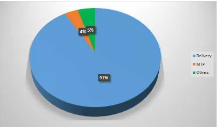
Risk Class Risk by Medical Condition
I No detectable increased risk of maternal mortality and
no/mild increase in morbidity.
II Small increased risk of maternal mortality or moderate
increase in morbidity.
III Significantly increased risk of maternal mortality or severe
morbidity. Expert counselling required. If pregnancy is
decided upon, intensive specialist cardiac and obstetric
monitoring needed throughout pregnancy, childbirth and
puerperium.
IV Extremely high risk of maternal mortality or severe
morbidity; pregnancy contraindicated. If pregnancy
occurs,termination should be discussed. If pregnancy
TABLE-4 PREDICTORS OF MATERNAL CARDIOVASCULAR EVENTS AND RISK SCORE FROM THE CARPREG STUDY
Prior cardiac event (heart failure, transient ischaemic attack, stroke before
pregnancy or arrhythmia)
Baseline NYHA functional class > II or cyanosis
Left heart obstruction (mitral valve area <2cm2, aortic valve area<1.5cm2, peak LV outflow tract gradient >30 mmHg by
echocardiography)
Reduced systemic ventricular systolic function (ejection fraction <40%) CARPREG risk score:For each above mentioned CARPREG predictor
that is present, a point is assigned. Risk estimation of cardiovascular
maternal complications
0 point 5%
1 point 27%
>1 point 75%
PRECONCEPTIONAL COUNSELLING
Women with cardiac conditions who desire or anticipate pregnancy
should be offered preconceptional counselling. The evolution of risk
provides a basis to explain to the patient the need for extensive testing
hospitalization and in some cases the need for surgical or medical
procedures before pregnancy. Risk assessment is also useful to determine
the type of facility where the patient should go for her delivery.
The need for induction of labour, shortening of the second stage of
labour, methods of anaesthesia used during labour and delivery,
endocarditis prophylaxis and anticoagulation therapy should also be a part
of the consultation. Patients with Eisenmenger syndrome, severe
pulmonary hypertension, severe left-sided obstructive lesions, and women
with Marfan syndrome and dilated aortic roots should be informed about
the high risk of maternal mortality and be counselled to consider avoiding
pregnancy and to choose adoption or other methods to have a family.
RHEUMATIC HEART DISEASE
Mitral stenosis
The normal Mitral valve surface area is 4cm2. Mitral stenosis
symptoms develops usually when the mitral valve surface area is less than
2.5cm2as described by Desai 200021. Due to tight stenosis left atrium is
dilated and left atrial pressure is chronically elevated resulting in fixed
cardiac output and development of pulmonary hypertension described by
stenosis develop cardiac failure for the first time in pregnancy. Rheumatic
endocarditis causes 75% of mitral stenosis.
Atrial tachycardia and atrial fibrillation are common in mitral
stenosis. Atrial fibrillation predisposes to mural thrombus and
cerebrovascular embolization that can cause stroke described by Hameed
and associate 200524.
Limited physical work, decreased sodium intake and addition of
diuretics described by Siva and Shah 200525. Alkasab and associates 1990,
Maxwell 2010 described the use of β blockers (usually given to decrease
the cardiac response during work and anxiety) in pregnancy.
Atrial fibrillation new onset - IV verapamil, electro cardio version, chronic – digoxin β blocker or calcium channel blocker, persistent atrial
fibrillation–heparin.
Hameed and co-workers described heparin for severe stenosis even
if there is a sinus rhythm. During labour, pain relief by epidural analgesia,
back rest, anxiolytic measures and IV fluid restriction to 75ml/hr are
necessary to avoid cardiac failure. Clark and colleague hypothesize
increase in pulmonary capillary wedge pressure during immediate
postpartum due to loss of the low resistance placental circulation along
with auto transfusion from lower extremities, increase in preload may lead
to avoid volume overload as described by Ramin and Gilstrap 1999.
Pregnancy outcomes were studied by Hammed 2001 and sawhney 200326
directly related with the degree of valvular stenosis.
AORTIC STENOSIS
Next to mitral stenosis, aortic stenosis is the common cardiac lesion.
The most common lesion is bicuspid aortic valve - Friedman and
co-workers 200827. Stenosis reduces the normal 2-3 cm aortic orifice and
creates resistance to ejection. Reduction in the valve area <1cm, pressure
gradient <5mmHg produces obstruction to flow and a progressive overload
on the left ventricle described by Carabello 200228, Roeder 201129. During
pregnancy some factors like haemorrhage, regional anaesthesia in labour
and vena caval occlusion by gravid uterus acutely decreases the preload
further and aggravates the fixed cardiac output. If the aortic valve area was
<1.5cm2 it will have increased complications reported by a study
conducted by Siu 2001. Women with valve gradient exceeding 100mmHg
appear to be at increased risk. Maternal mortality rate was 8% described by
Hameed and co-workers. If symptomatic bed rest, limitation of physical
activity and control of infection are suggested. Balloon valvotomy is
restricted because of grave complications like stroke, aortic rupture, aortic
1988, Lao and associates 1993, Datt 201031. Patient should be hydrated
sufficiently to prevent decrease in systemic vascular resistance. During
delivery, narcotic epidural analgesia is best as it avoids dangerous
hypotension a study conducted by Easterling and co-workers 198832. Slow
administration of dilute local anaesthesia was described by Camann and
Thornhill 1999, Xia and co-worker 200633. Tzemos and associate 200934
reported late cardiac events.
PULMONIC STENOSIS
It is usually congenital may be related with TOF or Noonan
syndrome pregnancy precipitate right side cardiac failure or atrial
arrhythmias described by Siv and Colman 2001. Cardiac complications
were infrequent during pregnancy described by a study conducted by
Drenthen 200635. If symptoms increased a balloon angioplasty was
necessary during pregnancy described by Siu 2001a36, Maxwell 201026.
MITRAL INSUFFICIENCY
Trivial mitral insufficiency is present in most normal patient by
Maxwell 201026. Mitral valve vegetation-Libman Sacs endocarditis are
common in patients with anti-phospholipid antibody described by Roldan
199637, Shroff 201238 Chronic mitral insufficient has a number of causes
which includes rheumatic fever, mitral valve prolapse ,etc. It is well
AORTIC INSUFFICIENCY
It is caused by rheumatic fever, connective tissue abnormalities and
congenital with marfan syndrome. The aortic root may dilate resulting in
aortic insufficiency which may also develop with bacterial endocarditis.
Aortic and mitral insufficiency have been related to appetite suppressants
like fenfluramine and ergot derived dopamine agonist like cabergoline
described by Gardin 200039, Schade 200740, Zanettini 200741. It is well
CONGENITAL HEART DISEASE
Cardiac Lesion
Congenital Heart Disease in Fetus (%) Previous sibling
Affected
Father Affected
Mother Affected
Marfan
syndrome
Not stated 50 50
Aortic stenosis 2 3 15-18
Pulmonary
stenosis
2 2 6-7
Ventricular
septal defect
3 2 10-16
Atrial septal
defect
2.5 1.5 5-11
Patent ductus
arteriosus
3 2.5 4
Coarctation of
the aorta
Not stated Not stated 14
Risks For Fetal Heart Lesionrelalated To Affected Family Members
The incidence in US is 8 / 1000 live birth, nearly one million adults
in that country described by Bashore 200742. Patients admitted for delivery
with congenital heart disease was 71.6 per 1,00,000 deliveries reported by
a study conducted by Optotowsky 201243. Arrythmias was the most
common and the mortality rate was nearly 1.5 per 1000 described by
Thompson and associates 201444.
Atrial septal defect:
One fourth of adults have a patent foramen ovale described by Kizer
200545. Ostium secundumtype defect is more common in nearly 70% of
cases. Pregnancy is well tolerated unless pulmonary hypertension develops
but this is uncommon described by Maxwell 201026 and Zuber 199946.
Aliago 200347 described risk of endocarditis was insignificant. Paradoxical
embolism is possible and may cause embolic stroke described by Erkut and
co-workers 200648. In asympomatic patients, prophylaxis for septal defect
is observation and low dose aspirin described by Kizer 200545and Maxwell
201025.Head and Thorne 200549 suggested compression stockings &
prophylactic heparin for a pregnant mother with ASD & concurrent
Ventricular septal defect:
These lesions usually close during paediatric period in 90%. If the
defect is <1.25 cm, pulmonary hypertension and cardiac failuredonot
develop. If the defect size is larger than aortic valve orifice, symptoms
rapidly develop. Patients with unrepaired large defects develop left
ventricular failure& pulmonary hypertension and increased incidence of
bacterial endocarditis described by Brickner 200050, Maxwell 2010. So IE
prophylaxis is often recommended in large unrepaired defects.
Atrioventricular septaldefect:
It accounts for 3% of all congenital heart disease charecterised by a
common ovoid AV junction associated with aneuploidy, Eisenmenger
syndrome. Compared with simple septal defect complications are more
frequent during pregnancy A Study conducted by Drenthen2005b51
concluded worsening of NYHA, arrhythmias and cardiac failure.
Patent dcutus arteriosus:
Functional closure of ductus from vasoconstriction occurs shortly
after term birth hypothesis described by Akintunde 201152. Most of the
lesions are repaired in paediatric period. In unrepaired patients mortality is
high after 50 years was described by Brickner 200050. Unrepaired ductus
cyanosis if, systemic BP falls & leads to shunt reversal of blood from
pulmonary artery into aorta described by Maxwell 2010. A sudden
decrease in blood pressure during labour due to conduction analgesia or
bleeding may result in total collapse therefore hypotension should be
avoided.
Cyanotic Heart disease:
Most commonly encountered lesion in pregnancy is Fallot tetralogy
described by Maxwell 2010. It is characterised by large ventricular septal
defect, pulmonary stenosis, right ventricular hypertrophy and an over
riding of aorta. Women who have undergone repair and who do not have
recurrence of cyanosis tolerate pregnancy well. A study conducted by Balci
201153 and Kamiya 201254 described surgically corrected TOF well
tolerated pregnancy and no maternal death. Oosterhof & associates 200655
described that pregnancy did not adversely affect graft function.
Single functional ventricle:
Fenistin 201256evaluated the treatment for patients with hypoplastic
left heart syndrome. Patients undergone a Fontan repair are at high risk of
atrial arrhythmias and cardiac failures described by Nitsche200959. Hoare
Ebstein Anomaly:
Malpositioned malformed tricuspid valve right ventricular failure
and cyanosis are common during pregnancy. In the absence of cyanosis,
patients tolerate pregnancy well.
Eisenmenger Syndrome:
The most common lesions are ASD VSD or PDA. Patients are
asymptomatic for years but pulmonary hypertension becomes severe to
cause right to left shunt described by Makaryus200659, Maxwell 2010.
Maternal and perinatal mortality – nearly 50% reported by
Gleicher197960.Weies and co-worker 1998 reported 36% maternal
mortality rate. Wang 201161reported one maternal death among 13 mothers
and five perinatal deaths in a study.
Pulmonary Hypertension:
Normal resting pulmonary artery pressure is 12-16 mm Hg. A study
conducted by Clark and colleagues 1989 pulmonary vascular resistance in
late pregnancy was 34% less than non-pregnant. PHT is a haemodynamic
observation not a diagnosis where mean pulmonary pressure is more than
25 mm Hg. WHO classification adopted by ACC EAHA Mclauchlin
One third of scleroderma have – PHT and 10% SLE have PHT
reported by Rich 200563. Sickle cell disease and thyrotoxicosis patients
have PHT but it is reversible with treatment–Cunnigham2004. Boggese &
colleague 199564described interstitial and restrictive lung disease have
varying degree of PHT. Patient with cirrhosis and portal HT have varying
degree of PHT and has been reported by Sigel 200765,cystic fibrosis
patients have PHT reported by Eden &co-workers 2000 pulmonary artery
pressure were over diagnosed by echo in 1/3 of cases reported by Penning
200166.Idiopathic PHT 3 years survival -60% Collagen vascular disease
causing PHT - only 35% reported by MC Laughlin 2004. Preconception
counselling is essential as highlighted by Easterling199967.
A study conducted by Curry 201268 and Weiss 199869reportedthe
mortality rate was nearly 30%. Bedard and associates 200970reported that
mortality statistics improved from 38% in 1996 to 25% in 2007.Pregnancy
is contraindicated with severe disease 80% death were during the first
month of puerperium.
Rest, oxygen, diuretics, vasodilators are standard treatments. Hsu
201171, Larson201072 recommended anticoagulation, Badalian200073,
Easterling1999,Garabedian201074 recommended I.V pulmonary artery
prostacyclin analogues. Lane201177 recommended inhaled nitric oxide is
useful for acute cardiopulmonary decompensation.
Parneix 200978 described low dose spinal or epidural analgesia for
LSCS, Weiss2000 & Lam 200179described inhaled nitric oxide or iloprost.
Peripartum cardiomyopathy:
It is like all other forms of dilated cardiomyopathy except for its
distinctive relationship with pregnancy described by Pyatt201180. Pearson
200081 established the diagnostics criteria After thorough search and
excluding the underlying cause for heart failure, idiopathic cardiomyopathy
may be considered which is described by Born and Bettelet 1998, Hibhaw
and associates 1999; various aetiologies like viral, abnormal immune
response , haemodynamic changes due to pregnancy, hormonal
interactions, malnutrition, inflammations, oxidative stress during late
pregnancy were described by Elkayam201182, Hilfiker- Kleiner200783,
Sliwa 201084, Patten 201285.Hypertension coexisting with peripartum
cardiomyopathy, is described by Cunningham 201286, Fong 201487,
Gunderson 201188 and they postulate peripartum cardiomyopathy to be a
vascular disease. Bultmen 200589and co-workers studied endomyocardial
biopsy specimen and reported evidence of myocarditis to be present due to
genomic materials of parvovirus-19. Prevalence of peripartum
Two other studies conducted by Gunderson 2011, Harper 201291 showed
the prevalance was lin 2000 and lin 2800 respectively. In earlier study by
Cunningham 198692 shows 1 in 15,000.So the prevalence was increased.
One year maternal mortality rate is 2-15% reported by Harper 2012, but in
those with persistence cardiac failure the maternal mortality was 85 %
over 5 years- Moioli 201093. In India, Mandal 201194 conducted a study
and reported the outcome of Peripartum cardiomyopathy -5 out of 36 was
died, another follow up study by Haiti, Fetl and co-worker 2009 shows
only 28% of these patients recovered. With a mean follow up of 39 months
De Souza and co-worker 200195 compared that 18% patients had died from
end stage cardiac failure.
Mitral valve prolapse:
A pathological connective tissue disorder – termed myxomatous
degeneration of the valve leaflet annulus and chordae tendinae. Mitral
insufficiency may develop. Most of them are asymptomatic, few of them
have symptoms like anxiety, palpitation , atypical chest pain, dysponea on
exertion& syncope hypothesized by Guy 201296. These patients have a
excellent pregnancy outcome - described by Lesniak-Sobelge 200497,
Rayburn 198798. In Taiwan, a study conducted by Chen 201199 reported
symptomatic patients according to ACOG 2011a& guidelines - is IE
Prophylaxis not indicated in MVP patients .
Diseases of the Aorta -:
Marfan syndrome & coarctation are two aortic diseases with
increased risk for aortic dissection, 50% of cases are related to pregnancy
hypothesized by O’ Gara 2004100. Other causes are bicuspid aortic valve,
Turner & Noonan syndrome. The differential diagnosis for it are
myocardial infarction, pulmonary embolism, pneumothorax, aortic valve
rupture , placental abruption and uterine rupture was described by Lang
1991101. Pepin102&collegues described a high rate of aortic dissection in
Ehlers danlos syndrome.
Marfan syndrome :
No racial predilection described by Ammash 2008103. In a study by
Rossiter 1995104 out of 21patients, 2 patients had dissection one died
during puerperium from graft infection. Rahman & coworekrs 2003103
hypothesized that no maternal death among 14 pregnant mothers. Aortic
dilatation reaches 5 to 6 cm elective surgery recommended before
pregnancy described by Gott 1999106 William 2002107. Simpson
20126reportedprophylactic B blockers reduces the haemodynamic stress
and slows the dilatation. Vaginal delivery - with aortic root dilatation was
aorta with prosthetic graft during puerperium is hypothesized by Simpson
2012, but the surgery is associated with fetal hypoxic ischemic cerebral
damage Mul 1998108 Seeburger 2007109. According to Goo 2011, Haas
2011, Papatsonis 2009110type A dissection was repaired favourably at the
time of Cesarean delivery. According to Meijboom 2006111,15% are
preterm deliveries.
Aortic coarctation:
Rare lesion - associated by abnormalities of large arteries. 25% have
bicuspid aortic valve, 10% have cerebral artery aneurysms. Others are
PDA, septal defects & Turner syndrome. Typical finding is hypertension in
upper limbs and normal pressure in lower limb. Coarctation is diagnosed
by MRI described by Dizon – Towson 1995112,sherer 2002113 zwiers
2006114. Beauchesue co-worker 2001115 described the outcome of 188
pregnancies. A study conducted by Krieger &colleagues 2011116 nearly 700
cases, 3 to 4 fold increase in HT, 5% adverse cardio vascular outcome,
41% underwent LSCS
Infective endocarditis:
Bacterial infection involves cardiac endothelium and usually end in
abusers& catheter related infections. Cox 1988118, Deger1992119 described
patients with antepartum endocarditis with Neisseria sicca and N.mucosa,
the latter causing maternal death .Beta haemolytic streptococcus infection
causes endocarditis have been described by Kangavari 2000120.E coli cause
endocarditis following LSCS was described by Kulas 2006121. The illness
is flu like described by karchmer 2012 diagnosed by Duke’s criteria
( Hoen 2013 , Pierce 2012) patients are treated according to microbial
sensitivity was described by Darouiche 2004122, Karchmer 2012). Cox
1988 described an incidence of 1 in 16,000 Cox1989 , Seaworth 1986123
IE PROPHYLAXIS:
American College of Obstetricians and Gynaecologists (ACOG) (2011a):
Standard (IV) : Ampicillin 2 g or cefazolin or ceftriaxone 1 g
Penicillin-allergic (IV) : Cefazolin or ceftriaxone 1g or Clindamycin
600mg
Oral : Amoxicillin 2g.
American Heart Association (Wilson, 2007)
Standard : Ampicillin 2g IV or IM or amoxicillin 2g (Oral)
Penicillin-allergic : Clarithromycin or azithromycin 500mg (Oral),
Cephalexin 500mg (Oral), Clindamycin 600 mg
(Oral), Intravenous, Intramuscular, or cefazolin
or ceftriaxone 1g intravenous or intramuscular.
Heart failure:
Women who develop cardiac failure almost always have obstetrical
complication like preeclampsia haemorrhage, Anaemia infection and
sepsis, chronic HT and obesity. Jess up 2003124described first sign in
Orthopnoea palpitations& chest pain – substernal are described by hibbard
1999. Ejection fraction <0.45%fractional shortening <30% end diastolic
dimension >2.7 cm/m2 were managed with diuretics and anti HT &
prophylactic heparin. Left ventricular outflow device are employed –
described by La Rue 2011125, sime 2011. Extracorporeal membrane
oxygenation reported to be life saving–smith 2009126.
Surgically corrected heart disease:
Most clinically significant CHD are repaired during child hood,
those frequently not diagnosed until adulthood include ASD, PS, Bicuspid
aortic valve & coarctation of aorta described by brickner co-worker 2000
Valve replacement:
Porcine type valves are much safer during pregnancy. However
valvular dysfunction with cardiac failure is common and these are not as
durable as mechanical one, valve replacement longevity averages 10-15
years. Cleuzious and co-workers 2010127 reported shunt pregnancy does
not accelerate the risk for replacement anticoagulation with heparin is less
risky for the foetus but the risk of maternal thromboembolic
complications are more described by MC Lintock 2011128. Chen and
coworkers reported warfarin during pregnancy has good pregnancy
outcome but the embryopathy rate was6.4 % chen 2000 Itur be alessio
UFH is inadequate & maternal mortality rate was increased Leyh
2002129.,2003 Rowan 2001130described both UFH & LMWH are associated
with valvular thrombosis anticoagulation with warfarin or heparin may
be restarted 6 years following vaginal delivery and 6-12 hrs following
LSCS recommended by AGOG 2011 b because they do not accumalate in
breast milk so that they do not induce an anticoagulant effects in the
infant ACOG 2011B.
Cardiac surgery during pregnancy:
Sutton and co-workers 2005131 described maternal mortality with
cardiopulmonary bypass are between 1.5 &2.5 % total mortality rate are
nearly 20% John-2011132and co-workers hypothesized that pregnant
mothers who underwent cardiothoracic surgery the urgency required
cardiopulmonary bypass Chandrasekar and associates 2009133
recommended surgery done electively when possible pump flow rate
maintained 7.25 L/ mt/mt2normothermic perfusion pressure 770mmHg
and haematocrit>28% Pavankumar 1988134conducted a study and reported
tight mitral stenosis require intervention during pregnancy which was
previously treated by closed mitral valvotomy percutaneous transcatheter
balloon dilatation is largely done described by Fawzy 2007135&Rahimtoole
Pregnancy after heart transplantation:
Lowenstein & co-workers 1988 138was reported successful
pregnancy after heart transplantation key 1989 and kim 1996139
hypothesized transplanted heart responds normally to haemodynamic
change during pregnancy. Dashe 1998140 described complication are
common. Arementi 2002 outcome of 37 heart recipient reported that 50%
developed HT , 22 % one rejection, usually delivered by LSCS at 37-38
was >5% infants are live born. Estensen & co-workers 2011 reported that
there was no maternal death by a study from sandinevia. Ethical
consideration and counselling were outlined by Rose 2016140.
Contraception:
This is much more important because they should limit their family
size and complete their families before there is serious cardiac
deterioration.
Oral contraceptives carry the risk of hypercoagulability,
thromboembolism, hypertension and hyperlipidemia IUCD carry the risk
of vaso vagal syncope due to pain during the time of insertion and
infection. It is prevented by smaller size Copper T and prophylactic
antibiotics- brebeer 1975.Barrier methods of contraception are safe but
have a low efficacy. These and low dose oral pills are preferable to IUCD.
MATERIALS AND METHODS
The study of maternal and perinatal outcome in heart disease
complicating pregnancy was carried out in Tirunelveli medical college .
This was done during a period of 14 months starting from June 2015 to
July 2016 and total of 107 mothers with heart disease complicating
pregnancy were studied.
Methodology:
1. Correct and careful history taking including H/O rheumatic fever,
H/O failure, pulmonary oedema etc., during antecedent pregnancies
details of recognised heart disease, details of treatment undergone
and H/O any surgery done for heart disease.
2. A through clinical examination.
3. Clinical diagnosis was made & the pregnant mothers were
investigated.
ECG and echocardiogram were done for all the mothers.
CBC,RFT,LFT, routine urine analysis were done for all patients.
TFT and coagulation profile were done for indicated cases. Pregnant
mothers were classified according to NYHA classification and
Inclusion Criteria:
All pregnant mothers with various heart disease.
Exclusion Criteria:
All pregnant women without any heart disease but having signs and
symptoms suggestive of heart disease were evaluated and newly diagnosed
heart disease patients were included in the study and mothers suny heart
disease were excluded.
Routine management: ANC
Patients with diagnosed heart disease were first admitted for correct
evaluation & clinical grading. The patients in functional class I &II were
discharged and advised to attend the OPD regularly. The frequency of
antenatal visits were determined by their cardiac status. They were
suggested to take sufficient rest to avoid sternous work, stress and take salt
restricted diet. Any infection if present was treated vigorously and were
advised to avoid contact with persons having respiratory infections
including common cold & to report at once if there was any evidence for
infection.
Patients were in functional class I &II were admitted at 32-34 weeks
, functional class III and IV were admitted immediately .Patients with
Provided strict bed rest, Oxygen and the treatment as advised by
cardiologist.
Labour:
In general, vaginal delivery is a better option so the main principle in
obstetric management was to wait for the spontaneous onset of labour.
Patients were in semi recumbent position with lateral tilt. They were
sedated in the first stage IV line was maintained. Prophylactic antibiotics
were given routinely. If the patients were on drugs, the drugs were
continued or else started as per cardiologist advice. Pulse rate, respiratory
rate blood pressure and foetal heart rate were monitored. Instruments were
used to cut short the second stage. Episiotomy and perineal lacerations
were sutured under local anaesthesia. LSCS was done for obstetric
indications only under epidural anaesthesia. After delivery the first 12-24
hours was carefully monitored for signs of failure & post partum
hemorrhage and they were kept in intensive care unit for 48 hrs and were
kept in postnatal ward for 14 days.
Babies were followed up by the paediatrician. Breast feeding was
allowed except in grade IV failure because it increases the cardiac output.
Patients were discharged after getting fitness for discharge from
OBSERVATION AND RESULTS
The present study was conducted in Tirunelveli Medical College,
Obstetrics and gynaecology department for a period of 14 months from
June 2015-July 2016 among the antenatal cases with heart disease. During
the study period total number of pregnant patients with heart disease were
107. Out of these 107 patients, 98 patients delivered and 4 underwent
MTP, 5 patients were delivered outside and referred here for further
management.
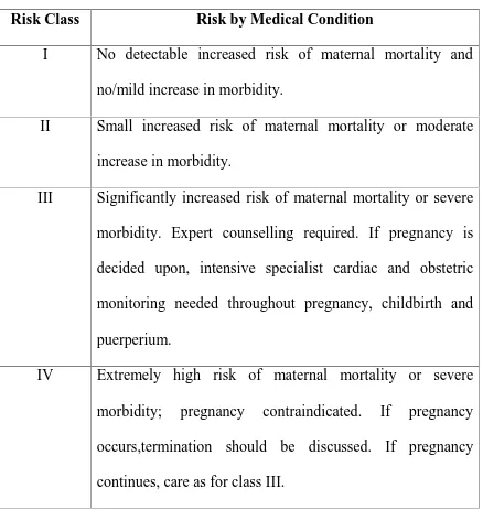
Total number of delivers in Tirunelveli Medical College Hospital
during the study period was 7830 out of which 98 deliveries were with
TABLE-1: INCIDENCE OF HEART DISEASE
Sl. No
Total No of delivery
Delivered mothers with Heart disease
Delivered mothers
with normal
CVS
Incidence of Heart disease
1 7830 98 7732 1.25%
FIG: 1 INCIDENCE OF HEART DISEASE
7830
98
7732
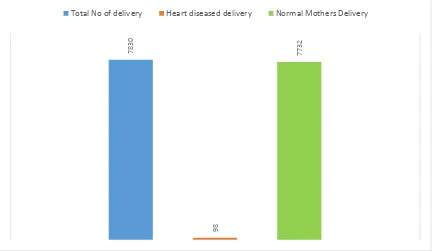
TABLE-2: DISTRIBUTION OF HEART DISEASE AMONG THE TOTAL DELIVERIES DURING STUDY PERIOD
A total of 107 cardiac mothers were admitted. They were classified
according to the nature of their disease. Among the 107 patients, CHD was
35 (32.7%), RHD was 53 (49.5 %) and others were 19 (17.8%).
Sl. No Category of Heart disease
No. and % of Heart disease deliveries
Heart diseases deliveries rate /
1000 (n=7830)
Frequency %
1 CHD 35 32.7 4.5
2 RHD 53 49.5 6.8
3 OTHERS 19 17.8 2.4
Total 107 100.0 13.7
The incidence of CHD, RHD and others were 0.4, 0.6 and 0.2
respectively.
FIG: 2: DISTRIBUTION OF HEART DISEASE AMONG THE TOTAL DELIVERIES DURING STUDY PERIOD
33%
49% 18%
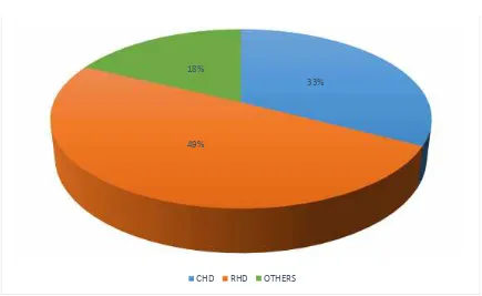
TABLE-3:DISTRIBUTION OF HEART DISEASE ACCORDING TO PURPOSE OF ADMISSION
Sl. No Purpose of
admission
Total No.of.cases Percentage
1 Delivery 98 91.58%
2 MTP 4 3.73%
3 Delivered outside 5 4.67%
FIG 3: DISTRIBUTION OF HEART DISEASE ACCORDING TO PURPOSE OF ADMISSION
91% 4% 5%
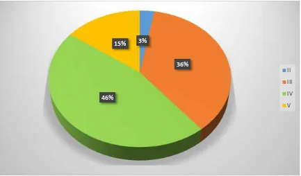
TABLE-4 : DISTRIBUTION OF MOTHERS ACCORDING TO THEIR SOCIO ECONOMICS Status
Sl No
Socio Economics
Status Frequency % χ
2 df Sig
1 II 3 2.8
49.523 3 P<0.001
2 III 39 36.4
3 IV 49 45.8
4 V 16 15.0
Total 107 100.0
Table 4 shows, 45.8% cases belong to class IV, 15% patients belong to
[image:51.595.100.533.435.689.2]classV and 36.4% patients belong to class III.
FIG: 4: DISTRIBUTION OF MOTHERS ACCORDING TO THEIR SOCIO ECONOMICS STATUS
3%
36%
46% 15%
TABLE-5: ASSOCIATION BETWEEN ANTENATAL REGISTRATION WITH HEART DISEASES:
Parity
CHD RH
D Others Total
χ2 d
f Sig
N
o % No %
N
o % No %
Booke
d 34 97.1 48
90. 6 19 100. 0 10 1 94.4 3.09 5 2 P>0.0 5 Un booke d
1 2.9 5 9.4 0
0.0
6 5.6
Total 35 100.
0 53 49. 5 19 17.8 10 7 100. 0
FIG:5:ASSOCIATION BETWEEN THE ANTENATAL
REGISTRATION WITH HEART DISEASES
Out of 107 patients, 101 were booked and 6 were unbooked. There
97. 1 2. 9 90. 6 9. 4 100 0
B O O K E D U N B O O K E D
ASSOCIATION BETWEEN THE ANTENATAL
REGISTRATION WITH HEART DISEASES
TABLE-6: AGE WISE DISTRIBUTION OF HEART DISEASES IN PERCENTAGE
Age group
CHD RHD Others Total
Frequenc
y %
Frequenc
y %
Frequenc
y % Frequency %
<20 1 2.9 1 1.9 1 5.3 3 2.8
20-24 18 51.4 19 35.8 5 26.9 42 39.3
25-29 9 25.7 22 41.5 9 47.4 40 37.4
30-34 6 17.1 9 17.0 3 15.8 18 16.8
35-39 1 2.9 2 3.8 1 5.3 4 3.7
Total 35 100 53 100 19 100 107 100
Mean±SD 25.3±4.3 26.0±3.8 26.7±4.2 25.9±4.0
Signifi F= 0.717and P>0.05 Range 36-19=17
Majority of cases were in the age group 20-30 years.
FIG:6:AGE WISE DISTRIBUTION OF HEART DISEASES IN PERCENTAGE 2. 9 51. 4 25. 7 17. 1 2. 9 1. 9 35. 8 41. 5 17 3. 8 5. 3 26. 9 47. 4 15. 8 5. 3
< 2 0 2 0 - 2 4 2 5 - 2 9 3 0 - 3 4 3 5 - 3 9
TABLE-7: DISTRIBUTION OF CHD, RHD AND OTHER MOTHERS ACCORDING TO THEIR PARITY
Parity CHD RHD Others Total χ2 df Sig
No % No % No % No %
Primi 18 16.8 20 18.7 6 5.6 44 41.1
2.501 1 P>0.05
Multi 17 15.9 33 30.8 13 12.1 63 58.9
Total 35 32.7 53 49.5 19 17.8 107 100.0
FIG 7:DISTRIBUTION OF CHD, RHD AND OTHER MOTHERS ACCORDING TO THEIR PARITY
16.
8
15.
9
18.
7
30.
8
5.
6
12.
1
P R I M I M U L T I
Diagnosis of heart disease
The practice of taking ECG and obtaining physician’s opinion for
all the antenatal mothers is now being followed in our hospital. Many
patients with MVPS with or without valvular dysfunction with
deterioration in their functional status are being diagnosed during
pregnancy.
TABLE-8 : TIME OF DIAGNOSIS OF HEART DISEASE:
Sl.No Time of diagnosis of heart disease Percentage
1 Heart disease diagnosed during
pregnancy
92.52%
2 Heart disease diagnosed before
pregnancy
7.4%
FIG:8: TIME OF DIAGNOSIS OF HEART DISEASE:
0.00% 10.00% 20.00% 30.00% 40.00% 50.00% 60.00% 70.00% 80.00% 90.00% 100.00%
Heart disease
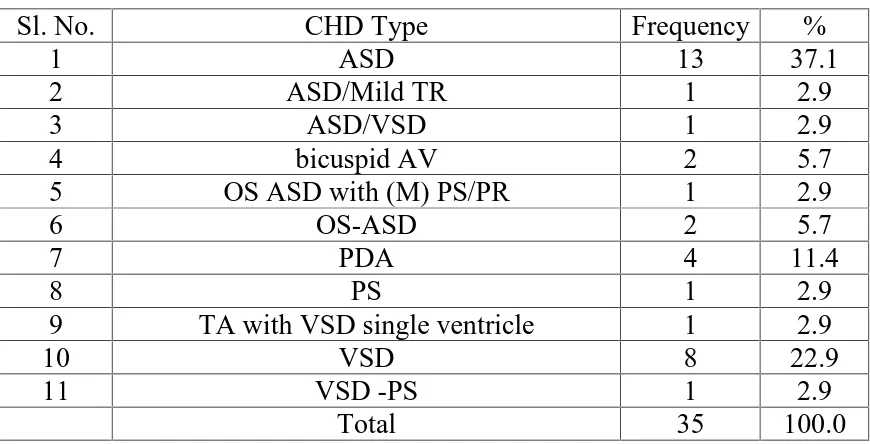
TABLE-9: PERCENTAGE DISTRIBUTION OF CHD ACCORDING TO THE SUB CLASSIFICATION CATEGORY:
Sl. No. CHD Type Frequency %
1 ASD 13 37.1
2 ASD/Mild TR 1 2.9
3 ASD/VSD 1 2.9
4 bicuspid AV 2 5.7
5 OS ASD with (M) PS/PR 1 2.9
6 OS-ASD 2 5.7
7 PDA 4 11.4
8 PS 1 2.9
9 TA with VSD single ventricle 1 2.9
10 VSD 8 22.9
11 VSD -PS 1 2.9
Total 35 100.0
FIG:9: DISTRIBUTION OF CHD ACCORDING TO THE SUB
CLASSIFICATION CATEGORY
37%
3% 3% 5% 3% 6% 11% 3% 3%
23%
3%
ASD ASD/Mild TR ASD/VSD bicuspid AV
OS ASD with (M) PS/PR OS-ASD
PDA PS
TA with VSD single ventricle VSD
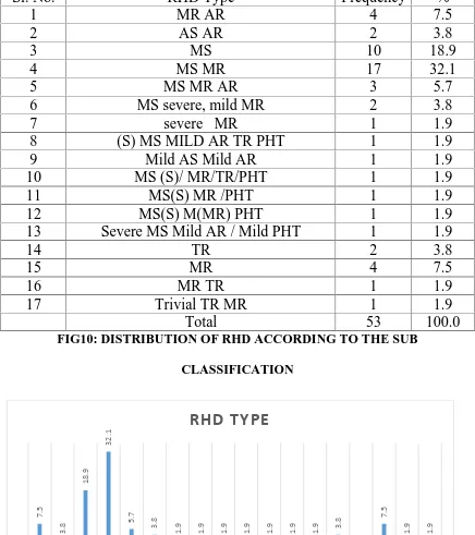
[image:56.595.99.533.508.763.2]TABLE-10: DISTRIBUTION OF RHD ACCORDING TO THE SUB CLASSIFICATION CATEGORY
Sl. No. RHD Type Frequency %
1 MR AR 4 7.5
2 AS AR 2 3.8
3 MS 10 18.9
4 MS MR 17 32.1
5 MS MR AR 3 5.7
6 MS severe, mild MR 2 3.8
7 severe MR 1 1.9
8 (S) MS MILD AR TR PHT 1 1.9
9 Mild AS Mild AR 1 1.9
10 MS (S)/ MR/TR/PHT 1 1.9
11 MS(S) MR /PHT 1 1.9
12 MS(S) M(MR) PHT 1 1.9
13 Severe MS Mild AR / Mild PHT 1 1.9
14 TR 2 3.8
15 MR 4 7.5
16 MR TR 1 1.9
17 Trivial TR MR 1 1.9
Total 53 100.0
FIG10: DISTRIBUTION OF RHD ACCORDING TO THE SUB
CLASSIFICATION 7. 5 3. 8 18. 9 32. 1 5. 7 3. 8 1. 9 1. 9 1. 9 1. 9 1. 9 1. 9 1.
9 3.8 7.
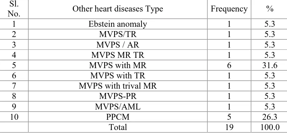
TABLE-11: DISTRIBUTION OF OTHER HEART DISEASES ACCORDING TO THE SUB CLASSIFICATION CATEGORY
Sl.
No. Other heart diseases Type Frequency %
1 Ebstein anomaly 1 5.3
2 MVPS/TR 1 5.3
3 MVPS / AR 1 5.3
4 MVPS MR TR 1 5.3
5 MVPS with MR 6 31.6
6 MVPS with TR 1 5.3
7 MVPS with trival MR 1 5.3
8 MVPS-PR 1 5.3
9 MVPS/AML 1 5.3
10 PPCM 5 26.3
Total 19 100.0
FIG: 11: DISTRIBUTION OF OTHER HEART DISEASES ACCORDING TO THE SUB CLASSIFICATION CATEGORY
5.
3
5.
3
5.
3
5.
3
31.
6
5.
3
5.
3
5.
3
5.
3
26.
[image:58.595.99.533.437.691.2]TABLE-12 : NYHA STATUS CLASSIFICATION OF CARDIAC SUBJECTS:
Parit y
CHD RHD Others Total
χ2 d
f Sig
N
o % No %
N
o % No %
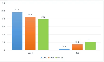
Good 34 97.1 45 84.9 15 78.
9 94 87.9
4.67
3 2
P>0.0 5
Bad 1 2.9 8 15.1 4 21.
1 13 12.1
Total 35 100.
0 53 100. 0 19 17. 8 10 7 100. 0
Majority of patients belong to NYHA class I and II.
Good prognosis–NYHA class I and II
Bad prognosis - NYHA Class III and IV
FIG:12 NYHA STATUS CLASSIFICATION OF CARDIAC SUBJECTS: 97.1 2.9 84.9 15.1 78.9 21.1 0 20 40 60 80 100 120 Good Bad
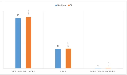
TABLE:13 :PREGNANCY OUTCOME IN PATIENTS ADMITTED FOR
DELIVERY
Pregnancy Outcome Number of Case Percentage
Vaginal delivery 70 71.42
LSCS 27 27.55
Died un delivered 1 1.02
This table shows majority of patients (71.42 % ) were delivered by normal vaginal
delivery.
FIG: 13 :PREGNANCY OUTCOME IN PATIENTS ADMITTED FOR
DELIVERY
70
27
1
71.
42
27.
55
1.
02
V A G I N A L D E L I V E R Y L S C S D I E D U N D E L I V E R E D
TABLE:14: DISTRIBUTION OF NORMAL AND INSTRUMENTAL VAGINAL
DELIVERY
S.No. Type of delivery No. Cases %
1. Labour natural 40 57.14
2. Outlet forceps 19 27.14
3. Vacuum extraction 9 12.85
4. Assisted Breech
Delivery
2 2.85
FIG:14: DISTRIBUTION OF NORMAL AND INSTRUMENTAL VAGINAL
DELIVERY
0 10 20 30 40 50 60
Labour natural Outlet forceps Vaccum extraction Assisted Breech Delivery
TABLE: 15: DISTRIBUTION OF LABOUR NATURAL
S.NO. Type of Delivery Number of Cases %
1. Labour Natural 17 42.5
2. LN with episiotomy 23 57.5
FIG:15: DISTRIBUTION OF LABOUR NATURAL
0 10 20 30 40 50 60 70
Labour Natural LN with episiotomy
TABLE: 16: DISTRIBUTION OF LSCS
S. No. Type of LSCS Cases %
1. Emergency primary LSCS 14 51.85
2. Emergency repeat LSCS 2 7.40
3. Elective primary LSCS 2 7.40
4. Elective repeat LSCS 9 32.33
FIG: 16: DISTRIBUTION OF LSCS
14
2 2
9
51.
85
7.
4
7.
4
32.
33
E M E R G E N C Y P R I M A R Y E M E R G E N C Y R E P E A T E L E C T I V E P R I M A R Y E L E C T I V E R E P E A T
TABLE 17: INDICATION OF PRIMARY EMERGENCY LOWER SEGMENT CAESAREAN SECTION
Indication Number of cases Percentage
Cephalopelvic Disproportion 4 28.57%
Premature rupture of membrane 2 14.28%
Breech 3 21.42%
Foetal distress 5 35.71%
FIG 17 : INDICATION OF PRIMARY EMERGENCY LOWER SEGMENT
CAESAREAN SECTION
4
2
3
5
28.
57%
14.
28%
21.
42% 35.71%
C E P H A L O M E T R I C
D I S P R O P O R T I O N P R E M A T U R E R U P T U R EO F M E M B R A N E B R E E C H F O E T A L D I S T R E S S
TABLE 18: INDICATION OF EMERGENCY REPEAT LOWER SEGMENT
CAESAREAN SECTION
Sl. No
Indication Number
of cases
Percentage
1 Previous LSCS with CPDI 1 50%
2 Previous LSCS with PROM 1 50%
FIG: 18:INDICATION OF EMERGENCY REPEAT LOWER SEGMENT
CAESAREAN SECTION
Previous LSCS with CPD minor
50% Previous LSCS with
PROM 50%
Percentage
TABLE 19: INDICATION OF PRIMARY ELECTIVE LOWER SEGMENT CAESAREAN SECTION
Sl. No
Indication Number of
cases
Percentage
1 CPD Major 1 50%
2 Flexed Breech 1 50%
FIG:19: INDICATION OF PRIMARY ELECTIVE LOWER SEGMENT
CAESAREAN SECTION
CPD Major 50% Flexed Breath
50%
TABLE: 20: INDICATION OF ELECTIVE REPEAT LOWER SEGMENT CAESAREAN SECTION
Sl. No
Indication Number of
cases
Percentage
1 Previous LSCS with CPD 8 88.88%
2 Previous LSCS with Breech 1 11.11%
FIG: 20: INDICATION OF ELECTIVE REPEAT LOWER SEGMENT CAESAREAN SECTION
Previous LSCS with CPD 89% Previous LSCS with
Breech 11%
TABLE 21: DISTRIBUTION OF COMPLICATION OF HEART DISEASE DEVELOPED DURING PREGNANCY
S.NO. Complication Number of
cases
Percentage
1. Severe Pulmonary
Hypertension
3 33.33
2. Acute pulmonary oedema 3 33.33
3. Congestive cardiac failure 1 11.11
4. Embolic manifestation 2 22.22
FIG: 21 DISTRIBUTION OF COMPLICATION OF HEART DISEASE
DEVELOPED DURING PREGNANCY
0 5 10 15 20 25 30 35
Severe Pulmonary
Hypertension Acute pulmonary oedema Congestive cardia failure Embolic manifestation
[image:68.595.98.534.470.723.2]TABLE 22 : DISTRIBUTION OF RISK FACTORS ASSOCIATED WITH THE CARDIAC DISEASE
S.No. Complication No. of
cases
%
1. Aanemia 13 34.21
2. Gestational hypertention 4 10.52
3. Gesational diabetes mellitus 9 7.89
4. Rh negative 10 26.31
5. Hypothyroid 4 10.25
6. Premature rupture of membrane 2 5.26
7. Preterm Premature rapture of membrane 1 2.63
FIG: 22 DISTRIBUTION OF RISK FACTORS ASSOCIATED WITH THE CARDIAC DISEASE
13
4
9 10
4
2 1 1
34.
21
10.
52
7.
89
26.
31
10.
25
5.
26
2.
63
2.
63
TABLE:23: DISTRIBUTION OF SURGICALLY CORRECTED HEART
DISEASE
S.No. Surgery No. of cases %
1. Closed Mitral commisurotomy 4 14.81
2. Baloon valvoplasty 2 7.4
3. ASD Closure 3 11.11
4. VSD closure 5 18.51
5. PDA ligation 3 11.11
6. Mitral valve replacement 6 22.22
7. As with AR–Sub Aortic excision 1 3.7
8. TOF Corrected 1 3.7
9. MS- MR Annuloplasty 1 3.7
FIG:23: DISTRIBUTION OF SURGICALLY CORRECTED HEART DISEASE
0 5 10 15 20 25
[image:71.595.99.533.484.741.2]TABLE-24: BIRTH WEIGHT OF BABIES ACCORDING TO THE TYPE OF HEART DISEASES
Sl. No
Type of Heart Disease
n Mean SD “F” df Significance
1 CHD 34 2.6 0.5
0.286 2, 96 P>0.05
2 RHD 46 2.6 0.4
3 Others 19 2.7 0.5
Total 99 2.6 0.4
FIG: 24: BIRTH WEIGHT OF BABIES ACCORDING TO THE TYPE OF HEART DISEASES
2.6 2.6
2.7
2.54 2.56 2.58 2.6 2.62 2.64 2.66 2.68 2.7 2.72
TABLE-25: ASSOCIATION BETWEEN HEART DISEASES AND MATURITY OF THE BABY
Heart
Disease Pre term Term Total χ2 df Sig
No % No % No %
CHD 3 3.0 31 31.3 34 34.3
0.361 2 P>0.05
RHD 6 6.1 40 40.4 46 46.5
Others 2 2.0 17 17.2 19 19.2
Total 11 11.1 88 88.9 99 100.0
FIG: 25: ASSOCIATION BETWEEN HEART DISEASES AND MATURITY OF THE BABY
0 5 10 15 20 25 30 35 40 45
CHD RHD Others
TABLE 26: DISTRIBUTION OF PERINATAL MORBIDITY
S.No. Reason for Admission No cases %
1. Respiratory distress syndrome 4 26.66
2. IUGR 1 6.66
3. MAS- Meconium aspiration syndrome 2 13.33
4. SGA- Small for gestational age 4 26.66
5. For maternal complication 4 26.66
Out of the 15 admission 1 baby expired in the early neonatal period
due to RDS
FIG: 26: DISTRIBUTION OF PERINATAL MORBIDITY
0 5 10 15 20 25 30
Respiration distress
syndrome IUGR MAS SGA For maternalcomplication
TABLE:27 DISTRIBUTION OF CASES ACCORDING TO ACCEPTANCE OF CONTRACEPTIVE METHODS
S.NO. Contracetptive method No. cases %
1. Copper T 23 23.46
2. Puerperal sterilisation 7 7.14
3. LSCS with Sterilisation 1 1.02
4. Barrier Methods 66 68.04
FIG:27 DISTRIBUTION OF CASES ACCORDING TO ACCEPTANCE OF CONTRACEPTIVE METHODS
4 patients were admitted for MTP. Out of 4 patients, one patient
died, 2 patients had copper T insertion, one underwent tubectomy. All
MTP were done by manual vacuum aspiration by MVA Syringe.
ECHO was done for all cardiac patients .
23
7
1
66
23.
46
7.
14
1.
02
68.
04
C O P P E R T P U E R P E R A L
S T E R I L I S A T I O N S T E R I L I S A T I O NL S C S W I T H B A R R I E R
TABLE 28: DISTRIBUTION OF CASES ACCORDING TO EJECTION FRACTION
E.F. Category CHD % RHD % Other %
>60% Normal 24 22.42 25 23.36 14 13.08
40-60 Mild 11 10.28 22 20.56 2 1.86
30-40 Moderate 0 0 3 2.80 1 0.93
<30 Severe 0 0 3 2.80 2 1.86
Total 35 53 19
FIG 28: DISTRIBUTION OF CASES ACCORDING TO EJECTION FRACTION
22.
42
10.
28
0 0
23.
36
20.
56
2.
8
2.
8
13.
08
1.
86
0.
93 1.86
> 6 0 % 4 0 - 6 0 3 0 - 4 0 < 3 0
TABLE-29:CATEGORY OF EF IN RELATION TO THE TYPES OF HEART DISEASE
Category CHD RHD Others Total χ2 df Sig
No % No % No % No %
Normal 24 68.6 25 47.2 14 73.7 63 58.9
2.018 1* P>0.05
Abnormal 11 31.4 28 52.8 5 26.3 44 41.1
Total 35 100.0 53 100 19 100 107 100
The table associates normal and abnormality.There was
[image:77.595.134.552.437.673.2]no statistically significant association between heart diseases (P>0.05).
FIG: 29: CATEGORY OF EF IN RELATION TO THE TYPES OF HEART DISEASE
68.6
31.4 47.2
52.8 73.7
26.3
0 10 20 30 40 50 60 70 80
Normal Abnormal
TABLE-30 : PREGNANCIES WITH OR WITHOUT CARDIOACTIVE DRUGS
Category CHD RHD Others Total χ2 Df Sig
No % No % No % No %
On drug 3 8.6 44 83.0 5 26.3 52 48.6
4.592 1* P<0.05
Not on drug 32 91.4 9 17.0 14 73.7 55 51.4
Total 35 100 53 100 19 100 107 100
In the above table the studied subjects had been classified according
to the drug administration. The patients on drugs were 48.6% and not on
drug were 51.4%.
FIG 30: PREGNANCIES WITH OR WITHOUT CARDIOACTIVE DRUGS
8.6
91.4 83
17 26.3
73.7
0 10 20 30 40 50 60 70 80 90 100
TABLE:31: ASSOCIATION BETWEEN TYPE OF HEART DISEASE AND MATERNAL MORTALITY
Heart disease
Alive Died Total X2 df Sig
No % No % No %
CHD 35 32.7 0 0.0 35 32.7 4.480 2 P>0.05
RHD 47 43.9 6 5.5 53 49.5
Other 18 16.8 1 0.9 19 17.8
Total 100 93.5 7 6.5 107 100.0
FIG: 31: ASSOCIATION BETWEEN TYPE OF HEART DISEASE AND MATERNAL MORTALITY
32.
7
43.
9
16.
8
0
5.
5
0.
9
C H D R H D O T H E R