Copyright © 2004, American Society for Microbiology. All Rights Reserved.
A Nucleotide Binding Motif in Hepatitis C Virus (HCV) NS4B
Mediates HCV RNA Replication
Shirit Einav,
1† Menashe Elazar,
1† Tsafi Danieli,
2and Jeffrey S. Glenn
1,3*
Division of Gastroenterology and Hepatology, Stanford University School of Medicine,
1and Veterans
Administration Medical Center,
3Palo Alto, California, and Protein Expression Facility, Wolfson
Centre for Applied Structural Biology, Alexander Silberman Institute of
Life Sciences, Hebrew University of Jerusalem, Jerusalem, Israel
2Received 1 March 2004/Accepted 6 June 2004
Hepatitis C virus (HCV) is a major cause of viral hepatitis. There is no effective therapy for most patients.
We have identified a nucleotide binding motif (NBM) in one of the virus’s nonstructural proteins, NS4B. This
structural motif binds and hydrolyzes GTP and is conserved across HCV isolates. Genetically disrupting the
NBM impairs GTP binding and hydrolysis and dramatically inhibits HCV RNA replication. These results have
exciting implications for the HCV life cycle and novel antiviral strategies.
Over 150 million people are infected with hepatitis C virus
(HCV) worldwide (1). Current therapies are inadequate for
most of these individuals (18). HCV is a positive
single-stranded RNA virus. Its 9.6-kb genome encodes a single
⬃
3,000-amino-acid polyprotein, which is proteolytically
pro-cessed into structural proteins, which are components of the
mature virus, and nonstructural proteins, which are involved in
replicating the viral genome (26). A characteristic feature of
positive-strand RNA viruses is their use of cytoplasmic
mem-branes as platforms for replication (27). These memmem-branes can
either be preexisting host cell compartments or novel
struc-tures induced by the virus (2, 4, 8, 17, 27). HCV is also believed
to replicate in association with intracellular membranes,
al-though how the RNA replication complex is assembled and
maintained remains unknown. Recently the HCV NS4B
pro-tein has been shown to induce the formation of a distinct
membranous structure designated the membranous web (5),
which represents the candidate site for HCV RNA replication
(12). The mechanism whereby NS4B mediates its function(s)
in membrane-associated RNA replication, however, remains
to be elucidated and may offer insights for the development of
novel antiviral strategies. Here we report the identification of
a nucleotide binding motif (NBM) within NS4B and show that
this motif mediates both binding and hydrolysis of GTP and
HCV RNA replication.
MATERIALS AND METHODS
Cell cultures.Cell monolayers of the human hepatoma cell line Huh-7 were routinely grown in complete medium consisting of equal volumes of Dulbecco’s modified minimal essential medium (Gibco) and RPMI 1640 (Gibco),
supple-mented with 1%L-glutamine (Gibco), 1% penicillin, 1% streptomycin, and 10%
fetal bovine serum. Cell lines were passaged twice weekly after treatment with 0.05% trypsin–0.02% EDTA and seeding at a dilution of 1:10.
Antibodies.A rabbit polyclonal antibody against green fluorescent protein (GFP) and an anti-rabbit secondary antibody were purchased from Molecular
Probes. A monoclonal antibody against glutathioneS-transferase (GST) was
purchased from Cell Signaling Technology.
Plasmids.Standard recombinant DNA technology was used to construct and purify all plasmids. All regions that were amplified by PCR were analyzed by automated DNA sequencing. Plasmid DNAs were prepared from large-scale bacterial cultures and purified by a Maxiprep kit (Marligen Biosciences). Re-striction enzymes were purchased from New England Biolabs.
The Bart79I plasmid was described previously. Briefly, it was made by PCR mutagenesis (9) of HCVrep1bBartMan/AvaII (3) such that nucleotide 5336 was changed from a G to T, resulting in a change in NS5A amino acid 1179 from serine to isoleucine. This mutation results in a dramatic increase in replication efficiency of the HCV subgenomic replicon (3). The NS4B NBM mutations of Bart79I (numbers represent the amino acid positions relative to amino acid 1 of NS4B), G129V, I131N, K135S, and K135R, were generated by site-directed mutagenesis using a PCR-based method. Briefly, complementary primers (for-ward primer 1, 3, 5, or 7 with primer 10 or reverse primer 2, 4, 6, or 8 with primer
9; Table 1) and the enzyme Platinum Pfx(Invitrogen) were used to generate by
PCR two DNA fragments with overlapping ends containing the mutation. These
ends were annealed to allow 3⬘extension of the complementary strand with the
3⬘overlap of each strand as a primer. The product was then further amplified by
PCR using primers 9 and 10 (Table 1). The PCR products and the Bart79I vector were cut with SspI and MluI, followed by ligation with T4 DNA ligase
(Invitro-gen) and transformation into chemically competentEscherichia coli(One Shot
Top10 competent cells; Invitrogen).
The plasmid PEF-NS4B-GFP was constructed in a two-step cloning procedure as follows. A PCR fragment of the NS4B gene amplified from the Bart79I plasmid with forward and reverse primers containing NcoI restriction sites (prim-ers 11 and 12; Table 1) was digested with NcoI and ligated with the NcoI-digested T7GFP plasmid (6) to generate the plasmid T7NS4BGFP. The plasmid T7NS4BGFP was digested with BglII-KpnI, and the fragment corresponding to NS4B-GFP was inserted into BamHI-KpnI-digested PEF6myc-HisA (Invitro-gen) to yield PEF-NS4B-GFP. To obtain the plasmids encoding mutations in the NBM in NS4B, coding sequences for proteins with G129V, I131N, K135S, and K135R mutations in the NBM (see above) were digested with NdeI-HpaI and the fragment corresponding to the mutated NBM was inserted into NdeI-HpaI-digested PEF-NS4B-GFP.
The GST-NS4B plasmid and those encoding the corresponding NBM mutant proteins were generated by using the Gateway technology (Invitrogen) according to the manufacturer’s protocol. In brief, a forward primer introducing a recom-bination site and a TEV protease cleavage site (primer 13; Table 1) and a reverse primer introducing a second recombination site and a stop codon (primer 14; Table 1) were used to generate a PCR product encoding wild-type or mutant NS4B flanked by the two recombination sites. This product was first introduced into a donor vector (pDonor 201), from which it was transferred to the destina-tion vector, pDEST15, to yield the GST-NS4B plasmid by a two-step recombi-nation procedure. The 5A-GFP plasmid was described previously (6).
* Corresponding author. Mailing address: Division of
Gastroenter-ology and HepatGastroenter-ology, Stanford University School of Medicine, CCSR
Building, Room 3115, 269 Campus Dr., Palo Alto, CA 94305-5187.
Phone: (650) 725-3373. Fax: (650) 723-3032. E-mail: jeffrey.glenn
@stanford.edu.
† S.E. and M.E. contributed equally to this work.
11288
on November 8, 2019 by guest
http://jvi.asm.org/
Infection and transfection.A vaccinia virus that expresses the T7 RNA poly-merase (T7RNAP) was used to infect Huh-7 cells. Following a 45-min incubation at 37°C the cells were washed twice with Optimem (Invitrogen) and subjected to transfection with the appropriate construct by using Lipofectamine 2000 (In-vitrogen) according to the manufacturer’s protocol. The cells were supplemented with growth medium and incubated for 5 h at 37°C.
GTP binding assay.Photoaffinity labeling of NS4B-GFP in membrane
prep-arations with32P-labeled GTP-␥-4-azidoanilide ([␥-32P]GTP␥AA; 38 Ci/mmol;
Affinity Labeling Technologies, Inc.) was carried out essentially as described previously (13). Cellular membrane preparations were prepared from vaccinia virus-infected and transfected Huh-7 cells. Following infection and transfection the cells were collected by trypsinization, washed once with phosphate-buffered saline (PBS), and resuspended in HME buffer (20 mM HEPES [pH 7.4], 1 mM
EDTA, 2 mM MgCl2), which was supplemented with phenylmethylsulfonyl
flu-oride to a final concentration of 1 mM and a protease inhibitor cocktail (Sigma). The cells were lysed by two cycles of freeze-thaw in dry ice-ethanol and then passaged through a 27.5-gauge needle 10 times. Nuclei were removed by
cen-trifugation at 250⫻gfor 10 min, and the postnuclear supernatant was subjected
to ultracentrifugation at 100,000⫻gfor 30 min to obtain the membrane
prep-aration. All steps were done at 4°C. One hundred and fifty micrograms of total membrane protein was resuspended in 20 mM Na-HEPES, pH 7.4. The assay
mixture containing a 30-l membrane preparation, 30l of 3⫻binding buffer (30
mM Na-HEPES [pH 7.4], 100 mM NaCl, 0.1 mM EDTA, 10 mM MgCl2), and
30l of [␥-32P]GTP␥AA (total of 15Ci) was incubated for 1 h at 30°C in the
dark. Samples were then irradiated with UV light at a 3-cm distance for 1 min
(2,000W, 254 nm; UVS-28; UV Products) to allow covalent attachment of the
bound radiolabeled guanine nucleotide. Unbound nucleotides were removed by
ultracentrifugation for 10 min at 100,000⫻g, and the membranes were
resus-pended in 1⫻binding buffer containing 2 mM dithiothreitol (for inactivation of
the unbound material) and irradiated on ice for an additional 3 min with UV light.
Immunoprecipitation of labeled NS4B-GFP.To identify the [␥-32P]GTP␥
AA-labeled NS4B-GFP, membrane preparations were incubated in 1 ml of TDB buffer (2.5% Triton X-100, 25 mM triethanolamine-Cl [pH 8.6], 20 mM NaCl, 0.5
M EDTA, 0.2% NaN3), followed by ultracentrifugation at 100,000⫻gfor 10
min. The supernatants were incubated overnight with a rabbit polyclonal anti-body directed against GFP (Molecular Probes) and protein A-Sepharose (Am-ersham Biosciences). Following three washes in NET buffer (150 mM NaCl, 0.5 mM EDTA, 50 mM Tris-HCl [pH 8.0]) immunoprecipitates were solubilized in sample buffer and analyzed by sodium dodecyl sulfate-polyacrylamide gel elec-trophoresis (SDS-PAGE) and autoradiography. Nitrocellulose membranes were also subjected to Western analysis with mouse anti-GFP antibodies (Roche) and horseradish peroxidase-conjugated donkey anti-mouse immunoglobulin G, fol-lowed by chemiluminescence (Amersham) development.
Transfection.DNA constructs were transfected into Huh-7 cells with Lipo-fectamine 2000 (Invitrogen) according to the manufacturer’s protocol.
Fluorescence microscopy.Cells expressing GFP fusion proteins were fixed in 4% formaldehyde 18 h posttransfection and mounted with polyvinyl alcohol (Mowiol) mounting medium. Fluorescence images were captured with a Nikon E600 fluorescence microscope equipped with a SPOT digital camera and the Openlab (Improvision) image acquisition software.
Expression and purification of wild-type and mutant GST-NS4B.Proteins
were expressed and purified as previously reported (30). Overnight cultures ofE.
colitransformed with parental or recombinant pDEST15 plasmids were diluted
1:100 in 400 ml of fresh medium and grown at 37°C to an optical density of 0.6.
Isopropyl--D-thiogalactopyranoside (IPTG; Invitrogen) was then added to a
final concentration of 0.1 mM. After 2 h of growth at room temperature, cells were pelleted and resuspended in 25 ml of lysis buffer (PBS [pH 7.3], 1% Triton
X-100 [J. T. Baker], 100 U of DNase [Sigma]/ml, 100g of Lysozyme [Sigma]/ml,
protease inhibitor cocktail [Sigma], 1 mM phenylmethylsulfonyl fluoride [Sigma],
2 mM MgCl2). After 15 min of incubation on ice, cells were lysed by one cycle in
a French press at a pressure of 10,000 lb/in2for 1 min, followed by centrifugation
at 12,000⫻gfor 5 min at 4°C. The supernatant was mixed at 4°C on a rotating
platform with 200l of 50% glutathione-agarose beads (Sigma). Beads were
then washed three times with PBS. GST-NS4B was eluted by a 10-min incubation
at room temperature in 100l of elution buffer (50 mM Tris-HCl [pH 8.0], 10
mM reduced glutathione, 0.1% Triton X-100). Elution was repeated twice. Glyc-erol was added to the pooled eluates at a final concentration of 20% and stored
at⫺20°C until use as described below. Expression and purification were
moni-tored by SDS-PAGE, followed by Coomassie staining or Western blot analysis with an anti-GST antibody. We estimate the maximum amount of
non-GST-containing protein to be⬍5%. In addition to the expected full-length GST-NS4B
band at 58 kDa, some faster-migrating GST-containing bands were detected. The latter appeared to be the result of premature termination, as their size correlated
with the positions of codons poorly recognized by standardE. colistrains, and
they were found to significantly decrease following the addition of appropriate tRNAs in an in vitro expression system (Rapid Translation system; Roche) (T.
Danieli, unpublished data). Typical final yields were 5g of total protein per
100-ml bacterial culture. Of note, there were no differences in yield or purity between the NBM mutant proteins and wild-type GST-NS4B.
GTPase assays.The standard GTPase assay was performed as previously
described (25). One-half microgram of purified protein was incubated in a 30-l
reaction mixture containing 20 mM HEPES-KOH (pH 6.8), 10 mM MgCl2, 2
mM dithiothreitol, 40M cold GTP (Promega), and 15Ci of [␥-32P]GTP/ml
(5,000 Ci/mmol; Amersham Biosciences). Serial aliquots were collected at dif-ferent incubation time intervals (5, 15, 30, 45, and 60 min), while the reaction was performed at 37°C. The reaction was terminated on ice by the addition of EDTA
to a final concentration of 5 mM. Aliquots (0.5l) were then spotted onto
[image:2.603.47.541.80.265.2]polyethyleneimine cellulose-coated thin-layer chromatography (TLC) plates (Merck). Plates were developed in 0.15 M LiCl–0.15 M formic acid (pH 3.5) in a TLC chamber, dried, and subjected to autoradiography and quantitative phos-phorimager analysis.
TABLE 1. Sequences of the oligonucleotides used in this study
Primer
no. Primer namea Sequence (5⬘33⬘)
1
G129V-for
CGCTGGAGCGGCTGTTGTCAGCATAGGCCTTGGGAAGG
2
G129V-rev
CCTTCCCAAGGCCTATGCTGACAACAGCCGCTCCAGCG
3
I131N-for
GCGGCTGTTGGCAGCAACGGCCTTGGGAAGGTGC
4
I131N-rev
GCACCTTCCCAAGGCCGTTGCTGCCAACAGCCGC
5
K135S-for
GCAGCATAGGCCTTGGGAGTGTGCTTGTGGATATTTTGG
6
K135S-rev
CCAAAATATCCACAAGCACACTCCCAAGGCCTATGCTGC
7
K135R-for
GCAGCATAGGCCTTGGGAGGGTGCTTGTGGATATTTTGG
8
K135R-rev
CCAAAATATCCACAAGCACCCTCCCAAGGCCTATGCTGC
9
3800sp-for
GTCATTGTGGGCAGGATCATCTTGTCCGGAAAGCC
10
5Right-rev
GTGACCCAACCAGGTATATTGATTGAGCCCGACCAGGAATGTGACC
11
Ncol-4B-for
CAGCCATGGCCTCACACCTCCCTTACATCG
12
4B-NcoI-rev
CATGCCATGGCGCATGGCGTGGAGCAGTCCTCG
13
Attb-TEV-4B-for
GGGGACAAGTTTGTACAAAAAAGCAGGCTTCGAAAACCTGTATTTTCAGGGCGC
CTCACACCTCCCTTACATCGAAC
14
4B-stop-attb-rev
GGGGACCACTTTGTACAAGAAAGCTGGGTTTAGCATGGCGTGGAGCAGTCCTCG
15
4Left-for
AGAGCGTCTTTACAGGCCTCACCCACATAGACGCCCATTTCTTGTCCCAG
16
4Right-rev
AGGGCGCCAGGGGAGAGGATAGCAGGGAGTAGGTTAACCAGGTCCTCGG
afor, forward primers; rev, reverse primers.
on November 8, 2019 by guest
http://jvi.asm.org/
In vitro RNA transcription.Plasmid DNA of the wild-type HCV (Bart79I) replicon and replicons encoding the various NS4B NBM mutations were linear-ized with ScaI and treated with proteinase K, followed by phenol-chloroform extraction and precipitation with ethanol. The DNA was resuspended in
RNase-free water to a final concentration of 1g/l. Four micrograms of DNA was used
as a template for transcription with the Ribomax RNA production kit (Promega) according to the manufacturer’s protocol. The template DNA was digested by
the addition of 5 U of RQ1 DNase (Promega) and a 15-min incubation at 37°C. The unincorporated ribonucleotides were removed by size exclusion with a Micro Bio-Spin P-30 column (Bio-Rad), and the transcribed RNA was extracted with phenol-chloroform, followed by precipitation in ethanol. The RNA pellet
was washed with 70% ethanol and resuspended in H2O. Determination of the
RNA concentration was performed by measurement of the optical density at 260 nm. The integrity of the RNA and its concentration were confirmed by 1% agarose gel electrophoresis and ethidium bromide staining.
Colony formation assays.The standard replicon colony formation assay was performed as previously described (3, 6). Briefly, subconfluent Huh-7 cells were
trypsinized and collected by centrifugation at 700⫻gfor 5 min. The cells were
then washed three times in ice-cold RNase-free PBS (BioWhittaker) and
resus-pended at 107cells/ml in PBS. Five micrograms of in vitro-transcribed RNA was
mixed with 0.4 ml of washed Huh-7 cells in a 2-mm-gap cuvette (BTX) and
immediately pulsed (0.68 kV, five 99-s pulses) with a BTX-830 electroporator.
After a 10-min recovery at room temperature, pulsed cells were diluted into 10
ml of prewarmed growth medium. Cells were plated in 10-cm3tissue culture
dishes at different densities (4⫻106, 4⫻105, 8⫻104, and 4⫻104cells per dish)
to permit accurate colony counting. Twenty-four hours postelectroporation, the
cells were supplemented with plain Huh-7 cells to a final density of 106cells/
plate. Following an additional 24 h, the selecting drug, G418 (Invitrogen), was added to the medium to a final concentration of 1 mg/ml. Growth medium supplemented with G418 was replaced every 4 days for 3 weeks. The plates were then washed twice with PBS and incubated in 1% crystal violet made in 20%
ethanol for 5 min, followed by three washes with H2O to facilitate colony
counting. The G418 transduction efficiency was calculated based on the number of G418-resistant colonies relative to the number of Huh-7 cells plated after electroporation. Results were expressed as number of colonies per microgram of transfected RNA of each mutant relative to the wild-type replicon.
RNA extraction, RT-PCR amplification, and sequencing.Several G418-resis-tant clones were isolated from colony formation assays performed with the replicon harboring the G129V mutation. Total cellular RNA of individual clones was extracted with TRIZOL reagent (Invitrogen) according to the
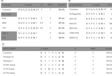
[image:3.603.95.488.76.344.2]manufactur-FIG. 1. Conserved sequence elements in NBM-containing proteins (10, 11, 31, 32). The most conserved element of the NBM is the so-called
A motif. Other conserved elements known from crystal structures to participate in nucleotide binding (G, PM2, and B motif) are also indicated.
(A and B) Consensus sequences of the NBM from some representative family members of the G protein superfamily of GTP-binding proteins
(A) and selected viruses with the indicated NBM-containing protein (B). (C) An NBM was located in HCV NS4B. The consensus amino acid
sequences of all HCV isolates available for examination, the genotype 1b clone used in this study, genotype 3, and the engineered G129V, I131N,
K135S, and K135R mutant NS4Bs are indicated. X, any amino acid. See text for details.
FIG. 2. HCV NS4B binds GTP. Membrane preparations from
Huh-7 cells transfected with plasmids encoding NS4B-GFP (lanes 1
and 5) or GFP (lanes 2 and 6), mock-transfected cells (lanes 3 and 7),
and cells transfected with the plasmid encoding 5A-GFP (lanes 4 and
8) were incubated with
32P-labeled photoactivatable GTP. (A)
Follow-ing 1 min of UV irradiation to activate covalent attachment of any
bound GTP, samples were washed and subjected to
immunoprecipi-tation with a rabbit anti-GFP antibody, SDS-PAGE, and
autoradiog-raphy. (B) Aliquots of the immunoprecipitates were also analyzed by
Western blotting. The blot was probed with a mouse GFP
anti-body, followed by chemiluminescence detection. Molecular mass
markers are indicated on the right.
on November 8, 2019 by guest
http://jvi.asm.org/
[image:3.603.50.278.509.614.2]er’s protocol. The reverse transcriptase reaction and PCR amplification were performed with the Superscript One-Step reverse transcriptase PCR (RT-PCR) kit (Invitrogen) according to the kit’s protocol. Briefly, the amplification reaction
mixture included 1g of total RNA as the template and 10 pmol of each primer.
Two sets of primers (4Left-for with 4Right-rev and 3800sp-for with 4Right-rev; Table 1) were used to amplify 1-kb and 650-bp segments, respectively, each containing the NBM region coding sequence. Performing two independent am-plification reactions for each clone provided further confirmation of the sequenc-ing results. The RT reaction was performed at 50°C for 30 min and was followed by incubation at 95°C for 2 min. The DNA was amplified by 28 cycles of 95°C for 15 s, 60°C for 30 s, and 68°C for 1 min. A final elongation step was performed at 68°C for 10 min. The PCR products were purified from agarose gels with the Ultra Clean 15 DNA purification kit (MoBio) and sent for automatic sequencing on an ABI Prism 377 DNA sequencer (Sequetech).
Protein assays.Concentrations of purified protein and protein content in membrane preparations were determined by the Bradford dye binding procedure using a Bio-Rad (Richmond, Calif.) protein assay kit.
RESULTS AND DISCUSSION
NS4B contains an NBM.
Inspection of the NS4B primary
sequence reveals the presence of an NBM in the middle of
NS4B. This motif consists of a set of conserved amino acids
found in both the GTP-binding members of the G protein
superfamily, as well as several viral proteins with
nucleotide-binding domains (10, 11, 31, 32). The most highly conserved
elements within these nucleotide-binding domains are the
so-called A motif and B motif (Fig. 1A and B). Because binding
and hydrolysis of nucleotides mediate a variety of critical
sig-naling, membrane trafficking, and membrane fusion events (7,
23, 24), we hypothesized that the NBM within NS4B may
similarly be important for NS4B’s role in HCV RNA
replica-tion.
NS4B binds GTP.
To determine the properties associated
with the wild-type and mutated versions of NS4B’s NBM, we
first constructed a plasmid, termed PEF-NS4B-GFP, which
encodes a NS4B protein with a C-terminal, in-frame GFP tag.
This tag allows for visualization in live cells and provides a
convenient epitope outside of any future field of mutagenesis
within NS4B. Importantly, GFP fusions to NS4B have been
previously reported to have no difference in intracellular
lo-calization patterns from wild-type NS4B (14, 19).
To test the hypothesis that NS4B can bind GTP, we
per-formed GTP-binding experiments using Huh-7 cells infected
with a T7RNAP-expressing vaccinia virus and transfected with
plasmids encoding NS4B-GFP or GFP or mock transfected.
Membrane preparations were prepared, and aliquots were
in-cubated with [
␥
-
32P]GTP
␥
AA (a UV-photoactivatable
nonhy-drolyzable GTP analog) essentially as described previously
(13). Following a brief pulse of UV irradiation to activate
covalent attachment of any bound GTP, pelleted membranes
were washed and subjected to immunoprecipitation with a
rabbit anti-GFP antibody, SDS-PAGE, transfer to
nitrocellu-lose, autoradiography, and Western blotting.
[image:4.603.138.447.69.225.2]As shown in Fig. 2A, NS4B-GFP, but not GFP, was
specif-ically labeled with GTP (Fig. 2A, lane 1 versus 2). Western
analysis with an antibody against GFP of the
immunoprecipi-tates revealed comparable expression levels of the two proteins
(Fig. 2B, lane 5 versus 6). To provide another measure of the
specificity of the observed labeling, we performed experiments
with the 5A-GFP plasmid, which encodes the first 31 amino
acids of HCV NS5A fused in frame to the N terminus of GFP
(6). The resulting fusion protein thus contains, like NS4B (6a),
a potent membrane-targeting N-terminal amphipathic helix yet
does not include a known nucleotide-binding element.
Essen-tially no GTP labeling of 5A-GFP was observed (Fig. 2A, lane
4) in spite of a larger amount of expressed protein (Fig. 2B,
FIG. 3. The NS4B NBM is specific for GTP and sensitive to genetic mutation. Each panel shows an autoradiograph (top), a Western blot
analysis with an anti-GFP antibody (middle), and a graph quantifying nucleotide binding relative to wild-type control (bottom). (A) Binding of
labeled GTP is progressively decreased in the presence of increasing concentrations of cold competitor nucleotide. Huh-7 cells were transfected
with a plasmid encoding NS4B-GFP. Membrane preparations were incubated with 10
M labeled GTP compound in the absence of cold GTP
␥
S
(lane 1) or presence of 1 mM (lane 2) or 100
M (lane 3) competing cold GTP
␥
S, followed by immunoprecipitation as in Fig. 2A. (B) NS4B-GFP
binds ATP significantly less efficiently than GTP. Membrane preparations prepared from Huh-7 cells transfected with plasmids encoding
NS4B-GFP (lanes 4 and 5) or GFP (lanes 6 and 7) were incubated with equal concentrations of labeled ATP (lanes 4 and 6) or GTP (lanes 5 and
7), followed by immunoprecipitation as in Fig. 2A. (C) Mutations within the NBM impair GTP binding. Huh-7 cells were transfected with plasmids
encoding wild-type NS4B-GFP (lane 8) or NS4B-GFP with one of the following NBM mutations: Ile131Asn (lane 9), Gly129Val (lane 10),
Lys135Ser (lane 11), or Lys135Arg (lane 12). As above, membrane fractions were incubated with labeled GTP, followed by immunoprecipitation.
Experiments were repeated between two and four times. When present, any detectable binding of GTP to the 5A-GFP negative control protein
was used for background subtraction purposes. Representative gels are shown. Mean values are plotted in the graphs, and error bars represent
standard errors.
on November 8, 2019 by guest
http://jvi.asm.org/
lane 8 versus 5). Labeling of 5A-GFP, detectable only after
extensive film exposure, was used for background subtraction
purposes in subsequent quantitative analyses. To our
knowl-edge, this is the first demonstration that NS4B has
GTP-bind-ing activity. In addition, these results indicate that such bindGTP-bind-ing
activity is preserved when NS4B is expressed in the form of a
fusion protein. The latter is a convenient although not
surpris-ing findsurpris-ing, as nucleoside triphosphate bindsurpris-ing activity remains
intact in fusion proteins made with other nucleotide-binding
proteins (21, 28).
We further evaluated the specificity of NS4B’s GTP binding
by performing binding experiments in the presence of excess
unlabeled ligand. Membrane aliquots of Huh-7 cells expressing
NS4B-GFP were incubated with a
32P-labeled GTP analog as
described above, except that cold guanosine 5
⬘
-
O
-(3-thiotri-phosphate) (GTP
␥
S) was added to the incubation mixture. As
shown in Fig. 3A, the binding of labeled GTP progressively
decreased in the presence of increasing concentrations of the
cold competitor nucleotide (Fig. 3A, top, lane 1 versus 3 and
2).
Although the NBM of poliovirus 2C can bind GTP, its
pref-erence is for ATP (25). We therefore asked whether NS4B
behaves similarly. Binding assays were performed using Huh-7
cells expressing NS4B-GFP as in Fig. 2. Membrane aliquots
were incubated with equal concentrations of
32P-labeled
ATP-␥
-4-azidoanilide or the GTP
␥
AA analog, followed by
immu-noprecipitation. As shown in Fig. 3B, although NS4B can bind
ATP, this appears to be significantly less efficient than GTP
binding (Fig. 3B, top, lane 4 versus 5). Again, no labeling of the
control GFP with either ATP or GTP was detected (Fig. 3B,
top, lanes 6 and 7). These results are in good agreement with
the observation that NS4B, unlike poliovirus 2C, contains
ad-ditional conserved amino acids implicated in GTP binding by
the members of the G protein family (31) (Fig. 1C).
Mutation of NS4B’s NBM impairs GTP binding.
Genetic
mutation of the NBM in a variety of nucleotide-binding
pro-teins can both impair nucleotide binding and disrupt
NBM-mediated functions. We therefore sought to test the hypothesis
that mutations within the NS4B NBM can similarly impair
GTP binding. For this, we designed four mutant proteins, each
harboring a single amino acid mutation within the NS4B NBM
(Fig. 1C). Ile131Asn is a single amino acid change at the X
2position of the NS4B NBM, a position that has been shown to
be critical for NBM function in other proteins (31, 29).
Lys135Ser and Lys135Arg are single amino acid changes at a
position previously reported to confer an intermediate
pheno-type when mutated in the poliovirus system (31). Gly129Val is
a single amino acid mutation at the highly conserved first
position of the NBM A motif consensus sequence. These
mu-tations were introduced into NS4B-GFP, and GTP binding
assays were performed. As shown in Fig. 3C, top, the G129V
and I131N NBM mutant proteins exhibited a two- to threefold
reduction in GTP binding on average compared to the
wild-type NS4B-GFP protein (lanes 10 and 9 versus 8). In contrast,
the K135S and K135R NBM mutant proteins reduced GTP
binding activity to a lesser degree (lanes 11 and 12 versus 8).
This was not simply the result of an obvious gross effect of the
mutations on folding, as the apparent intracellular expression
levels and distribution patterns of mutant and wild-type
pro-teins appeared identical by fluorescence microscopy (Fig. 4).
Moreover, Western analysis with an anti-GFP antibody of the
immunoprecipitates again revealed comparable levels of
ex-pression of these proteins (Fig. 3C).
NS4B has GTPase activity which is mediated by the NBM.
[image:5.603.46.281.71.370.2]We next sought to test the hypothesis that the NS4B NBM
mediates, in addition to GTP binding, GTP hydrolysis. For
this, wild-type NS4B and the four NBM mutant proteins were
fused in frame with N-terminal GST tags. The resultant fusion
proteins, termed GST-NS4B, (IN),
GST-NS4B-(GV), GST-NS4B-(KS), and GST-NS4B-(KR), were expressed
in
E. coli
BL21 and purified with glutathione beads, as
de-scribed previously (30). The purified proteins were then tested
for their ability to hydrolyze GTP by a standard GTPase assay
wherein release of phosphate from [
␥
-
32P]GTP was monitored
by quantitative TLC, essentially as described previously (25).
Not only does NS4B have GTPase activity, but also the GTPase
activity is sensitive to disruption of the NBM (Fig. 5). Indeed,
the targeted mutations could either partially (K135S and K135R)
or nearly completely (G129V and I131N) abolish GTPase
activity. Mutations that mildly affect nucleoside
triphos-phate binding in other proteins have been reported to affect
FIG. 4. Mutations within NS4B’s NBM are not associated with
obvious changes in protein expression level or intracellular distribution
pattern. Huh-7 cells plated on coverslips were transfected with
plas-mids encoding wild-type NS4B-GFP (WT) or NS4B-GFP with one of
the following NBM mutations: Gly129Val (GV), Ile131Asn (IN),
Lys135Ser (KS), or Lys135Arg (KR). Eighteen hours posttransfection
the cells were fixed and imaged by a fluorescence microscope. Note
that all of these proteins display the same reticular membrane
local-ization pattern with distinct foci located in the cytoplasm that is
char-acteristic of wild-type NS4B.
on November 8, 2019 by guest
http://jvi.asm.org/
nucleotide hydrolysis more dramatically (21). It appears that a
similar situation exists for NS4B.
Disrupting NS4B’s NBM inhibits HCV RNA replication.
To
test the hypothesis that the NS4B NBM is important for HCV
replication, we introduced the above series of point mutations
within the NBM into high-efficiency HCV replicons (Fig. 6B).
The latter were then assayed in standard replicon colony
for-mation assays, as previously described (3, 6). A replicon
car-rying a lethal mutation in the active site of the viral polymerase
(GDD
3
AAG), NS5B, was used as a negative control (16).
The tested mutations had a variety of significant effects on
replication (Fig. 7). While mutating the A motif Lys to either
Ser (K135S) or Arg (K135R) was associated with intermediate
levels of replication (2 and 18% respectively), mutating the
first Gly of the A motif to Val (G129V) dramatically inhibited
replication with only rare colony formation. No colonies could
be isolated when the A motif Ile was changed to Asn (I131N).
The appearance of colonies on the G129V plates provided us
with the opportunity to perform reversion analyses. RNA was
therefore extracted from the rare colonies isolated from such
experiments, amplified by RT-PCR, and sequenced. While it is
possible that secondary-site revertants may appear, our
se-quence analysis to date has revealed the presence of only
primary-site revertants. The latter do, however, provide
addi-tional evidence for the requirement of maintaining a funcaddi-tional
NBM.
Our results suggest that the NBM within NS4B is essential
for mediating NS4B’s role in HCV replication in vitro. The
requirement of a NBM for productive viral infection in vivo is
further suggested by the conservation of this motif across
nat-ural HCV isolates of all genotypes (Fig. 1C; data not shown).
The dramatic effect on replication would not appear to be
simply the result of altered expression, misfolding, or
mislo-calization of the mutant proteins. Indeed Western blot
analy-ses of proteins expressed in transfected cells and fluorescence
analysis of the wild-type protein and various NBM mutant
NS4B proteins fused to GFP revealed no obvious differences in
protein levels (Fig. 3C, middle) and intracellular distribution
(Fig. 4), respectively. In addition, the mutations in the NBM do
not seem to disrupt targeting of the protein to the endoplasmic
reticulum (ER) (14, 19), as the colocalization of mutant NS4Bs
with the ER marker PDI is similar to that of wild-type protein
(data not shown). Finally, examination of HCV sequences
from clinical isolates published in public databases reveals that
wobble mutations occur at the codon sites that we mutated,
strongly suggesting that it is unlikely that our mutations disrupt
FIG. 5. NS4B has GTPase activity, which is mediated by the NBM. Equal amounts of purified GST, GST-NS4B, and the NBM mutant proteins
GST-NS4B(GV), GST-NS4B(IN), GST-NS4B(KS), and GST-NS4B(KR) were incubated with [
␥
-
32P]GTP. Aliquots were collected every 15 min
and subjected to TLC to allow separation of hydrolyzed
32P
i
from GTP, followed by autoradiography and phosphorimager analysis. (A)
Repre-sentative TLC plate. Locations of GTP and
32P
i
standards are indicated on the left. (B) GTPase activity of wild-type NS4B (
}
) and G129V (
Œ
),
I131N (
■
), K135S (
F
), and K135R (
⫻
) mutant proteins is plotted as a function of time. When present, any detectable hydrolysis of GTP in the
[image:6.603.82.501.68.247.2]GST control was used for background subtraction purposes. Each data point represents the average of at least four independent determinations.
The error bars represent standard deviations.
FIG. 6. Engineered mutations in NS4B’s NBM. (A) Consensus
se-quences for all NBM-containing proteins, all HCV isolates, and HCV
genotype 1b. X, any amino acid. (B) Single-amino-acid point mutations
(gray) were engineered into the NBM of NS4B at the indicated
posi-tions. (C) Effect on poliovirus replication of mutations engineered at
the same positions in the poliovirus 2C protein’s NBM (22, 31). N.D.,
not done.
on November 8, 2019 by guest
http://jvi.asm.org/
a critical RNA structure or sequence. Rather, these subtle
mutations appear to affect a key regulatory site in the NS4B
amino acid sequence. Interestingly, when analogous mutations
were introduced into the A motif of poliovirus 2C, similar
quantitative and qualitative effects on viral replication were
observed (Fig. 6C) (22, 31). Although speculative, the
de-creased replication potential of the K135R mutant replicon
compared to that of the parental genotype 1b replicon is also
intriguing. Arginine at this position of the NS4B NBM is found
in HCV genotype 3, a genotype that clinically is much more
responsive to interferon therapy than genotype 1b (15).
Current structure prediction models do not offer a
conclu-sive assignment of the secondary structure of NS4B in the
NBM region. This likely reflects the relative paucity of solved
crystal structures of membrane proteins such as NS4B within
the databases used to construct the prediction program
algo-rithms. Nevertheless, it may well be that the NBM is located
within a globular cytoplasmic domain. Certainly, recent
empir-ical studies using glycosylation markers suggest that this
do-main is within the cytoplasm as opposed to the ER lumen (19).
Alternatively, the NBM may reside close to the interface
be-tween the cytoplasm and ER membrane in a “cytoplasmic
pocket,” perhaps created by NS4B oligomerization, within
which the NBM is exposed to nucleotides.
Our results suggest that efficient binding and hydrolysis of
nucleotides by NS4B are required for viral replication. These
results do not rule out the possibility that the NS4B NBM
mediates binding of nucleotides not only as single molecules
but also as part of a polynucleotide structure (such as RNA).
In the latter scenario, by simultaneously binding cellular
mem-branes and RNA, NS4B might serve in channeling the
repli-cation complex through the vesicular membranous structure or
contribute to the structural integrity of the replication complex
by anchoring it to membranes.
GTPases typically are associated with a variety of regulatory
factors (7). Based on our characterization of NS4B we would
now expect it to also have such partners of cellular and/or viral
origin. GTPases, such as Dynamin, can also hydrolyze other
nucleotides (20). We recently became aware that an ATPase
function has been proposed for NS4B (WO 99/01582), and this
is consistent with our ATP binding data. That different types of
nucleotides can bind NS4B suggests that nucleoside analogs
traditionally contemplated for use against the NS5B
polymer-ase activity may now be considered for targeting the GTPpolymer-ase
function of NS4B as well.
Finally, these results reveal that the NBM within NS4B
rep-resents an attractive new potential target for anti-HCV therapy
in its own right. Because the amino acid sequence immediately
adjacent to either side of the NBM region that we mutated is
highly conserved across HCV isolates, yet very different from
that contained in known host cell GTP-binding proteins, highly
selective inhibitors can be readily envisaged. Such compounds
represent an exciting potential addition to current anti-HCV
combination therapy regimens.
ACKNOWLEDGMENTS
We thank Benjamin Aroeti, Allen Cooper, Harry Greenberg, Karla
Kirkegaard, and Charles Rice for helpful discussions and critical
read-ing of the manuscript.
[image:7.603.86.498.68.261.2]S.E. is a recipient of the Stanford Dean’s fellowship award. M.E. is
a recipient of an American Liver Foundation Award. This work was
also supported by RO1DK066793 and a Burroughs Welcome Career
Award (to J.S.G.).
FIG. 7. Genetic disruption of NS4B’s NBM impairs HCV RNA replication. Replication of HCV replicons harboring the mutations depicted
in Fig. 5B were assayed by colony formation assays. (A) Wild-type and mutant replicons were electroporated into Huh-7 cells, and G418-resistant
colonies were selected and stained with crystal violet. These replicons contain the gene for neomycin phosphotransferase (6). Each dot represents
a colony of Huh-7 cells that was able to grow in the presence of G418 due to the presence of efficiently replicating intracellular replicons. WT,
Bart79I (wild type) (3, 6); Pol
⫺, Bart79I with a lethal mutation in NS5B (16); KR, Bart79I with a Lys135Arg point mutation; KS, Bart79I with a
Lys135Ser point mutation; GV, Bart79I with a Gly129Val point mutation; IN, Bart79I with an Ile131Asn point mutation. A representative plate
is shown. (B) Percentages of colonies relative to wild-type control. Note that rare colonies were obtained with the G129V mutant protein (
ⴱ
) and
that none were obtained with the Pol
⫺or I131N mutant proteins.
on November 8, 2019 by guest
http://jvi.asm.org/
REFERENCES
1. Alter, M. J., D. Kruszon-Moran, O. V. Nainan, G. M. McQuillan, F. Gao, L. A. Moyer, R. A. Kaslow, and H. S. Margolis.1999. The prevalence of hepatitis C virus infection in the United States, 1988 through 1994. N. Engl.
J. Med.341:556–562.
2. Bienz, K., D. Egger, Y. Rasser, and W. Bossart.1980. Kinetics and location of poliovirus macromolecular synthesis in correlation to virus-induced
cyto-pathology. Virology100:390–399.
3. Blight, K.J., and A. Kolykhalov, and C. M. Rice.2001. Efficient initiation of
HCV RNA replication in cell culture. Science290:1972–1974.
4. Chu, P. W., and E. G. Westaway.1992. Molecular and ultrastructural analysis of heavy membrane fractions associated with the replication of Kunjin virus
RNA. Arch. Virol.125:177–191.
5. Egger, D., B. Wolk, R. Gosert, L. Bianchi, H. E. Blum, D. Moradpour, and K. Bienz.2002. Expression of hepatitis C virus proteins induces distinct membrane alterations including a candidate viral replication complex. J.
Vi-rol.76:5974–5984.
6. Elazar, M., K. H. Cheong, P. Liu, H. B. Greenberg, C. M. Rice and J. S. Glenn.2003. The amphipathic helix-dependent localization of NS5A
medi-ates HCV RNA replication. J. Virol.77:6055–6061.
6a.Elazar, M., P. Liu, C. M. Rice, and J. S. Glenn.2004. An N-terminal amphipathic helix in hepatitis C virus (HCV) NS4B mediates membrane association, correct localization of replication complex proteins, and HCV
RNA replication. J. Virol.78:11393–11400.
7. Etienne-Manneville, S., and A. Hall.2002. Rho GTPases in cell biology.
Nature420:629–635.
8. Froshauer, S., J. Kartenbeck, and A. Helenius.1988. Alphavirus RNA rep-licase is located on the cytoplasmic surface of endosomes and lysosomes.
J. Cell Biol.107:2075–2086.
9. Glenn, J. S., J. C. Marsters, and H. B. Greenberg.1998. Use of a prenylation
inhibitor as a novel antiviral agent. J. Virol.72:9303–9306.
10. Gorbalenya, A. E., and E. V. Koonin.1989. Viral proteins containing the
purine NTP-binding sequence pattern. Nucleic Acids Res.17:8413–8440.
11. Gorbalenya, A. E., E. V. Koonin, and Y. I. Wolf.1990. A new superfamily of putative NTP-binding domains encoded by genomes of small DNA and
RNA viruses. FEBS Lett.262:145–148.
12. Gosert, R., D. Egger, V. Lohmann, R. Bartenschlager, H. E. Blum, K. Bienz, and D. Moradpour.2003. Identification of the hepatitis C virus RNA repli-cation complex in huh-7 cells harboring subgenomic replicons. J. Virol.
77:5487–5492.
13. Gudi, S. R. P., B. C. Craig, and J. A. Frangos.1996. Fluid flow rapidly activates G proteins in human endothelial cells—involvement of G proteins
in mechanochemical signal transduction. Circ. Res.79:834–839.
14. Hugle, T., F. Fehrmann, E. Bieck, M. Kohara, H. G. Krausslich, C. M. Rice, H. E. Blum, and D. Moradpour.2001. The hepatitis C virus nonstructural protein 4B is an integral endoplasmic reticulum membrane protein. Virology
284:70–81.
15. Knolle, P. A., S. Kremp, T. Hohler, F. Krummenauer, P. Schirmacher, and G. Gerken.1998. Viral and host factors in the prediction of response to
interferon-alpha therapy in chronic hepatitis C after long-term follow-up. J.
Viral Hepat.5:399–406.
16. Kolykhalov, A. A., K. Mihalik, S. M. Feinstone, and C. M. Rice.2000. Hepatitis C virus-encoded enzymatic activities and conserved RNA elements
in the 3⬘nontranslated region are essential for virus replication in vivo.
J. Virol.74:2046–2051.
17. Lazarus, L. H., and R. Barzilai.1974. Association of foot-and-mouth disease virus replicase with RNA template and cytoplasmic membranes. J. Gen.
Virol.23:213–218.
18. Liang, T. J., B. Rehermann, L. B. Seeff, and J. H. Hoofnagle.2000. Patho-genesis, natural history, treatment, and prevention of hepatitis C. Ann.
Intern. Med.132:296–305.
19. Lundin, M., M. Monne, A. Widell, G. von Heijne, and M. A. A. Persson.2003. Topology of the membrane-associated hepatitis C virus protein NS4B. J.
Vi-rol.77:5428–5438.
20. Maeda, K., T. Nakata, Y. Noda, R. Sato-Yoshitake, and N. Hirokawa.1992. Interaction of Dynamin with microtubules: its structure and GTPase activity
investigated by using highly purified Dynamin. Mol. Biol. Cell3:1181–1194.
21. Marin, M. S., R. Casais, J. M. Alonso, and F. Parra.2000. ATP binding and ATPase activities associated with recombinant rabbit hemorrhagic disease
virus 2C-like polypeptide. J. Virol.74:10846–10851.
22. Mirzayan, C., and E. Wimmer.1992. Genetic analysis of an NTP-binding
motif in poliovirus polypeptide 2C. Virology189:547–555.
23. Neves, S. R., P. T. Ram, and R. Iyengar.2002. G protein pathways. Science
296:1636–1639.
24. Pfeffer, S.2003. Membrane domains in the secretory and endocytic pathways.
Cell112:507–517.
25. Pfister, T., and E. Wimmer.1999. Characterization of the nucleoside triphos-phatase activity of poliovirus protein 2C reveals a mechanism by which
guanidine inhibits poliovirus replication. J. Biol. Chem.274:6992–7001.
26. Reed, K. E., and C. M. Rice.2000. Overview of hepatitis C virus genome structure, polyprotein processing, and protein properties. Curr. Top.
Micro-biol. Immunol.242:55–84.
27. Rice, C.M.1996.InB. N. Fields, D. M. Knipe, and P. M. Howley (ed.), Fields virology, p. 931–959. Lippincott-Raven Publications, Philadelphia, Pa. 28. Rodriguez, P. L., and L. Carrasco.1993. Poliovirus protein 2C has ATPase
and GTPase activities. J. Biol. Chem.268:8105–8110.
29. Seeburg, P. H., W. W. Colby, D. J. Capon, D. V. Goeddel, and A. D. Levinson.
1984. Biological properties of human c-Ha-ras1 genes mutated at codon 12.
Nature312:71–75.
30. Smith, D. B., and K. S. Johnson.1988. Single-step purification of
polypep-tides expressed inEscherichia colias fusions with glutathioneS-transferase.
Gene67:31–40.
31. Teterina, N. L., K. M. Kean, A. E. Gorbalenya, V. I. Agol, and M. Girard.
1992. Analysis of the functional significance of amino acid residues in the putative NTP-binding pattern of the poliovirus 2C protein. J. Gen. Virol.
73:1977–1986.
32. Valencia, A., P. Chardin, A. Wittinghofer, and C. Sander.1991. The ras protein family: evolutionary tree and role of conserved amino acids.
Bio-chemistry30:4637–4648.
on November 8, 2019 by guest
http://jvi.asm.org/