0022-538X/09/$08.00⫹0 doi:10.1128/JVI.00251-09
Copyright © 2009, American Society for Microbiology. All Rights Reserved.
MINIREVIEW
Advances in RNA Structure Prediction from Sequence: New
Tools for Generating Hypotheses about Viral RNA
Structure-Function Relationships
䌤
†
Susan J. Schroeder*
Department of Chemistry and Biochemistry, University of Oklahoma, 620 Parrington Oval, Norman, Oklahoma 73019
Advances in sequencing technology have made abundant RNA sequence information available, but the challenge of how to interpret these data remains. The RNA sequence contains many layers of information. RNA sequences code for proteins and small RNAs, such as microRNAs or transacting small interfering RNAs (siRNAs). RNA encodes information about both structure and function. Viral RNA structures, such as riboswitches, internal ribosome entry sites (75), and panhan-dles (71), regulate the stages of the viral life cycle, including replication (100), transcription (99), splicing (40, 48), amino-acylation (31, 55), translation (12, 75, 98), and encapsidation (27, 60, 71, 84). Because viral RNAs are structurally dynamic, current prediction methods focusing on a single minimum free-energy structure may not always identify functionally relevant structures without additional experimental restraints. Because RNA structure determination is often experimentally difficult despite tremendous advances in RNA crystallography, nuclear magnetic resonance spectroscopy, and chemical modification, RNA structure prediction is an important tool for generating hypotheses about structure-function relationships in RNA. RNA structure prediction can be useful for interpreting or designing mutagenesis experiments, identifying conserved structural features, and designing siRNA strategies. This review will briefly outline the basic ideas and assumptions underlying RNA structure prediction, compare different approaches to RNA structure prediction from a user’s perspective, and discuss some applications of RNA structure prediction to viral RNA interfer-ence (RNAi) research.
The RNA sequence, or primary structure, determines the secondary structure, or pattern of canonical Watson-Crick pairs forming duplexes and irregular regions, such as loops and single-stranded regions. The secondary structure then deter-mines the tertiary structure or overall three-dimensional shape of the molecule. The tertiary structure contains the interaction sites for proteins, other RNA molecules, carbohydrates, or other small molecules and thus determines the quaternary or higher-order structure. The greater thermodynamic stability of RNA helices than that of tertiary interactions makes the RNA
folding process hierarchical (35). For example, the favorable free energy involved in forming two stacked base pairs (0.4 to 0.9 kcal/mol per nucleotide) (105) is larger than the favorable free energy to form tertiary interactions, such as ribose zippers or tetraloop-receptor interactions (0.1 to 0.5 kcal/mol per nu-cleotide) (25, 97). Note that 1.4 kcal/mol is equivalent to 1 order of magnitude in a binding constant at 37°C. The hierar-chical nature of RNA folding makes RNA structure prediction a tractable challenge (92).
WHAT IS RNA STRUCTURE PREDICTION?
Phylogenetic analysis of RNA sequences remains the gold standard of RNA structure prediction. Phylogenetic analysis involves generating an alignment of many RNA sequences and looking for patterns of covariation between two nucleotides. Two nucleotides would covary if, for example, nucleotide 1 was usually a G but sometimes a U, if nucleotide 2 was a C when-ever nucleotide 1 was a G, and if nucleotide 2 was an A whenever nucleotide 1 was a U. This pattern of variation main-tains the formation of an isosteric Watson-Crick base pair. Phylogenetic analysis requires a good alignment of many RNA sequences over a diverse range of species with enough varia-tion to observe covariavaria-tion patterns but also enough conserva-tion to establish a good alignment. When regions of 100% conserved nucleotides are shown as large loops in secondary structure diagrams generated by phylogenetic analysis, no in-formation is known about the secondary structure. This does not mean that the nucleotides must necessarily be single stranded. Because RNA-protein interactions and the RNA tertiary structure are also often conserved in evolution, phylo-genetic analysis indirectly accounts for such tertiary and qua-ternary structures. However, sometimes only one or a few sequences are available or the sequences show too little or too much variation for a good alignment. In these cases, compu-tational approaches can generate possible RNA secondary structures.
Computational approaches typically use a recursive algo-rithm to evaluate possible base pairs for a given RNA se-quence. There are 1.8N possible structures for any given
se-quence whereNis the number of nucleotides; thus, there are 3.37⫻1025possible structures for a 100-mer RNA. This is far
more structures than even the fastest computer today could ever calculate in anyone’s lifetime. All computational ap-proaches make some approximations or simplifications in
or-* Mailing address: Department of Chemistry and Biochemistry, Uni-versity of Oklahoma, 620 Parrington Oval, Norman, OK 73019. Phone: (405) 325-3092. Fax: (405) 325-6111. E-mail: [email protected]. † Supplemental material for this article may be found at http://jvi .asm.org/.
䌤Published ahead of print on 15 April 2009.
6326
on November 8, 2019 by guest
http://jvi.asm.org/
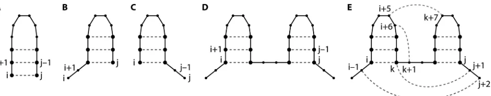
der to reduce the complexity of the RNA folding problem. In a stochastic context-free grammar (SCFG), the most common approximation in RNA structure prediction programs, nucle-otide pairings are evaluated as one of four possibilities: paired with each other, two cases where one nucleotide is paired but the other nucleotide is unpaired, or both nucleotides paired but not to each other (Fig. 1A to D) (29, 72). A recursive algorithm systematically evaluates each possible pair of nucle-otides. This approach makes computation of RNA secondary structures practical and reasonable but excludes the possibility of nonnested pairing interactions, such as pseudoknots, kissing hairpins, and base triples (Fig. 1E). All prediction program listed in Tables 1 and 2 are capable of predicting the two hairpins (Fig. 1D) which are compatible with the formation of nonnested pairing interactions and tertiary structures. In ad-dition, several programs specifically address the computational challenges of nonnested pairing and the limitations of SCFG grammars.
The next step is to rank possible pairs and structures and store this information in what is often called a “fill step.” Free energy is a common ranking system. The free energies of RNA base pair stacks can be measured experimentally. The free energy of a stack of two base pairs accounts for both the hydrogen bonds in Watson-Crick pairs and the interactions between stacked bases, which include electrostatic interac-tions, dipole-dipole interacinterac-tions, van der Wals forces, and hy-drophobic effects. This smallest unit of an RNA double helix is referred to as a nearest neighbor (7, 32, 93, 105). Expanded nearest neighbors describe irregular or single-stranded re-gions; for example, the nucleotides in a hairpin loop and the nucleotides in the closing base pair and all the effects of changes in the phosphodiester backbone as the RNA strand makes this hairpin turn would be grouped together as the smallest unit of which to measure the loop free energy. The nearest-neighbor approximation assumes that the free energy of each smallest unit or motif, for example, a stack of base pairs or a hairpin loop, is the same in any secondary structure con-text. For example, a GNRA tetraloop hairpin would have the same energetic stability in a group I intron, 23S rRNA, or the human immunodeficiency virus (HIV) genome. This approxi-mation is most often reasonable, although non-nearest-neigh-bor effects have been observed for some noncanonical pairs,
especially GU pairs, and single nucleotide bulges (43, 52, 85, 86). This approximation allows the free energy of a secondary structure to be calculated by adding together the energetic stabilities of each smallest unit or motif.
The Turner laboratory members and associates have made extensive free-energy measurements of RNA motifs (32, 49, 66, 67), and these thermodynamic parameters form the core of many RNA structure prediction programs. In order to exper-imentally measure the free energies of RNA motifs, the UV absorbance of small synthetic oligonucleotide duplexes (typi-cally 8 to 12 nucleotides) is recorded as a function of temper-ature in an optical melting experiment. One assumption of these experiments is that the RNA exists in only two states, duplex or single stranded; thus, intermediates occur very quickly and do not contribute significantly to the UV absor-bance. This assumption often does not hold true for complex motifs, such as large loops, multibranch loops, or pseudoknots, and complicates the thermodynamic analysis of these motifs (17, 33, 65). Differential scanning calorimetry (8) or single-molecule force-extension experiments (57) can measure RNA free energies without using a two-state assumption. The free-energy measurements of hairpin, internal, and bulge loops enable predictions for unmeasured loop sequences, but not every possible sequence of every motif has been measured yet. Loop sequences with surprisingly stable energies are still being discovered. Thus, improvements in the collection of RNA free-energy parameters continue as an active area of research.
[image:2.585.48.542.66.163.2]Free-energy minimization approaches to RNA structure prediction assume that the lowest free-energy structure will form. Often, this assumption works well; for example, the base pairing in tRNA and group II introns are correctly predicted 83% and 88% of the time, respectively, by using free-energy minimization and the Zuker algorithm (67). However, the low-est free-energy structure may not always be the functional structure. The conformation of the RNA may be determined by folding kinetics rather than thermodynamic free energy (11, 106). Also, no RNA structure prediction program yet incorpo-rates RNA-protein interactions, which can dramatically alter the folding landscape. Many prediction programs also do not consider pseudoknots or other stabilizing RNA tertiary inter-actions. In addition, a very large number of structures may exist within a small range defined by the error of the free-energy
FIG. 1. Four possible relationships between nucleotides i and j. Dots represent nucleotides i⬍k⬍j. Lines represent sequential covalent connections. Dashed lines indicate hydrogen-bonded base pairing. (A) i and j are paired. (B) i is unpaired and j is paired. (C) i is paired and j is unpaired. (D) Both i and j are paired but not to each other (29). (E) A nested base pair, such as a base pair between nucleotides i and k in panel E, forms a helical stem. Nonnested interactions, such as a base pair between j⫹1 and k (a base triple), a base pair between k⫹1 and i⫹6 (a pseudoknot), or a base pair between i⫹5 and k⫹7 (a kissing hairpin interaction), are not allowed in an SCFG and require a higher-level grammar, more memory, and more run time. A base pair between nucleotides j⫹2 and i⫺1 would create a multibranch loop. Consideration of possible multibranch loops requires an additional array. Vienna Websuite and RNAstructure calculate the stabilities of multibranch loops differently. Three-dimensional motifs, such as ribose zippers and tetraloop-receptor interactions, are built from many pairing interactions.
on November 8, 2019 by guest
http://jvi.asm.org/
calculation, and sometimes those structures can be very differ-ent from one another. This becomes more problematic as the lengths of the RNAs increase. For example, when tRNA (76 nucleotides) and satellite tobacco mosaic virus RNA (1,058 nucleotides) are folded with the Wuchty algorithm in the Vi-enna suite, which calculates all possible structures within a small energy range, tRNA can form 13 different structures within a 1-kcal/mol range, and the structure most different from the minimum energy structure has 23 nucleotides paired differently. In contrast, satellite tobacco mosaic virus can form
42,768 structures within 1 kcal/mol; the structure most differ-ent from the minimum free-energy structure has 560 nucleo-tides paired differently but is only 0.2 kcal/mol greater in free energy, a free-energy difference well within error of the calcu-lation (see the supplemental material). Additional experimen-tal data that further defines possible RNA conformations can address this problem in RNA structure prediction and be-comes even more important for longer RNA sequences.
[image:3.585.41.549.81.260.2]Free-energy minimization is only one approach to analyzing the array of ranked possible pairs and structures for a given
TABLE 1. Characteristics of programsa
Program
Access Algorithm
Free Code
available
Online
submission Windows Linux Zuker Wuchty McCaskill
Ding and
Lawrence Nussinov
mfold X X X X X X
RNAstructure X X X X X X X
Vienna X X X X X X X X X
Sfold X X X
RNA STAR X
PKNOTS X X X
pknotsRG X X X X
NUPACK X X X X
ILM X X X X X
HotKnots X X X X X
ConStruct X X X X
Pfold X X
CONTRAfold X X X X X
Rosetta X X X
MC-Sym X X X X X
aX, feature is available; Free, free for academic use; 3D, three dimensional.
TABLE 2. Websites and key references for programs
Program Website Key references
mfold http://mfold.bioinfo.rpi.edu/ 64, 107–109
RNAstructure http://rna.urmc.rochester.edu/rnastructure.html 61, 66, 68
Vienna http://rna.tbi.univie.ac.at/ 6, 38, 101, 104
Sfold http://sfold.wadsworth.org/ 18, 19, 59
RNA STAR http://wwwbio.leidenuniv.nl/⬃batenburg/STRAbout.html 1, 39, 41, 91, 96
PKNOTS http://selab.janelia.org/software.html 80, 81
pknotsRG http://bibiserv.techfak.uni-bielefeld.de/download/tools/pknotsrg.html 77
NUPACK http://nupack.org/ 22
ILM http://cic.cs.wustl.edu/RNA/ 82, 83
HotKnots http://www.cs.ubc.ca/labs/beta/Software/HotKnots/ 47, 78
ConStruct http://www.biophys.uni-duesseldorf.de/construct3/ 62, 103
Pfold http://www.daimi.au.dk/⬃compbio/pfold/ 53, 54
CONTRAfold http://contra.stanford.edu/contrafold/ 23, 24
Rosetta http://www.rosettacommons.org/tiki/tiki-index.php 14
MC-Sym http://www.major.iric.ca/MC-Pipeline/ 63, 74
on November 8, 2019 by guest
http://jvi.asm.org/
[image:3.585.42.539.441.727.2]RNA sequence. Many different prediction programs use dif-ferent algorithms to analyze or “trace back” through this array to generate not only a minimum free-energy structure but also a set of low-energy structures. In addition, experimental data such as chemical modification or phylogenetic covariation can be used in addition to free-energy minimization. Alternatively, possible pairs can be ranked using criteria other than free energy, such as the maximum number of base pairs, or the base pairing can be described using concepts other than SCFGs, such as nucleotide cyclic motifs.
Different prediction programs have developed different strategies for addressing the limitations of RNA structure pre-diction by free-energy minimization alone. Because even slight differences in calculation methods or the set of thermodynamic parameters can alter the minimum free-energy prediction, comparison of the results from more than one program is a good approach to generating an informed hypothesis about RNA structure and function. Table 1 lists some of these pro-grams and their characteristics, such as access, options for algorithms, and options for including experimental data. Refer to each prediction program’s website or primary citation for a complete description of user options and an evaluation of performance. The evaluation criteria and databases of known secondary structures used to evaluate prediction accuracy vary substantially between different research groups and make di-rect comparisons complex. Results of comparative tests of dif-ferent RNA structure prediction programs have been reviewed elsewhere (26, 42, 78). Prediction programs aim to predict structures based on a set of rules and assumptions rather than optimize for particular types of structures, although several programs are designed to address the computational difficul-ties associated with predicting pseudoknotted, nonnested pair-ing (Fig. 1E). This list is not exhaustive, and the followpair-ing discussion only highlights some practical considerations for selecting an RNA structure prediction program. The “best” program is the user’s decision that depends on the RNA stud-ied, the questions asked, the available experimental data and resources, and the intended applications of the structure pre-diction.
APPROACHES TO RNA STRUCTURE PREDICTION
mfold and UNAFold.The popular mfold program has been expanded, updated, and renamed UNAFold (64). The Quik-fold option in DINAmelt, the online version of the program, contains the traditional format where a user submits a se-quence and chooses RNA or DNA folding, temperature, salt conditions, and a free-energy range for suboptimal structures. UNAFold and mfold use the Zuker algorithms (108, 109) to compute the minimum free-energy structure for a given se-quence and systematically sample structures within a percent-age of free-energy range to create a set of diverse suboptimal structures. Users can specify nucleotides to be single stranded or in a particular base pair if information from chemical mod-ification, enzymatic probing, compensatory mutations, or phy-logenetic covariation is available. The options for hybridization analysis can be applied to the design of siRNA or antisense RNA.
[image:4.585.43.542.82.259.2]RNAstructure.RNAstructure 4.6 is a Windows implemen-tation of the Zuker algorithm and includes additional options for other folding algorithms and incorporation of experimental data. The authors of RNAstructure collaborate very closely with the Turner laboratory and keep the most up-to-date ther-modynamic parameters (66). The OligoWalk program can be used for siRNA design (61). Two unique ways of incorporating experimental data in the RNA folding is done with Dynalign (42, 68) and chemical modification (15, 66). The Dynalign program computes the lowest free-energy sequence alignment and secondary structure common to two RNA sequences. The two sequences need not be aligned, but the maximum number of sequences to be folded at one time is two. RNAstructure also uses a more-advanced definition of chemical modification restraints for traditional chemical modification reagents and new selective 2⬘-hydroxyl acylation analyzed by primer exten-sion (SHAPE) data (70). Chemical modification reagents, such as dimethyl sulfate, methylate accessible adenine nucleotides and cause stops in reverse transcription primer extension re-actions. This type of chemical modification occurs not only at single-stranded adenines but also at adenines in AU pairs that
TABLE 1—Continued
Experimental data
siRNA
design Pseudoknots
3D
prediction Unique feature(s)
Turner
rules Phylogeny
Restrain as single stranded
Restrain pair
X X X X First popular folding program
X X X X X Advanced chemical modification and SHAPE
X X X X RNAalifold, RNAz
X X Advanced siRNA design
X X Genetic, Monte Carlo, and greedy algorithms
X X Pseudoknot grammar
X X Focus on three types of pseudoknots
X X Pseudoknot pair probabilities
X X X Iterative loop matching algorithm
X X Algorithm to find hot spots
X X X Alignment editor, user interface
X SCFG training
X Conditional training
X X X Fragment assembly
X X X X X Nucleotide cyclic motifs
on November 8, 2019 by guest
http://jvi.asm.org/
are at the end of a helix, next to a GU pair, or next to bulged nucleotides; and RNAstructure allows for all these possibilities when a nucleotide is marked as chemically modified (66). SHAPE detects flexibility in the RNA backbone that correlates with helical or irregular motifs in secondary structure forma-tion, and these data can be included as a restraint or as a pseudofree energy in ranking possible base pairs (15).
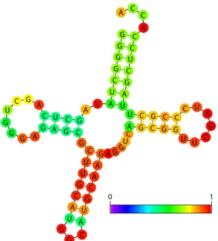
Vienna RNA Websuite.The Vienna RNA Websuite intro-duced the Wuchty algorithm, developed applications of the McCaskill algorithm, and also offers a wide variety of algo-rithms and functions. The Wuchty algorithm computes all pos-sible secondary structures within a narrow free-energy range (104). The Wuchty algorithm generates a small but complete set of suboptimal structures that may include some very dif-ferent secondary structures but also very many highly similar structures. However, structures containing more than one sub-optimal region may occur in the Wuchty set of structures but would be absent if the Zuker method for sampling suboptimal structures were used. The McCaskill algorithm evaluates the statistical probabilities of base pairs and secondary structures by using a partition function calculation (69). “Statistical” re-fers to the thermodynamic and statistical mechanics definition
based on a Boltzmann distribution. This information can be overlaid in different colors on the lowest-energy structure and thus provides an estimate of confidence in the predicted struc-ture (Fig. 2). For example, one region of a secondary strucstruc-ture (red) may be exceptionally thermodynamically stable, occur frequently in all suboptimal structures, have a high statistical probability, and thus be more likely to be a correct prediction. In contrast, another region of the secondary structure (purple or dark blue at the left end of the scale near zero probability) may have multiple thermodynamically equivalent possible con-formations in the suboptimal structures and a low statistical probability and thus be less clearly a correct prediction.
[image:5.585.135.451.66.414.2]The Vienna Websuite includes RNAalifold and RNAz, pro-grams that combine phylogenetic covariation information with thermodynamic stability and base pair statistical probabilities when ranking possible base pairs. RNAalifold accepts any number of aligned sequences in the standard ClustalW and FASTA formats and generates a lowest-energy consensus structure by weighting the ranking of base pairs with covaria-tion data (6). RNAz searches for noncoding RNAs (101) or conserved secondary structure elements within long RNA se-quences, such as viral genomes (45, 46), by weighting base pair
FIG. 2. The minimum free-energy secondary structure of tRNAala fromShigella sonneiwith the base pair probabilities shown in color. The red pairs have a high probability of forming, green pairs have a medium probability of forming, and blue pairs (none shown) have a low probability of forming. The normalized scale showing zero (dark blue) to one (red) probability is shown on the lower right. Figure produced using Vienna Websuite with default parameters.
on November 8, 2019 by guest
http://jvi.asm.org/
statistical probabilities and thermodynamic stabilities with co-variation data in a combined Z score for possible base pairs. The RNAup and RNAxs functions are used to design siRNA strategies.
Sfold.The Sfold algorithm uses a unique algorithm to aid in the design of siRNA. The algorithm combines thermodynamic stabilities (67), calculations of target accessibility (59, 88), and empirical rules for efficient siRNA developed by the Zamore, Amgen, and Dharmacon groups (3, 51, 79, 87, 94). The website offers specialized programs for the design of siRNA, antisense RNA, trans-cleaving RNA, and mRNA-microRNA interac-tions as well as a general program for statistically sampling suboptimal RNA structures. The algorithm uses a partition function calculation and then groups suboptimal structures by similarity (9, 19, 20). The centroid structure is the most-repre-sentative structure that is closest in similarity to all the other structures. If the centroid structure is different from the min-imum free-energy structure, the centroid structure is often closer to the phylogenetic prediction and contains fewer base pairs, or fewer false-positive base pair predictions, than the minimum free-energy prediction. The point is to show a struc-ture that represents a group of strucstruc-tures rather than a single predicted structure. Many long RNA sequences, such as viral genomes or mRNA, may not have a single structure but in-stead have a dynamic structure that has some conserved fea-tures but also varies and changes, and these many conforma-tions may all exist simultaneously in the cell. Thus, the centroid structure may better describe the overall average prediction for all these conformations and may better show whether a target site is accessible. Sfold also outputs a probability profile of nucleotide accessibility, loop profiles, and an estimate of siRNA potency.
PROGRAMS FOR PREDICTING SECONDARY STRUCTURES WITH PSEUDOKNOTS
RNA STAR.The problem of predicting pseudoknots (Fig. 1) has inspired a wide variety of solutions and adaptations of existing algorithms. RNA STAR offers three algorithms that simulate the folding of RNA and include pseudoknot struc-tures. The greedy algorithm, based on the idea that maximizing each step of the calculation will produce the optimal result, adds the most stable stem helix onto a growing structure (1). A Monte Carlo algorithm generates a set of random structures and then selects a probability-weighted stem helix to add onto a growing structure (39). The genetic algorithm simulates an evolution of RNA folding by starting with unfolded structures and increasing the fitness criterion, low free energy, in each generation (41, 96). The genetic algorithm allows stem helices to be added or removed and allows “crossovers,” structures containing the best pieces of previous generations. Because these programs are simulations rather than calculations, the output should also be evaluated on how well the program converges to a single structure and the reproducibility of the final structure. The user chooses the number of rounds in the simulation.
PKNOTS and pknotsRG.PKNOTS uses free-energy mini-mization and a higher-level grammar that removes the “con-text-free” restrictions shown in Fig. 1 (80, 81). The computa-tional cost of allowing pseudoknots [O(N6) in time and O(N4)
in space], however, makes the program computationally unrea-sonable for sequences greater than approximately 100 nucleo-tides. The program pknotsRG restricts the types of possible pseudoknots to three common simple types, with at most two helices, and does not allow complex interactions, such as kiss-ing hairpins or triple helices. This approximation improves the run time [O(N4) in time and O(N2) in space] so that sequences
of up to 800 nucleotides are computationally reasonable (77). The O(Nx) function describes the computational complexity
and program run time, depending onN, the number of nucle-otides in the sequence. The actual time necessary for the com-putation will also depend on the type of computer used and the processor speed.
NUPACK.The NUPACK program provides probabilities of pseudoknotted base pair formation (22). These base pair prob-abilities can predict free energies of pseudoknot stprob-abilities that match experimental values (21). Base pair probabilities are useful for analyses of how pseudoknots may be in equilibrium between two conformations and how mutations can shift that equilibrium. Optical melting experiments and single-molecule pulling experiments can provide measurements of pseudoknot interaction energies (for examples, see references 10, 34, and 76).
ILM. The ILM program uses an iterative loop matching algorithm to maximize base pairs and allows pseudoknots to form by allowing base pairs to be added or removed in succes-sive rounds (83). The Nussinov algorithm, or maximum loop matching algorithm, is the basic framework for generating a structure with the most possible base pairs (73). The base pairs are ranked using both thermodynamic parameters and covaria-tion data for aligned sequences. ILM requires the RnaViz program (16) to visualize the RNA secondary structure with pseudoknots.
HotKnots. The HotKnots program takes advantage of the hierarchical nature of RNA secondary and tertiary structure folding (47, 78). A hot spot is a particularly stable branch point in a tree of possible structures. The program generates a tree of structures using the Zuker algorithm and restrains pseudoknot “hot spots” to be single stranded and then allows structures with pseudoknots to form. The HotKnots program is a heuristic that does not guarantee the lowest energy structure but quickly predicts many reasonably good structures.
ConStruct. The program ConStruct simultaneously aligns multiple RNA sequences and finds a consensus RNA struc-ture, thus combining information on thermodynamic stability and phylogeny (103). Because sequence alignment often re-quires manual editing, ConStruct offers an interactive user interface that allows manual editing of the alignment. Thus, the alignment can be improved with preliminary information about secondary structure. A new additional step then maxi-mizes base pairing and thus allows for pseudoknot, base triple, and other tertiary interactions.
NONTHERMODYNAMIC APPROACHES
Pfold and CONTRAfold.Not all folding programs use lowest free energy as the criteria for forming base pairs in an RNA secondary structure. Pfold uses a sequence alignment and a set of base pair probabilities determined from a set of known tRNA and 23S rRNA secondary structures to predict RNA
on November 8, 2019 by guest
http://jvi.asm.org/
secondary structure (53). CONTRAfold (24) uses a condi-tional training method and a database of 151 consensus struc-tures from known secondary strucstruc-tures in the Rfam database (36, 37) to generate parameters for RNA folding. The param-eters for base stacking interactions are surprisingly propor-tional to the experimentally measured values. RNA alignment and folding (RAF), an option in CONTRAfold, accepts mul-tiple unaligned sequences and simultaneously aligns the se-quences and computes a consensus secondary structure (23).
PREDICTING THREE-DIMENSIONAL STRUCTURE
Rosetta.FARNA, fragment assembly of RNA using Rosetta software, samples different possible conformations of RNA to predict three-dimensional structure (14). Computational anal-ysis of the crystal structure of the 50S ribosomal subunit of
Haloarcula marismortui (4) produced a library of RNA frag-ments, which is a collection of torsion angles for all possible conformations of three nucleotide pieces, and an energy func-tion, which includes terms for hydrogen bonding, base stack-ing, compactness, steric clashes, and planar base pairs. The only additional input is the sequence of the RNA to be pre-dicted. The Monte Carlo sampling method limits the length of the RNA to approximately 40 nucleotides. This method accu-rately predicts the noncanonical regions for 13 out of 20 test cases, including conformations not in the ribosome, such as the base triple in the SL2 stem-loop of the HIV psi packaging signal (2) and the pseudoknot in beet western yellow mosaic virus (30).
MC-Sym.MC-Sym, macromolecular conformation by sym-bolic generation, uses a hierarchical approach to three-dimen-sional structure prediction (74). MC-Fold predicts a secondary structure; MC-Sym predicts a three dimensional structure from a given secondary structure. The program is based on nucleo-tide cyclic motifs, the smallest nondivisible unit in a graph grammar description of RNA structure which includes back-bone torsion angles, base pairing, and base stacking interac-tions (56, 89). The program also incorporates experimental data from chemical modification, SHAPE, hydroxyl radical footprinting, phylogenetic alignments, and distance restraints from nuclear magnetic resonance spectroscopy. The program is capable of predicting structures of 150 nucleotides and cor-rectly predicts 11 of 13 test sequences, which contain a maxi-mum of 47 nucleotides and include the pseudoknot in yellow leaf virus (13).
APPLICATIONS OF RNA STRUCTURE PREDICTION TO RNAi
One of the most exciting applications of RNA structure prediction is toward understanding and manipulating the abil-ity of small RNAs to regulate gene expression. RNA structure prediction of the secondary structure of the mRNA target, the pre-microRNA, or short hairpin RNA can improve microRNA identification and siRNA design strategies (44, 50, 59, 88, 90). The accessibility of the mRNA target site for base pairing and the energetic cost of rearranging the RNA secondary structure can be important for estimating the effectiveness of the small RNA for regulating gene expression. RNA structure predic-tion can improve identificapredic-tion of viral sequences that are
Dicer substrates to be processed as viral siRNA and also iden-tification of viral siRNA target sites (28, 44, 50, 88, 90, 95). Sequence complementarity alone does not provide sufficient specificity for effective siRNA strategies. Higher-order RNA structure can provide the specificity in molecular recognition to reduce off-target effects (44, 50, 59). RNA structure predic-tion can also facilitate the interpretapredic-tion of mutapredic-tions. For example, in studies of RNAi targeting HIV type 1 (HIV-1) in human cells (102), some resistance mutations in the HIV-1 sequence emerged that were not at the siRNA target site. When the secondary structure prediction of the target site region was recalculated with the resistance mutation sequence, an alternate fold was revealed. Mutations that cause changes in secondary structure in HIV-1 RNA and MS2 bacteriophage RNA occur in forced-evolution experiments in which a dele-terious mutation is introduced and the progeny are sequenced to identify compensating mutations (5, 58). Accurate structure prediction methods can thus facilitate future RNAi technolo-gies and viral RNA research.
ACKNOWLEDGMENTS
The author is supported by grants from the Oklahoma Center for the Advancement of Science and Technology and the Pharmaceutical Research and Manufacturers of America Foundation and by an insti-tutional research grant from the American Cancer Society to the University of Oklahoma Health Sciences Center.
The author thanks Douglas H. Turner and Kay Sheez for critical reading of the manuscript.
REFERENCES
1.Abrahams, J., M. van den Berg, E. van Batenburg, and C. Pleij.1990. Prediction of RNA secondary structure, including pseudoknotting, by com-puter simulation. Nucleic Acids Res.18:3035–3044.
2.Amarasinghe, G., R. De Guzman, R. Turner, and M. Summers.2000. NMR structure of the stem-loop SL2 of the HIV-1 psi RNA packaging signal reveals a novel A-U-A base-triple platform. J. Mol. Biol.299:145–156. 3.Amarzguioui, M., and H. Prydz.2004. An algorithm for selection of
func-tional siRNA sequences. Biochem. Biophys. Res. Commun.316:1050–1058. 4.Ban, N., P. Nissen, J. Hansen, P. B. Moore, and T. A. Steitz.2000. The complete atomic structure of the large ribosomal subunit at 2.4 Å resolu-tion. Science289:905–920.
5.Berkhout, B., and A. T. Das.2009. Virus evolution as a tool to study HIV-1 biology. Methods Mol. Biol.485:436–451.
6.Bernhart, S., I. Hofacker, S. Will, A. Gruber, and P. Stadler.2008. RNAali-fold: improved consensus structure prediction for RNA alignments. BMC Bioinformatics9:474.
7.Borer, P. N., B. Dengler, I. Tinoco, Jr., and O. C. Uhlenbeck.1974. Stability of ribonucleic acid-double-stranded helices. J. Mol. Biol.86:843–853. 8.Breslauer, K. J.1995. Extracting thermodynamic data from equilibrium
melting curves for oligonucleotide order-disorder transitions. Methods En-zymol.259:221–241.
9.Carvalho, L. E., and C. E. Lawrence.2008. Centroid estimation in discrete high-dimensional spaces with applications in biology. Proc. Natl. Acad. Sci. USA105:3209–3214.
10.Chen, G., J. Wen, and I. Tinoco, Jr. 2007. Single molecule mechanical unfolding and folding of a pseudoknot in human telomerase RNA. RNA 13:2175–2188.
11.Chen, S., and K. Dill.2000. RNA folding energy landscapes. Proc. Natl. Acad. Sci. USA97:646–651.
12.Costantino, D. A., J. S. Pfingsten, R. P. Rambo, and J. S. Kieft.2008. tRNA-mRNA mimicry drives translation initiation from a viral IRES. Nat. Struct. Mol. Biol.15:57–64.
13.Cornish, P., S. Stammler, and D. Giedroc.2006. The global structures of a wild-type and poorly functional plant luteoviral mRNA pseudoknot are essentially identical. RNA12:1959–1969.
14.Das, R., and D. Baker.2007. Automated de novo prediction of native-like RNA tertiary structures. Proc. Natl. Acad. Sci. USA104:14664–14669. 15.Deigan, K., T. Li, D. Mathews, and K. Weeks.2009. Accurate
SHAPE-directed RNA structure determination. Proc. Natl. Acad. Sci. USA106:97– 102.
16.De Rijk, P., J. Wuyts, and R. De Wachter.2003. RnaViz 2: an improved representation of RNA secondary structure. Bioinformatics19:299–300.
on November 8, 2019 by guest
http://jvi.asm.org/
17.Diamond, J., D. Turner, and D. Mathews.2001. Thermodynamics of three-way multibranch loops in RNA. Biochemistry40:6971–6981.
18.Ding, Y., C. Y. Chan, and C. E. Lawrence.2004. Sfold web server for statistical folding and rational design of nucleic acids. Nucleic Acids Res. 32:W135–W141.
19.Ding, Y., and C. E. Lawrence.2004. A statistical sampling algorithm for RNA secondary structure prediction. Nucleic Acids Res.31:7280–7301. 20.Ding, Y., and C. E. Lawrence.2001. Statistical prediction of single-stranded
regions in RNA secondary structure and application to predicting effective antisense target sites and beyond. Nucleic Acids Res.29:1034–1046. 21.Dirks, R. M., and N. A. Pierce.2004. An algorithm for computing nucleic
acid base-pairing probabilities including pseudoknots. J. Comput. Chem. 25:1295–1304.
22.Dirks, R. M., and N. A. Pierce.2003. A partition function algorithm for nucleic acid secondary structure including pseudoknots. J. Comput. Chem. 24:1664–1677.
23.Do, C. B., C.-S. Foo, and S. Batzglou. 2008. A max-margin model for efficient simultaneous alignment and folding of RNA sequences. Bioinfor-matics24:i68–i76.
24.Do, C. B., D. A. Woods, and S. Batzglou.2006. CONTRAfold: RNA sec-ondary structure prediction without physics-based models. Bioinformatics 22:e90–e98.
25.Doherty, E. A., R. T. Batey, B. Masquida, and J. A. Doudna.2001. A universal mode of helix packing in RNA. Nat. Struct. Biol.8:339–343. 26.Dowell, R., and S. Eddy.2004. Evaluation of several lightweight stochastic
context-free grammars for RNA secondary structure prediction. BMC Bioinformatics5:71.
27.D’Souza, V., and M. F. Summers.2004. Structural basis for packaging the dimeric genome of Moloney murine leukemia virus. Nature431:586–590. 28.Du, Q.-S., C.-G. Duan, Z.-H. Zhang, Y.-Y. Fang, R.-X. Fang, Q. Xie, and H.-S. Guo.2007. DCL4 targetsCucumber mosaic virussatellite RNA at novel secondary structures. J. Virol.81:9142–9151.
29.Eddy, S. R.2004. How do RNA folding algorithms work? Nat. Biotechnol. 22:1457–1458.
30.Egli, M., G. Minasov, L. Su, and A. Rich.2002. Metal ions and flexibility in a viral RNA pseudoknot at atomic resolution. Proc. Natl. Acad. Sci. USA 99:4302–4307.
31.Felden, B., C. Florentz, A. McPherson, and R. Giege.1994. A histidine accepting tRNA-like fold at the 3⬘end of satellite tobacco mosaic virus RNA. Nucleic Acids Res.22:2882–2886.
32.Freier, S. M., R. Kierzek, J. A. Jaeger, N. Sugimoto, M. H. Caruthers, T. Neilson, and D. H. Turner.1986. Improved free-energy parameters for predictions of RNA duplex stability. Proc. Natl. Acad. Sci. USA83:9373– 9377.
33.Giedroc, D., C. Theimer, and P. Nixon. 2000. Structure, stability, and function of RNA pseudoknots involved in stimulating ribosomal frameshift-ing. J. Mol. Biol.298:167–185.
34.Green, L., C. Kim, C. Bustamante, and I. Tinoco, Jr.2008. Characterization of the mechanical unfolding of RNA pseudoknots. J. Mol. Biol.375:511– 528.
35.Greenleaf, W., K. Frieda, D. Foster, M. Woodside, and S. Block.2008. Direct observation of hierarchical folding in single riboswitch aptamers. Science319:630–633.
36.Griffiths-Jones, S., A. Bateman, M. Marshall, A. Khanna, and S. R. Eddy. 2003. Rfam: an RNA family database. Nucleic Acids Res.31:439–441. 37.Griffiths-Jones, S., S. Moxon, M. Marshall, A. Khanna, S. R. Eddy, and A.
Bateman.2005. Rfam: annotating non-coding RNAs in complete genomes. Nucleic Acids Res.33:D121–D124.
38.Gruber, A. R., R. Lorenz, S. H. Bernhart, R. Neubock, and I. L. Hofacker. 2008. The Vienna RNA Websuite. Nucleic Acids Res.36:W70–W74. 39.Gultyaev, A. P.1991. The computer simulation of RNA folding involving
pseudoknot formation. Nucleic Acids Res.19:2489–2494.
40.Gultyaev, A. P., H. A. Heus, and R. C. Olsthoorn.2007. An RNA confor-mational shift in recent H5N1 influenza A viruses. Bioinformatics23:272– 276.
41.Gultyaev, A. P., F. H. van Batenburg, and C. W. Pleij.1995. The computer simulation of RNA folding pathways using a genetic algorithm. J. Mol. Biol. 250:37–51.
42.Harmanci, A., G. Sharma, and D. Mathews.2007. Efficient pairwise RNA structure prediction using probabilistic alignment constraints in Dynalign. BMC Bioinformatics8:130.
43.He, L., R. Kierzek, J. SantaLucia, Jr., A. E. Walter, and D. H. Turner.1991. Nearest-neighbor parameters for GU mismatches. Biochemistry30:11124– 11132.
44.Hofacker, I.2007. How microRNAs choose their targets. Nat. Genet.39: 1191–1192.
45.Hofacker, I., M. Fekete, C. Flamm, M. Huynen, S. Rauscher, P. Stolorz, and P. Stadler.1998. Automatic detection of conserved RNA structure elements in complete RNA virus genomes. Nucleic Acids Res.26:3825– 3836.
46.Hofacker, I., and P. Stadler.1999. Automatic detection of conserved base pairing patterns in RNA virus genomes. Comput. Chem.23:401–414.
47.Jabbari, H., A. Condon, and S. Zhao. 2008. Novel and efficient RNA secondary structure prediction using hierarchical folding. J. Comput. Biol. 15:139–163.
48.Jablonski, J. A., E. Buratti, C. Stuani, and M. Caputi.2008. The secondary structure of the human immunodeficiency virus type 1 transcript modulates viral splicing and infectivity. J. Virol.82:8038–8050.
49.Jaeger, J., D. Turner, and M. Zuker.1989. Improved predictions of sec-ondary structures for RNA. Proc. Natl. Acad. Sci. USA86:7706–7710. 50.Kertesz, M., N. Iovino, U. Unnerstall, U. Gaul, and E. Segal.2007. The role
of site accessibility in microRNA target recognition. Nature Genetics39: 1278–1284.
51.Khvorova, A., A. Reynolds, and S. D. Jayasena.2003. Functional siRNas and miRNas exhibit strand bias. Cell115:209–216.
52.Kierzek, R., M. E. Burkard, and D. H. Turner.1999. Thermodynamics of single mismatches in RNA duplexes. Biochemistry38:14214–14223. 53.Knudsen, B., and J. Hein.2003. Pfold: RNA secondary structure grammar
prediction using stochastic context-free grammars. Nucleic Acids Res.31: 3423–3428.
54.Knudsen, B., and J. Hein.1999. RNA secondary structure prediction using stochastic context-free grammars and evolutionary history. Bioinformatics 15:446–454.
55.Koenig, R., S. Barends, A. Gultyaev, D. Lesemann, H. Vetten, S. Loss, and C. Pleij.2005. Nemesia ring necrosis virus: a new tyomovirus with genomic RNA having a histylatable tobamovirus-like 3⬘end. J. Gen. Virol.86:1827– 1833.
56.Lemieux, S., and F. Major.2006. Automated extraction and classification of RNA tertiary structure cyclic motifs. Nucleic Acids Res.34:2340–2346. 57.Li, P. T., J. Vieregg, and I. Tinoco, Jr.2008. How RNA unfolds and refolds.
Annu. Rev. Biochem.77:77–100.
58.Licis, N., and J. van Duin.2006. Structural constraints and mutational bias in the evolutionary restoration of a severe deletion in RNA phage MS2. J. Mol. Evol.63:314–329.
59.Long, D., R. Lee, P. Williams, C. Chan, V. Ambros, and Y. Ding.2007. Potent effect of target structure on microRNA function. Nat. Struct. Mol. Biol.14:287–294.
60.Loo, L., R. Guenther, S. Lommel, and S. Franzen.2007. Encapsidation of nanoparticles by red clover necrotic mosaic virus. J. Am. Chem. Soc.129: 11111–11117.
61.Lu, Z., and D. Mathews.2008. OligoWalk: an online siRNA design tool utilizing hybridization thermodynamics. Nucleic Acids Res. 36:W104– W108.
62.Luck, R., S. Graf, and G. Steger.1999. ConStruct: a tool for thermodynamic controlled prediction of conserved secondary structure. Nucleic Acids Res. 27:4208–4217.
63.Major, F., M. Turcotte, D. Gautheret, G. Lapalme, E. Fillion, and R. Cedergren.1991. The combination of symbolic and numerical computation for three-dimensional modeling of RNA. Science253:1255–1260. 64.Markham, N. R., and M. Zuker.2008. UNAFold: software for nucleic acid
folding and hybridization. Methods Mol. Biol.453:3–31.
65.Mathews, D. H., and D. H. Turner.2002. Experimentally derived nearest-neighbor parameters for the stability of RNA three-and four-way multi-branch loops. Biochemistry41:869–880.
66.Mathews, D. H., M. D. Disney, J. L. Childs, S. J. Schroeder, M. Zuker, and D. H. Turner.2004. Incorporating chemical modification constraints into a dynamic programming algorithm for prediction of RNA secondary struc-ture. Proc. Natl. Acad. Sci. USA101:7287–7292.
67.Mathews, D. H., J. Sabina, M. Zuker, and D. H. Turner.1999. Expanded sequence dependence of thermodynamic parameters improves prediction of RNA secondary structure. J. Mol. Biol.288:911–940.
68.Mathews, D. H., and D. H. Turner.2002. Dynalign: an algorithm for finding the secondary structure common to two RNA sequences. J. Mol. Biol. 317:191–203.
69.McCaskill, J.1990. The equilibrium partition function and base pair bind-ing probabilities for RNA secondary structure. Biopolymers29:1105–1119. 70.Merino, E. J., K. A. Wilkinson, J. L. Coughlan, and K. M. Weeks.2005. RNA structure analysis at single nucleotide resolution by selective 2⬘ -hydroxyl acylation and primer extension (SHAPE). J. Am. Chem. Soc. 127:4223–4231.
71.Mir, M., B. Brown, B. Hjelle, W. Duran, and A. Panganiban.2006. Han-tavirus N protein exhibits genus-specific recognition of the viral RNA pan-handle. J. Virol.80:11283–11292.
72.Nussinov, R., and A. Jacobson.1980. Fast algorithm for predicting the secondary structure of single-stranded RNA. Proc. Natl. Acad. Sci. USA 77:6309–6313.
73.Nussinov, R., G. Pieczenik, J. Griggs, and D. Kleitman.1978. Algorithms for loop matchings. SIAM J. Appl. Math.35:68–82.
74.Parisien, M., and F. Major.2008. The MC-Fold and MC-Sym pipeline infers RNA structure from sequence data. Nature452:51–55.
75.Pfingsten, J., and J. Kieft.2008. RNA Structure-based ribosome recruit-ment: lessons from theDicistroviridaeintergenic region IRESes. RNA14: 1255–1263.
on November 8, 2019 by guest
http://jvi.asm.org/
76.Puglisi, J., J. Wyatt, and I. Tinoco, Jr.1988. A pseudoknotted RNA oligo-nucleotide. Nature331:283–286.
77.Reeder, J., and R. Giegerich.2004. Design, implementation and evaluation of a practical pseudoknot folding algorithm based on thermodynamics. BMC Bioinformatics5:104.
78.Ren, J., B. Rastegari, A. Condon, and H. Hoos.2005. HotKnots: heuristic prediction of RNA secondary structures including pseudoknots. RNA11: 1491–1504.
79.Reynolds, A., D. Leake, Q. Boese, S. Scaringe, W. Marshall, and A. Khvorova. 2004. Rational siRNA design for RNA interference. Nat. Biotechnol.22:326–330.
80.Rivas, E., and S. Eddy.2000. The language of RNA: a formal grammar that includes pseudoknots. Bioinformatics16:334–340.
81.Rivas, E., and S. R. Eddy.1999. A dynamic programming algorithm for RNA structure prediction including pseudoknots. J. Mol. Biol.285:2053– 2068.
82.Ruan, J., G. Stormo, and W. Zhang.2004. ILM: a web server for predicting RNA secondary structures with pseudoknots. Nucleic Acids Res.32:W146– W149.
83.Ruan, J., G. Stormo, and W. Zhang.2004. An iterated loop matching approach to the prediction of RNA secondary structures with pseudoknots. Bioinformatics20:58–66.
84.Schneemann, A.2006. The structural and functional role of RNA in icosa-hedral virus assembly. Annu. Rev. Microbiol.60:51–67.
85.Schroeder, S. J., and D. H. Turner.2000. Factors affecting the thermody-namics stability of small asymmetric internal loops in RNA. Biochemistry 39:9257–9274.
86.Schroeder, S. J., and D. H. Turner.2001. Thermodynamic stabilities of internal loops with GU closing pairs in RNA. Biochemistry 40:11509– 11517.
87.Schwarz, D. S., G. Hutvagner, T. Du, Z. Xu, N. Aronin, and P. D. Zamore. 2003. Asymmetry in the assembly of the RNAi enzyme complex. Cell 115:199–208.
88.Shao, Y., C. Chan, A. Maliyekkel, C. Lawrence, I. Roninson, and Y. Ding. 2007. Effect of target secondary structure on RNAi efficiency. RNA13: 1631–1640.
89.St-Onge, K., P. Thibault, S. Hamel, and F. Major.2007. Modeling RNA tertiary structure motifs by graph-grammars. Nucleic Acids Res.35:1726– 1736.
90.Tafer, H., S. L. Ameres, G. Obernosterer, C. A. Gebeshuber, R. Schroeder, J. Martinez, and I. L. Hofacker.2008. The impact of target accessibility on the design of effective siRNAs. Nat. Biotechnol.26:578–583.
91.Taufer, M., A. Licon, R. Araiza, D. Mireles, F. van Batenburg, A. Gultyaev, and M. Leung.2009. PseudoBase⫹⫹: an extension of PseudoBase for easy searching, formatting, and visualization of pseudoknots. Nucleic Acids Res. 37:D127–D135.
92.Tinoco, I., Jr., and C. Bustamante.1999. How RNA folds. J. Mol. Biol. 293:271–281.
93.Tinoco, I., Jr., O. C. Uhlenbeck, and M. D. Levine.1971. Estimation of secondary structure in ribonucleic acids. Nature230:362–367.
94.Ui-Tei, K., Y. Naito, F. Takahashi, T. Haraguchi, H. Ohki-Hamazaki, A. Juni, R. Ueda, and K. Saigo.2004. Guidelines for the selection of highly effective siRNA sequences for mammalian and chick RNA interference. Nucleic Acids Res.32:936–948.
95.Uzilov, A. V., J. M. Keegan, and D. H. Mathews.2006. Detection of non-coding RNAs on the basis of predicted secondary structure formation free energy change. BMC Bioinformatics7:173–203.
96.van Batenburg, F., A. Gultyaev, and C. Pleij.1995. An APL-programmed genetic algorithm for the prediction of RNA secondary structure. J. Theor. Biol.174:269–280.
97.Vander Meulen, K., J. Davis, T. Foster, M. Record, Jr., and S. Butcher. 2008. Thermodynamics and folding pathway of tetraloop receptor-mediated RNA helical packing. J. Mol. Biol.384:702–717.
98.van Lipzig, R., A. Gultyaev, C. Pleij, M. van Montagu, M. Cornelissen, and F. Meulewaeter.2002. The 5⬘and 3⬘extremities of the satellite tobacco necrosis virus translational enhancer domain contribute differentially to stimulation of translation. RNA8:229–236.
99.Wang, S., L. Mortazavi, and K. A. White.2008. Higher-order RNA struc-tural requirements and small-molecule induction of tombusvirus sub-genomic mRNA transcription. J. Virol.82:3864–3871.
100.Wang, S., and K. A. White.2007. Riboswitching on RNA virus replication. Proc. Natl. Acad. Sci. USA104:10406–10411.
101.Washietl, S., I. Hofacker, and P. Stadler.2005. Fast and reliable prediction of noncoding RNAs. Proc. Natl. Acad. Sci. USA102:2454–2459. 102.Westerhout, E. M., M. Ooms, M. Vink, A. T. Das, and B. Berkhout.2005.
HIV-1 can escape from RNA interference by evolving an alternative struc-ture in its RNA genome. Nucleic Acids Res.33:796–804.
103.Wilm, A., K. Linnenbrink, and G. Steger.2008. ConStruct: improved con-struction of RNA consensus structures. BMC Bioinformatics9:219. 104.Wuchty, S., W. Fontana, I. L. Hofacker, and P. Schuster.1999. Complete
suboptimal folding of RNA and the stability of secondary structures. Biopolymers49:145–165.
105.Xia, T., J. SantaLucia, Jr., M. E. Burkard, R. Kierzek, S. J. Schroeder, X. Jiao, C. Cox, and D. H. Turner.1998. Thermodynamic parameters for an expanded nearest-neighbor model for formation of RNA duplexes with Watson-Crick base pairs. Biochemistry37:14719–14735.
106.Zarrinkar, P., and J. Williamson. 1994. Kinetic intermediates in RNA folding. Science265:918–924.
107.Zuker, M.2003. Mfold server for nucleic acid folding and hybridization prediction. Nucleic Acids Res.31:3406–3415.
108.Zuker, M.1989. On finding all suboptimal foldings of an RNA molecule. Science244:48–52.
109.Zuker, M., and P. Stiegler.1981. Optimal computer folding of large RNA sequences using thermodynamics and auxiliary information. Nucleic Acids Res.9:133–148.
Susan J. Schroederbegan exploring the RNA world as an undergrad-uate in Douglas H. Turner’s laboratory at the University of Rochester in New York. She completed her doctoral thesis in the Turner labo-ratory and studied thermodynamic stabilities of RNA asymmetric in-ternal loops, RNA structure prediction, and nuclear magnetic reso-nance spectroscopy of a loop in a group I intron active site. As an NIH Ruth L. Kirchstein postdoctoral fellow in Peter B. Moore’s laboratory at Yale University, she studied ribosome crystallography, antibiotic resistance, and rRNA mutagenesis. Now an assistant professor in the Chemistry and Biochemistry Department at the University of Okla-homa, Dr. Schroeder focuses on viral RNA structure, function, and energetics. Dr. Schroeder and members of her laboratory study satel-lite tobacco mosaic virus RNA and prohead RNA from bacteriophage packaging motors in order to improve RNA structure prediction from sequence and discover the fundamental knowledge necessary to solve the RNA folding problem.