0022-538X/10/$12.00 doi:10.1128/JVI.02012-09
Copyright © 2010, American Society for Microbiology. All Rights Reserved.
A Mouse Model of Lethal Infection for Evaluating Prophylactics
and Therapeutics against Monkeypox Virus
䌤
Jennifer Stabenow,
1† R. Mark Buller,
1* Jill Schriewer,
1Cheri West,
2John E. Sagartz,
3and Scott Parker
1Department of Molecular Microbiology and Immunology,1and Department of Comparative Medicine,2
Saint Louis University Medical School, 1100 S. Grand Blvd., St Louis, Missouri 63104, and
Seventh Wave Laboratories, LLC, 743 Sprint 40 Park Drive, Suite 209, Chesterfield, Missouri 630053
Received 24 September 2009/Accepted 22 January 2010
Monkeypox virus (MPXV) is an orthopoxvirus closely related to variola, the etiological agent of smallpox. In humans, MPXV causes a disease similar to smallpox and is considered to be an emerging infectious disease. Moreover, the use of MPXV for bioterroristic/biowarfare activities is of significant concern. Available small animal models of human monkeypox have been restricted to mammals with poorly defined biologies that also have limited reagent availability. We have established a murine MPXV model utilizing the STAT1-deficient C57BL/6 mouse. Here we report that a relatively low-dose intranasal (IN) infection induces 100% mortality in
the stat1ⴚ/ⴚmodel by day 10 postinfection with high infectious titers in the livers, spleens, and lungs of
moribund animals. Vaccination with modified vaccinia virus Ankara (MVA) followed by a booster vaccination is sufficient to protect against an intranasal MPXV challenge and induces an immune response more robust than that of a single vaccination. Furthermore, antiviral treatment with CMX001 (HDP-cidofovir) and ST-246 protects when administered as a regimen initiated on the day of infection. Thus, thestat1ⴚ/ⴚmodel provides
a lethal murine platform for evaluating therapeutics and for investigating the immunological and pathological responses to MPXV infection.
During the early smallpox-free epoch, the orthopoxviruses were of minor bioterroristic concern due to the largely vacci-nated population; however, this has changed with the in-creased risk of bioterrorism, and variola virus (VARV) and monkeypox virus (MPXV) are considered to have significant potential to become bioterror agents (36, 37). VARV, the etiological agent of smallpox, is officially stored at two WHO secure laboratories in the United States and Russia; however, there is concern that covert stocks exist. Furthermore, we are currently faced with the possibility of intentional release of wild-type or genetically modified VARV. Of most concern would be viruses encoding human interleukin-4 (IL-4), which could significantly increase virulence, as demonstrated with the mousepox/ectromelia virus (ECTV) model (18). As a result of the cessation of routine vaccination and the high number of individuals that are contraindicated for vaccination, the human population lacks solid “herd immunity” to naturally circulating orthopoxviruses. One such virus that is of particular concern is MPXV, due to its ability to infect humans, its mortality rate of approximately 10% (depending on the strain), its propensity to infect a large number of species, its apparently increasing transmissibility in the human population, and its reportedly expanding host range (36). One such example of increasing host range was observed during 2003 in the United States,
where imported African rodents transmitted MPXV to native prairie dogs, which acted as an “amplification reservoir” that allowed for the transmission of MPXV to humans (14).
To date, MPXV animal models for efficacy testing of pro-phylactics and therapeutics have been restricted to nonhuman primates and nonmurine small animal models, such as the 13-lined ground squirrel (Spermophilus tridecemlineatus) (49, 55), the black-tailed prairie dog (Cynomys ludovicianus) (14, 17, 22, 60), and the African dormouse (Graphiurus kelleni) (50). Because the ground squirrel and the prairie dog are difficult to propagate, have low fecundity rates, and have com-plex husbandry requirements, they must be obtained from their natural habitat and therefore have unknown health statuses (16, 58). Conversely, the African dormouse has many charac-teristics similar to those of laboratory mice and can be easily propagated in a research vivarium. The disadvantage to this model is that there are few commercially available reagents for characterizing the animals’ response to infection, and their biology is poorly understood (50).
Suckling white mice have been shown to be highly suscepti-ble to MPXV inoculations by various routes. Eight-day-old white mice developed disease and died following intraperito-neal or intranasal (IN) inoculations with 1.2⫻106 PFU.
In-jection into the footpad also induced severe disease and death following 6 ⫻ 102 PFU inoculations. Disease symptoms
cluded flabbiness; loss of appetite; and following footpad in-fections, edema of the foot. Similar symptoms were observed following inoculation by the oral route, which induced 40% lethality. Intradermal inoculations with MPXV resulted in 50% death. The intranasal route of infection was determined to induce the highest level of lethality, causing 100% death in mice as old as 15 days, compared to only 14% and 60% * Corresponding author. Mailing address: Department of Molecular
Microbiology and Immunology, Saint Louis University Medical School, 1100 S. Grand Blvd., St Louis, MO 63104. Phone: (314) 977-8870. Fax: (314) 977-8717. E-mail: [email protected].
† Present address: University of Tennessee Health Science Center, Regional Biocontainment Laboratory, 858 Madison Ave., Memphis, TN 38163.
䌤Published ahead of print on 3 February 2010.
3909
on November 8, 2019 by guest
http://jvi.asm.org/
against viral challenge; thus, antiviral efficacy cannot be prop-erly evaluated. Third, the relatively short susceptible time win-dow of birth to 15 days old makes large-scale experiments impractical. Fourthly, young immunoimmature mice cannot be used to study vaccination efficacy. To this end, we sought to identify adult mice that are susceptible to lethal MPXV chal-lenges and can be used for antiviral and vaccination efficacy studies.
In the present study we found that most common strains of adult immunocompetent laboratory mice are resistant to MPXV. We also found that type 1 and type 2 interferon (IFN) receptor-null mice were resistant. Because strains lacking STAT1, a key protein involved in type 1 and 2 IFN signaling networks, have been shown to be sensitive to a wide number of viral and bacterial infections (13, 15, 30, 46, 52–54), we evalu-ated their sensitivities to MPXV challenges. We found that C57BL/6 mice lacking stat1 (C57BL/6stat1⫺/⫺) were highly
sensitive to MPXV and that 129 mice lackingstat1were sen-sitive but to a lesser degree than the C57BL/6stat1⫺/⫺animals.
In this report, we show that the disease course in MPXV-infected C57BL/6stat1⫺/⫺mice, that is, weight loss and death
by day 10 postinfection, is similar to that observed in wild-type mice infected with ECTV, the etiological agent of mousepox (11). Further, we reveal that antiviral therapy with CMX001 or ST-246 protects mice to a degree similar to that of vaccination with Dryvax or modified vaccinia virus Ankara (MVA), sup-porting the use of the C57BL/6stat1⫺/⫺as a model to evaluate
orthopoxvirus prophylactics and therapeutics.
MATERIALS AND METHODS
Mice. The Institutional Animal Care and Use Committee at Saint Louis University School of Medicine approved all experimental protocols. C57BL/6 mice were purchased from the National Cancer Institute (Frederick, MD). SCID, DBA, A/Ncr, C3HeJ, IFN-␥R⫺/⫺, and BALB/c mice were acquired from
Jackson Laboratories (Bar Harbor, ME). IFN-␣/R⫺/⫺mice were provided by
Lynda Morrison (Saint Louis University), who acquired them from Michel Aguet (29). The 129stat1⫺/⫺mouse strain was acquired from Taconic (Hudson, NY);
it was originally developed in the laboratory of Robert Schreiber at Washington University School of Medicine (St. Louis, MO) (26). C57BL/6 mice carrying a stat1⫺/⫺mutation were provided by Michael Holtzman (Washington University
School of Medicine), who acquired them from Joan Durbin (New York Univer-sity School of Medicine, NY) (10). It was noted during husbandry of the C57BL/6 stat1⫺/⫺mouse strain that with age (⬃12 months of age) this line showed a
propensity to develop tumors of distinct lineages (data not shown).
All experimental and animal procedures were completed at biosafety level 3 (BSL-3) or animal BSL-3 (ABSL-3). A standard rodent diet (Teklad Global 18% protein rodent diet) and water were providedad libitum. Corn cob bedding was
Grand Island, NY). Virus plaque assays were carried out on BSC-1 cell mono-layers as previously described (6). Virus was purified through a sucrose cushion as described elsewhere (27).
Interferon gamma cytokine assay.Antigen-presenting cells were generated by infecting naïve splenocytes overnight at a multiplicity of infection (MOI) of 0 or 0.1. The cells were then irradiated at 2,500 rads, labeled with carboxyfluorescein diacetate succinimidyl ester (CFSE) (BD Biosciences), and cocultured with re-sponder populations overnight. Samples were incubated for 4 to 6 h with Golgi Plug (BD Biosciences), washed with phosphate-buffered saline (PBS)-1% Fetal Clone II, and stained for surface markers. The cells were fixed using Cytofix (BD Biosciences). For intracellular staining, the cells were washed twice in Permwash buffer (BD Biosciences) and incubated with anti-IFN-␥ antibody (clone XMG1.2). The cells were washed twice in Permwash buffer and analyzed on a BD LSRII flow cytometer.
Antiviral compounds.CMX001, was a gift from Chimerix, Inc. (Durham, NC). Solutions of CMX001 were prepared by dissolving the compound in normal saline to make a stock solution of 25 mg/ml, which was frozen at⫺20°C. The stock solution was diluted in normal saline to obtain working stocks.
ST-246 was a gift from SIGA Technologies (Corvallis, OR). The compound was prepared by adding 0.75% carboxyl methyl cellulose and 1% Tween (CMC) to make a final concentration of 10 mg/ml and was stored at 4°C on a stir plate. For CMX001 and ST-246, animals were dosed via gastric gavage beginning at time (T) zero approximately 4 h p.i.
Vaccination.For Dryvax vaccinations, mice were vaccinated with 2.5l (which is the volume that fills the bifurcated needle) of PBS (without Ca2⫹and Mg2⫹)
containing approximately 2.5⫻105PFU Dryvax at the base of the tail with 15
punctures from a bifurcated needle (Precisions Medical Products, Inc., Denver, PA). For MVA vaccinations, mice were intramuscularly injected with 50l (1⫻
108
50% tissue culture infective dose [TCID50] per 0.5 ml in Tris-buffered saline
[TBS] [pH 7.7]) of undiluted Immavune (MVA-BN; Bavarian Nordic, Germany). Animal infection.At day of infection (T⫽0), mice were anesthetized by intraperitoneal injection of 9 mg/ml ketamine HCl and 1 mg/ml xylazine at a ratio of 0.1 ml to 10 g body weight. Intranasal infections with 5l/nare of MPXV-ZAI-79 were used to seed the upper respiratory tract as described previously (39).
Histopathology.Tissues from each of two infected C57BL/6stat1⫺/⫺and 129
stat1⫺/⫺mice and each of one uninfected C57BL/6stat1⫺/⫺and 129stat1⫺/⫺
control mice were collected for microscopic examination. The following tissues were collected in 10% neutral buffered formalin, fixed for 24 h, and then trans-ferred to 70% ethanol prior to trimming, processing, and embedding in paraffin: adrenal gland, aorta, bone marrow (femur), bone marrow (sternum), bone (fe-mur), bone (sternum), brain, cecum, colon, duodenum, esophagus, eye, gallblad-der, Harderian gland, heart, ileum, jejunum, kidney, larynx, liver, lung, lymph node (mandibular and mesenteric), mammary gland, nasal cavity, optic nerve, ovary, oviduct, pancreas, parathyroid, pituitary gland, rectum, salivary gland (mandibular), skeletal muscle, skin, spinal cord, spleen, stomach, thymus, thy-roid, tongue, trachea, urinary bladder, uterus with cervix, and vagina. Paraffin sections were stained with hematoxylin and eosin (H and E) and examined microscopically.
Plaque neutralization assay.Vaccinia virus (VACV; strain WR) was diluted in Dulbecco modified Eagle medium 2 (DMEM-2) to a concentration of approxi-mately 1,000 PFU/ml and aliquoted in 100-l samples. The serum-virus mixture was incubated for 2 h and then plated onto BSC-1 cells along with a virus-only control, a medium-only control, and positive and negative controls of pooled serum samples from A/Ncr mice percutaneously vaccinated with a 1:10 dilution
on November 8, 2019 by guest
of Dryvax and mock vaccinated with PBS, respectively. The plate was incubated for 1 h and then overlaid with overlay media. The plates were incubated for 48 h at 37°C, stained with crystal violet, and examined for plaque formation. The neutralization titer was taken as the reciprocal of the highest dilution of serum samples that caused a 50% reduction in the number of virus plaques, as long as the titer was twofold more than that of the serum samples from negative-control mice.
ELISAs.To determine the level of orthopoxvirus-specific antibodies in serum samples collected in the mouse study, a direct anti-vaccinia virus enzyme-linked immunosorbent assay (ELISA) was performed using lysates from BSC-1 cells infected with VACV-WR. Clarified cell lysate was diluted in 50 mM carbonate-bicarbonate buffer (pH 9.6) at 1:2,500 and used to coat 96-well microtiter ELISA plates at 4°C overnight. Plates were blocked with blocking buffer (for 100 ml, 98 ml PBS-T [5 ml 10% Tween 20, 1 liter PBS, pH 7.2] and 2 ml normal goat serum [Vector, Burlingame, CA]) at room temperature for 30 min, and serial dilutions of mouse serum samples were added to wells. Following incubation at room temperature for 1 h, wells were washed with PBS-T. Bound antibody was de-tected by using biotin-conjugated goat anti-mouse IgG (Invitrogen, Carlsbad, CA) at a 1:2,500 dilution followed by streptavidin-horseradish peroxidase (HRP) (Invitrogen, Carlsbad, CA) at 1:4,000 and orthophenylenediamine (0.4 mg/ml) in 50 mM citrate buffer (pH 5.0) as a chromogen. Optical density was measured at 490 nm.
Statistics.The Mann-Whitney test was used to compare the means of two groups of mice for percent weight changes and for comparison of the geometric means of the viral titers in tissue samples. Mortality rates were analyzed using Fisher’s exact test.ttests were used in all other cases.Pvalues below 0.05 were considered statistically significant.
RESULTS
MPXV is not lethal to immunocompetent adult mice.
MPXV is capable of infecting a broad number of species, including rodents, some monkeys, and humans. Adult mice have heretofore been reported as resistant. However, weanling mice (ⱕ15 days old) have been reported to be susceptible to MPXV infections (24). Because MPXV and VARV are trans-mitted by the respiratory route and infection with these viruses are a major health concern, we investigated murine models of lethal infection following intranasal inoculation with MPXV. Table 1 summarizes the response of a number of mouse strains to various doses of MPXV following intranasal and footpad inoculations. We found that the 129, C57BL/6, DBA, A/Ncr, C3HeJ, IFN-␥R⫺/⫺, and IFN-␣/R⫺/⫺mice were not
suscep-tible to MPXV at doses from 102 to 104 PFU, but all mice
seroconverted. In contrast, the immunodeficient SCID strain
was susceptible to an intranasal MPXV infection, as were the C57BL/6stat1⫺/⫺and 129stat1⫺/⫺strains, which are
charac-terized by their failure to respond to type 1 or 2 IFN-induced STAT1-dependent signaling pathways (Table 1).
STAT1-null mice are susceptible to intranasal MPXV chal-lenges.To explore the response ofstat1⫺/⫺mice to an
intra-nasal MPXV challenge, we intraintra-nasally infected C57BL/6
stat1⫺/⫺ mice with 4.7 to 4,700 PFU and evaluated their
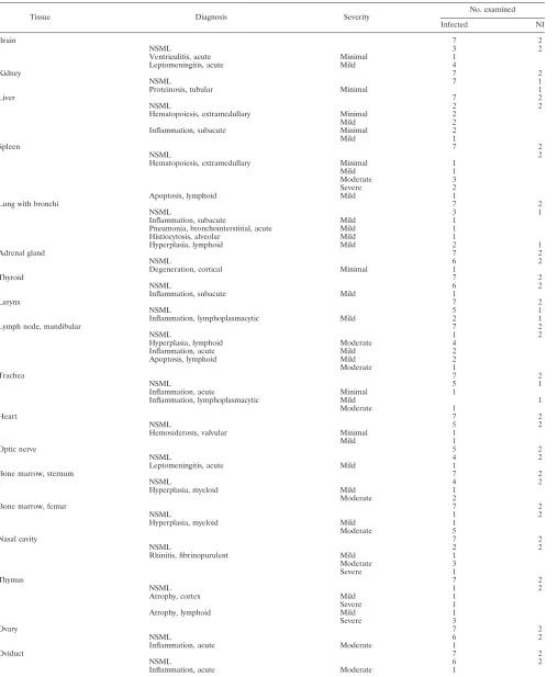
re-sponse to infection. At the 47-PFU dose, 25% of female mice died by day 21 p.i. (P⫽0.048, compared to noninfected [NI]), and 50% of male mice died by day 12 p.i. (P⫽0.17, compared to NI) (Fig. 1A and C). When infected with the highest dose of 4,700 PFU, all animals died by 9 days p.i. (P⫽0.008, compared to NI). No animals died at the lowest dose of 4.7 PFU, and any weight loss was insignificant (Fig. 1D). With the female mice, weight loss was not observed at the 4.7-PFU dose, but it was significant at the 47-PFU dose in female mice (P⫽ 0.0071, 0.0012, and 0.0036 for days 9, 10, and 11 p.i., respectively, compared to uninfected) (Fig. 1B). We calculated the 50% lethal dose (LD50) values to be 213 and 47 PFU for females and males, respectively (Fig. 1A and C) (44).
The failure of MPXV to lethally infect the C57BL/6 mice suggests that the susceptibility of the C57BL/6stat1⫺/⫺mice is
due to thestat1mutation. To confirm that the observed phe-notype was due to the loss ofstat1and was not due to some other genetic polymorphism, we tested the susceptibility of the
129stat1⫺/⫺mouse strain to an intranasal MPXV challenge.
These mice were resistant to doses at or below 470 PFU (Table 1). At the highest dose of 4,700 PFU, 20% of males died and 40% of females died by day 10 p.i.; no wild-type 129 mice died (P⫽ 1.0 and 0.44 compared to NI for male and female 129
stat1⫺/⫺ mice, respectively) (data not shown). Because the
stat1 mutant phenotype in the C57BL/6 stat1⫺/⫺ strain was
more acute than that in the 129stat1⫺/⫺strain, we selected the
C57BL/6stat1⫺/⫺strain to further evaluate the pathogenicity
of MPXV in a murine model.
We next determined the relative levels of virus replication. Female C57BL/6stat1⫺/⫺mice were infected with 4,600 PFU
of MPXV, and all died by day 9 p.i. The titers of infectious virus in spleen, liver, and lung samples from animals sacrificed 8 days p.i. were determined. Titrations revealed that the lung had the largest amount of infectious virus (1.7⫻105PFU/g),
followed by the spleen (1.7⫻104PFU/g) and liver (1.8⫻102
PFU/g) (Fig. 2). With 129 stat1⫺/⫺animals, we detected no
virus in spleen, liver, or lung samples at 8 days following an intranasal infection with the same virus dose (data not shown). This finding could be due to the 2- or 3-log higher LD50in 129
stat1⫺/⫺mice, which also have a longer disease course.
Nec-ropsies from infected C57BL/6 stat1⫺/⫺ mice 8 days p.i.
re-vealed that the spleens were enlarged approximately fourfold and that the lungs were red and mottled. Animals were ema-ciated (as determined by low levels of abdominal fat) with distended abdomens, and the entire gastrointestinal (GI) tract, including the stomach, was filled with gas. Results of his-topathologic examination of tissues from several sacrificed an-imals are described in Table 2.
Histopathology in C57BL/6 stat1ⴚ/ⴚand 129stat1ⴚ/ⴚmice
infected with MPXV.Histopathology results considered to be related to infection or potentially related to infection are sum-TABLE 1. Disease resistance of 4- to 8-week-old
immunocompetent murine strains infected with MPXVa
Strain Dose
(PFU) Route
Day of death p.i.
(% mortality) Seroconversion
SCID 275 IN 16.8⫾0.8 (100) NA
SCID 27.5 IN 31⫾11.3 (40) NA
DBA 5⫻104 FP/IN Survive ⫹
A/Ncr 5⫻104 FP/IN Survive ⫹
C3HeJ 5⫻104 FP/IN Survive ⫹
BALB/c 990 IN Survive ⫹
IFN-␥R⫺/⫺ 990 IN Survive ⫹
Type 1 IFNR⫺/⫺ 6⫻103 FP Survive ⫹
C57BL/6 5⫻104 FP/IN Survive ⫹
C57BL/6stat1⫺/⫺ 470 IN 9.3⫾0.7 (90) ⫹
129 5⫻104 FP/IN Survive ⫹
129stat1⫺/⫺ 470 IN Survive ⫹
129stat1⫺/⫺ 4,700 IN 10 (40) ⫹
a
FP, footpad injection; NA, not applicable.
on November 8, 2019 by guest
http://jvi.asm.org/
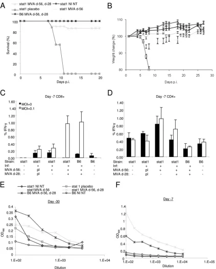
[image:3.585.42.288.97.245.2]marized in Table 2. No specific microscopic lesion (NSML) changes were observed with C57BL/6 and 129 mice.
Microscopic changes associated with infection were gener-ally comparable between the C57BL/6 stat1⫺/⫺ and 129
stat1⫺/⫺(not shown) strains. With mice of each strain, minimal
or mild acute leptomeningitis in the cerebrum and optic nerve was observed (Fig. 3A and B). In a single infected C57BL/6
stat1⫺/⫺ mouse, minimal acute ventriculitis within the brain
was also observed.
Acute inflammation of the upper respiratory tract was ob-served with mice of both strains. Mild to severe exudation or fibrinopurulent rhinitis in the nasal turbinates and nasal cavity of all infected mice was observed (Fig. 3C and D). Mild sub-acute inflammation was present in the larynx of one infected C57BL/6 stat1⫺/⫺ mouse. In addition, in the lungs, a single
infected C57BL/6stat1⫺/⫺mouse exhibited mild
bronchoint-erstitial pneumonia, and two mice exhibited hyperplasia of the peribronchiolar lymphoid tissue.
In the liver and spleen of mice of both stat1⫺/⫺ strains,
extramedullary hematopoiesis was present (minimal to mod-erate in the liver, mild to severe in the spleen). In addition, mild or moderate bone marrow hyperplasia was present in all infected mice.
Lymphoid depletion (mild or severe) resulting in cortical atrophy was present in the thymus of infected C57BL/6
stat1⫺/⫺mice. In the mandibular lymph nodes, mild increased
[image:4.585.81.502.74.427.2]lymphocyte apoptosis was observed with infected C57BL/6 FIG. 1. Dose response to MPXV challenge in female and male C57BL/6stat1⫺/⫺mice. Male and female mice were inoculated with 4.7 to 4,700
PFU of MPXV and monitored for death (A and C) and weight loss (B and D). The higher doses of 4,700 and 470 PFU induced significant weight loss from approximately 4 days p.i. * indicates 1 death at day 21 (75% survival).n⫽5 mice per group.
FIG. 2. Day 8 tissue titers. Liver, spleen, and lung titers from mice sacrificed on day 8 p.i.n⫽5 mice per group.
on November 8, 2019 by guest
TABLE 2. Tissue necropsy report for MPXV-infected C57BL/6stat1⫺/⫺micea
Tissue Diagnosis Severity No. examined
Infected NI
Brain 7 2
NSML 3 2
Ventriculitis, acute Minimal 1
Leptomeningitis, acute Mild 4
Kidney 7 2
NSML 7 1
Proteinosis, tubular Minimal 1
Liver 7 2
NSML 2 2
Hematopoiesis, extramedullary Minimal 2
Mild 2
Inflammation, subacute Minimal 2
Mild 1
Spleen 7 2
NSML 2
Hematopoiesis, extramedullary Minimal 1
Mild 1
Moderate 3
Severe 2
Apoptosis, lymphoid Mild 1
Lung with bronchi 7 2
NSML 3 1
Inflammation, subacute Mild 1
Pneumonia, bronchointerstitial, acute Mild 1
Histiocytosis, alveolar Mild 1
Hyperplasia, lymphoid Mild 2 1
Adrenal gland 7 2
NSML 6 2
Degeneration, cortical Minimal 1
Thyroid 7 2
NSML 6 2
Inflammation, subacute Mild 1
Larynx 7 2
NSML 5 1
Inflammation, lymphoplasmacytic Mild 2 1
Lymph node, mandibular 7 2
NSML 1 2
Hyperplasia, lymphoid Moderate 4
Inflammation, acute Mild 2
Apoptosis, lymphoid Mild 2
Moderate 1
Trachea 7 2
NSML 5 1
Inflammation, acute Minimal 1
Inflammation, lymphoplasmacytic Mild 1
Moderate 1
Heart 7 2
NSML 5 2
Hemosiderosis, valvular Minimal 1
Mild 1
Optic nerve 5 2
NSML 4 2
Leptomeningitis, acute Mild 1
Bone marrow, sternum 7 2
NSML 4 2
Hyperplasia, myeloid Mild 1
Moderate 2
Bone marrow, femur 7 2
NSML 1 2
Hyperplasia, myeloid Mild 1
Moderate 5
Nasal cavity 7 2
NSML 2 2
Rhinitis, fibrinopurulent Mild 1
Moderate 3
Severe 1
Thymus 7 2
NSML 1 2
Atrophy, cortex Mild 1
Severe 1
Atrophy, lymphoid Mild 1
Severe 3
Ovary 7 2
NSML 6 2
Inflammation, acute Moderate 1
Oviduct 7 2
NSML 6 2
Inflammation, acute Moderate 1
aNo significant microscopic lesions (NSML) reported in adrenal gland, aorta, cecum, colon, duodenum, esophagus, eye, gallbladder, Harderian gland, heart, ileum, jejunum, kidney, lymph node (mesenteric), mammary gland, optic nerve, pancreas, parathyroid, prostate, rectum, salivary gland, skeletal muscle, tongue, urinary bladder, penis, seminal vesicle, skin, skeletal muscle, spinal cord, stomach, testes, thyroid, tongue, trachea, urinary bladder, uterus with cervix, and vagina.
on November 8, 2019 by guest
http://jvi.asm.org/
stat1⫺/⫺mice. Lymphoid apoptosis and/or depletion is a
com-mon nonspecific finding in mice experiencing stress associated with debilitation from a variety of causes. In contrast, in one of the infected 129 mice, mild lymphoid hyperplasia was present (not shown). Two mice of each strain exhibited mild acute inflammation in the mandibular lymph node.
One C57BL/6 infected mouse exhibited moderate acute in-flammation affecting both the ovary and oviduct. Although an unusual lesion, the singular incident of this finding makes it of uncertain relationship to infection.
MVA vaccination protects mice from a lethal MPXV chal-lenge.To test the efficacy of the third-generation MVA small-pox vaccine, groups of male and female C57BL/6stat1⫺/⫺mice
were vaccinated once with MVA via intramuscular injections at day⫺56 before MPXV infection or twice via intramuscular injection at day ⫺56 before infection with an MVA booster vaccination at day⫺28 before infection. All infected mice (day 0) vaccinated with placebo died by day 11 p.i. following an MPXV challenge of 4.2⫻ 104PFU (P⫽1.0) (Fig. 4A), but
mice vaccinated either once with MVA (day ⫺56) or with MVA plus a booster vaccination (day⫺56 and day⫺28) had similar survival levels of approximately 90% (P⫽0.0001 and 0.0001, respectively, compared to NI); however, mice that also
received the booster vaccination at⫺28 days before infection experienced little weight loss compared to mice vaccinated once, which experienced significant weight loss on days 6, 7, and 8 p.i (P⫽0.0002, 0.0002, and 0.0004, respectively, com-pared to untreatedstat1⫺/⫺mice) (Fig. 4A and B).
We next measured immunologic memory by intracellular IFN-␥staining in CD4⫹and CD8⫹cells from mice bled at 7 days before infection. We found that C57BL/6 and C57BL/6
stat1⫺/⫺ mice that received a vaccination and a booster had
significantly elevated amounts of intracellular IFN-␥in CD8⫹ cells when exposed to viral antigenin vitro (C57BL/6, 1.0 ⫾ 0.29, and placebo, 0.24⫾ 0.09,P ⫽ 0.02; C57BL/6stat1⫺/⫺,
1.15⫾ 0.4, and placebo, 0.24 ⫾ 0.08, P ⫽ 0.05) (Fig. 4C). Conversely, C57BL/6stat1⫺/⫺animals that received only one
vaccination failed to significantly increase their levels of intra-cellular IFN-␥in CD8⫹cells, compared to placebo controls (C57BL/6stat1⫺/⫺, 0.26⫾0.11, and placebo, 0.23⫾0.09,P⫽
0.84) (Fig. 4C). Thus, the absence of STAT1 had no effect on CD8⫹immunologic memory when mice were vaccinated and received a booster compared to wild-type mice. Levels of CD4⫹intracellular IFN-␥did not change significantly for any of the groups (Fig. 4D).
We also used ELISAs to check the antibody responses of FIG. 3. Histopathology (H and E) of the cerebrum, optic nerve, nasal turbinates, and nasal cavity. (A) Ventral cerebrum and optic nerve from C57BL/6stat1⫺/⫺mice. Acute leptomeningitis (arrow) characterized by neutrophil and macrophage infiltration and fibrin deposition was observed
with infected mice of both strains (magnification,⫻200). (B) Cerebrum from the C57BL/6stat1⫺/⫺strain presented with mild acute leptomeningitis
(arrow) characterized by neutrophil and macrophage infiltration, which was observed with infected mice of both strains (magnification,⫻100). (C) Nasal turbinate from the C57BL/6stat1⫺/⫺ strain presented with fibrinopurulent exudate (arrow) adherent to nonulcerated respiratory
epithelium in infected mouse (magnification,⫻200). (D) Nasal turbinate from the C57BL/6stat1⫺/⫺strain presented with fibrinopurulent rhinitis
with inflammatory cells extending throughout the respiratory submucosa. Fibrinopurulent exudate (circled E) occludes the lumen of much of nasal cavity (magnification,⫻200). Pictures are typical results from experiments in whichn⫽7.
on November 8, 2019 by guest
mice following the primary vaccination and following the booster vaccination. To this end, we bled mice at day⫺30 and day⫺7 before infection (Fig. 4E and F). C57BL/6 mice bled following the primary vaccination (day⫺30) had significantly
increased antibody levels compared to unvaccinated C57BL/6 controls (0.34⫾0.04 and 0.07⫾0.02, respectively,P⫽0.0006 at a 1:200 dilution), and their levels were similar to those of vaccinated C57BL/6stat1⫺/⫺mice (Fig. 4 E), which were
gen-FIG. 4. Protection of single and double MVA-vaccinated male and femalestat1⫺/⫺mice following MPXV infection. (A) Survival of C57BL/6 stat1⫺/⫺mice following a primary MVA vaccination at 56 days before MPXV challenge (d-56) or following a primary vaccination with a booster
vaccination at 28 days before infection (d-56, d-28). (B) Weight change in vaccinated and infected animals (n⫽21, 7, 9, 19, and 8 for stat1 MVA d-56, d-28; stat1 NI NT; stat1 placebo; stat1 MVA d-56; and B6 MVA d-56, d-28, respectively). (C and D) Infected and noninfected CD8⫹and CD4⫹intracellular IFN-␥levels from blood taken 7 days before infection. (E and F) Antibody responses at day⫺30 and day⫺7 following the primary MVA vaccination and following a booster vaccination. NI indicates not infected; NT indicates no treatment; d-56 indicates MVA primary vaccination at 56 days before infection; d-28 indicates MVA booster vaccination at 28 days before infection; pl indicates placebo; an MOI of 0 is mock infected; and B6 indicates C57BL/6 wild type; OD490, optical density at 490 nm.
on November 8, 2019 by guest
http://jvi.asm.org/
[image:7.585.81.504.66.592.2]erally higher than the antibody responses in placebo and un-treated C57BL/6stat1⫺/⫺mice. At the day⫺7 time point, the
C57BL/6stat1⫺/⫺group that received the booster vaccination
had significantly more antibodies (0.8⫾0.06) than both the C57BL/6 mice receiving booster vaccinations and the C57BL/6
stat1⫺/⫺ mice (0.14 ⫾ 0.02, P ⫽ 0.001) receiving only the
primary vaccination (Fig. 4F), indicating an antibody response in C57BL/6stat1⫺/⫺mice following vaccination that is stronger
than that of wild-type animals.
To measure the duration of the immune response in C57BL/6stat1⫺/⫺mice vaccinated with MVA on day⫺56 and
day⫺28, we bled mice at days 42, 72, and 105 and measured their serum antibody levels using ELISAs. We found no sig-nificant differences between the MVA-vaccinated C57BL/6
stat1⫺/⫺ mice and the wild-type C57BL/6 mice at any of the
time points (data not shown). We also found that the antibody levels between day 42 and 105 significantly decreased in the C57BL/6stat1⫺/⫺animals (0.95⫾0.11 at day 42, 0.49⫾0.14
at day 105,P⫽0.03) but did not significantly decrease in the C57BL/6 animals (0.76⫾0.11 at day 42, 0.82⫾0.1 at day 105,
P⫽0.66).
Antiviral therapy protects C57BL/6stat1ⴚ/ⴚmice against a
lethal MPXV challenge.Previously it has been shown that mice can be protected from lethal orthopoxvirus disease by the administration of antivirals, such as CMX001 and ST-246 (3, 39, 41, 62). To test whether C57BL/6stat1⫺/⫺mice could be
used to evaluate the efficacy of antivirals following MPXV challenges, we infected and treated C57BL/6 stat1⫺/⫺ mice
with CMX001, which is a DNA polymerase inhibitor, or with ST-246, which prevents viral release from infected cells (35, 62). Following a 5,000-PFU intranasal MPXV infection, groups of mice were treated with a 10-mg/kg dose of CMX001 on the day of infection, followed by every-other-day dosings with 2.5 mg/kg until day 14 p.i. All mice survived the MPXV challenge, had negligible weight loss, and seroconverted; how-ever, when mice were rechallenged at day 38 p.i. we found that 20% died by 8 days postrechallenge (Fig. 5A). We also noted
significant weight-loss in this group following rechallenge (Fig. 5B).
Mice treated with daily 100-mg/kg administrations of ST-246 for 10 days following infection also survived the initial infection and had negligible weight loss (Fig. 5A and B); however, as with the CMX001 treatment group, 20% of these animals died by 15 days postrechallenge; unlike mice treated with CMX001, these mice experienced negligible weight loss (Fig. 5B). All infected mice treated with either saline or CMC vehicles were dead by 9 days p.i.
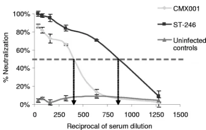
[image:8.585.82.500.65.238.2]Finally, we tested the neutralization values of antivirally treated mice. At 28 days p.i., we bled mice from groups treated with CMX001 or ST-246 and determined the 50% neutraliza-tion values. We found that mice treated with ST-246 had al-most double the 50% neutralization value compared to mice treated with CMX001 (Fig. 6); however, the increased titer of antibody did not correlate with increased survivability follow-ing rechallenge.
FIG. 5. CMX001 and ST-246 partially protect against lethal MPXV challenges. C57BL/6stat1⫺/⫺mice were intranasally infected with 5,000
PFU of MPXV and treated with 10 mg/kg of CMX001 on the day of infection followed by every-other-day dosing of 2.5 mg/kg until 14 days p.i., or C57BL/6stat1⫺/⫺mice were treated with 100 mg/kg of ST-246 daily for 10 days starting on the day of infection. Survival curves and weight
changes are shown (A and B, respectively).Vehicle (veh) mice received saline gavages according to the CMX001 dosing regimen or according to the ST-246 dosing regimen (CMC veh). NI indicates noninfected mice. Arrows indicate rechallenge with MPXV at day 38 p.i.n⫽5 per group.
FIG. 6. Anti-orthopoxvirus serum neutralization in mice treated with CMX001 compared to those treated with ST-246. Serum from mice 28 day p.i. was used to determine the 50% serum neutralization values of VACV-infected BSC-1 cells. The dashed line indicates the 50% neutralization value.
on November 8, 2019 by guest
[image:8.585.316.523.546.677.2]DISCUSSION
In this study we found that tested immunocompetent mouse strains, as well as type 1 and type 2 IFN receptor knockout strains, were resistant to MPXV infection. The immunocom-promised SCID mouse is sensitive to an IN infection at⬍275 PFU and has also been shown to be sensitive to IP infections at 105PFU (33) but is limited by its lack of a fully functional
immune system. We next evaluated STAT1-null mice, which are defective in their ability to initiate transcription of many type 1 and type 2 receptor IFN-stimulated genes (reviewed in reference 43). STAT1-null mice are highly sensitive to a num-ber of viral and bacterial infections and have been used as the foundation for developing several disease models, including respiratory syncytial virus, influenza, Listeria monocytogenes,
Leishmania major, vesicular stomatitis virus, Sendai virus,
mouse hepatitis virus, and others (4, 10, 12, 19, 20, 30, 45, 46, 53).
The 129stat1⫺/⫺mice revealed some sensitivity to intranasal
MPXV infections with doses higher than 470 PFU; however, the C57BL/6 stat1⫺/⫺ strain was highly sensitive to infection
with LD50 values of 47 and 213 PFU for males and females, respectively. One possible explanation for the higher LD50for
females could be explained by their propensity to produce high levels of CD8⫹ intracellular IFN-␥ (data not shown). The differences in 129stat1⫺/⫺and C57BL/6stat1⫺/⫺strain
sensi-tivities indicate that background, strain, and potentially gen-der-specific alleles have an important role in determining host susceptibility to MPXV infection. Also, thestat1mutation is different between the mouse strains. The C57BL/6 stat1⫺/⫺
strain produces no detectable STAT1 antigen, whereas the 129
stat1⫺/⫺strain, which has a deletion in the N-terminal domain,
produces a limited amount of abnormal STAT1 that might contribute to this strain’s decreased sensitivity to MPXV (10, 26). This is underpinned by the finding that 129stat1⫺/⫺mice
had no detectable virus in their spleens, livers, or lungs at day 8 compared to C57BL/6stat1⫺/⫺animals; however, some of
the 129stat1⫺/⫺mice did become sick and die. It should be
noted that the lack of detectable virus in the tested tissue from the 129stat1⫺/⫺mice could indicate that mortality was due to
pathology in other tissues, such as the brain, as leptomeningitis was reported for these mice. Furthermore, the LD50for this strain is likely 2 or 3 logs higher than that of the C57BL/6
stat1⫺/⫺mice. That said, H-and-E data does indicate
involve-ment of the spleen and liver in the 129stat1⫺/⫺animals (data
not shown).
In contrast to our findings, cynomolgus monkeys infected with MPXV-ZAI-79 had papulovesicular lesions primarily in the lymph nodes and thymus. Gross lesions with a granuloma-tous appearance were demonstrated in the GI tract organs, such as the stomach, small intestine, and colon (47, 63). More-over, infectedstat1⫺/⫺mice were associated with
histopatho-logic changes that were comparatively less severe than those previously reported for dormice infected with MPXV-ZAI-79 (50). In dormice, acute hemorrhage was present in numerous tissues, and significant hepatocellular necrosis was observed. While both species exhibited rhinitis, in dormice, nasal muco-sal syncytial cell formation with intracytoplasmic viral inclu-sions was also observed. Leptomeningitis was not a feature of infection in dormice. In summary, the presence of the stat1
mutation changes a relatively low-dose, intranasal infection from one characterized by no apparent disease signs to one characterized by fulminant and lethal disease. It would be interesting to test under appropriate containment conditions adultstat1⫺/⫺mice for susceptibility to severe disease
follow-ing VARV infection.
The sensitivity ofstat1⫺/⫺mice to MPXV and myxoma virus
infections suggests IFNs may be critical to recovery from pox-virus infections in general (59). Poxpox-viruses, like most pox-viruses, encode proteins that antagonize antiviral mechanisms medi-ated by IFN (reviewed in reference 25). VACV, an orthopox-virus closely related to MPXV, encodes an inhibitor (H1) of phosphorylation of STAT1 and STAT2 following type 1 or type 2 IFN stimulationin vitro(23, 31). VACV encodes IFN-bind-ing proteins (B19 and B8) that block selected type 1 and 2 IFN-mediated induction of both STAT1-dependent and -inde-pendent genes and are both essential for virulence (7, 28, 32, 48, 57, 61). Finally, VACV encodes a phosphorylation-resis-tant homologue of eIF2␣ (K3) and a double-stranded RNA binding protein (E3) that targets the PKR and 2⬘-5⬘OAS/ RNaseL systems (21). MPXV, like other examined orthopox-viruses, encodes apparently intact orthologues of H1, B19, and B8; however, the K3 orthologue is fragmented and the amino terminal domain of the E3 orthologue is missing, which likely restricts function. The N-terminal domain of E3L shows se-quence similarity with a group of host Z-DNA-binding pro-teins, ADAR-1 and Dlm, and has been shown to be important in the spread of VACV in the mouse (2). We speculate that the loss of the N-terminal domain of the MPXV E3 orthologue may contribute to the attenuated phenotype of MPXV in mice. Treatment of C57BL/6stat1⫺/⫺mice with CMX001 or
ST-246 provided solid protection against MPXV challenges, with negligible weight loss. The higher neutralization value from ST-246 treated and infected mice could be a result of an increasing amount of viral antigen which would likely be present because this drug has antiviral activity at the viral egress stage, whereas CMX001 has activity at the DNA repli-cation stage. Consideration should be given to the fact that MPXV is a complex virus that will induce the generation of a wide variety of antibodies, and the ELISAs used in these ex-periments measure all anti-orthopoxvirus antibodies and not just those that are protective. Of some concern is the obser-vation that some mice died following a rechallenge initiated several weeks after the cessation of drug treatment. This result suggests that surviving a drug-treated MPXV infection does not provide full immunity against subsequent MPXV challenge in the C57BL/6stat1⫺/⫺mouse, and therefore, antibody titers
alone do not directly correlate with protection. In contrast, C57BL/6 mice treated with CMX001 following an intranasal infection with ECTV are protected following rechallenge (data not shown). With regard to these data, consideration should be given to the quantity of animals being tested (n⫽5). That said, these data suggest that MPXV-infected STAT1-deficient hu-mans successfully treated with antiviral therapy may be suscep-tible to MPXV reinfection.
It is exciting to report in this study that we could successfully vaccinatestat1⫺/⫺mice and protect them from a lethal MPXV
challenge. A similar response has also been reported with rotavirus infections in stat1⫺/⫺ mice, which are resistant to
subsequent challenge (56). Vaccinating C57BL/6stat1⫺/⫺mice
on November 8, 2019 by guest
http://jvi.asm.org/
level of protection. From the literature, others have shown that robust cell-mediated and humoral responses can be generated in the absence of signaling pathways requiring STAT1 (re-viewed in reference 43). Vaccinating C57BL/6stat1⫺/⫺mice
with a single Dryvax vaccination also provided partial protec-tion (approximately 80%) against subsequent MPXV chal-lenges, with negligible weight loss (data not shown); that said, this vaccination did cause severe necrosis at the base of the tail where Dryvax was administered, making it of limited utility and suggesting thatstat1⫺/⫺mice are likely highly susceptible to
nonvaccine strains of VACV (e.g., Copenhagen or Western Reserve strains). These data suggest that humans with muta-tions instat1could be contraindicated to vaccination with Dry-vax and ACAM2000 and that following MVA vaccination, they may not receive the same level of protection as fully immuno-competent individuals.
Humans with heterozygousstat1mutations have been shown to be unusually sensitive to mycobacterial but not viral infec-tions; however, all patients recovered from infection (8). Ho-mozygous mutations in stat1, which completely abrogate STAT1 function, have been described for at least 3 infants who developed disseminatedMycobacterium bovis BCG infections from which they subsequently recovered; however, all died at a later date from viral disease (4, 5, 9). Humans with thestat1
mutations have differential susceptibility to severe virus dis-ease. In one example, a human carrying the homologousstat1
mutations could mount a protective immune response to clear the attenuated polio vaccine, a rhinovirus infection acquired after a bone marrow transplant, and a parainfluenza type II infection; however, the patient was not able to survive follow-ing an Epstein-Barr virus infection (5). One reason for the differential response to viral infections of humans with thestat1
mutation could be the existence of IFN-induced STAT1-inde-pendent pathways, which have been studied in detail with mice. Approximately 500 genes are regulated by the IFN-␥/STAT1 pathway (1); however, it has been demonstrated that STAT1-independent responses to IFNs exist. For example, IFN-␥ -treated, wild-type, bone marrow-derived macrophages (BMMs) have expression changes in 216 transcripts, as detected by microarrays, compared to expression changes of 150 different transcripts in STAT1-null BMMs. Moreover, IFN-␥ -depen-dent gene expression in serum-starved STAT1-null fibroblasts have been shown to control expression of several protein groups (42, 43). The effective IFN-stimulated STAT-1-inde-pendent responses may be deSTAT-1-inde-pendent on the virus or stage of
acts early and is required for controlling initial viral replica-tion; however, the STAT1-independent pathway functions later and is required for complete viral clearance (53).
In summary, advantages to using a murine MPXV model for therapeutic development include: (i) biomarkers of disease progression have been established in the mousepox model and can be readily applied to the MPXV stat1⫺/⫺mouse model
(38); (ii) reagents are readily available, and the biology and genetics of the mouse are well understood; (iii) early in the drug development plan, mice are invariably used to acquire efficacy, toxicity, and pharmacokinetic/pharmacodynamic data; and (iv) MPXV itself causes a natural disease in humans, unlike VACV and ECTV, which are used in other mouse models.
In the ECTV/mousepox model, we and others have evalu-ated successful, delayed dosings with CMX001 and ST-246 (39, 40). Further evaluation of dosing regimens would be a logical next step in evaluating drug efficacy against MPXV in C57BL/6
stat1⫺/⫺mice.
ACKNOWLEDGMENTS
This work was supported by NIAID grant NOI-AI-15436 and U54-AI-057169 from the NIAID to the Midwestern Regional Center of Excellence for Biodefense and Emerging Infectious Diseases (MRCE).
We thank Mike Bray for critically reviewing the manuscript.
REFERENCES
1.Boehm, U., T. Klamp, M. Groot, and J. C. Howard.1997. Cellular responses to interferon-gamma. Annu. Rev. Immunol.15:749–795.
2.Brandt, T., M. C. Heck, S. Vijaysri, G. M. Jentarra, J. M. Cameron, and B. L. Jacobs.2005. The N-terminal domain of the vaccinia virus E3L-protein is required for neurovirulence, but not induction of a protective immune re-sponse. Virology333:263–270.
3.Buller, R. M., G. Owens, J. Schriewer, L. Melman, J. R. Beadle, and K. Y. Hostetler.2004. Efficacy of oral active ether lipid analogs of cidofovir in a lethal mousepox model. Virology318:474–481.
4.Chapgier, A., S. Boisson-Dupuis, E. Jouanguy, G. Vogt, J. Feinberg, A. Prochnicka-Chalufour, A. Casrouge, K. Yang, C. Soudais, C. Fieschi, O. F. Santos, J. Bustamante, C. Picard, L. de Beaucoudrey, J. F. Emile, P. D. Arkwright, R. D. Schreiber, C. Rolinck-Werninghaus, A. Rosen-Wolff, K. Magdorf, J. Roesler, and J. L. Casanova.2006. Novel STAT1 alleles in otherwise healthy patients with mycobacterial disease. PLoS Genet.2:e131. 5.Chapgier, A., R. F. Wynn, E. Jouanguy, O. Filipe-Santos, S. Zhang, J. Feinberg, K. Hawkins, J. L. Casanova, and P. D. Arkwright.2006. Human complete Stat-1 deficiency is associated with defective type I and II IFN responses in vitro but immunity to some low virulence viruses in vivo. J. Im-munol.176:5078–5083.
6.Chen, N., G. Li, M. K. Liszewski, J. P. Atkinson, P. B. Jahrling, Z. Feng, J. Schriewer, C. Buck, C. Wang, E. J. Lefkowitz, J. J. Esposito, T. Harms, I. K. Damon, R. L. Roper, C. Upton, and R. M. Buller.2005. Virulence differences between monkeypox virus isolates from West Africa and the Congo basin. Virology340:46–63.
on November 8, 2019 by guest
7.Colamonici, O. R., P. Domanski, S. M. Sweitzer, A. Larner, and R. M. Buller.1995. Vaccinia virus B18R gene encodes a type I interferon-binding protein that blocks interferon alpha transmembrane signaling. J. Biol. Chem. 270:15974–15978.
8.Dupuis, S., C. Dargemont, C. Fieschi, N. Thomassin, S. Rosenzweig, J. Harris, S. M. Holland, R. D. Schreiber, and J. L. Casanova.2001. Impair-ment of mycobacterial but not viral immunity by a germline human STAT1 mutation. Science293:300–303.
9.Dupuis, S., E. Jouanguy, S. Al Hajjar, C. Fieschi, I. Z. Al Mohsen, S. Al Jumaah, K. Yang, A. Chapgier, C. Eidenschenk, P. Eid, A. Al Ghonaium, H. Tufenkeji, H. Frayha, S. Al Gazlan, H. Al Rayes, R. D. Schreiber, I. Gresser, and J. L. Casanova.2003. Impaired response to interferon-alpha/beta and lethal viral disease in human STAT1 deficiency. Nat. Genet.33:388–391. 10.Durbin, J. E., R. Hackenmiller, M. C. Simon, and D. E. Levy.1996. Targeted
disruption of the mouse Stat1 gene results in compromised innate immunity to viral disease. Cell84:443–450.
11.Esteban, D. J., and R. M. Buller.2005. Ectromelia virus: the causative agent of mousepox. J. Gen. Virol.86:2645–2659.
12.García-Sastre, A., R. K. Durbin, H. Zheng, P. Palese, R. Gertner, D. E. Levy, and J. E. Durbin. 1998. The role of interferon in influenza virus tissue tropism. J. Virol.72:8550–8558.
13.Gavrilescu, L. C., B. A. Butcher, L. Del Rio, G. A. Taylor, and E. Y. Denkers. 2004. STAT1 is essential for antimicrobial effector function but dispensable for gamma interferon production during Toxoplasma gondii infection. In-fect. Immun.72:1257–1264.
14.Guarner, J., B. J. Johnson, C. D. Paddock, W. J. Shieh, C. S. Goldsmith, M. G. Reynolds, I. K. Damon, R. L. Regnery, and S. R. Zaki.2004. Mon-keypox transmission and pathogenesis in prairie dogs. Emerg. Infect. Dis. 10:426–431.
15.Hogan, R. J., G. Gao, T. Rowe, P. Bell, D. Flieder, J. Paragas, G. P. Kob-inger, N. A. Wivel, R. G. Crystal, J. Boyer, H. Feldmann, T. G. Voss, and J. M. Wilson.2004. Resolution of primary severe acute respiratory syn-drome-associated coronavirus infection requires Stat1. J. Virol.78:11416– 11421.
16.Hoogland, J. L.1995. The black-tailed prairie dog: social life of a burrowing mammal, p. 221–259. The University of Chicago Press, Chicago, IL. 17.Hutson, C. L., V. A. Olson, D. S. Carroll, J. A. Abel, C. M. Hughes, Z. H.
Braden, S. Weiss, J. Self, J. E. Osorio, P. N. Hudson, M. Dillon, K. L. Karem, I. K. Damon, and R. L. Regnery.2009. A prairie dog animal model of systemic orthopoxvirus disease using West African and Congo Basin strains of monkeypox virus. J. Gen. Virol.90:323–333.
18.Jackson, R. J., A. J. Ramsay, C. D. Christensen, S. Beaton, D. F. Hall, and I. A. Ramshaw.2001. Expression of mouse interleukin-4 by a recombinant ectromelia virus suppresses cytolytic lymphocyte responses and overcomes genetic resistance to mousepox. J. Virol.75:1205–1210.
19.Johnson, L. M., and P. Scott.2007. STAT1 expression in dendritic cells, but not T cells, is required for immunity to Leishmania major. J. Immunol. 178:7259–7266.
20.Karst, S. M., C. E. Wobus, M. Lay, J. Davidson, and H. W. Virgin.2003. STAT1-dependent innate immunity to a Norwalk-like virus. Science299: 1575–1578.
21.Langland, J. O., J. M. Cameron, M. C. Heck, J. K. Jancovich, and B. L. Jacobs.2006. Inhibition of PKR by RNA and DNA viruses. Virus Res. 119:100–110.
22.Langohr, I. M., G. W. Stevenson, H. L. Thacker, and R. L. Regnery.2004. Extensive lesions of monkeypox in a prairie dog (Cynomys sp). Vet. Pathol. 41:702–707.
23.Mann, B. A., J. H. Huang, P. Li, H. C. Chang, R. B. Slee, A. O’Sullivan, M. Anita, N. Yeh, M. J. Klemsz, R. R. Brutkiewicz, J. S. Blum, and M. H. Kaplan.2008. Vaccinia virus blocks Stat1-dependent and Stat1-independent gene expression induced by type I and type II interferons. J. Interferon Cytokine Res.28:367–380.
24.Marennikova, S. S., and E. M. Seluhina.1976. Susceptibility of some rodent species to monkeypox virus, and course of the infection. Bull. World Health Organ.53:13–20.
25.McFadden, G.2005. Poxvirus tropism. Nat. Rev. Microbiol.3:201–213. 26.Meraz, M. A., J. M. White, K. C. Sheehan, E. A. Bach, S. J. Rodig, A. S.
Dighe, D. H. Kaplan, J. K. Riley, A. C. Greenlund, D. Campbell, K. Carver-Moore, R. N. DuBois, R. Clark, M. Aguet, and R. D. Schreiber.1996. Targeted disruption of the Stat1 gene in mice reveals unexpected physiologic specificity in the JAK-STAT signaling pathway. Cell84:431–442. 27.Moss, B., and P. L. Earl.1998. Expression of proteins in mammalian cells
using vaccinia virus vectors. Overview of the vaccinia virus expression system, p. 16.15.1–16.15.5.InCurrent protocols in molecular biology. Wiley, New York, NY.
28.Mossman, K., C. Upton, R. M. Buller, and G. McFadden.1995. Species specificity of ectromelia virus and vaccinia virus interferon-gamma binding proteins. Virology208:762–769.
29.Muller, U., U. Steinhoff, L. F. Reis, S. Hemmi, J. Pavlovic, R. M. Zinkerna-gel, and M. Aguet.1994. Functional role of type I and type II interferons in antiviral defense. Science264:1918–1921.
30.Mumphrey, S. M., H. Changotra, T. N. Moore, E. R. Heimann-Nichols, C. E.
Wobus, M. J. Reilly, M. Moghadamfalahi, D. Shukla, and S. M. Karst.2007. Murine norovirus 1 infection is associated with histopathological changes in immunocompetent hosts, but clinical disease is prevented by STAT1-depen-dent interferon responses. J. Virol.81:3251–3263.
31.Najarro, P., P. Traktman, and J. A. Lewis.2001. Vaccinia virus blocks gamma interferon signal transduction: viral VH1 phosphatase reverses Stat1 activation. J. Virol.75:3185–3196.
32.Nuara, A. A., H. Bai, N. Chen, R. M. Buller, and M. R. Walter.2006. The unique C termini of orthopoxvirus gamma interferon binding proteins are essential for ligand binding. J. Virol.80:10675–10682.
33.Osorio, J. E., K. P. Iams, C. U. Meteyer, and T. E. Rocke.2009. Comparison of monkeypox viruses pathogenesis in mice by in vivo imaging. PLoS One 4:e6592.
34.Panchanathan, V., G. Chaudhri, and G. Karupiah. 2008. Correlates of protective immunity in poxvirus infection: where does antibody stand? Im-munol. Cell Biol.86:80–86.
35.Parker, S., L. Handley, and R. M. Buller.2008. Therapeutic and prophylactic drugs to treat orthopoxvirus infections. Future Virol.3:595–612. 36.Parker, S., A. Nuara, R. M. Buller, and D. A. Schultz.2007. Human
mon-keypox: an emerging zoonotic disease. Future Microbiol.2:17–34. 37.Parker, S., D. A. Schultz, H. Meyer, and R. L. Buller.2008. Smallpox and
monkeypox viruses, p. 639–644.InB. W. J. Mahy and N. H. V. Van Regen-mortel (ed.), Encyclopedia of virology, Academic Press, Boston, MA. 38.Parker, S., A. M. Siddiqui, C. Oberle, E. Hembrador, R. Lanier, G. Painter,
A. Robertson, and R. M. Buller.2009. Mousepox in the C57BL/6 strain provides an improved model for evaluating anti-poxvirus therapies. Virology 385:11–21.
39.Parker, S., E. Touchette, C. Oberle, M. Almond, A. Robertson, L. C. Trost, B. Lampert, G. Painter, and R. M. Buller.2008. Efficacy of therapeutic intervention with an oral ether-lipid analogue of cidofovir (CMX001) in a lethal mousepox model. Antiviral Res.77:39–49.
40.Quenelle, D. C., R. M. Buller, S. Parker, K. A. Keith, D. E. Hruby, R. Jordan, and E. R. Kern.2007. Efficacy of delayed treatment with ST-246 given orally against systemic orthopoxvirus infections in mice. Antimicrob. Agents Che-mother.51:689–695.
41.Quenelle, D. C., D. J. Collins, W. B. Wan, J. R. Beadle, K. Y. Hostetler, and E. R. Kern.2004. Oral treatment of cowpox and vaccinia virus infections in mice with ether lipid esters of cidofovir. Antimicrob. Agents Chemother. 48:404–412.
42.Ramana, C. V., M. P. Gil, Y. Han, R. M. Ransohoff, R. D. Schreiber, and G. R. Stark.2001. Stat1-independent regulation of gene expression in re-sponse to IFN-gamma. Proc. Natl. Acad. Sci. U. S. A.98:6674–6679. 43.Ramana, C. V., M. P. Gil, R. D. Schreiber, and G. R. Stark.2002.
Stat1-dependent and -inStat1-dependent pathways in IFN-gamma-Stat1-dependent signaling. Trends Immunol.23:96–101.
44.Reed, L. J., and H. A. Muench.1938. A simple method of estimating fifty percent endpoints. Am. J. Hygiene27:493–497.
45.Rosas, L. E., T. Keiser, R. Pyles, J. Durbin, and A. R. Satoskar.2003. Development of protective immunity against cutaneous leishmaniasis is de-pendent on STAT1-mediated IFN signaling pathway. Eur. J. Immunol.33: 1799–1805.
46.Rothfuchs, A. G., C. Trumstedt, F. Mattei, G. Schiavoni, A. Hidmark, H. Wigzell, and M. E. Rottenberg.2006. STAT1 regulates IFN-alpha beta- and IFN-gamma-dependent control of infection with Chlamydia pneumoniae by nonhemopoietic cells. J. Immunol.176:6982–6990.
47.Saijo, M., Y. Ami, Y. Suzaki, N. Nagata, N. Iwata, H. Hasegawa, I. Iizuka, T. Shiota, K. Sakai, M. Ogata, S. Fukushi, T. Mizutani, T. Sata, T. Kurata, I. Kurane, and S. Morikawa.2009. Virulence and pathophysiology of the Congo Basin and West African strains of monkeypox virus in non-human primates. J. Gen. Virol.90:2266–2271.
48.Sakala, I. G., G. Chaudhri, R. M. Buller, A. A. Nuara, H. Bai, N. Chen, and G. Karupiah. 2007. Poxvirus-encoded gamma interferon binding protein dampens the host immune response to infection. J. Virol.81:3346–3353. 49.Sbrana, E., S. Y. Xiao, P. C. Newman, and R. B. Tesh.2007. Comparative
pathology of North American and central African strains of monkeypox virus in a ground squirrel model of the disease. Am. J. Trop. Med. Hyg.76:155– 164.
50.Schultz, D. A., J. E. Sagartz, D. L. Huso, and R. M. Buller.2009. Experi-mental infection of an African dormouse (Graphiurus kelleni) with monkey-pox virus. Virology383:86–92.
51.Shchelukhina, E. M., and S. S. Marennikova.1975. [Generalized monkeypox in orally infected rabbits and white mice]. Vopr. Virusol. \?\November-December:703–705. (In Russian.)
52.Shornick, L. P., A. G. Wells, Y. Zhang, A. C. Patel, G. Huang, K. Takami, M. Sosa, N. A. Shukla, E. Agapov, and M. J. Holtzman.2008. Airway epithelial versus immune cell Stat1 function for innate defense against respiratory viral infection. J. Immunol.180:3319–3328.
53.Shresta, S., K. L. Sharar, D. M. Prigozhin, H. M. Snider, P. R. Beatty, and E. Harris. 2005. Critical roles for both dependent and STAT1-independent pathways in the control of primary dengue virus infection in mice. J. Immunol.175:3946–3954.
54.Sugawara, I., H. Yamada, and S. Mizuno.2004. STAT1 knockout mice are