A Dissertation on
‘’ “THE ROLE OF LOW MOLECULAR WEIGHT HEPARIN IN TREATMENT OF ACUTE PANCREATITIS’’
Dissertation Submitted to
THE TAMIL NADU Dr.M.G.R. MEDICAL UNIVERSITY
CHENNAI- 600032
with partial fulfillment of the regulations for the award of the degree of
M.S. GENERAL SURGERY
(BRANCH 1)
COIMBATORE MEDICAL COLLEGE,
COIMBATORE
CERTIFICATE
This is to certify that this dissertation titled “THE ROLE OF LOW MOLECULAR WEIGHT HEPARIN IN TREATMENT OF ACUTE
PANCREATITIS ” is the bonafide work of Dr.ARAVIND. M Post Graduate student in M.S General Surgery, Coimbatore Medical College and Hospital, Coimbatore. This study was undertaken in the Department of General Surgery, Coimbatore Medical College and Hospital, Coimbatore during the period June 2016 to August 2017 in the partial fulfillment of the requirement of the “The Tamil Nadu Dr. M.G.R. Medical University” for the award of M.S. Degree in General Surgery. This dissertation has not been submitted in part or fully to any other University or Board. It gives me great pleasure to forward this dissertation.
HOD GUIDE
Prof. Dr. V. Elango M.S., Prof.Dr.S.Balasubramanian M.S., Head of the Department Professor of Surgery
Department of General Surgery Department of General Surgery Coimbatore Medical College Coimbatore Medical College
Coimbatore Coimbatore
Dr.B.Asokan M.S., MCh.,
The Dean
DECLARATION
The dissertation titled “THE ROLE OF LOW MOLECULAR WEIGHT
HEPARIN IN TREATMENT OF ACUTE PANCREATITIS” is being
submitted by me to “The Tamil Nadu Dr. M.G.R. Medical University” in partial fulfillment of the regulation for the for award of of M.S. Degree in General Surgery (Branch – 1). This work has been carried out in the Department of General Surgery, Coimbatore Medical College and Hospital, Coimbatore under the guidance of Dr.S.Balasubramanian M.S, Professor of General Surgery, Coimbatore Medical College and Hospital, Coimbatore.
Date:
ACKNOWLEDGEMENT
The success behind any project is not the sole effort of a single person but an endeavor where many minds and hands are put together. It is time for me to remember one and all at the end of the fruitful completion of this project.
I express my gratitude to Dr.B.Asokan M.S., MCh., Dean, Coimbatore Medical College Hospital for permitting me to use the clinical material for the study.
I would like to express my gratitude to Prof. Dr. V. Elango M.S., Professor & Head of Department, Department of General Surgery who allowed me to carry out this dissertation in his department for his excellent guidance and valuable suggestions.
I am greatly indebted to my guide and teacher Prof.
Dr.S.Balasubramanian. M.S., It is because of his constant guidance and immense support the completion of this project was possible. His innovative thinking made me understand the basics of clinical research and implications in clinical practice.
My sincere thanks and gratitude to Prof.Dr.D.N.Renganathan M.S., Prof.S.Natarajan M.S., Prof Dr.Nirmala M.S., Prof. S.Shanthi M.S.,for their suggestions and helping hands by allowing me to include patients under their care and making me able to complete this work.
I am deeply indebted to my Assistant Professors
Dr.S.Karthikeyan M.S., Dr. V. Umamaheshwari M.S., DGO.
Dr. A.M. Umashankar .M.S., DMRD, for their priceless support and sparing their valuable time in correcting the manuscript of my dissertation.
I would like to thank my colleagues for their valuable guidance and priceless support.
CERTIFICATE – II
This is to certify that this dissertation work titled “THE ROLE OF LOW MOLECULAR WEIGHT HEPARIN IN TREATMENT OF ACUTE
PANCREATITIS” of the candidate Dr.ARAVIND. M with registration Number
221511302 for the award of M.S in the branch of General Surgery,I personally verified the urkund.com website for the purpose of plagiarism Check. I found that the uploaded thesis file contains 110 pages from introduction to conclusion and the result shows 0% (Zero) percentage of plagiarism in the dissertation.
ABBREVIATIONS
APACHE : Acute Physiology And Chronic Health Evaluation
BISAP : Blood Urea Impaired Mental Status SIRS Age Pleural Effusion CARS : Counter Anti Inflammatory Response Syndrome
CBD : Common Blie Duct
CECT : Contrast Enhanced Computed Tomography CKD : Chronic Kidney Disease
CTSI : Computed Tomograph Severity Index
ERCP : Endoscopic Retrograde Cholangio Pancreatography MCTSI : Modified Computed Tomograph Severity Index MODS : Multi Organ Dysfunction
MRI : Magnetic Resonance Imaging SAP : Severe Acute Pancreatitis
INDEX
SR.NO CONTENT PAGE NO.
I INTRODUCTION 1
II AIMS AND OBJECTIVES 3
III REVIEW OF LITERATURE 4
IV MATERIALS AND METHODS 89
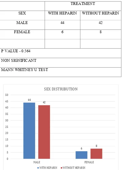
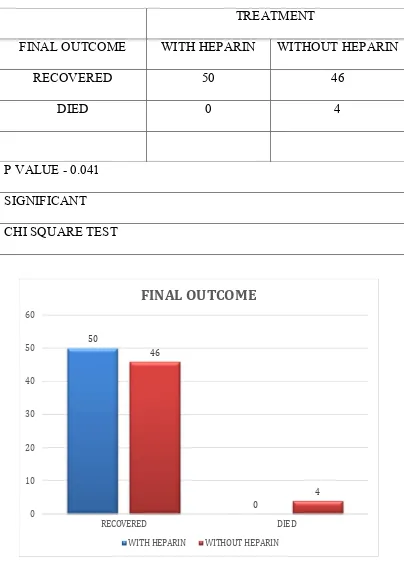
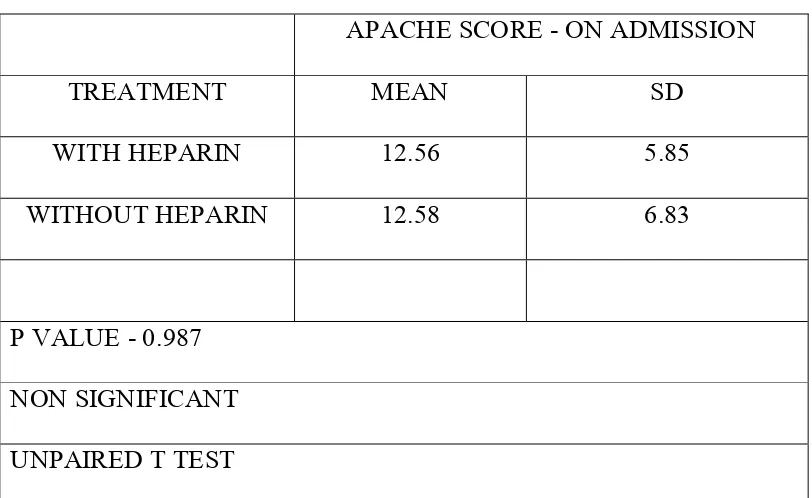
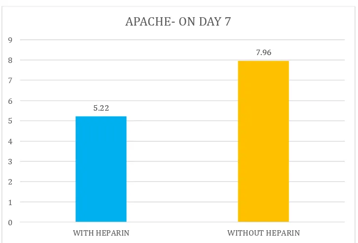
V RESULTS 92
VI DISCUSSION 107
VII CONCLUSION 110
VIII BIBLIOGRAPHY
IX
ANNEXURES PROFORMA CONSENT FORM
1
INTRODUCTION
Acute pancreatitis is a disease which has many etiologies. Each etiology seems to affect the pancreatic acinar cell in some way that results in premature activation and retention of potent proteolytic enzymes
In early stages of pancreatitis, macrophages, neutrophils, endothelial cells are activated. Preinflammatory cytokines are released and inflammation factors re elevated during acute pancreatitis and have been implicated in progression of pancreatitis associated microvascular disturbance and hemorrhagic necrosis. Ischemia, reperfusion injury and tiny thrombosis are closely associated with pancreatic microcirculation disturbance1.
Severe acute pancreatitis [SAP] is severe and frequently a lethal disorder. Its mortality rate reaches upto 25 to 40%.2.SAP is usually complicated with systemic inflammatory cascades and microcirculatory disturbances – related morbidity due to infected pre pancreatic necrosis.
2
Low molecular weight heparin (LMWH) is known to posses a special anti-thrombin activity which is stronger and safer than unfrationated heparin. LMWH can reduce the release of cytokines and inflammatory mediators, resulting in an improvement of the microcirculation of pancreas.
3
AIM OF THE STUDY
Effect of LMWH in improvement of microcirculation in acute pancreatitis.
OBJECTIVES OF THE STUDY
4
REVIEW OF LITERATURE
A. Role Of Low Molecular Weight Heparin In Treatment Of
Acute Pancreatitis
A multiple centre prospective clinical study done to assess the effect
5
obviously in C group, but not in LT group, and no hemorrhagic complications occurred. So, it was concluded that LMWH can enhance the effect of conventional treatment for SAP, and can markedly decrease the mortality of SAP. LMWH is a simple, safe, economic and effective method for treatment of SAP(5).
A study to explore the clinical effects of low-molecular-weight
6
295 u/l, 16 ± 1.60 s, 3 ± 0.60 g/l, 39.80 ± 5.60 s, and 294 ± 49 × 109/l, respectively, all of which were similar or superior to those in the control group (1738 ± 346 u/l, 2453 ± 473 u/l, 15 ± 1.50 s, 2.50 ±
0.50, 39.80 ± 5.90, and 192 ± 37 × 109/l)). Apache ii scores and ctspn after 2 weeks of treatment in the observation group were 8.50 ± 1.80 and 2.10 ± 1, respectively, which were superior to those in the control group (9.60 ± 2.40 and 4.30 ± 2.60, respectively; p < 0.05). Moreover, the incidence of complications, mortality rate, and average duration of the hospital stay in the observation group were lower than those in the control group (p > 0.05). The cure rate in the observation group was higher than that in the control group. So, it was concluded lmwh combined with uti enhances the efficacy of conventional treatment and reduces mortality. Thus, it is a potentially effective treatment strategy for severe acute pancreatitis in children(6).
7
8
Embryology
The pancreas develops as 2 buds (outpouchings) of endoderm from
10
Gross Anatomy
The pancreas is prismoid in shape and appears triangular in cut section with superior, inferior, and anterior borders as well as anterosuperior, anteroinferior, and posterior surfaces.
The head of the pancreas lies in the duodenal C loop in front of the inferior vena cava (IVC) and the left renal vein (see the following images). The uncinate process is an extension of the lower (inferior)
11
The lower (terminal) part of the CBD runs behind (or sometimes through) the upper half of the head of pancreas before it joins the main pancreatic duct (MPD) of Wirsung to form a common channel (ampulla).
The neck of the pancreas lies in front of the superior mesenteric vein, splenic vein and portal vein junction. The body and tail of the pancreas run obliquely upward to the left in front of the aorta and left kidney. The pancreatic neck is the arbitrary junction between the head and body of the pancreas. Portal vein lies behind the neck of the pancreas. The narrow tip of the tail of the pancreas reaches the splenic hilum in the splenorenal (lienorenal) ligament.
The pancreatic head constitutes about 50% and the body and tail the remaining 50% of the pancreatic parenchymal mass.
12 Blood supply
Pancreas derives a rich blood supply from both celiac axis and superior mesenteric artery; that is why when angiography is done for bleeding as a complication of acute pancreatitis, chronic pancreatitis or pancreatoduodenectomy both celiac axis and superior mesenteric artery should be evaluated.
13
the proximal body of the pancreas, and the splenic artery runs toward the left on the superior border of the distal body and tail of the pancreas. [1, 2, 3, 4, 5]
The superior mesenteric artery (SMA) comes off from the anterior surface of the aorta just below the origin of the celiac trunk at the level of L1 behind the neck of the pancreas. Then, it descends down in front of the uncinate process and the 3rd (horizontal) part of the duodenum to enter the small bowel mesentery.
The gastroduodenal artery (GDA), a branch of the CHA, runs down behind the first part of the duodenum in front of the neck of the pancreas and divides into the right gastro-omental (gastroepiploic) artery (RGEA) and superior pancreaticoduodenal artery (SPDA), which further bifurcates into anterior and posterior branches. The inferior pancreaticoduodenal artery (IPDA) arises from the SMA and also bifurcates into anterior and posterior branches.
14
arteria magna pancreatica) of the splenic artery supply the pancreatic body and tail. Multiple, small pancreatic branches of a dorsal pancreatic artery from the splenic artery and an inferior pancreatic artery from the superior mesenteric artery supply the body and tail of pancreas.
The arterial supply of the pancreas forms an important collateral circulation between the celiac axis and superior mesenteric artery.
Veins accompany the SPDA and IPDA. Superior pancreaticoduodenal veins (SPDVs) drain into the portal vein and inferior pancreaticoduodenal veins (IPDVs) drain into the superior mesenteric vein (SMV). A few small, fragile uncinate veins drain directly into the SMV. Some veins from the head of the pancreas drain into the gastrocolic trunk. Numerous small, fragile veins drain directly from the pancreatic body and tail into the splenic vein.
The SMV lies to the right of the SMA in front of the uncinate process and the 3rd part of the duodenum. The splenic vein arises in the splenic hilum behind the tail of the pancreas and runs from left to right on the posterior surface of the pancreatic body. Union of the horizontal splenic vein and the vertical SMV forms the portal vein behind the neck of the pancreas.
15
SPDVs, right gastro-omental (gastroepiploic vein, left gastric vein (LGV), and right gastric vein (RGV); then, it runs up (superiorly) behind the first part of the duodenum in the hepatoduodenal ligament behind (posterior to) the common bile duct on the right and proper hepatic artery on the left.
The portal venous system (splenic vein, SMV, and portal vein) has no valves.
Lymphatic drainage
The head of the pancreas drains into pancreaticoduodenal lymph nodes and lymph nodes in the hepatoduodenal ligament, as well as prepyloric and postpyloric lymph nodes. The pancreatic body and tail drain into mesocolic lymph nodes (around the middle colic artery) and lymph nodes along the hepatic and splenic arteries. Final drainage occurs into celiac, superior mesenteric, and para-aortic and aortocaval lymph nodes.
Nerve supply
16 Natural and Pathophysiologic Variants
Natural variants
The main pancreatic duct and common bile duct may not unite to form a common channel and open separately at the major duodenal papilla. In addition, an aberrant (normal vessel is not present) right hepatic artery (RHA) may arise from the superior mesenteric artery (SMA) and accessory RHA (in addition to the normal one from common hepatic artery [CHA]) from the SMA.
Pathophysiologic variants
An annular pancreas is caused by failure of rotation of the ventral bud of the pancreas. A ring of pancreas is present around and obstructs the second part (C loop) of the duodenum. Neonates with this pancreatic variant present with vomiting; abdominal x-rays show a double-bubble (gastric and duodenal) appearance. Treatment includes dudodeno-jejunostomy and not division of the pancreatic ring because it may result in pancreatic juice leak and fistula.
17
may not be adequate (because of the smaller size of the accessory duct) and may cause functional obstruction, resulting in recurrent attacks of acute pancreatitis. The main pancreatic duct (of Wirsung) drains only the lower (inferior) half of the head and uncinate process and does not communicate with the accessory duct.
A long (> 15 mm common channel of pancreatic duct and common bile duct is described as anomalous pancreatio biliary ductal junction/ union (APBDJ/ APBDU) - it is associated with choledochal cyst and carries a higher risk of biliary malignancy.
Accessory pancreatic tissue may be present in the stomach, small intestine, Meckel diverticulum, omentum, and hilum of spleen as soft yellow nodules/lobules.
Physiology
Pancreas is one of the organs in the body that has both exocrine and endocrinal functions. The function of the pancreas is to make digestive enzymes which digest food materials in the small intestines. In addition the pancreas also makes insulin which controls the blood glucose levels.
Exocrine Function
18
enzymes such as – amylase, lipase, proteases and trypsinogen which help digest the fat, protein as well as carbohydrates from the food that we eat. The alkaline juice helps to neutralize the acid secretions of the stomach. It secretes about 1.5 liters of these juices in a day. The stimulants for enzymes secretion are secretin, cholecystokinin and acetylcholine.
The enzymes are conveyed to the upper part of the small intestine called duodenum via a tube called the pancreatic duct.
Endocrine Function
19 B. Definition of acute pancreatitis
The International Symposium on Acute Pancreatitis (Atlanta, September 1992) defined Acute Pancreatitis as “An acute inflammatory process of the pancreas, with variable involvement of other regional tissues or remote organ systems” (8)
20 C. Classification of Acute Pancreatitis
A commonly used classification system (the Atlanta classification)
(8)
divides AP into two broad categories. The classification was developed following 3 days of group meetings and open discussions, with unanimous consensus on a series of definitions for a clinically based classification system for acute pancreatitis by a diverse group of 40 international authorities from six medical disciplines and 15 countries. The proposed classification system was to be of value to practicing clinicians in the care of individual patients and to academicians seeking to compare inter-institutional data.
The Atlanta Classification:
(1)Mild : (edematous and interstitial) acute pancreatitis
(2)Severe: (usually synonymous with necrotizing) acute pancreatitis The criteria for severe AP included any of the following:
21
2) An APACHE II score of 8 or more within the first 48 hours 3) Organ failure (respiratory, circulatory, renal, and/or GI bleeding) 4) Local complications (pancreatic necrosis, abscess or pseudocyst)
Most attacks of AP are mild with recovery occurring within five to seven days. Death is unusual (less than 3 percent) in such patients. (9)
Approximately 15 to 25 percent of all cases are severe. Severe necrotizing pancreatitis is associated with a high rate of complications (local and systemic) and mortality (approximately 17%)
Severe Acute Pancreatitis as defined by Atlanta Symposium:
Early prognostic signs
Ranson’s score >3
APACHE II score >8
Organ failure and/or Local complications
Necrosis
Abscess
22
However some patients with local complications in the absence of organ failure are seen to have low mortality rates, like patients with mild acute pancreatitis, but have prolonged hospitalizations, like patients with severe acute pancreatitis in a new subgroup called “moderately severe acute pancreatitis”(10)
23
It fails to recognize multiple and persistent (> 48 hours) organ
failure, which is more predictive of severity than transient organ failure.
The nomenclature developed to describe local complications
cannot be used to describe findings such as walled off pancreatic necrosis and does not allow for the differentiation of infected from sterile necrosis
It combines predicting systems (early) and local complications
(late) to define severe acute pancreatitis.
Revised Atlanta Classification:
The Revised Atlanta Classification was initiated as an international, web-based process that began in a clinical symposium in 2007 at the Digestive Diseases Week (12).
24 Components
The Revised Atlanta Classification dealt primarily with two broad areas, namely,
a) discrete definitions of organ failure and local complications (including necrosis) and
b) classification of severity of the disease.
The revised classification categorizes AP into interstitial edematous (IEP) and necrotizing pancreatitis based on contrast enhanced computed tomography (CECT) imaging. IEP constitutes 80-90% of AP, in which the pancreas appears relatively homogenously enhanced on CECT with or without mild peripancreatic stranding or peripancreatic fluid collection. Necrotizing pancreatitis on the other hand is characterized by lack of enhancement of the pancreas and/or (peri) pancreatic tissues on CECT. Both the pancreatic parenchyma and peripancreatic tissues together are involved more frequently than involvement of either alone. Recognition of the degree of necrosis (pancreatic alone, peripancreatic alone, or both) is important since the prognosis varies.
25
Organ failure may be transient (resolves within 48 h of onset) or persistent (persists for 48 h and more). Local complications include fluid collections, gastric outlet dysfunction, splenic and portal vein thrombosis, and colonic necrosis. Four discrete types of collections have been described, namely, acute peripancreatic fluid collection (APFC), pancreatic pseudocyst (PP), acute necrotic collection (ANC) and walled off necrosis (WON).
27 Determinant based classification:
29 Determinants
The Determinant Based Classification primarily centers on causally associated factors (or determinants) for mortality. The determinants could be
1) local, i.e. (peri)pancreatic necrosis or 2) systemic, i.e. organ failure.
(Peri) pancreatic necrosis is defined as nonviable tissue located in the pancreas alone, or in the pancreas and peripancreatic tissues, or in the peripancreatic tissues alone. (Peri) pancreatic necrosis could be sterile or infected. Infected (peri) pancreatic necrosis is defined by the presences of either gas bubbles within necrotic areas on computed tomography, a positive culture of (peri) pancreatic necrosis obtained by image guided fine-needle aspiration, or positive culture of (peri) pancreatic necrosis obtained during the first drainage and/or necrosetomy. Organ failure is defined as a score of 2 or more according the Sequential Organ Failure Assessment (SOFA) system (14) ; or if there is a need for inotropic support and/or serum creatinine of > 2 mg/dl, and/or PaO2/FiO2 < 300 mm Hg. Organ failure for 48 h or more is defined as persistent, while it is defined as transient if less than 48 h.
The four categories in the Determinant Based Classification include:
30
2) moderate - sterile (peri) pancreatic necrosis and/or transient organ failure
3) severe - presence of either infected (peri) pancreatic necrosis or persistent organ failure
4) critical AP - presence of both infected (peri) pancreatic necrosis and persistent organ failure.
31 D. Etiology of Acute Pancreatitis :
Acute Pancreatitis is an inflammatory condition of the pancreas characterized clinically by abdominal pain and elevated levels of pancreatic enzymes in the blood. (15,16) In mild AP, the pancreas recovers its normal endocrine and exocrine functions and histology. Patients with severe acute pancreatitis develop permanent endocrine and exocrine insufficiencies depending on the extent of pancreatic injury and necrosis, irrespective of the etiology. Scarring of the pancreatic ducts can persists in some patients, mimicking the ductal changes of chronic pancreatitis.
32
33
A review on the epidemiology of acute pancreatitis found an increasing incidence (especially that due to alcohol and gallstones in some areas) and decreasing case-fatality rate(18)
Gallstones and other causes of mechanical ampullary obstruction:
Mechanical ampullary obstruction can be induced by gallstones and a variety of disorders. The most common cause of acute pancreatitis in most areas of the world is gallstones (including microlithiasis), which accounts for 35 to 40 % of cases.(19) Cholecystectomy and clearing the common bile duct of stones prevents recurrence, confirming the cause and effect relationship. (20)
The mechanism by which the passage of gallstones induces pancreatitis is unknown. Two factors have been suggested as the possible initiating event in gallstones pancreatitis:
a) Reflux of bile into the pancreatic duct due to transient obstruction of the ampulla during passage of gallstones(21)
35 Gallstone Ultra sonogram
Incidence of gallstones (biliary pancreatitis):
Although 35 percent of attacks of Acute Pancreatitis are caused by gallstones, only 3 to 7 % of patients with gallstones eventually develop pancreatitis. (20,23) Gender and stone size may be risk factors for gallstone pancreatitis. The risk of developing AP in patients with gallstones is greater in men; however, more women develop this disorder since gallstones occur with increased frequency in women.
36
are more likely than larger stones to pass through the cystic duct and cause obstruction at the ampulla. (24,25)
Management of Biliary Pancreatitis
37
aminotransferase (ALT) concentration is the most clinically useful parameter in predicting a gallstone etiology in patients with acute pancreatitis. (26)
If gallstone pancreatitis is suspected on the basis of imaging and laboratory findings and there is no history of alcohol abuse, cholecystectomy with cholangiography is recommended during the same hospitalization. ERCP is performed after laproscopic cholecystectomy if a common bile duct stone is found and not removed at surgery.
Biliary sludge and Microlithiasis
Biliary sludge is a viscous suspension in the gallbladder bile that may contain small stones (<5 mm in diameter). It is formed by modification of hepatic bile by gallbladder mucosa; thus hepatic bile samples may be insufficient for its diagnosis.(27)
Most patients with biliary sludge are asymptomatic. (27) Sludge appears as a mobile, low- amplitude echo on ultrasound that layers in the most dependent part of the gallbladder and is not associated with shadowing. Microscopic analysis of bile in patients with sludge often shows cholesterol monohydrate crystals or calcium bilirubinate granules.
38
Sludge is typically found in patients with functional or mechanical bile stasis, such as those undergoing a prolonged fast, with distal bile duct obstruction, or on total parenteral nutrition. In addition, certain drugs that excreted by hepatocytes, such as ceftriaxone, can complex with bile to form sludge within the biliary system when its solubility in bile is exceeded.
Biliary sludge is commonly found in patients with acute pancreatitis with no obvious cause. However, the association between biliary sludge and acute pancreatitis is unproven. Because of the high risk of recurrence, cholecystectomy is recommended in patients who have had an episode of pancreatitis and have biliary sludge.(29)
Other causes of Acute Pancreatitis secondary to Ampullary
Obstruction
39 Alcohol and Acute Pancreatitis
Acute alcoholic pancreatitis, along with gallstones pancreatitis, is the most common diagnosis among patients hospitalized with pancreatic disease.(30-34) Approximately 10 % of chronic alcoholics develop attacks of clinically acute pancreatitis. Studies have shown that not all patients progress to chronic pancreatitis, even with continued alcohol abuse. (35)
Why only a small proportion of all alcoholics develop pancreatitis, what genetic and environmental factors influence the development of pancreatitis in alcoholics, and what is the exact mechanism of pancreatic injury by alcohol remain unanswered in spite of extensive and ongoing research.
Role of smoking in the etiology of Acute Pancreatitis
Until recently, smoking was thought to be a risk factor due to its association with alcohol. However, at least three large studies have suggested that cigarette smoking is an independent risk factor for acute pancreatitis by mechanisms that are unclear. (36,37,38)
Hypertriglyceridemia
40
inflammation in this setting is unclear.(39) Hypertriglyceridemia may account for 1.3 to 3.8 percent of cases of acute pancreatitis. (40)
Hypertriglyceridemia, with concentrations severe enough to trigger attacks of acute pancreatitis, may even be present in children as a part of a spectrum of inherited lipoprotein metabolism disorders. Acquired causes of hypertriglyceridemia include obesity, diabetes mellitus, hypothyroidism, pregnancy, estrogen or tamoxifen therapy, glucocorticoid excess, nephrotic syndrome and beta blockers.
Hypercalcemia
Hypercalcemia of any cause can lead to acute pancreatitis, although actual incidence is low. Proposed mechanisms include deposition of calcium in the pancreatic duct and calcium activation of trypsinogen within the pancreatic parenchyma. (41)
Genetic mutations
41
(SPINK1), which may act as a disease modifier). In addition, certain mutations in the cystic fibrosis gene (CFTR) have been associated with acute pancreatitis.
Inherited forms of pancreatitis, which may present as recurrent acute pancreatitis but eventually progresses to chronic pancreatitis, may be inherited as autosomal dominant, autosomal recessive or be a mutagenic disorder as a result of mutations in these or yet unidentified genes.
Drug induced Acute Pancreatitis
Acute pancreatitis related to drug or medication use is uncommon. The literature on drug-induced pancreatitis mostly consists of case reports and anecdotal account. Many drugs have been implicated as etiologic agents, and the list continues to grow. (42)
The following drugs were definitely associated with pancreatitis by at least two of the three reviews of this subject (42):
AIDS therapy-didanosine, pentamidine
Antimicrobial agents-metronidazole, stibogluconate, sulfonamides,
tetracycline
Diuretics-furosemide, thiazides
42
Immunosuppressive agents –L-asparaginase, azathioprine
Neuropsychiatric agents-valporic acid
Anti-inflammatory drugs-sulindac, salicylates
Others-calcium, estrogen,tamoxifen
The pathogenesis of drug-induced pancreatitis may be due to an idiosyncratic response in some cases (eg, 6-mercaptopurine, aminosalicylates, and sulfonamides) or due to a direct toxic effect (eg, diuretics, sulfonamides).
A high index of suspicion and careful drug history are essential for making the diagnosis. The time course of developing the disorder depends upon the drug involved.
Infectious causes of Acute Pancreatitis
There are numerous case reports of acute pancreatitis due to a wide variety of infectious agents.
Cases of definite pancreatitis were associated with the following organisms(43):
Viruses–mumps, Coxsackie virus, Hepatitis B, Cytomegalo virus,
43
Bacteria-Mycoplasma, legionella, Leptospira, Salmonella
Fungi-Aspergillus
Parasites-Toxoplasma, Cryptosporidium, Ascaris
The frequency with which these infections lead to pancreatitis is not known.
Trauma
Blunt or penetrating trauma can damage the pancreas and cause acute pancreatitis, although these injuries are uncommon due to the retroperitoneal location of the gland. The diagnosis of traumatic pancreatitis is difficult and requires a high degree of suspicion.
Vascular disease
Pancreatic ischemia is an uncommon cause of clinically significant pancreatitis. However, ischemia with resultant pancreatitis has been reported in the following circumstances(45):
Vasculitis (SLE and PAN)
Atheroembolism
Intraoperative hypotension
44 Acute Pancreatitis Post-ERCP
Asymptomatic hyperamylasemia occurs in 35 to 70 percent of patients undergoing ERCP. A diagnosis of post-ERCP pancreatitis is generally made if the hyperamylasemia is accompanied by persistent severe upper abdominal pain, often with nausea and vomiting. Acute pancreatitis occurs in about 3 % of patients undergoing diagnostic ERCP, 5% of patients undergoing therapeutic ERCP, and up to 25 % undergoing sphincter of Oddimanometric studies. (46)
Idiopathic Acute Pancreatitis
45 E. Pathogenesis of acute pancreatitis:
46
syndrome (SIRS). An excessive SIRS leads to distant organ damage and multiple organ dysfunction syndrome (MODS).MODS associated with acute pancreatitis is the primary cause of morbidity and mortality in this condition. (48)
Animal Models Demonstrate Pathogenesis of Inflammatory Response
Although a number of animal models have been developed to understand the pathogenesis of acute pancreatitis, none is strictly comparable to the human condition.(49) Alcohol abuse and gall stones are implicated in the etiopathogenesis of 90% of cases of acute pancreatitis in India (50) which is comparable to their role stated in western literature.
(33,34,51-53)
However, none of the existing animal models duplicates these situations. In addition, the commonly used agents for inducing pancreatitis in animal models, such as cerulin and a choline-deficient ethionine-supplemented diet, are not recognized causes of human acute pancreatitis. (49)
47
pancreatitis. Furthermore, the clinical and pathologic features of human acute pancreatitis, regardless of the inciting event, are very similar.(49,50,51)
Despite the limitations of animal models, the data suggest that a similar cascade of events occurs once pancreatitis begins independent of the inciting event or initial mechanism. (50) Animal studies have shown that this cascade cannot be halted successfully unless therapy is initiated either prophylactically or within a few hours of the initiating event. It is not clear from these studies why some individuals develop interstitial or edematous pancreatitis, while others go on to develop the necrotizing form of the disease. (52)
Thus animal models help us understand the mechanisms and subsequent consequences of intra-pancreatic digestive enzyme activation, the generation and role of cytokines and other inflammatory mediators in the pancreatic acinar cell, and the role of extra-acinar players such as inflammatory cells in pancreatic inflammation. Further, mechanistic advances have also been made in understanding the modes of cell death, including apoptosis and necrosis, and their relevance to pancreatitis.
48
molecule mediators such as nitric oxide (57), reactive oxygen species (43), polyamine depletion (58), and cyclooxygenase (COX) – 2 (59). While pancreatitis may be due to several of these factors acting in different ways, the disease frequently develops in severity over time and thus it is important to understand the initial events that trigger or exacerbate it, so as to design treatments that are beneficial if administered in the early stages of presentation.
Triggering event:
Only a small percent of people with predisposing factors actually develop acute pancreatitis. For example, several large population based studies have drawn the conclusion that only 3-7 % with gallstones, 10% of alcoholics and <1% of patients with hypercalcemia eventually contract pancreatitis. (60)
The exact mechanism of induction of acute pancreatitis is not known. However several theories have been put forward.
In alcohol induced acute pancreatitis ongoing studies are being conducted to elucidate the following possible triggering pathways (30):
1) sensitization of acinar cells to CCK by the activation of zymogens 2) Potentiation of the effect of CCK on the activation of transcription
49
3) Generation of toxic metabolites such as acetaldehyde and fatty acid ethyl esters
4) Sensitization of the pancreas to the toxic effects of coxsackie virus B3
5) Activation of pancreatic stellate cells by acetaldehyde and oxidative stress and subsequent increased production of collagen and other matrix proteins.
Two factors have been suggested as the possible initiating events in gallstones pancreatitis:
1) reflux of bile into the pancreatic duct due to transient obstruction of the ampulla during passage of gallstones
2) obstruction at the ampulla secondary to stone(s) or edema resulting from the passage of a stone
50 Early Acute Changes
Intra-acinar activation of proteolytic enzymes:
One of the earliest events seen in different models of acute pancreatitis is blockade of secretion of pancreatic enzymes while synthesis continues.
The central requirement for induction of acute pancreatitis is intraacinar activation of these proteolytic enzymes, which ultimately leads to autodigestive injury to the gland.
A proposed mechanism by which intra-acinar activation occurs and leads to pancreatic destruction in animal models of pancreatitis is as follows: (60)
1) a devastating event occurs very early which allows generation of large amounts of active trypsin within the pancreas.
2) Collection of lysosomal enzymes such as cathepsin B and digestive vacuoles within the acinar cell/ in the normal acinar cell, these two groups of enzymes are carefully sorted by the Golgi network.
51
4) The normal defense mechanisms of the pancreas are overwhelmed by the large amounts of trypsin released.
5) The intra pancreatic release of trypsin leads to activation of more trypsin, and other pancreatic enzymes such as phospholipase, chymotrypsin, and elastase.
6) Trypsin also activates other enzymes cascades including complement, kallikrein- kinin, coagulation and fibrinolysis.
7) The intra pancreatic release of active pancreatic enzymes leads to pancreatic autodigestion
This sets up a vicious cycle of active enzymes damaging cells, which then release more active enzymes. The destruction spreads along the gland and into the peripancreatic tissue.
Microcirculation injury:
52
There is also speculation about the role of ischemia- reperfusion injury in the pancreas. Reperfusion of damaged tissues leads to the release of free radicals and inflammatory cytokines into the circulation, which could cause further injury.(61) The importance of microcirculatory injury can be appreciated by the importance of aggressive fluid replacement in the management of acute pancreatitis, which minimizes this injury.
Leucocyte chemoattraction and release of cytokines
Microscopic and radionuclide studies using Indium – 111 tagged leukocytes show marked glandular invasion by macrophages and polymorphonuclear leukocytes in early stages of animal and human pancreatitis. Activation of complement and the subsequent release of C5a have a significant role in the recruitment of these inflammatory cells.
53
Activated pancreatic enzymes, microcirculatory impairment, and the release of inflammatory mediators lead to rapid worsening of pancreatic damage and necrosis. This interaction makes it difficult to estimate the individual roles of these factors in inducing pancreatic damage. In addition, approximately 80 percent with pancreatitis develop only interstitial pancreatitis rather than necrotizing pancreatitis; the factors involved in limiting the pancreatic damage are not well understood.
Systemic Response
Some patients with severe pancreatic damage develop systemic complications including fever, acute respiratory distress syndrome (ARDS), pleural effusions, renal failure, shock, and myocardial depression.
This systemic inflammatory response syndrome (SIRS) is probably mediated by activated pancreatic enzymes (phospholipase, elastase, trypsin, etc) and cytokines (tumor necrosis factor, platelet activating factor) released into the circulation from the inflamed pancreas.
54
depression and shock are thought to be secondary to vasoactive peptides and a myocardial depressant factor. Acute renal failure has been explained on the basis of hypovolemia and hypotension. Metabolic complications include hypocalcemia, hyperlipidemia, hyperglycemia, hypoglycemia, and diabetic ketoacidosis. The pathogenesis of hypocalcemia is multifactorial and includes calcium- soap formation, hormonal imbalances (eg, pararthyroid hormone, calcitonin, glucagon), binding of calcium by free fatty acid-albumin complexes, and intracellular translocation of calcium.
55 Acute Respiratory Distress syndrome
Bacterial translocation – The normal human gut prevents the translocation of bacteria into the systemic circulation through a complex barrier that consists of immunologic, bacteriologic, and morphologic components. During the course of acute pancreatitis, the gut barrier is compromised, leading to translocation of bacteria, which can result in local and systemic infection. The breakdown in the gut barrier is thought to be a consequence of ischemia due to hypovolemia and pancreatitis- induced gut arteriovenous shunting.
56
The consequences of bacterial translocation the gut in acute pancreatitis can be lethal. Local bacterial infection of pancreatic and peripancreatic tissues occurs in approximately 30 percent of patients with severe acute pancreatitis, potentially resulting in multiorgan failure and its sequelae.
The Inflammatory Cascade in Acute Pancreatitis:
Triggering events:
A number of situations can precipitate acute pancreatitis in humans, but only a small fraction of patients with these predisposing factors ultimately develop the disease. For example, only 3 to 7 percent of people with gallstones (23); 10 percent of alcoholics; and a few patients with hypercalcemia(60) eventually develop acute pancreatitis.
Alcoholic Pancreatitis
57
1) Sensitization of acinar cells to CCK – induced premature activation of zymogens
2) Potentiation of the effect of CCK on the activation of transcription factor, nuclear factor kB, and activating protein – 1
3) Generation of toxic metabolites such as acetaldehyde and fatty acid ethyl esters
4) Sensitization of the pancreas to the toxic effects of coxsackie virus B3
5) Activation of pancreatic stellate cells by acetaldehyde and oxidative stress and subsequent increased production of collagen and other matrix proteins.
Different mechanisms have been proposed for other forms of pancreatitis.
Gallstones Pancreatitis
1) reflux of bile into the pancreatic duct due to transient obstruction of the ampulla during passage of gallstones (47)
2) obstruction at the ampulla secondary to stone(s) or edema resulting from the passage of a stone (48)
3) Another defense mechanism involves mesotrypsin and enzyme Y, which lyses and inactivates trypsin.
58
To summarize, cationic trypsin is the most abundant form of trypsin produced by the pancreas and is the primary catalyst for the conversion of pancreatic zymogens into pancreatic digestive enzymes after they are secreted into the duodenum. Premature activation of digestive enzymes in the pancreas is the major cause of pancreatic injury and immune system activation, leading to acute pancreatitis and later chronic pancreatitis. The primary defense against pancreatitis is to control trypsin activity, either through prevention of premature activation of trypsinogen to trypsin, or by the destruction, inhibition, or elimination of trypsin from the pancreas. (63)
Intra- acinar activation of proteolytic enzymes
59
Proposed cascade of events in Intra-acinar activation of proteolytic
enzymes:
Co localization of lysosomal enzymes, such as cathepsin B and
digestive enzymes including trypsinogen, occurs in unstable vacuoles within the acinar cell (64). In the normal acinar cell, the golgi network carefully sorts these two groups of enzymes. In early pancreatitis, however, cathepsin B cleaves the trypsinogen activation peptide from trypsinogen within the acinar vacuoles, leading to intrahepatic activation of trypsin (64)
The normal defense mechanisms of the pancreas are overwhelmed
by the large amounts of trypsin released. In addition, the intrapancreatic release of trypsin leads to activation of more trypsin, and other pancreatic enzymes such as phospholipase, chymotrypsin, and elastase. Trypsin also activates other enzyme cascades including complement, kallikrein – kinin, coagulation and fibrinolysis. (29)
The intrapancreatic release of active pancreatic enzymes leads to
60
The activation of trypsinogen occurs before either biochemical or morphological injury to acinar cell is evident.
An in vitro model found that complete inhibition of pancreatic cathepsin B activity with E-644(a specific potent and irreversible cathepsin B inhibitor) prevented cerulean-induced trypsinogen activation(66)
This observation supports the significance of cathepsin B activation of trypsinogen, and the importance of co-localization of pancreatic digestive enzymes and lysosomal hydrolases. In addition, it suggest that complete inhibition of cathepsin B may be of benefit in either the prevention or treatment of acute pancreatitis
Other mechanism besides cathepsin B have also been suggested to have a role like trypsinogenautoactivation or activation by other lysosomal proteinases
Microcirculation injury:
61
swelling of the gland (edematous or interstitial pancreatitis). Vascular injury could lead to local microcirculatory failure and amplification of the pancreatic injury. (19,37)
There is also speculation about the role of ischemia- reperfusion injury in the pancreas. Reperfusion of damaged tissues leads to the release of free radicals and inflammatory cytokines into the circulation, which could cause further injury.(37) The importance of microcirculatory injury can be appreciated by the importance of aggressive fluid replacement in the management of acute pancreatitis, which minimizes this injury.
Release of inflammatory mediators
Microscopic and radionuclide studies using Indium – 111 tagged leukocytes show marked glandular invasion by macrophages and polymorphonuclear leukocytes in early stages of animal and human pancreatitis. Activation of complement and the subsequent release of C5a have a significant role in the recruitment of these inflammatory cells.
62
oxygen metabolites which overwhelm the scavenging capacity of endogenous antioxidant systems. These substances also interact with the pancreatic microcirculation to increase vascular permeability and induce thrombosis and hemorrhage, leading to pancreatic necrosis.
Activated pancreatic enzymes, microcirculatory impairment, and the release of inflammatory mediators lead to rapid worsening of pancreatic damage and necrosis. This interaction makes it difficult to estimate the individual roles of these factors in inducing pancreatic damage. In addition, approximately 80 percent with pancreatitis develop only interstitial pancreatitis rather than necrotizing pancreatitis; the factors involved in limiting the pancreatic damage are not well understood
Complications
Pancreatitis can cause serious complications, including:
63 Terminology
The following are the latest terms according to the updated Atlanta classification to describe fluid collections associated with acute pancreatitis 10,11:
fluid collections in interstitial oedematous pancreatitis
o acute peripancreatic fluid collections (APFC): in the first 4 weeks:
non-encapsulated peripancreatic fluid collections
o pseudocysts: develop after 4 weeks; encapsulated peripancreatic or
remote fluid collections
fluid collections in necrotising pancreatitis
o acute necrotic collections (ANCs): in the first 4 weeks;
non-encapsulated heterogeneous non-liquefied material
o walled-off necrosis (WON or WOPN): develop after 4 weeks;
encapsulated heterogeneous non-liquefied material
Clinical presentation
64 mass effect
o biliary obstruction
o gastric outlet obstruction
secondary infection
Pathology
Pseudocysts occur from disruption of pancreatic duct structure with resulting leakage and accumulation of pancreatic juice resulting in haemorrhagic fat necrosis. A severe inflammatory reaction that is incited by this results in encapsulation of the cyst by fibrous tissue. This usually takes 4-6 weeks. In approximately 50% of cases the cyst retains a communication with the pancreatic duct Such cysts are more problematic to treat, and are more likely to recur.
Aetiology
acute or chronic pancreatitis (most common)
pancreatic trauma
iatrogenic, e.g. post partial gastrectomy
Radiographic features
65
located in the pancreatic bed. However, they can be found anywhere from the groin to the mediastinum and even in the neck, having ascended in the retroperitoneum via the diaphragmatic hiatuses into the mediastinum 5.
It is not possible to reliably distinguish infected from non-infected pseudocysts on imaging alone .
Ultrasound
66 CT
Pseudocysts appear as well-circumscribed, usually round or oval peripancreaticfluid collections of homogeneously low attenuation, that are usually surrounded by a well-defined enhancing wall .
According to the revised Atlanta classification, pseudocysts contain no non-liquefied components within the fluid collection .
MRI
T1
o hypo-intense (fluid signal) centre
o wall demonstrates mild early enhancement, which progressively
becomes more intense
T2
o hyperintense (fluid signal)
o layering or dependent debris, highly specific
Treatment and prognosis
67
spontaneously. Approximately half of all pseudocysts resolve spontaneously . Indications for drainage include
infection
large size: > 4-6 cm
mass effect
o gastric outlet obstruction o hydronephrosis
o biliary obstruction
growth on serial scanning
Treatment options include:
open surgical debridement, or cystenterostomy with a Roux-en-Y
jejunal loop
endoscopic drainage into the stomach (or duodenum)
percutaneous drainage
o remains somewhat controversial, although increasingly accepted o many centres report high safety and efficacy
o critics raise concern regarding potential reaccumulation and fistula
68
Octreotide infusion: decreases amount of pancreatic secretions
Cysts that do not communicate with the pancreatic duct usually do not recur, and are unlikely to create fistulae
Infection. Acute pancreatitis can make your pancreas vulnerable to
bacteria and infection. Pancreatic infections are serious and require intensive treatment, such as surgery to remove the infected tissue.
Kidney failure. Acute pancreatitis may cause kidney failure, which
can be treated with dialysis if the kidney failure is severe and persistent.
Breathing problems. Acute pancreatitis can cause chemical changes
in your body that affect your lung function, causing the level of oxygen in your blood to fall to dangerously low levels.
Diabetes. Damage to insulin-producing cells in your pancreas from
chronic pancreatitis can lead to diabetes, a disease that affects the way your body uses blood sugar.
Malnutrition. Both acute and chronic pancreatitis can cause your
69
Pancreatic cancer. Long-standing inflammation in your pancreas caused by chronic pancreatitis is a risk factor for developing pancreatic cancer
F. Management of Acute pancreatitis :
Acute pancreatitis can be divided into two broad categories:
1) Edematous or mild acute pancreatitis and 2) Necrotizing or severe acute pancreatitis
70
complications and significant mortality. One study characterized an intermediate group called “moderately severe acute pancreatitis”, which comprised patients with local complications but no organ failure. (10) These patients had low mortality like mild acute pancreatitis but morbidity (requiring prolonged hospital stay and interventions) like severe acute pancreatitis. A subgroup of patients has early severe acute pancreatitis characterized by extended pancreatic necrosis with organ failure at admission called fulminant acute pancreatitis because of organ failure either at admission or within 72 hours (mortality 90 percent). (67)
71 Supportive Care
The first step in managing patients with acute pancreatitis is determining whether the pancreatitis is likely to be mild or severe.
Mild acute pancreatitis is treated with supportive care including pain control, intravenous fluids, and correction of electrolyte and metabolic abnormalities. The majority of patients require no further therapy, and recover and eat within three to seven days.
In severe acute pancreatitis, intensive care unit monitoring and support of pulmonary, renal, circulatory, and hepatobiliary function may minimize systemic sequelae. (68)
Vital signs and urine output should be monitored every few hours in the first 24-48 hours. Patients with severe pancreatitis will need ongoing monitoring for other complications that might arise.
72
hemoconcentration at 24 hours) was associated with development of necrotizing pancreatitis. (69)
The exact amount and composition of fluid resuscitation that is required has not been extensively studied but several approaches have been published(70). Adequate fluid replacement can be assessed by improvement of vital signs and urine output and reduction in hematocrit and blood urea nitrogen over 24 hours, particularly if they were high at the onset. Monitoring the blood urea nitrogen may be particularly important, as both the BUN at the time of admission and the change in BUN during the first 24 hours of hospitalization predict mortality. Increased fluid resuscitation should be considered in patients whose BUN levels stay the same or increases.
73
Oxygen saturation needs to be assessed routinely and supplemental oxygen administrated to maintain arterial oxygen saturation of greater than 95 percent.
Deep vein thrombosis prophylaxis should be considered in bedridden patients.
Pain Management
Abdominal pain is often the dominant symptom. Uncontrolled pain can contribute to the hemodynamic instability. Adequate pain control is mandatory. Meperidine has been favored over morphine for analgesia in pancreatitis because studies showed that morphine caused an increase in sphincter of Oddipressure(73) . Fentanyl (intravenous) is being used increasingly for pain relief for all cases of mild AP due to its better safety profile, especially in renal impairment.
Nutrition
Patients with mild pancreatitis can often be managed with intravenous hydration alone since recovery often occurs rapidly, allowing patients to resume an oral diet. Nutritional support is often required in patients with severe pancreatitis.
74
elemental or semi-elemental formula) is preferred to total parenteral nutrition.
Enteral
A benefit of early enteral nutrition is its ability to maintain the intestinal barrier. Bacterial translocation from the gut is probably a major cause of infection.
In a meta-analysis of eight trials, enteral nutrition significantly reduced mortality, multiple organ failure, systemic infections, and the need for surgery compared with those who received parenteral nutrition(74). Guidelines issued by the American College of Gastroenterology and the American Gastroenterological Association recommended enteral feeding for severe acute pancreatitis .
Radiologic or endoscopic placement of a jejunal feeding tube beyond the ligament of Treitz and enteral feeding should be attempted. If not possible, nasogastric feeding has been proposed as an easier alternative. A controlled trial comparing nasogastric with nasojejunal feedings found no significant differences in any of the clinical outcomes measured (75).
75
a subgroup of patients there is clear correlation of pain, recurrence of pancreatitis, or worsening of fluid collections to feeding, either oral or enteral. These patients often have disrupted pancreatic ducts with fluid collections. Drainage of fluid collections may allow resumption of oral intake. If the fluid collections are not considered suitable for drainage, total parenteral nutrition will be needed to maintain nutrition. If the target rate of enteral feeding is not achieved within 48 to 72 hours, supplemental parenteral nutrition should be provided.
Parenteral
Parenteral nutrition should be initiated in patients who do not tolerate enteral feeding or in whom nutritional goals cannot be reached within two days.
Control of Infection
The occurrence of pancreatic infection is a leading cause of morbidity and mortality in acute necrotizing pancreatitis. Approximately one-third of patients with pancreatic necrosis develop infected necrosis(11). Patients who develop infection tend to have more extensive necrosis. Although infection can occur early in the course of necrotizing pancreatitis, it is more often seen late in the clinical course (after 10 days)
(76)
76
The important organisms causing infection in necrotizing pancreatitis are predominantly gut-derived, including Escherichia coli, Pseudomonas, Klebsiella, and Enterococcus. The majority of infections (about 75 percent) are monomicrobial. Fungal infection and infection with gram-positive organisms are uncommon but occur more frequently in the setting of prophylactic antibiotic use for severe acute pancreatitis, especially when used for more than 10 to 14 days. Fungal infections occur in approximately 9 percent of necrotizing pancreatitis and it is not clear if they are associated with higher mortality.
Approaches taken to decrease bacterial infections in acute necrotizing pancreatitis include enteral feeding, systemic antibiotics, percutaneous computerized tomography (CT) guided aspiration, and necrosectomy.
Role of Systemic antibiotics
The role of prophylactic systemic antibiotics in acute pancreatitis is unsettled since studies evaluating its benefits and harms(77) have produced contradictory results. Guidelines have been issued by multiple societies and differ in their recommendations
77
Guidelines from the American Gastroenterological Association(153) do not make a firm recommendation with regard to prophylactic antibiotics, but note that “Antibiotic prophylaxis, if used, should be restricted to patients with substantial pancreatic necrosis (>30 percent of the gland necrotic by CT criteria) and should continue for no more than 14 days.”
Guidelines from the Italian Association for the Study of the Pancreas(79) recommend them for patients with CT-proven necrosis.
Protease inhibitors
Protease inhibitors, a class of drugs used to treat or prevent infection by viruses, have been described in the treatment of acute pancreatitis in observational studies, but their role remains unclear (80).
Percutaneous CT-guided aspiration
78 Necrosectomy
Surgical debridement of infected necrosis (necrosectomy) can be accomplished by open surgery or a minimally invasive approach (endoscopic or percutaneous radiologic). Indications for necrosectomy include a failure to improve after antibiotics and CT-guided aspiration or if the patient becomes unstable from pulmonary, cardiovascular, or renal complications.
Because of the high mortality and morbidity associated with early necrosectomy, patients may benefit from continued conservative management. (81,82)
Treatment of Associated Conditions
In addition to the above treatment for pancreatic inflammation, treatment of acute pancreatitis is aimed at correcting any underlying predisposing factors, such as gallstones, hypertriglyceridemia, and complications of splenic vein thrombosis.
Gallstone Pancreatitis
79 Endoscopic retrograde cholangiography
Early endoscopic retrograde cholangiography (ERCP) with papillotomy or surgical intervention to remove bile duct stones may lessen the severity of gallstone pancreatitis. Multiple studies suggest that early endoscopic papillotomy is of benefit in humans with acute biliary pancreatitis.
ERCP should be performed within 24 hours if there is concomitant cholangitis. In general, ERCP should be performed within 72 hours in those with a high suspicion of persistent bile duct stones (ie, visible common bile duct stone on noninvasive imaging, persistently dilated common bile duct, and jaundice).
80 Cholecystectomy
Cholecystectomy should be performed after recovery in all patients with gallstone pancreatitis prior to hospital discharge. It is indicated only after an attack of acute pancreatitis since the incidence of pancreatitis from gallstones is only 3 to 7 percent. Failure to perform cholecystectomy is associated with a 25 to 30 percent risk of recurrent acute pancreatitis, cholecystitis, or cholangitis within 6 to 18 weeks. The risk is highest in patients who did not undergo asphincterotomy.
81
other hand, in patients who have had severe necrotizing pancreatitis, delaying cholecystectomy for at least three weeks may be reasonable because of an increased risk of infection. There is some controversy about cholecystectomy after sphincterotomy in elderly patients.
Pancreatitis occurring in patients with gallstones suggests that there has been migration of stones into the common bile duct. Thus, a cholangiogram and clearance of the common bile duct if stones present either before or during surgery is mandatory to prevent recurrence after cholecystectomy. If the clinical suspicion of common bile duct stones is high (eg, in those with persistent or worsening liver test abnormalities or cholangitis), a preoperative ERCP is the best test as there is a high likelihood that therapeutic intervention (sphincterotomy, stone extraction) will be required. On the other hand, if the suspicion of persistent common bile duct stone is low (eg, if liver test normalize), an intraoperative cholangiogram during cholecystectomy may be preferable to avoid the morbidity associated with ERCP.
82 Splenic Vein Thrombosis
83 Abdominal compartment syndrome
Patients with severe pancreatitis are at increased risk for intraabdominal hypertension and abdominal compartment syndrome. Factors that can contribute to abdominal compartment syndrome in patients with acute pancreatitis include tissue edema from aggressive fluid resuscitation, peripancreatic inflammation, ascites, and ileus(87). If abdominal compartment syndrome is confirmed, either percutaneous catheter-based or surgical decompression is indicated .
Chronic pancreatitis
84 Pain Management
85
1. Pain medication begins with nonopioids (like acetaminophen, ibuprofen, or both).
2. If nonopioids do not relieve pain, mild opioids (like codeine) are given.
3. If mild opioids do not relieve pain, strong opioids (like morphine) are given.
Many patients with chronic pancreatitis receive antioxidants with their pain medicine, which has been shown to help with pain relief.3-5 There are other options for pain relief, such as a celiac plexus block, which may provide another option for significant pain relief. The celiac plexus block is achieved via injection and prevents the nerves that travel from the pancreas from reporting pain signals back to the brain.
If there is a narrowing of the pancreatic duct, placement of a plastic tube called a stent into the duct can be helpful in alleviating pain symptoms.
Limited Role of Endoscopic Retrograde
Cholangiopancreatography (ERCP)