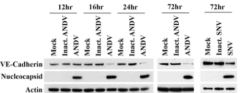
Andes Virus Disrupts the Endothelial Cell Barrier by Induction of Vascular Endothelial Growth Factor and Downregulation of VE-Cadherin
Full text
Figure


Related documents
Our data demonstrate that IL-6 released by JEV-infected pericytes is critical for proteasomal degradation of ZO-1 and the accompanying disruption of endo- thelial barrier
CM pre- pared from cells infected with WT, ovVEGF- ⌬ , or the Lister strain of vaccinia virus or mock infected with PBS was sub- jected to three assays of VEGF-like
Adenosine agonists stimulate vascular endothelial growth factor (VEGF) protein expression, and A^R antago- nists prevent hypoxia-induced VEGF protein expression. Compounds were added
Previous studies have shown that treatment of endothelial cells with VEGF causes interaction between VE- cadherin and VEGFR-2 [32] and that VEGF can stimulate the
Downregulation of vascular endothelial growth factor receptors by tumor necrosis factor-alpha in cultured human vascular endothelial
ology, St. Abbreviations used in this paper: bFGF, basic fibroblast growth fac- tor; CM, conditioned medium; HMECs, human microvascular endo- thelial cells; M199, medium 199;
The present study investigated the interplay of these factors on junctional occupancy of VE-cadherin and macromolecular leakage in human en- dothelial monolayers and the
A strong in- duction of endothelial fenestrations was observed in cocultures of endothelial cells with choroid plexus epi- thelial cells, or mammary epithelial cells stably
