R E S E A R C H
Open Access
Evolution of the Pax-Six-Eya-Dach network: the
calcisponge case study
Sofia AV Fortunato
1,2, Sven Leininger
1,3and Maja Adamska
1*Abstract
Background:ThePax-Six-Eya-Dachnetwork (PSEDN) is involved in a variety of developmental processes, including well documented roles in determination of sensory organs and morphogenesis in bilaterian animals. Expression of PSEDN components in cnidarians is consistent with function in sensory organ development. Recent work in demosponges demonstrated the presence of single homologs ofPaxandSixgenes, and their possible involvement in morphogenesis, but the absence of the remaining network components. Calcisponges are evolutionarily distant from demosponges, and the developmental toolkits of these two lineages differ significantly. We used an emerging model system,Sycon ciliatum, to identify components of the PSEDN and study their expression during embryonic and postembryonic development.
Results:We identified twoPax, threeSixand oneEyagenes in calcisponges, a situation strikingly different than in the previously studied demosponges. One of the calcispongePaxgenes can be identified asPaxB, while the second Paxgene has no clear affiliation. The three calcispongeSixgenes could not be confidently classified within any known family ofSixgenes. Expression analysis in adultS. ciliatumdemonstrated that representatives ofPax,Six andEyaare expressed in patterns consistent with roles in morphogenesis of the choanocyte chambers. Distinct paralogues ofPaxandSixgenes were expressed early in the development of the putative larval sensory cells, the cruciform cells. While lack of known photo pigments in calcisponge genomes precludes formal assignment of function to the cruciform cells, we also show that they express additional eumetazoan genes involved in specification of sensory and neuronal cells:ElavandMsi.
Conclusions:Our results indicate that the role of aPax-Six-Eyanetwork in morphogenesis likely predates the animal divergence. In addition,PaxandSix, as well asElavandMsiare expressed during differentiation of cruciform cells, which are good candidates for being sensory cells of the calcaronean sponge larvae.
Keywords:Calcisponges, Sycon,Eyes absent, Pax,Six, Sensory cells
Background
In insect and vertebrate model systems,Pax,Six,Eyes ab-sent(Eya) andDachshund(Dach) form a network (PSEDN) interconnected by a series of protein and protein-DNA interactions [1,2]. This network is often referred to as Retinal Determination Gene Network (RDGN), although it is involved in a variety of developmental processes in addition to eye development, including roles in morpho-genesis of other sensory organs, kidneys and the branchial arches [3-5]. It has been suggested that the insect and verte-brate PSEDN/RDGN are not homologous [6], although studies in cnidarians indicate deep evolutionary roots of
this network. In particular, it has been demonstrated that members of the Pax, Six and Eya families are expressed during neural development and sensory organ formation in a wide range of cnidarians [7-15]. In demosponges, two components of the PSEDN have been identified,PaxBandSix1/2[16,17]. A recent study in the freshwater demosponge Ephydatia muelleri shows that these genes are co-expressed, potentially interact and are likely involved in juvenile/adult morphogenesis [16,17].
While sponges lack a nervous system, larvae of some species have well defined sensory cells, organized into simple organ-like structures [18,19]. For example, the parenchymella-type larvae ofAmphimedon queenslandica have a pigmented ring equipped with long“steering” cilia at their posterior pole known as the sensory organ of the larva. Although opsin is not found in theA. queenslandica * Correspondence:[email protected]
1
Sars International Centre for Marine Molecular Biology, University of Bergen, Thormøhlensgt. 55, Bergen 5008, Norway
Full list of author information is available at the end of the article
© 2014 Fortunato et al.; licensee BioMed Central Ltd. This is an Open Access article distributed under the terms of the Creative Commons Attribution License (http://creativecommons.org/licenses/by/4.0), which permits unrestricted use, distribution, and reproduction in any medium, provided the original work is properly credited. The Creative Commons Public Domain Dedication waiver (http://creativecommons.org/publicdomain/zero/1.0/) applies to the data made available in this article, unless otherwise stated.
Fortunatoet al. EvoDevo2014,5:23
genome, the larval phototactic behavior [20] is likely me-diated by cryptochrome [21], which has also been sug-gested to participate in light reception in adult tissue of another demosponge, Suberites domuncula [22]. Signifi-cantly, AmqCry2 expression is associated with the pig-ment ring [23]. Unfortunately, no information regarding expression ofPaxorSixgenes during development of the pigment ring is published, making it impossible to predict whether the ancestral PSEDN function was related to morphogenesis only or both morphogenesis and sensory organ formation.
We have recently began developingSycon ciliatum as a model representing calcisponges (subclass Calcaro-nea), a lineage evolutionarily distant from demosponges and appearing to significantly differ from demosponges in its gene content [24-27]. Embryonic and postembryo-nic development of syconoid calcaronean species is well studied, allowing us to relate gene expression patterns to developmental events. Importantly, different stages of ra-dial (choanocyte) chamber morphogenesis can be com-pared in a single specimen fixed during the growth phase: when the asconoid body plan of the juvenile gives rise to the syconoid body plan of the adult, radial chambers form around the original central choanocyte chamber and continue to develop sequentially from bottom to top, with the region just under the osculum remaining in asconoid organization [28]. The amphiblastulae larvae of calcisponges from the subclass Calcaronea are strikingly different from the parenchymellae [25,29,30]. Amphiblas-tulae are composed of only three cell types of embryonic origin: macromeres, micromeres, and four cruciform cells distributed around the “equator” and conveying unique tetra-radial symmetry to the larva [24,31]. While the mac-romeres and micmac-romeres participate in formation of the juvenile body upon metamorphosis, the cruciform cells degenerate upon settlement [32]. The function of cruci-form cells has not been studied experimentally, but based on ultrastructure examination it has been suggested that they might act as photoreceptors [33]. Intriguingly, differ-entiating cruciform cells ofS. ciliatumexpressSoxB[24], a transcription factor involved in bilaterian neurogenesis [34,35] and expressed in cnidarian neurosensory cells [36]. In addition, they express several genes which, while clearly having multitude of roles in animal development, are also implicated in specification of neuronal cell types in eume-tazoans: components of the Wnt pathway (dvl, tcf and beta-catenin),Smad1/5andnanos, lending support to the notion that they could be sensory cells [25,37-39].In this study we chose to address the evolution of the PSEDN by studying expression of potential components of this net-work inS. ciliatum, focusing on the cruciform cells as the likely sensory cells of the larvae, and on the adult morpho-genesis represented by formation of the radial (choano-cyte) chambers.
We searched the genomic and transcriptomic datasets of S. ciliatum and a second calcaronean species, Leuco-solenia complicata, for genes encoding the components of the PSED network. To gain additional insight into identity of the cruciform cells, we also searched for genes encoding known proteins involved in photorecep-tion (opsin and cryptochrome), and the RNA binding proteins Elav and Musashi, which are involved in specifi-cation of neurosensory cells in eumetazoans. In this paper, we report that calciponge genomes contain an ortholog of theEya gene, which has not been previously reported in demosponges. We have not identified opsin and dachshund in calcisponges, which is consistent with the absence of these genes in demosponges. On the other hand, cryptochrome, which is present in demos-ponges, and likely responsible for light perception in the demosponge larvae, is absent from the calcisponge ge-nomes. Expression ofPax, Six, Eya, Msi and Elavgenes in Sycon ciliatum was studied by in situ hybridization.
Here we show that Pax, Six and Eya genes are
co-expressed during morphogenesis of the radial chambers, and that Pax and Six, as well as Elav and Msi are co-expressed during formation of cruciform cells.
Methods
Sequence retrieval, alignment and phylogenetic analyses Sycon ciliatum and Leucosolenia complicata Pax, Six and Eyagenes were identified by BLAST searches ofSyconand Leucosoleniadraft genomes (a preliminary draft of Leucoso-lenia) and transcriptomes as previously described [24] using specific domains from the following taxa: Bilateria, Bran-chiostoma floridaeandMus musculus; Porifera, Amphime-don queenslandica; Cnidaria,Nematostella vectensis.
For Eya, alignments were performed using the con-served ED domain. For Pax genes two alignments were performed: in the first alignment, the complete paired domain (PD) was included and in the second alignment the truncated RED-PD domain was used. Lack of home-odomains in the calcisponge Pax sequences precludes homeodomain-based phylogenetic analyses. ForSixgenes, the homeodomain along with the extendedsine oculis do-main was used. A combination of ClustalX and MUSCLE was used for the alignments, which were manually cor-rected where necessary.
Figure 1(See legend on next page.)
Fortunatoet al. EvoDevo2014,5:23 Page 3 of 12
sampled every 1,000th generation. Two Bayesian ana-lyses were run for each dataset from 1 to 10 million generations, depending on the dataset. Convergence was assessed by plotting the log likelihood against the number of generations using Tracer v1.4 [42]. The ana-lysis were stopped when the split frequency between the two runs was lower than 0.01. After the removal of an appropriate burn-in (20 to 25% in most cases), the consensus trees were visualized with FigTree v1.4.0 [43]. The ML analysis was performed using PhyMl 3.0 [44] as follows: To provide a starting tree for the boot-strap analysis, two rounds of PhyMl analysis, each starting from five random trees, were run using the following command line: −i align.phy –d aa –f e –m LG –c 4–a value –v value –rand_start –s NNI. The better of the two resulting ML trees (the tree with better likelihood value) was selected as an input tree for 1,000 bootstrap analysis using the following command line: -i align.phy -d aa -f e -m LG -c 4 -a value -v value -u best_random_tree. nwk -b 1,000 -s NNI.
Molecular analyses: RT-PCR andin situhybridization
Sample collection, fixation, PCR amplification of genes, se-quencing, probe production and in situ hybridization in sliced sponges containing different reproductive stages and in small adultSycon ciliatumspecimens were performed as described previously [24].
Results
Two Pax genes are found inSyconand one inLeucosolenia
TwoPaxgenes were found inSyconand one in Leucoso-lenia. As the Leucosolenia dataset is less extensive than theSyconone, it is possible that our current analysis can miss aLeucosolenia sequence. In contrast to the demos-ponges’ PaxB with a recognizable partial homeodomain and an octapeptide, the Pax genes in calcisponges do not appear to contain a homeodomain or octapeptide. Both Sycon Pax genes contain an intron in the PD do-main (Additional file 1) corresponding to the intron-exon boundaries found in cnidarians [45] and in other sponges [16,46]. The phylogenetic analysis of the PD of Pax (Figure 1C, Additional file 2) shows that both
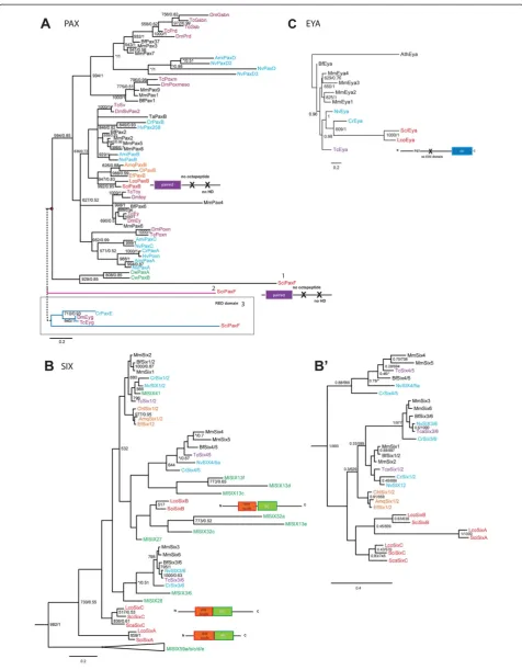
calcisponges have a single ortholog of PaxB and con-firms the affiliation of demosponges PaxBgenes as pre-viously reported [16,46]. Several phylogenetic analyses were performed to determine affiliation of the second Pax gene inSycon(Figure 1A). The first analysis used an alignment of the complete PD domain (Figure 1A, pos-ition 1), and indicated affiliation of this gene with the ctenophore Coeloplana willeyi Pax genes [47]. For the second analysis we removed the C. willeyi sequences from the alignment, and in this analysis the second Sycon Pax gene did not affiliate with any subfamily of Pax genes (Figure 1A, position 2). Finally, in the third analysis (Figure 1A, position 3), we used the partial PD domain (RED motif ) and included arthropod eye gone (Eyg) genes and Cladonema radiatum PaxE gene. The result of the analysis shows that this second Pax gene fell within the PaxE subfamily (theEygsubfamily) but this association was not supported (see also Additional file 3). Due to the unclear affiliation of the secondPax gene in Sycon, we decided to name itPaxF, following the next let-ter in the classification ofPaxgenes. Importantly, in none of our analyses SciPaxF affiliated with Pax1/9 and/or Pax3/7 subfamilies and thus it does not provide additional support for the notion of Pax duplication before diver-gence of Porifera [48].
SINE class family is expanded in calcisponges
We found threeSixgenes in each of theSyconand Leu-cosolenia genomes corresponding to the SINE class of homeobox genes. All of them had the characteristic Six homeodomain with lysine at position 50 and thesine oc-ulisDNA binding domain situated at the -N terminal to the homeodomain (Additional file 4), as seen in previ-ously classified Six genes [49]. Additionally, we found two genes (SciHD35531and LcoHD71216) that contained partial sine oculis domains and homeodomains which displayed similarity to both the TALE and SINE gene classes. The homeodomains of SciHD35531 had a four amino acid insertion, instead of the three typically ob-served in TALE homeodomains (Additional file 5).
The first phylogenetic analysis was based on the homeo-domain sequences and included the entire expanded (See figure on previous page.)
Figure 1Phylogenetic analyses of the Six,EyaandPaxgenes. A, Bayesian phylogenetic tree of Pax genes inferred from the paired domain. The red dots indicate the different positions ofSciPaxF: 1, position ofSciPaxFwhen the complete the PD domain was used to infer the phylogeny; 2, position ofPaxFwhen removing ctenophore sequences and 3, when using the RED domain (S6) and includingCrPaxEand ArthropodEyggenes.
B, The maximum likelihood tree was inferred from the Six homeodomain. The Six tree was rooted using the TALE class homeodomain as an outgroup.
complement of ctenophore Six genes [50] (Figure 1B). This analysis correctly separated the cnidarian and bilaterian sequences into the three recognized families (Six1/2, Six3/6 and Six4/5) and confirmed the affiliation of demospongeSix genes within the Six1/2 family [16,17,46] albeit with low support value (Figure 1B). Among the ctenophore
sequences, MlSix41 nested within the Six1/2 family, consistent with the presence of the ETSY motif in its
homeodomain, while MlSix36 and MlSix28 grouped
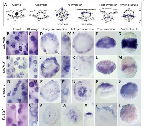
[image:5.595.57.539.91.510.2]with the Six3/6 family as described [50]. Surprisingly, none of the calcisponge sequences associated with demosponge sequences or any other recognized Six Figure 2Expression ofSycon PaxandSixgenes during embryogenesis. A, Summary of embryogenesis inSycon, with consecutive stages depicted from left to right: oocyte, cleavage, pre-inversion, post-inversion, larva. Abbreviations: pin, pinacocytes; ch, choanocytes; ac, accessory cells; cc, cruciform cells; mi, micromeres ma, macromeres; and mc, maternal cells.B-G,SciPaxBexpression is detectable in the oocytes (B), and in all blastomeres during late cleavage (C). During early pre-inversion it is detectable in the micromeres and cruciform cells and in late pre-inversion (E), post-inversion (F) and in larvae (G)SciPaxBexpression is restricted to the equatorial micromeres.H-M,SciPaxFexpression is detectable in the oocytes (H) and is gradually restricted to become predominant in the cross cells (J to K), and then the equatorial micromeres in the larva (M).N-S, SciSixCexpression is present in the oocytes (N) and then all blastomeres (O), but strongest in the cruciform cells by early pre-inversion (P). During late pre-inversion (Q) the expression is localized to the cruciform cells and the macromeres, and becomes limited to the macromeres in the post-inversion embryos and larva (R,S).T-Z,SciSixAis expressed in the oocytes (T), but not in cleavage stages or early pre-inversion stage embryos (U, V), when strong expression in a ring of accessory cells is apparent (U). Transient expression in the anterior micromeres is detectable in post-inversion stage embryos (W-Y) but not larvae (Z). All images are of whole mounted, glycerol-cleared specimens, except Y, which is a resin section. Embryos in pre-inversion, post-inversion and larvae were isolated from the tissue, except from K. The asterisk indicates macromeres at the posterior pole of the embryo and larva. Black arrows, white arrows and white arrowheads indicate the cruciform cells, micromeres and the accessory cells, respectively.
Fortunatoet al. EvoDevo2014,5:23 Page 5 of 12
families. Because of unclear affiliation, calcisponge Six genes are referred to as SixA, SixB and SixC (with the
SixC sequences being most similar to the SixC
se-quence fromSycon calcaravis,ScaSixC[47]). In the tree presented on Figure 1B, Sci/LcoSixB genes associated with several of the ctenophore sequences, and this grouping receives moderate support in the ML analysis; whileSciSixAandSciSixCfell completely outside of the recognized Six families. Given the difficulty in assigning the ctenophore Six genes and the fact that many of them are on long branches, we suspected that affilia-tions of calcisponge and Mnemiopsis Six genes might represent an artefact of long-branch attraction. We have thus carried out additional analyses with limited comple-ments or completely without the ctenophore sequences, utilizing either only the homeodomain (Figure 1B’ and Additional file 6), or the SINE domain together with the homeodomain (Additional file 7). In particular, we were hoping to differentiate between scenarios in which the three calcispongeSixgenes are all descended from aSix1/ 2ancestral sequence, and are a result of family expansion in the calcisponge lineage, or are remnants of ances-tral sequences, which are preserved in ctenophore and
calcisponge genomes and lost in all others. In the homeodomain-only analysis without the ctenophore sequences, the calcisponge sequences clustered to-gether, although this grouping did not receive signifi-cant support (Figure 1B’). In the tree utilizing the SINE domain, the calcisponge sequences did not form a monophyletic clade, but all of them branched basally (Additional file 7). Significantly, the grouping of the demospongeSixgenes with the Six1/2 family was sen-sitive to taxon sampling. However, all demospongeSix and the calcispongeSixBhomeodomains have the charac-teristic Six1/2 family 'ETSY' motif, and SixC sequences have a similar ‘ETNY’motif ([50] and Additional file 5). Altogether, the results of the phylogenetic analyses indi-cate that the calcispongeSixgenes may represent a spe-cific diversification of the SINE class in calcareous sponges, similar to the one observed in the ctenophores [50], although the exact relationships between the para-logs remain unclear.
Eyes absent(Eya) is present in calcisponges
We found single orthologs of the eyes absent gene,Eya,in the two calcisponges. The Eya gene has not been previ-ously reported in demosponges. In both species, the pre-dicted proteins have a conserved C-terminal amino-acid domain, theEyadomain (ED) but not the N-terminal ED2 domain (Additional files 8 and 9). The phylogenetic ana-lysis in Figure 1C shows that the calcispongeEyagenes af-filiate with the cnidarianEyagenes with moderate support values. Eya gene containing the ED domain and P/S/T rich region is found in the choanoflagelateMonosiga bre-vicollis[13], indicating thatEyawas lost in demosponges. The presence of the ED domain and the P/S/T region but not the ED2 region of theEyagene corroborates the no-tion that the ED2 domain was established in the last com-mon ancestor of bilaterians and cnidarians [13].
Pax and Six genes, but not Eya are expressed during embryogenesis and in the larvae
[image:6.595.56.292.372.635.2]As described for multiple species of calcaronean sponges [25,29,51] and schematically represented on Figure 2A, embryogenesis ofSycontakes place in the mesohyl. Sym-metric cleavage followed by cell differentiation leads to formation of a cup-shaped embryo composed of three cell types: macromeres, micromeres and cross cells. Macro-meres are cells which are large, non-ciliated and granular in appearance, located close to the choanocytes of the par-ent sponge. Numerous micromeres are small and ciliated and located closer to the pinacocytes. The four cruciform cells are similar in size to the macromeres and are sym-metrically distributed among the micromeres on the equa-tor of the embryo. The embryo undergoes inversion while it translocates to the radial chamber and the mature larva swims through the oscular opening.
Figure 4(See legend on next page.)
Fortunatoet al. EvoDevo2014,5:23 Page 7 of 12
The expression ofSciPaxB(Figure 2B-G) begins in the oocytes and continues during early and late cleavage, with transcripts present in all blastomeres (Figure 2C). During early pre-inversion,SciPaxBexpression is evident in the cruciform cells and micromeres (Figure 2D, D’). In the larvae, SciPaxB expression is high in a broad band of equatorial micromeres (Figure 2F). The expression of SciPaxF begins in the oocytes (Figure 2H) and is also initially uniform in all blastomeres, but by early pre-inversion the expression is stronger in micromeres and evident in the cruciform cells (Figure 2J-K). In the lar-vae, theSciPaxFexpression is weak in the posterior mi-cromeres, and expression in the cruciform cells is no longer apparent (Figure 2M). Expression of SciSixC is detected in the oocytes (Figure 2N), and during early pre-inversion it becomes most prominent in the cruci-form cells (Figure 2P), resolving to strong expression in the macromeres and weaker in the cruciform cells dur-ing late pre-inversion (Figure 2Q). In post-inversion stages and larva, only macromere expression is appar-ent (Figure 2R, S).
The expression ofSciSixAis strong in oocytes (Figure 2T), but not detectable in the cleavage stage embryos (Figure 2U-V), instead becoming prominent in a ring of accessory cells (which are derived from choanocytes) surrounding the embryos (Figure 2U, Y). In the late pre-inversion stage embryos, SciSixA expression is de-tectable in the anterior micromeres of the embryos (Figure 2W-Y) although this expression is no longer de-tectable in the larvae (Figure 2Z). Finally, the SciSixB gene expression was weak and appeared ubiquitous throughout all cell types with no distinct expression pattern (data not shown). No expression of SciEya was observed during embryogenesis, although its expression is detectable in choanocytes (Additional file 10 and see below).
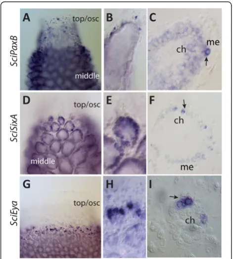
Pax-Six-Eya are co-expressed in choanocyte chambers
In addition to the embryonic expression described above, SciPaxB, SciSixA and SciEya transcripts are detected in adult cell types, being most prominent in the choanocytes (the innermost epithelial cells responsible for feeding) (Figure 3). Expression of SciPaxB is uniform throughout the choanocytes and is strong in scattered mesohyl cells,
particularly at the tips of the radial chambers and in the area of osculum (Figure 3A-C). SciSixA expression is stronger in choanocytes of the uppermost, forming cham-bers than in the older chamcham-bers, with somewhat heteroge-neous expression among the choanocytes (Figure 3D-F). Notably,SciSixA expression is not detectable in choano-cytes which are not organized in chambers, but located in the uppermost area of the sponge in the region of ascon-oid organization (Figure 3D). The expression of SciEya is similar to that of SciSixA in that the choanocytes of the oscular region are negative, the choanocytes located in the uppermost, forming choanocyte chambers dis-play the most prominent expression, and the choano-cytes of the older chambers display low level of expression (Figure 3G-I).
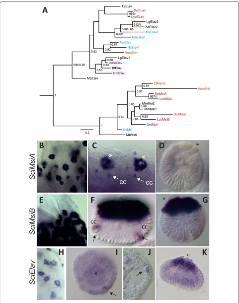
Elav and Musashi are expressed in the cruciform cells and macromeres
We identified one Elavand two Musashi genes in each of the two analyzed sponges (Additional file 11). The phylogenetic analyses were performed using the con-served RRM2 domain present in both gene families (Additional file 12). The calcispongeElavgenes affiliated with the Trichoplax adhaerens Elav gene, albeit with poor support. Affiliation of this clade with either Elav1 or Elav2 subfamily differed between ML and Bayesian analysis, never reaching significant support (Figure 4A). The calcisponge MsiA sequences clustered with demos-pongeMsigenes, while theMsiBpair affiliated with bila-terian sequences (Figure 4A).
Both ofMsigenes and theElavgene display similar ex-pression patterns, being expressed strongly and uniformly in the oocytes and during cleavage, and gradually becom-ing most prominent in the cruciform cells (Figure 4B, E, H, I and Additional file 13). During pre-inversion, strong expression of SciMsiA and weaker expression of both SciMsiBandSciElavis also detectable in the macromeres, in addition to continued expression in the cruciform cells (Additional file 13 and Figure 4C, J). Among the three genes, SciMsiB is detectable until the latest (post-inver-sion) stages in the cruciform cells (Figure 4F). Finally, SciMsiB and SciElav are detectable in the larval macro-meres (Figure 4G, K).
(See figure on previous page.)
Discussion
Pax,Six, Eyes absentand Dachshundgenes are involved in a variety of developmental processes, including the determination and morphogenesis of eyes and other sen-sory organs such as the ear [3,52]. In addition to over-lapping expression patterns, protein-protein interactions and direct expression regulation demonstrate that PSED genes function as a network in a variety of animals [1,3]. Although many of the regulatory relationships are con-served in the PSED network, the interactions between network components can vary during development and in different animal lineages [6,53].
The origin of the PSED network has been previously in-vestigated in non-bilaterian animals, including cnidarians [7-9,13,54] and demosponges [16,47]. As demosponges contain PaxBand Six1/2genes, but not Eya or Dach, it has been proposed that the ancestral network was com-posed of these two genes only. However, absence ofEyain demosponges appears secondary, as it is present in the choanoflagellates [13]. Here we show that the genomes of calcisponges also containEya, suggesting that its loss oc-curred in the lineage leading to demosponges and is thus not representative of all sponges. A question remains whether the three genes (Pax-Six-Eya) indeed do interact, and thus form a network, or whether the network arose later during evolution from components working indi-vidually in sponges. Importantly, a recent study in the demospongeEphydatia mulleriindicated that in this spe-cies expression of Six1/2 might be controlled by PaxB [17]. Unfortunately, it is not possible at the moment to functionally test interactions of the potential network components in calcisponges. However, their co-expression in several developmental contexts in Sycon suggests that these genes might also operate as a network, and in fact might control similar developmental processes as in other animals: sensory cell specification and morphogenesis. With the exception ofEya, all of the identified genes are co-expressed in the oocytes and in embryos undergoing cleavage, suggesting they are maternally expressed and have early developmental roles. In subsequent stages of development, the most striking and potentially inform-ative co-expression is in the embryonic cruciform cells and later during adult morphogenesis, as summarized on Figure 5.
The cruciform cells have been suggested to act as lar-val photoreceptors [33] and the amphiblastulae have been shown to respond to light [55]. Surprisingly, we found that not only opsin, but also cryptochrome genes, which likely convey light sensitivity to the demosponge larvae, are absent fromSyconandLeucosoleniagenomes, indicating that calcisponges have lost cryptochrome. Thus, calcisponge larvae might be able to detect light using another, yet unclear mechanism. Intriguingly, ho-mologues of multiple genes involved in specification of
neural and sensory cells in other animals are expressed during cross cell differentiation. These include SciPaxB, SciPaxF,SciSixC,SciElav,SciMsiAandSciMsiBpresented
Figure 5Summary of the expression patterns. A,SciPaxBin cruciform cells and equatorial micromeres during pre-inversion and micromeres in the larvae;B,SciPaxFin cruciform cells during pre-inversion and equatorial micromeres of the larva;C,SixAin anterior micromeres,D,SciSixC,SciElav,SciMsiAandSciMsiBin the cruciform cells and in the macromeres of the post-inversion stage embryos and/ or larvae;E, Gradients ofSciSixAandSciEyain choanocyte cells organized in chambers,SciPaxBin all choanocytes and in scattered mesohyl cells.
Fortunatoet al. EvoDevo2014,5:23 Page 9 of 12
in this study (see Figure 5A),SciSoxB[24], components of the Wnt pathway (Dvl, Tcf and Beta-catenin), Smad1/5 andNanos[25].
The observed co-expression indicates that SciSixC, Sci-PaxBandSciPaxFmay potentially interact during embryo-genesis, whileSciPaxB, SciSixA and SciEyamay potentially interact during adult morphogenesis (Figure 5). Among the three potential components of the PSEN in Sycon expressed in the choanocytes,SciPaxBhas the broadest ex-pression, with transcripts uniformly detected in all cho-anocytes. In contrast, SciSixA and SciEya transcripts are conspicuously absent from the uppermost choano-cytes remaining in asconoid organization, but are par-ticularly strongly expressed in the choanocytes of the uppermost chambers which are undergoing morpho-genesis, with SciEya expression diminishing along a somewhat steeper gradient than SciSixA, so that both are expressed at low levels in the already formed cham-bers (Figure 5b). SciPaxB, SciSixA and SciEya expres-sion patterns are thus consistent with interaction of these three genes during formation and maintenance of the organization of the radial chambers, and thus with a concerted role in morphogenesis. Despite lack ofEya of the demosponge Ephydatia, the morphogenetic role of the potential network might be a conserved feature, as knock down ofEmPaxBandEmSix1/2results in ap-parent dysmorphogenesis of the juveniles [17].
Conclusions
Overall, the presence of the sponge Eya gene and co-expression ofPax,SixandEyagenes in calcisponges indi-cate that thePax-Six-Eyanetwork may have already been established in the last common ancestor of sponges and eumetazoans, withEyasubsequently lost in demosponges. Based on gene expression during adult body plan forma-tion inSycon, we propose that this network had an ancient role in morphogenesis. Additionally, co-expression ofPax andSixwith conserved eumetazoan neural genesElavand Msiin candidate larval sensory cells, suggests these genes could be ancestrally involved in the determination of sen-sory cell types. We envisage a scenario in which a simple PSE network was active in early metazoans, and that add-itional genes, such as Dachshund, were then later co-opted into the network to expand its regulatory capacity in more complex animals.
Additional files
Additional file 1:The predicted Pax protein sequences and intron-exon boundaries.Exon-intron boundaries are highlighted in yellow, the paired domain is red.
Additional file 2:Alignment of the PD domain.This alignment was used for the phylogenetic analyses displayed in Figure 1C. The red box
indicates the location of the RED motif used for the phylogenetic analyses in Additional file 3. Abbreviations are as in Figure 1.
Additional file 3:Bayesian phylogenetic tree of thePaxgene family inferred from the RED motif of the PRD domain.Support values on nodes are as follows: left, bootstrap (BT) values obtained from ML analysis; right, posterior probability from the Bayesian analysis. For abbreviations of species names see Figure 1.
Additional file 4:Sixgenes protein sequences inSyconand
Leucosolenia.Exon-intron boundaries are indicated by highlighting. Sine oculis domain is red, the homeodomain is underlined.
Additional file 5:Alignment of the homeodomain of the SINE and TALE classes including all of Six genes and selected TALE genes identified in calcisponges.Abbreviations are as in Figure 1.
Additional file 6:Maximum likelihood tree of the SINE class.
Phylogenetic tree inferred from the homeodomain of Six and TALE genes. Bootstrap values are displayed on each node. Names are prefixed as in Figure 1. The tree was rooted with a selection of TALE class of homeobox genes.MnemiopsisSix genes found in long branches on the tree from Figure 1B were not included in this analysis.
Additional file 7:Maximum likelihood phylogenetic analyses of sine oculis domain and homeodomain of the Six class.ML bootstrap values greater than 500 are displayed. Names are prefixed as in Figure 1.
Additional file 8:EYA protein sequences.Red indicates the location of the ED domain. Exon-intron boundaries are highlighted.
Additional file 9:Alignment of the ED domain.This alignment was used for the phylogenetic analyses displayed in Figure 1B. Abbreviations are as in Figure 1.
Additional file 10:TheSciEyagene is not expressed during embryogenesis.A, oocytes; B, embryos during pre-inversion and C, post-inversion.
Additional file 11:Elav and Msi protein sequences.Exon-intron boundaries are highlighted. Red indicates the location of the RMM2 domain.
Additional file 12:RRM2 motif alignment for Msi and Elav sequences.This alignment, without gaps, was used for the phylogenetic analyses displayed in Figure 4.
Additional file 13:Predominant expression ofSciMsiAandSciElav in the cruciform cells.Late cleavage and pre-inversion stage embryos are shown forSciMsiAandSciElav, respectively.
Abbreviations
BLAST:Basic Local Alignment Search Tool; ED:Eyadomain; ML: Maximum Likelihood; PD: Paired domain; PSEDN:Pax-Six-Eya-Dachnetwork; RDGN: Retinal Determination Gene Network.
Competing interests
The authors declare that they have no competing interests.
Authors’contributions
SF and MA conceived and designed the study. MA and SL carried out field collection. SF performed phylogenetic analyses and analyzed expression of all genes presented in this manuscript exceptMusashi, which were studied by SL. SF drafted and MA edited the manuscript with input from SL. All authors read and approved the final manuscript.
Acknowledgements
We thank M. Adamski for sharing unpublished sequence assemblies and participation in field collections and G. Richards for insightful comments on the manuscript. This work was supported by the core budget of the Sars Centre to MA.
Author details
1
Sars International Centre for Marine Molecular Biology, University of Bergen, Thormøhlensgt. 55, Bergen 5008, Norway.2Department of Biology, University
Received: 10 March 2014 Accepted: 15 May 2014 Published: 23 June 2014
References
1. Donner A, Maas R:Conservation and non-conservation of genetic pathways in eye specification.Int J Dev Biol2004,48:743–753. 2. Friedrich M:Ancient mechanisms of visual sense organ development
based on comparison of the gene networks controlling larval eye, ocellus, and compound eye specification in drosophila.Arthropod Struct Dev2006,35:357–378.
3. Silver S, Rebay I:Signaling circuitries in development: insights from the retinal determination gene network.Dev Suppl2005,132:3–13. 4. Kozmik Z, Holland N, Kreslova J, Oliveri D, Schubert M, Jonasova K, Holland
L, Pestarino M, Benes V, Candiani S:Pax-Six-Eya-Dach network during amphioxus development: conservationin vitrobut context specificity in vivo.Dev Biol2007,306:143–159.
5. Bassham S, Postlethwait JH:The evolutionary history of placodes: a molecular genetic investigation of the larvacean urochordateOikopleura dioica.Development2005,132:4259–4272.
6. Wagner G:The developmental genetics of homology.Nat Rev Genet2007, 8:473–479.
7. Matus D, Pang K, Daly M, Martindale M:Expression of Pax gene family members in the anthozoan cnidarian,Nematostella vectensis.Evol Dev
2007,9:25–38.
8. Suga H, Tschopp P, Graziussi DF, Stierwald M, Schmid V, Gehring WJ: Flexibly deployed Pax genes in eye development at the early evolution of animals demonstrated by studies on a hydrozoan jellyfish.Proc Natl Acad Sci U S A2010,107:14263–14268.
9. Kozmik Z, Daube M, Frei E, Norman B, Kos L, Dishaw L, Noll M, Piatigorsky J: Role of Pax genes in eye evolution: a cnidarian PaxB gene uniting Pax2 and Pax6 functions.Dev Cell2003,5:773–785.
10. Kumar J:The sine oculis homeobox (SIX) family of transcription factors as regulators of development and disease.Cell Mol Life Sci2009,66:565–583. 11. Kozmik Z:The role of Pax genes in eye evolution.Brain Res Bull2008,
75:335–339.
12. Kozmik Z:Pax genes in eye development and evolution.Curr Opin Genet Dev2005,15:430–438.
13. Graziussi DF, Suga H, Schmid V, Gehring WJ:The“Eyes absent”(eya) gene in the eye-bearing hydrozoan jellyfishCladonema radiatum:
conservation of the retinal determination network.J Exp Zool B Mol Dev Evol2012,318:257–267.
14. Galliot B, Quiquand M, Ghila L, De Rosa R, Miljkovic-Licina M, Chera S: Origins of neurogenesis, a cnidarian view.Dev Biol2009,332:2–24. 15. Sinigaglia C, Busengdal H, Leclère L, Technau U, Rentzsch F:The bilaterian
head patterning genesix3/6controls aboral domain development in a cnidarian.PLoS Biol2013,11(2):e1001488.
16. Hill A, Boll W, Ries C, Warner L, Osswalt M, Hill M, Noll M:Origin of Pax and Six gene families in sponges: SinglePaxBandSix1/2orthologs in Chalinula loosanoffi.Dev Biol2010,343:106–123.
17. Rivera A, Winters I, Rued A, Ding S, Posfai D, Cieniewicz B, Cameron K, Gentile L, Hill A:The evolution and function of the Pax/Six regulatory network in sponges.Evol Dev2013,15:186–196.
18. Maldonado M:The ecology of the sponge larva.Can J Zool2006, 84:175–194.
19. Ludeman D, Farrar N, Riesgo A, Paps J, Leys S:Evolutionary origins of sensation in metazoans: functional evidence for a new sensory organ in sponges.BMC Evol Biol2014,14:3.
20. Leys S, Cronin T, Degnan B, Marshall J:Spectral sensitivity in a sponge larva.J Comp Physiol Neuroethol Sens Neural Behav Physiol2002, 188:199–202.
21. Feuda R, Hamilton S, McInerney J, Pisani D:Metazoan opsin evolution reveals a simple route to animal vision.Proc Natl Acad Sci U S A2012, 109:18868–18872.
22. Müller WE, Wang X, Schröder HC, Korzhev M, Grebenjuk VA, Markl JS, Jochum KP, Pisignano D, Wiens M:A cryptochrome-based photosensory system in the siliceous spongeSuberites domuncula(Demospongiae).
FEBS J2010,277:1182–1201.
23. Rivera A, Ozturk N, Fahey B, Plachetzki D, Degnan B, Sancar A, Oakley T: Blue-light-receptive cryptochrome is expressed in a sponge eye lacking neurons and opsin.J Exp Biol2012,215:1278–1286.
24. Fortunato S, Adamski M, Bergum B, Guder C, Jordal S, Leininger S, Zwafink C, Rapp HT, Adamska M:Genome-wide analysis of the sox family in the calcareous spongeSycon ciliatum: multiple genes with unique expression patterns.EvoDevo2012,3:14.
25. Leininger S, Adamski M, Bergum B, Guder C, Liu J, Laplante M, Bråte J, Hoffman NF, Fortunato S, Jordal S, Rapp HT, Adamska M:Developmental gene expression provides clues to relationships between sponge and eumetazoan body plans.Nat Commun2014,5:3905.
26. Riesgo A, Farrar N, Windsor PJ, Giribet G, Leys SP:The analysis of eight transcriptomes from all poriferan classes reveals surprising genetic complexity in sponges.Mol Biol Evol2014,31:1102–1120.
27. Sebé-Pedrós A, Ariza-Cosano A, Weirauch M, Leininger S, Yang A, Torruella G, Adamski M, Adamska M, Hughes T, Gómez-Skarmeta J, Ruiz-Trillo I:Early evolution of the T-box transcription factor family.Proc Natl Acad Sci U S A
2013,110:16050–16055.
28. Maas O:Die Weiterentwicklung der Syconen nach der metamorphose.
Zeutsch wiss Zool1900,67:215–240.
29. Ereskovsky AV:The Comparative Embryology of Sponges.Dordrecht Heidelberg London New York: Springer; 2010.
30. Maldonado M, Bergquist P:Atlas of Marine Invertebrate Larvae.San Diego, CA: Academic Press; 2002.
31. Manuel M:Early evolution of symmetry and polarity in metazoan body plans.C R Biol2009,332:184–209.
32. Amano S, Hori I:Metamorphosis of calcareous sponges. 2. Cell rearrangement and differentiation in metamorphosis.Invert Reprod Dev
1993,24:13–26.
33. Tuzet O:Éponges calcaires. InTraité de Zoologie Anatomie, Systématique, Biologie Spongiaires.Edited by Grassé P-P. Paris: Masson et Cie; 1973:27–132. 34. Phochanukul N, Russell S:No backbone but lots of Sox: invertebrate Sox
genes.Int J Biochem Cell Biol2010,42:453–464.
35. Watanabe H, Fujisawa T, Holstein TW:Cnidarians and the evolutionary origin of the nervous system.Dev Growth Differ2009,51:167–183. 36. Jager M, Queinnec E, Le Guyader H, Manuel M:Multiple Sox genes are
expressed in stem cells or in differentiating neuro-sensory cells in the hydrozoanClytia hemisphaerica.EvoDevo2011,2:12.
37. Kanska J, Frank U:New roles for Nanos in neural cell fate determination revealed by studies in a cnidarian.J Cell Sci2013,126:3192–3203. 38. Grigoryan T, Wend P, Klaus A, Birchmeier W:Deciphering the function of
canonical Wnt signals in development and disease: conditional loss- and gain-of-function mutations of beta-catenin in mice.Genes Dev2008, 22:2308–2341.
39. Hegarty S, O'Keeffe G, Sullivan A:BMP-Smad 1/5/8 signalling in the development of the nervous system.Prog Neurobiol2013,109:28–41. 40. Abascal F, Zardoya R, Posada D:ProtTest: selection of best-fit models of
protein evolution.Bioinformatics2005,21:2104–2105.
41. Ronquist F, Huelsenbeck JP:MrBayes 3: Bayesian phylogenetic inference under mixed models.Bioinformatics2003,19:1572–1574.
42. Rambaut A, Drummond AJ:Tracer v1.5.; 2009. http://beast.bio.ed.ac.uk/ software/tracer/.
43. Rambaut A:Figtree v1.4.0.; 2012. http://tree.bio.ed.ac.uk/software/figtree/. 44. Guindon S, Dufayard J-F, Lefort V, Anisimova M, Hordijk W, Gascuel O:New
algorithms and methods to estimate maximum-likelihood phylogenies: assessing the performance of PhyML 3.0.Syst Biol2010,59:307–321. 45. Ryan J, Burton P, Mazza M, Kwong G, Mullikin J, Finnerty J:The
cnidarian-bilaterian ancestor possessed at least 56 homeoboxes: evidence from the starlet sea anemone.Nematostella vectensis. Genome Biol2006,7:R64. 46. Larroux C, Luke G, Koopman P, Rokhsar D, Shimeld S, Degnan B:Genesis
and expansion of metazoan transcription factor gene classes.Mol Biol Evol2008,25:980–996.
47. Hoshiyama D, Iwabe N, Miyata T:Evolution of the gene families forming the Pax/Six regulatory network: isolation of genes from primitive animals and molecular phylogenetic analyses.FEBS Lett2007,581:1639–1643. 48. Breitling R, Gerber JK:Origin of the paired domain.Dev Genes Evol2000,
210:644–650.
49. Takatori N, Butts T, Candiani S, Pestarino M, Ferrier D, Saiga H, Holland P: Comprehensive survey and classification of homeobox genes in the genome of amphioxus,Branchiostoma floridae.Dev Genes Evol2008,218:579–590. 50. Ryan J, Pang K, Program NCS, Mullikin J, Martindale M, Baxevanis A:The
homeodomain complement of the ctenophoreMnemiopsis leidyi suggests that Ctenophora and Porifera diverged prior to the ParaHoxozoa.EvoDevo2010,1:9.
Fortunatoet al. EvoDevo2014,5:23 Page 11 of 12
51. Franzen W:Oogenesis and larval development ofScypha Ciliata(Porifera, Calcarea).Zoomorphology1988,107:349–357.
52. Rebay I, Silver S, Tootle T:New vision from Eyes absent: transcription factors as enzymes.Trends Genet2005,21:163–171.
53. Schlosser G, Wagner GP:Modularity in development and evolution.Chicago: University of Chicago Press; 2004.
54. Stierwald M, Yanze N, Bamert R, Kammermeier L, Schmid V:The Sine oculis/Six class family of homeobox genes in jellyfish with and without eyes: development and eye regeneration.Dev Biol2004,274:70–81. 55. Elliot GR, Macdonald TA, Leys SP:Sponge larval phototaxis: a comparative
study.Bollettino dei Musei e degli Istituti Biologici dell’Universita di Genova
2004,68:291–300.
doi:10.1186/2041-9139-5-23
Cite this article as:Fortunatoet al.:Evolution of the Pax-Six-Eya-Dach network: the calcisponge case study.EvoDevo20145:23.
Submit your next manuscript to BioMed Central and take full advantage of:
• Convenient online submission
• Thorough peer review
• No space constraints or color figure charges
• Immediate publication on acceptance
• Inclusion in PubMed, CAS, Scopus and Google Scholar
• Research which is freely available for redistribution