Growth Factor-Beta Polymorphisms and Complications
of Coronary Heart Disease
Dylan R. Morris, Joseph V. Moxon, Erik Biros, Smriti M. Krishna, Jonathan Golledge* Vascular Biology Unit, School of Medicine and Dentistry, James Cook University, Townsville, Australia
Abstract
Objective: To investigate the association between common transforming growth factor beta (TGF-b) single nucleotide
polymorphisms (SNP) and significant complications of coronary heart disease (CHD).
Method:We performed a meta-analysis of published case-control studies assessing the association ofTGF-bSNPs with a
range of CHD complications. A random effects model was used to calculate odds ratios and confidence intervals. Analyses were conducted for additive, dominant and recessive modes of inheritance.
Results:Six studies involving 5535 cases and 2970 controls examining the association of common SNPs inTGF-b1with CHD
were identified. Applying a dominant model of inheritance, three TGF-b1SNPs were significantly associated with CHD complications: The T alleles ofrs1800469 (OR = 1.125, 95% CI 1.016–1.247, p = 0.031) andrs1800470 (OR = 1.146, 95% CI 1.026–1.279, p = 0.021); and the C allele ofrs1800471(OR = 1.207, 95% CI 1.037–1.406, p = 0.021).
Conclusion:This meta-analysis suggests that common genetic polymorphisms inTGF-b1are associated with complications
of CHD.
Citation:Morris DR, Moxon JV, Biros E, Krishna SM, Golledge J (2012) Meta-Analysis of the Association between Transforming Growth Factor-Beta Polymorphisms and Complications of Coronary Heart Disease. PLoS ONE 7(5): e37878. doi:10.1371/journal.pone.0037878
Editor:Weili Zhang, FuWai Hospital - Chinese Academy of Medical Sciences, China
ReceivedJanuary 8, 2012;AcceptedApril 25, 2012;PublishedMay 25, 2012
Copyright:ß2012 Morris et al. This is an open-access article distributed under the terms of the Creative Commons Attribution License, which permits unrestricted use, distribution, and reproduction in any medium, provided the original author and source are credited.
Funding:DM and EB were supported by the National Heart Foundation of Australia (http://www.heartfoundation.org.au/Pages/default.aspx). JG holds a Practitioner Fellowship from the National Health and Medical Research Council, Australia (http://www.nhmrc.gov.au/), a Senior Clinical Research Fellowship, and National and International Research Alliance Program (NIRAP) grant, both from the Queensland Government (http://www.business.qld.gov.au/science/scientific-research/scientific-government-support.html). The funders had no role in study design, data collection and analysis, decision to publish, or preparation of the manuscript.
Competing Interests:The authors have declared that no competing interests exist.
* E-mail: [email protected]
Introduction
Coronary heart disease (CHD) is the leading cause of mortality in the western world. In the U.S.A. CHD is responsible for 1 in every 6 deaths and is estimated to cost over US$177.5 billion per annum. [1] Early stage CHD is asymptomatic, but advanced disease is associated with severe complications including myocar-dial infarction, heart failure, unstable angina and sudden cardiac arrest. [2] Well defined risk factors for CHD include smoking, diabetes, hypertension, dyslipidaemia, male gender and age. A family history of CHD also confers significant risk, demonstrated by an eightfold increase in monozygotic twins and a fourfold increase in dizygotic twins of affected individuals. [3] The genetic mechanisms underlying CHD predisposition are poorly under-stood despite recent findings from genome-wide association studies (GWAS) (discussed in detail by Patelet al.(2011)). [4].
Data from independent gene studies suggest a putative role for
TGF-b signalling in CHD. [5,6] A range of potentially athero-protective roles have been described for the TGF-b isoforms, favouring a stable fibrous plaque over an unstable inflammatory plaque. In vitro studies have shown TGF-b1 to upregulate extracellular matrix (ECM) production, inhibit vascular smooth
muscle cell proliferation and impair division and migration of endothelial cells. [7] Additionally, TGF-b1 modulates the local inflammatory response by down-regulating expression of endothelial adhesion molecules, inhibiting foam cell formation and impeding proliferation of CD4 and CD8 T lymphocytes. [7] Dysregulation of
TGF-bsignalling resulting from genetic mutations, particularly single nucleotide polymorphisms (SNPs), within components of theTGF-b signalling pathway have been linked to an increased risk of developing common vascular diseases. [8,9] Some but not all studies suggest a positive association between CHD and several SNPs within genes encoding TGF-b cytokines. [5,6,10,11,12,13] Given the current discordance in published studies, the involvement ofTGF-bSNPs in CHD pathogenesis remains unclear. Accordingly, we conducted a meta-analysis combining publically available data with the aim of clarifying whether TGF-b SNPs are associated with clinically significant complications of CHD.
Methods
Search Strategy
with end-stage complications of CHD. The search followed the guidelines of the 2009 preferred reporting items for systematic reviews and meta-analysis (PRISMA) statement and was under-taken in two stages. [14] Initially, the MEDLINE database was searched using the terms ‘‘transforming growth factor’’ or ‘‘TGF’’ and ‘‘SNP’’ or ‘‘polymorphism’’ and ‘‘coronary heart disease’’, limits human. Focused searches were also undertaken as above, including ‘‘myocardial infarction’’, ‘‘unstable angina’’, ‘‘heart failure’’ or ‘‘sudden cardiac arrest’’ as specific complications of CHD. Hand-searching of references and use of the related articles function in Pubmed was performed to identify any additional studies. Finally, following the recommendations of Stern and Simes (1997), we minimized the possibility of publication bias in our meta-analysis by searching the National Institute of Health clinical trials registry (http://clinicaltrials.gov/) using original search terms to identify unpublished studies eligible for inclusion in the current analysis. These searches were restricted to studies that started before 2010 to avoid the problem of the delayed publication of studies with negative results and included both large and small scale investigations. [15].
Study Selection
Studies included were case-control investigations comparing the association ofTGF-b1, TGF-b2and TGF-b3SNPs with clinical endpoints of CHD. Studies in which subjects did not have documented symptoms and diagnostic evidence of CHD were excluded. Studies which included cases with mixed types of heart disease, such as congenital heart disease, were excluded. Animal studies were excluded. Abstracts were initially screened by the primary author (DM). If a decision could not be made regarding inclusion, the full text of the article was examined.
Data Extraction
The following data were independently extracted from selected studies by two authors (DM & JM) using a standardised data extraction form: 1) study design; 2) number of participants in case and control groups; 3) patient characteristics of each group; 4) method of screening for CHD; 5) SNP(s) assessed; and 6) odds ratio (OR) and 95% confidence interval for association with CHD. Limitations or biases stated by the authors of each study were also recorded. SNP annotation was standardised using RS numbers as stated by the publishing authors. If this detail was not provided, RS numbers were assigned using the dbSNP resource on the NCBI server where possible. [16] After completion of the data extraction, the two reviewers compared their findings and resolved any disagreements at a consensus meeting. The data extraction prototol utilised in this study, and proof of compliance with PRISMA guidelines are provided as Text S1 and Table S1 respectively.
Quantitative Data Synthesis
Combined unadjusted ORs for the association of TGF-b SNPs with CHD complications were generated using additive, dominant and recessive inheritance under a random effects model. For the rs1800469, rs1800470 and rs1800471 SNPs publication bias was assessed using funnel plots. Heterogeneity analysis was performed using the Cochran Q test and I2 index. Sensitivity analyses were performed using the one-study remove approach to assess the impact of each study on the combined effect as previously described. [9] All computations were performed using the Comprehensive Meta Analysis v2 software package (Biostat, Inc.).
Results
Flow of Included Studies
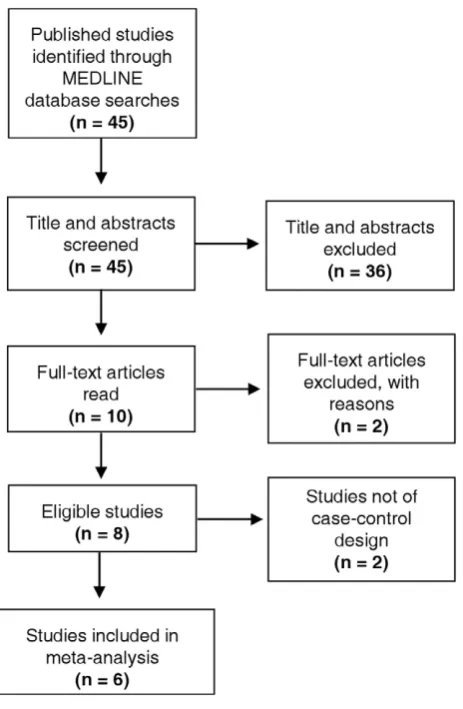
From 45 publications identified by initial data searches, 10 studies fulfilled inclusion criteria for the systematic review and were selected for further assessment (Figure 1). The full texts of these 10 studies were evaluated, after which 2 articles were excluded. One study was not included as it assessed patients only with end stage renal disease. [17] Furthermore, the case population included patients with non-cardiac atherosclerotic disease. [17] The second study assessed the prevalence of a selectedTGF-b2SNP in CHD patients, however, the SNP that was assessed was not named. [10] The remaining 8 studies focussed solely onTGF-b1SNPs in cases and controls (Table 1). Furthermore the study of Sie et al. (2006) utilised a prospective cohort design and that of Manginaset al. (2008)did not include a control group with which to compare genotype distributions. [5,6] Accordingly, these two studies were also excluded, leaving 6 independent case-control investigations in the current meta-analysis (Figure 1).
Study Characteristics
A collective total of 8,505 patients were included across the 6 studies (cases n = 5535, controls n = 2970). Five investigations were conducted in patients from a Western European background and one study was conducted using Japanese recruits. [18] Population sizes of the independent studies ranged from 238 to 4868 participants. Cases included patients with myocardial infarction (4 studies), ischemic heart failure (1 study) or patients with angiographically-proven CHD (1 study). [13] Three studies utilised coronary angiography to confirm a positive diagnosis of CHD whereas two studies relied on the World Health Organi-sation diagnostic criteria for MI, employing clinical history, ECG and cardiac biomarkers. Likewise, there was also considerable inter-study variation in screening of controls, with only two studies utilising coronary angiography to confirm a negative diagnosis of CHD. [13,19] Common CHD risk factors for cases and controls are shown in Table 1. With the exception of age and gender, studies did not match cases and controls for conventional CHD risk factors. Moreover, the distribution of risk factors was not uniformly reported across studies and two studies failed to document the occurrence of environmental risk factors. [12,19] Reported data suggested that CHD populations demonstrated a higher prevalence of hypertension, smoking, diabetes and dyslipidaemia. Only two investigations sub-divided population groups by sex. [18,19].
found that rs1800471 was associated with CHD complications (odds ratio 1.40, 95% CI 1.02–1.92, P = 0.036), however this was not supported by data generated by subsequent investigations into this polymorphism. [5,6,12,13,19,20] It is important to note that Koch et al. (2006) and Yokata et al. (2000) separated their results by sex, and found positive associations only in the male populations. [18,19] The association identified by Cambien et al. (1996) was also restricted to a male population, as this study did not include female participants. [20].
Quantitative Data Synthesis
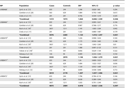
The association between reportedTGF-b1 SNPs and compli-cations of CHD was assessed using additive, dominant and recessive models of inheritance (Table 2 and Table S2). Analyses revealed no significant association of theTGF-b1rs1800468 and rs1800472 under any model, and these SNPs were not further investigated. Heterogeneity assessments for rs1800469, rs1800470 and rs1800471 demonstrated significant inter-study variation, particularly for rs1800470, under additive and recessive models (Table 3 and Table S2), thus data from these models were
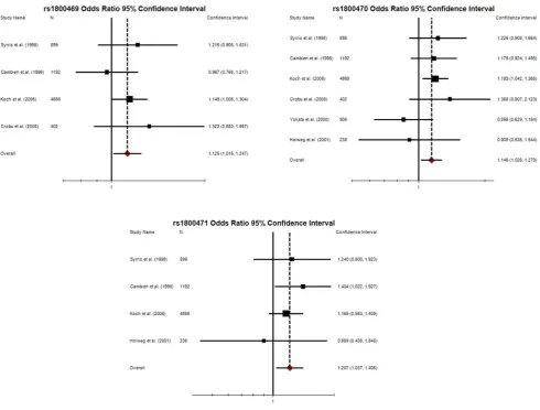
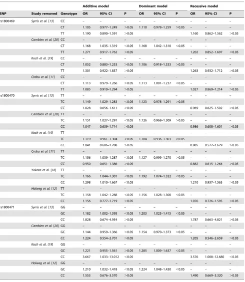
disregarded. In contrast, no heterogeneity was observed for calculations made using the dominant model of inheritance suggesting that conclusions made applying this algorithm were relatively robust. Findings from the dominant model of inheritance were therefore selected for further assessment. Significant positive associations were observed between CHD complications and the minor alleles of rs1800469 (OR 1.125, 95% CI 1.016–1.247, P = 0.031), rs1800470 (OR 1.146, 95% CI 1.026–1.279, P = 0.021) and rs1800471 (OR 1.207, 95% CI 1.037–1.406, P = 0.021; Table 2). As expected, odds ratios generated by the current meta-analysis differed from findings of previous studies (Figure 2). Funnel plots suggested no publication bias was observed for these SNPs (Figure S1). Sensitivity analyses confirmed that multiple studies contributed to the observed significant associations between rs1800469, rs1800470 and end-stage CHD under a dominant model of inheritance (Table 4). In contrast, the significant association between rs1800471 and CHD end points was only lost if the individual study by Cambienet al.(1996) was removed. [20].
Discussion
The association between SNPs of the TGF-b1 cytokine and clinically significant cardiac events was previously unclear due to conflicting data generated by a range of independent studies. To clarify this we conducted a meta-analysis of publically available data, confirming significant associations between the minor alleles ofTGF-b1 rs1800469,rs1800470andrs1800471with CHD when applying a dominant model of inheritance. Odds ratios generated for these SNPs were relatively low (1.125 – 1.207), as expected for a multifactorial disease such as CHD.
Odds ratios generated by our meta-analysis for TGF-b1 rs1800469, rs1800470 and rs1800471 are lower than previously reported, [18,19,20] possibly reflecting the larger, more genetically diverse population analysed in this study. Odds ratios may also have been lowered as we assessed patient populations of mixed gender. Previously Cambienet al.(1996), Yokataet al.(2000) and Koch et al. (2006) independently identified positive associations between these SNPs and end-stage CHD in their male recruits only. [18,19,20] These associations were not observed in cohorts comprising both male and female subjects. [11,12,13] This tentatively suggests that these SNPs may be more relevant to CHD progression in male carriers although this hypothesis could not be assessed in the current study as sex-specific data were not provided by all investigators.
Mechanisms underpinning the positive associations ofTGF-b1 rs1800649, rs1800470 and rs1800471 with development of end-stage CHD remain unclear. These SNPs are located within functional regions of the TGF-b1 gene and it is therefore plausible to suggest that they may directly impact on TGF-b1
signalling. For example, rs1800469 is situated within the promoter region of the TGF-b1 gene and has been reported to alter secretion ofTGF-b1in vitro. [21] Interestingly, Yokotaet al.
(2000) reported reduced serum concentrations of TGF-b1 in subjects carrying theTCorTTgenotypers1800469compared to the CC genotype. [18] No significant difference in circulating
TGF-b1 was observed between case (myocardial infarction) or control recruits of the same genotype, suggesting that altered circulating levels ofTGF-b1are not the principal cause of CHD complications in this population. Rs1800470and rs1800471 are both located within exons, resulting in amino acid changes of
[image:4.612.65.297.59.414.2]Leu10ProandArg25Prorespectively. Coxet al.(2007) reported that the proline substitution at codon 10 is associated with higher circulating concentrations of acid-activatable TGF-b1 and Figure 1. Flow diagram showing studies included.45 published
studies assessing the association of TGF-b SNPs and CHD were identified by searching the MEDLINE database. Appraisal of the abstracts identified 10 articles eligible for full-text appraisal. From these, a further 2 articles were excluded, leaving 8 articles for inclusion in the systematic review. Six of these studies adopted a case-control design, and data from these were extracted for further assessment in the meta-analysis.
Table 2.Meta-analysis of published associations betweenTGF-b1SNPs and complications of CHD.
SNP Population Cases Controls OR* 95% CI p value
rs1800468, Syrris et al.[13] 655 244 1.246 0.826–1.80 0.294
Cambien et al.[20] 563 629 1.084 0.792–1.482 0.615
Crobu et al.[11] 201 201 0.756 0.434–1.319 0.325
*Combined 1419 1074 1.064 0.846–1.339 0.346
rs18004696
Syrris et al.[13] 655 244 1.215 0.905–1.631 0.194
Cambien et al.[20] 563 629 0.967 0.768–1.217 0.776
Koch et al.[19]{
3657 1211 1.145 1.005–1.304 0.042
Crobu et al.[11] 201 201 1.322 0.880–1.987 0.179
*Combined 5076 2285 1.125 1.016–1.247 0.031
rs18004701 Syrris et al.[13] 655 244 1.226 0.908–1.654 0.183
Cambien et al.[20] 563 629 1.175 0.924–1.465 0.188
Koch et al.[19]{
3657 1211 1.193 1.042–1.365 0.01
Crobu et al.[11] 201 201 1.388 0.907–2.123 0.131
Yokota et al.[18]{
315 591 0.856 0.629–1.164 0.322
Holweg et al.[12] 144 94 0.909 0.535–1.544 0.724
*Combined 5535 2970 1.146 1.026–1.279 0.021
rs1800471¤
Syrris et al.[13] 655 244 1.24 0.800–1.923 0.337
Cambien et al.[20] 563 629 1.404 1.022–1.927 0.036
Koch et al.[19]{ 3657 1211 1.159 0.953–1.409 0.14
Holweg et al.[12] 144 94 0.899 0.438–1.848 0.772
*Combined 5019 2178 1.207 1.037–1.406 0.021
rs1800472u Syrris et al.[13] 655 244 1.796 0.784–4.116 0.166
Cambien et al.[20] 563 629 0.638 0.389–1.044 0.074
Koch et al.[19]{
3657 1211 1.033 0.791–1.349 0.812
*Combined 4875 2084 0.978 0.622–1.536 0.397
OR, odds ratio; CI, 95% confidence intervals. ,Rs1800468: promoter, in terms of minor A allele;6
Rs1800469: promoter, in terms of minor T allele;1Rs1800470: coding sequence, in terms of minor T allele; ¤Rs1800471: coding sequence, in terms of minor C allele;uRs1800472: coding sequence, in terms of minor T allele.
*Combined, indicates meta-analysis data calculated for a Random Effects model, assuming dominant inheritance. Data for additive and recessive models displayed in Table S1.
{
Odds ratio given for combined male and female population, not just male population as provided by author.
{
[image:5.612.61.557.553.712.2]Odds ratio given in terms of minor allele, not major allele as stated in publication. doi:10.1371/journal.pone.0037878.t002
Table 3.Heterogeneity analysis of combined effect size.
Additive model Dominant Model Recessive Model
SNP Genotype Q df (Q) P I2
Q df (Q) P I2
Q df (Q) P I2
rs1800469 CC – – – – – – – – – – – –
CT 2.351 3 0.503 0.000 2.595 3 0.458 0.000 – – – –
TT 4.017 3 0.260 0.253 4.634 3 0.201 0.353
rs1800470 TT – – – – – – – – – – – –
TC 0.767 5 0.979 0.000 5.532 5 0.354 0.096 - – – –
CC 25.854 5 ,0.001 0.807 31.145 5 ,0.001 0.839
rs1800471 GG – – – – – – – – – – – –
GC 1.870 3 0.600 0.000 1.694 3 0.638 0.000 – – – –
CC 2.747 3 0.432 0.000 2.731 3 0.435 0.000
Q, Cochran Q test; df, degrees of freedom; I2
, I2
increased secretion in vitro. [22] This may be a result of altered intracellular trafficking, or increased transcription. Substitution of proline residues are also likely to alter the chemical properties and tertiary structure of TGF-b1 protein, potentially interfering with receptor interactions. However, these effects are yet to be described.
Limitations
This study has several limitations. Firstly, only 6 published studies fulfilled our selection criteria, thus the meta-analysis was restricted to a relatively small patient population primarily comprising subjects from Western Europe. Importantly, these studies focused solely on TGF-b1SNPs and we were therefore unable to investigate the contribution of TGF-b2 or TGF-b3
polymorphisms to the development of end-stage CHD. Secondly, we were unable to adjust the meta-analysis to correct for common cardiovascular risk factors (e.g. gender or smoking) as these were not uniformly reported in the independent investigations and could not be easily obtained. This was further complicated by significant variation in clinical definitions of hypertension and dyslipidaemia. [13] It is therefore possible that calculated ORs and P values are over-estimates of the true biological situation. Thirdly, significant heterogeneity was observed between studies suggesting
that these data could not be investigated using the additive and recessive models of inheritance. Similarly, we noted significant inter-study variation in methods employed to screen control populations. Only 2 of the analysed studies provided a definitive negative CHD diagnosis in controls via coronary angiography, [13,19] whereas other studies employed non-specific methodolo-gies, such as electrocardiography, or a complete lack of screening. [11,12,18,20] Consequently, there is potential for false negative diagnoses in some studies, and it is feasible to suggest that control subjects may not truly represent healthy (CHD-free) individuals. Finally, data collected from patients presenting with a range of cardiovascular complications were appraised in this analysis, thereby assuming that similar pathological mechanisms underpin distinct CHD endpoints. Subsequently, we are unable to suggest specific contributions of TGF-b1 SNPs to the development of discrete events such as myocardial infarction and end stage heart failure.
[image:6.612.60.551.58.430.2]The authors attempted to include SNPs related to the TGF-b genes from previous GWAS in the present meta-analysis. Prior to data extraction, the Catalogue of Published Genome-Wide Association Studies (genome.gov) was searched and 16 CHD GWAS were identified. [23] The full text and supplementary data of these studies were hand-searched by the primary author. The Figure 2. Forest plots detailing unadjusted odds ratios and 95% confidence intervals for the association between TGFb1 rs1800469, rs1800470 and rs1800471 with end-stage CHD calculated using the dominant model of inheritance.The dotted line represents overall odds ratio calculated in the current meta-analysis for each SNP.
risk alleles identified in this meta-analysis ofTGF-bgene studies have not been highlighted in recent GWAS focused on the presence or complications of CHD. The reasons for differences between findings of candidate gene and genome wide association
[image:7.612.65.556.87.653.2]studies are currently unexplained. Some possible reasons include: risk alleles only acting in certain populations due to variation in other genetic factors or environmental exposure, false discovery, confounding effects of combining samples and data from very Table 4.Leave-one-out sensitivity analysis of combined effect size.
Additive model Dominant model Recessive model
SNP Study removed Genotype OR 95% CI P OR 95% CI P OR 95% CI P
rs1800469 Syrris et al.[13] CC – – – – – – – – –
CT 1.105 0.977–1.249 .0.05 1.110 0.978–1.259 .0.05 – – –
TT 1.190 0.890–1.591 .0.05 1.160 0.862–1.562 .0.05
Cambien et al.[20] CC – – – – – – – – –
CT 1.168 1.035–1.319 ,0.05 1.168 1.042–1.310 ,0.05 – – –
TT 1.271 0.917–1.762 .0.05 1.202 0.852–1.697 .0.05
Koch et al.[19] CC – – – – – – – – –
CT 1.052 0.883–1.253 .0.05 1.106 0.918–1.333 .0.05 – – –
TT 1.301 0.922–1.837 .0.05 1.263 0.932–1.712 .0.05
Crobu et al.[11] CC – – – – – – – – –
CT 1.113 0.979–1.266 .0.05 1.113 1.001–1.237 ,0.05 – – –
TT 1.085 0.910–1.294 .0.05 1.027 0.869–1.214 .0.05
rs1800470 Syrris et al.[13] TT – – – – – – – – –
TC 1.149 1.029–1.283 ,0.05 1.123 0.978–1.291 .0.05 – – –
CC 1.028 0.656–1.611 .0.05 0.969 0.625–1.502 .0.05
Cambien et al.[20] TT – – – – – – – – –
TC 1.151 1.027–1.291 ,0.05 1.126 0.968–1.309 .0.05 – – –
CC 1.047 0.639–1.714 .0.05 0.986 0.608–1.601 .0.05
Koch et al.[19] TT – – – – – – – – –
TC 1.119 0.961–1.304 .0.05 1.104 0.936–1.303 .0.05
CC 1.041 0.606–1.788 .0.05 0.985 0.577–1.679 .0.05
Crobu et al.[11] TT – – – – – – – – –
TC 1.156 1.039–1.287 ,0.05 1.127 0.999–1.270 .0.05 – – –
CC 0.950 0.651–1.386 .0.05 0.882 0.615–1.264 .0.05
Yokota et al.[18] TT – – – – – – – – –
TC 1.166 1.044–1.301 ,0.05 1.192 1.074–1.322 ,0.05 – – –
CC 1.298 1.010–1.667 ,0.05 1.210 0.937–1.563 .0.05
Holweg et al.[12] TT – – – – – – – – –
TC 1.158 1.042–1.288 ,0.05 1.156 1.028–1.300 ,0.05 – – –
CC 1.156 0.777–1.719 .0.05 1.076 0.726–1.595 .0.05
rs1800471 Syrris et al.[13] GG – – – – – – – – –
GC 1.182 1.002–1.395 ,0.05 1.203 1.023–1.415 ,0.05 – – –
CC 1.828 0.674–4.954 .0.05 1.787 0.663–4.821 .0.05
Cambien et al.[20] GG – – – – – – – – –
GC 1.144 0.959–1.366 .0.05 1.154 0.970–1.373 .0.05 – – –
CC 1.224 0.554–2.701 .0.05 1.205 0.546–2.659 .0.05
Koch et al.[19] GG – – – – – – – – –
GC 1.221 0.955–1.561 .0.05 1.285 1.009–1.637 ,0.05 – – –
CC 3.667 1.033–13.012 ,0.05 3.576 1.008–12.680 ,0.05
Holweg et al.[12] GG – – – – – – – – –
GC 1.210 1.032–1.418 ,0.05 1.224 1.048–1.430 ,0.05 – – –
CC 1.553 0.676–3.570 .0.05 1.490 0.669–3.320 .0.05
different studies where information is collected under different definitions, variation in demographic profile of control popula-tions, and heterogeneity of data collection methods across studies.
Conclusion
In conclusion, we confirm significant positive associations between theTGF-b1SNPsrs1800469,rs1800470and rs1800471
and complications of CHD. Further work is required to determine the clinical relevance of these findings.
Supporting Information
Figure S1 Funnel plots detailing publication bias for TGF-b1 SNPs rs1800469, rs1800470 and rs1800471 using the dominant model of inheritance.
[image:8.612.312.553.285.578.2](TIF)
Table S1 Checklist to confirm compliance with PRISMA guidelines for systematic reviews and meta-analyses.
(DOC)
Table S2 Meta-analysis of published associations between
TGF-b1 SNPs and CHD complications under additive, dominant and recessive models of inheritance.
(DOC)
Text S1 Protocol used for systematic data collection from eligible articles.
(DOC)
Acknowledgments
The authors thank Dr. Cecile Holweg for providing additional data to facilitate the analysis.
Author Contributions
Conceived and designed the experiments: DM EB JVM JG. Performed the experiments: DM EB SMK JVM JG. Analyzed the data: DM EB JVM. Contributed reagents/materials/analysis tools: DM JVM EB SMK JG. Wrote the paper: DM JVM EB JG.
References
1. Roger VL, Go AS, Lloyd-Jones DM, Adams RJ, Berry JD, et al. (2011) Heart disease and stroke statistics–2011 update: a report from the American Heart Association. Circulation 123: e18–e209.
2. Libby P (2008) Braunwald’s heart disease : a textbook of cardiovascular medicine. Philadelphia: Saunders/Elsevier.
3. Marenberg ME, Risch N, Berkman LF, Floderus B, de Faire U (1994) Genetic susceptibility to death from coronary heart disease in a study of twins. N Engl J Med 330: 1041–1046.
4. Patel RS, Ye S (2011) Genetic determinants of coronary heart disease: new discoveries and insights from genome-wide association studies. Heart 97: 1463–1473.
5. Manginas A, Tsiavou A, Chaidaroglou A, Giamouzis G, Degiannis D, et al. (2008) Inflammatory cytokine gene variants in coronary artery disease patients in Greece. Coronary artery disease 19: 575–582.
6. Sie MP, Uitterlinden AG, Bos MJ, Arp PP, Breteler MM, et al. (2006) TGF-beta 1 polymorphisms and risk of myocardial infarction and stroke: the Rotterdam Study. Stroke 37: 2667–2671.
7. Grainger DJ (2007) TGF-beta and atherosclerosis in man. Cardiovasc Res 74: 213–222.
8. Bertolino P, Deckers M, Lebrin F, ten Dijke P (2005) Transforming growth factor-beta signal transduction in angiogenesis and vascular disorders. Chest 128: 585S–590S.
9. Biros E, Norman PE, Jones GT, van Rij AM, Yu G, et al. (2011) Meta-analysis of the association between single nucleotide polymorphisms in TGF-beta receptor genes and abdominal aortic aneurysm. Atherosclerosis.
10. Biggart S, Chin D, Fauchon M, Cardew G, du Fou L, et al. (1998) Association of genetic polymorphisms in the ACE, ApoE, and TGF beta genes with early onset ischemic heart disease. Clin Cardiol 21: 831–836.
11. Crobu F, Palumbo L, Franco E, Bergerone S, Carturan S, et al. (2008) Role of TGF-beta1 haplotypes in the occurrence of myocardial infarction in young Italian patients. BMC medical genetics 9: 13.
12. Holweg CT, Baan CC, Niesters HG, Vantrimpont PJ, Mulder PG, et al. (2001) TGF-beta1 gene polymorphisms in patients with end-stage heart failure. The Journal of heart and lung transplantation : the official publication of the International Society for Heart Transplantation 20: 979–984.
13. Syrris P, Carter ND, Metcalfe JC, Kemp PR, Grainger DJ, et al. (1998) Transforming growth factor-beta1 gene polymorphisms and coronary artery disease. Clin Sci (Lond) 95: 659–667.
14. Moher D, Liberati A, Tetzlaff J, Altman DG (2009) Preferred reporting items for systematic reviews and meta-analyses: the PRISMA statement. BMJ (Clinical research ed ) 339: b2535.
15. Stern JM, Simes RJ (1997) Publication bias: evidence of delayed publication in a cohort study of clinical research projects. BMJ (Clinical research ed ) 315: 640–645.
16. Database of Single Nucleotide Polymorphisms (dbSNP). Bethesda (MD): National Center for Biotechnology Information, National Library of Medicine. (dbSNP Build ID: {build 134})).
17. Rao M, Guo D, Jaber BL, Tighiouart H, Pereira BJ, et al. (2004) Transforming growth factor-beta 1 gene polymorphisms and cardiovascular disease in hemodialysis patients. Kidney Int 66: 419–427.
18. Yokota M, Ichihara S, Lin TL, Nakashima N, Yamada Y (2000) Association of a T29–.C polymorphism of the transforming growth factor-beta1 gene with genetic susceptibility to myocardial infarction in Japanese. Circulation 101: 2783–2787.
19. Koch W, Hoppmann P, Mueller JC, Schomig A, Kastrati A (2006) Association of transforming growth factor-beta1 gene polymorphisms with myocardial infarction in patients with angiographically proven coronary heart disease. Arteriosclerosis, thrombosis, and vascular biology 26: 1114–1119.
20. Cambien F, Ricard S, Troesch A, Mallet C, Generenaz L, et al. (1996) Polymorphisms of the transforming growth factor-beta 1 gene in relation to myocardial infarction and blood pressure. The Etude Cas-Temoin de l’Infarctus du Myocarde (ECTIM) Study. Hypertension 28: 881–887.
21. Grainger DJ, Heathcote K, Chiano M, Snieder H, Kemp PR, et al. (1999) Genetic control of the circulating concentration of transforming growth factor type beta1. Hum Mol Genet 8: 93–97.
22. Cox A, Dunning AM, Garcia-Closas M, Balasubramanian S, Reed MWR, et al. (2007) A common coding variant in CASP8 is associated with breast cancer risk. Nature genetics 39: 352–358.