Exhibit Distinct Protease Preferences
John Misasi,a,bKartik Chandran,a* Jin-Yi Yang,aBryden Considine,a,bClaire Marie Filone,a,cMarceline Côté,aNancy Sullivan,d Giulia Fabozzi,dLisa Hensley,cand James Cunninghama,e
Division of Hematology, Department of Medicine, Brigham & Women’s Hospital, Boston, Massachusetts, USAa
; Division of Infectious Diseases, Children’s Hospital Boston, Boston, Massachusetts, USAb
; U.S. Army Medical Research Institute of Infectious Diseases, Ft. Detrick, Maryland, USAc
; Vaccine Research Center, National Institute for Allergy and Infectious Diseases, National Institutes of Health, Bethesda, Maryland, USAd
; and Department of Microbiology and Immunobiology, Harvard Medical School, Boston, Massachusetts, USAe
Filoviruses are enveloped viruses that cause sporadic outbreaks of severe hemorrhagic fever [CDC, MMWR Morb. Mortal. Wkly.
Rep. 50:73–77, 2001; Colebunders and Borchert, J. Infect. 40:16 –20, 2000; Colebunders et al., J. Infect. Dis. 196(Suppl. 2):S148 –
S153, 2007; Geisbert and Jahrling, Nat. Med. 10:S110-S121, 2004]. Previous studies revealed that endosomal cysteine proteases
are host factors for ebolavirus Zaire (Chandran et al., Science 308:1643–1645, 2005; Schornberg et al., J. Virol. 80:4174 – 4178,
2006). In this report, we show that infection mediated by glycoproteins from other phylogenetically diverse filoviruses are also
dependent on these proteases and provide additional evidence indicating that they cleave GP1 and expose the binding domain
for the critical host factor Niemann-Pick C1. Using selective inhibitors and knockout-derived cell lines, we show that the
ebola-viruses Zaire and Cote d’Ivoire are strongly dependent on cathepsin B, while the ebolaebola-viruses Sudan and Reston and Marburg
virus are not. Taking advantage of previous studies of cathepsin B inhibitor-resistant viruses (Wong et al., J. Virol. 84:163–175,
2010), we found that virus-specific differences in the requirement for cathepsin B are correlated with sequence polymorphisms
at residues 47 in GP1 and 584 in GP2. We applied these findings to the analysis of additional ebolavirus isolates and correctly
predicted that the newly identified ebolavirus species Bundibugyo, containing D47 and I584, is cathepsin B dependent and that
ebolavirus Zaire-1995, the single known isolate of ebolavirus Zaire that lacks D47, is not. We also obtained evidence for
virus-specific differences in the role of cathepsin L, including cooperation with cathepsin B. These studies strongly suggest that the use
of endosomal cysteine proteases as host factors for entry is a general property of members of the family
Filoviridae.
E
bdaviruses and the closely related marburgvirus comprise the
family
Filoviridae
(6, 8, 9, 16). Several lines of recent
investi-gation have elucidated key steps in the pathway for ebolavirus
entry into cells. Ebolavirus particles attach to cells through the
binding of their glycoprotein (GP) to cell surface receptors or
lectins, such as TIM-1 and DC-SIGN, expressed on the plasma
membrane (1, 22, 27, 29, 37). Membrane-bound particles are
taken up into cells by a macropinocytosis-like mechanism and
transported to late endosomes/lysosomes (LE/LY) (20, 30, 31, 34),
which contain essential entry host factors. We previously showed
that cleavage of ebolavirus Zaire-Mayinga (EBOV-May) GP by
endosomal cysteine proteases is required for infection (7). More
recent work has revealed a second host factor in LE/LY that is
broadly required by filoviruses: Niemann-Pick C1 (NPC1) (5, 10),
a multipass transmembrane protein that resides in the limiting
membrane (44). According to a recently proposed model, virus
GP is cleaved by endosomal cysteine proteases and binds to NPC1
(10).
Several studies have examined the role of protease cleavage in
more detail for EBOV-May. They show that cathepsin L functions
in concert with cathepsin B to cleave the GP1 subunit of virus GP
(7, 35). Structural and functional studies reveal that proteases
re-move the heavily glycosylated carboxyl-terminal domain of GP1
to expose a more conserved domain that is closely associated with
GP2 (12, 19, 25) and that is proposed to contain the binding site
for the filovirus receptor (4, 13, 24, 28). Further, we recently
showed that cleaved, but not uncleaved, GP1 binds to purified
LE/LY membranes in an NPC1-dependent manner and
coimmu-noprecipitates with NPC1 (10). We identified small molecules
that target NPC1, inhibit infection, and block the binding of
cleaved GP1 to NPC1-containing membranes (10), strongly
sug-gesting that the conserved N-terminal domain of GP1 is a ligand
for NPC1. Taken together, these previous findings suggest a model
in which proteolytic cleavage of GP to remove the
carboxyl-terminal domain of GP1 and expose its N-carboxyl-terminal domain may
be functionally analogous to the role of CD4 binding to HIV
gp120 to displace highly variable loops and create the
coreceptor-binding site (18). Our recent studies show that NPC1 expression is
also essential for infection by virus isolates from the other species
that make up the family
Filoviridae
(23), including the Cote
d’Ivoire, Sudan, and Reston ebolaviruses, the new ebolavirus
spe-cies Bundibugyo (BDBV), and Marburg virus (5, 10). In this
study, we tested this model of infection by examining the
endo-somal cysteine protease requirements for infection by these
vi-ruses.
Received19 September 2011Accepted18 December 2011
Published ahead of print11 January 2012
Address correspondence to James Cunningham, jcunningham
@rics.bwh.harvard.edu, or Kartik Chandran, [email protected]. * Present address: Department of Microbiology and Immunology, Albert Einstein College of Medicine, Bronx, New York, USA.
John Misasi and Kartik Chandran made equivalent contributions to this study. Copyright © 2012, American Society for Microbiology. All Rights Reserved.
doi:10.1128/JVI.06346-11
on November 7, 2019 by guest
http://jvi.asm.org/
MATERIALS AND METHODS
Cells and cell culture conditions.Vero, 293T, and mouse embryonic fibroblast (MEF) cell lines were maintained in Dulbecco’s modified Ea-gle’s medium (DMEM), 100g/ml penicillin-streptomycin, and 2 mM
L-glutamine (Invitrogen). 293T and Vero cells were carried in either 10% fetal bovine serum (FBS) or 5% FBS–5% FetalPlex (Gemini). MEF cells were carried in 10% FBS. During virus production and at least 18 h prior to infection, Vero, 293T, and MEF cells were grown in 10% FBS-containing medium. MEF cells were described previously (7). NPC1⫺/⫺
and NPC1⫺/⫺ Chinese hamster ovary (CHO) cells stably expressing
mouse NPC1 were described previously (10).
Expression plasmids.Expression plasmids for mouse cathepsin B, mouse cathepsin L, EBOV-May GP, and mucin deletion-containing (⌬Muc) EBOV-May GP were described previously (7). Plasmids encod-ing ebolavirus Cote d’Ivoire GP, ebolavirus Sudan-Boniface GP, ebolavi-rus Reston-Pennsylvania GP, and Lake Victoria marburgviebolavi-rus Musoke GP were obtained from Anthony Sanchez (CDC) and subcloned into pCAGGS.⌬Muc GP constructs were created using PCR to remove⌬Muc EBOV-May GP amino acids (aa) 309 to 489,⌬Muc ebolavirus Cote d’Ivoire GP aa 310 to 489,⌬Muc ebolavirus Sudan GP aa 309 to 490, ⌬Muc ebolavirus Reston GP aa 310 to 490, and ⌬Muc Bundibugyo (BDBV) GP aa 309 to 489. This PCR added a silent XbaI site at the site of the deletion in each GP. Empty plasmid pCAGGS or a plasmid encoding -galactosidase was used as a sham vector control.
Chimeric EBOV-May GP1/Reston ebolavirus GP2 and Reston ebola-virus GP1/EBOV-May GP2 were created by restriction digestion of the parental plasmids with XbaI (New England BioLabs) to liberate GP1- and GP2-containing fragments for each virus GP. The fragments were gel purified and ligated using T4 DNA ligase (New England BioLabs). Vectors were verified by DNA sequencing.
EBOV-May and Reston ebolavirus⌬Muc mutations were made using site-directed PCR mutagenesis. The sequences of the primers used were 5=-CTCTAGAGCCTCTGCTAACCA, 5=CATTACAGGTTAGTGAAGT CGACAAACTAGTTTGT, 5=-GTCGACTTCACTAACCTGTAATGT, 5=-ACTCTCAAAGCAACAGATATTGATCAATTGGT, 5=-TCAATATC TGTTGCTTTGAGAGT, 5=-GCTACGCACCTTTTCACTCCTCAACCG TAAGGCAATTGA, 5=-AGTGAAAAGGTGCGTAGCTCA, 5=-CTGAGG ACTTACTCAATTCTTAACAGAAAAGCT, 5=-GAATTGAGTAAGTCC TCAGT, 5=-TATACTCGAGCTCGGTACCTCAAACCT, and 5=-GCTAG CTCGAGAAATCAACACA. Chimeric viruses were made as described above.
Infection assays.Pseudotyped vesicular stomatitis virus (VSV) was created as previously described (7). Pseudotyped VSV particles encoding green fluorescent protein (GFP) (VSVGFP) were added to cells in serial
10-fold dilutions and assayed using fluorescence microscopy or flow cy-tometry. When using fluorescence microscopy, GFP-positive cells were counted manually and an infectious unit (IU) was defined as one GFP-expressing cell when within the linear range of dilutions. Titer was defined as IU/ml of virus added and was determined using the following formula: (number of GFP-expressing cells⫻dilution factor)/volume (in ml) of virus added to the well. Two-color flow cytometry was used to determine the relative ratio of infected to uninfected cathepsin B⫺/⫺cathepsin L⫹/⫹
MEF cells in cells that were cotransfected with an mRFP-expressing plas-mid and a sham plasplas-mid, mouse cathepsin B, or mouse cathepsin L. De-tails of this protocol were described previously (7).
Cathepsin B⫺/⫺ cathepsin L⫺/⫺MEF cell rescue assays were
per-formed with six-well dishes. Sixteen to 20 h after seeding of the dishes, the medium was changed to low (0.2%) serum. After 4 h, cells were trans-fected using 4g total DNA and 10l Lipofectamine 2000 (Invitrogen) that were diluted into Opti-MEM (Invitrogen). The plasmids used were pCAGGS (4g), mouse cathepsin B (4g), mouse cathepsin L (4g), and both cathepsin B (2g) and cathepsin L (2g). Five hours following transfection, cells were washed once with phosphate-buffered saline (PBS) and fresh medium containing 10% FBS was added. Twenty-four hours posttransfection, cells were removed with trypsin-EDTA solution
(Invitrogen), spun down, and resuspended in fresh medium and 8,000 cells were added to each well of a 48-well dish. Following an additional 18 h, the medium was changed. Four hours later, pseudotyped VSV encoding luciferase (VSVluc) was added at dilutions within the linear range of
infec-tion and sufficient to give signals of 104to 105relative luminescence units
(RLU) on cells transfected with pCAGGS. Twenty-four hours after the addition of the virus, cells were lysed using standard conditions for the firefly luciferase kit (Promega). Lysates were transferred to a 96-well plate, luciferin reagent was added, and after 1 min of incubation, RLU were measured on an EnVison plate reader (Perkin-Elmer).
Protease inhibitors and protease activity assays.Protease inhibitors E-64, E-64d, and CA074 (Sigma) were dissolved in dimethyl sulfoxide (DMSO) and dispensed into culture medium immediately before use. The final DMSO concentration in medium was always 1% (vol/vol). Cell monolayers were preincubated with inhibitors or DMSO for 3 to 4 h at 37°C. Viruses were added directly to the culture medium containing DMSO or inhibitors, and infectivities were measured as already described. The enzymatic activities of cathepsins B and L in acidified lysates (100 mM NaCl, 50 mM Na acetate, 0.5% Triton X-100, pH 5.5) of Vero and MEF cells were assayed as previously described (7, 46). The absence of cathepsin B activity in cathepsin B⫺/⫺cathepsin L⫹/⫹MEF cells, the lack
of cathepsin B and L activities in cathepsin B⫺/⫺cathepsin L⫺/⫺MEF
cells, and the presence of similar levels of cathepsin L activity in cathepsin B⫺/⫺cathepsin L⫹/⫹and wild-type MEF cells were confirmed.
Ebolavirus Sudan infection.Vero cells were treated with E-64d (300 M) or vehicle (1% DMSO) for 4 h and then infected with EBOV-May or ebolavirus Sudan-Gulu (multiplicity of infection [MOI], 0.1). After 1 h, the virus inoculums were removed by washing and fresh medium con-taining E-64d or vehicle was added. Cell supernatant was collected on day 3. RNA was isolated from the supernatant using Virus RNA Extraction kits (Qiagen), and ebolavirus NP RNA was measured using a quantitative reverse transcription (RT)-PCR assay (45). Virus titer was calculated us-ing a standard curve obtained usus-ing a virus stock of known titer as deter-mined by plaque assay.
Marburg virus infection.Vero cells were seeded into six-well plates at a density of 2⫻105/well. One day later, the medium was removed and
replaced with fresh medium containing E64d (300M). After 4 h, Ebola virus Zaire (MOI, 0.2) or Marburg virus Ci67 (Popp; MOI, 0.2) was added to cells and drug. One hour later, medium containing drug and virus was removed, cells were washed with fresh medium, and fresh medium con-taining drug was added to each well. Time zero samples were harvested immediately, and remaining samples were incubated for 72 h. Superna-tants were harvested for determination of virus by quantitative RT-PCR, and cells were lysed for determination of viral protein by Western blot assay (14).
Protease assay. VSV particles bearing mucin domain deletion-containing GPs from the indicated viruses were incubated in the presence of 0.2 mg/ml chymotrypsin in reaction buffer (10 mM Tris-HCl, 135 mM NaCl, 1 mM EDTA, pH 7.5). After 1 h, the digestion was stopped by the addition of phenylmethylsulfonyl fluoride (PMSF) to a final concentra-tion of 1 mM. Virus particles were deglycosylated by overnight incubaconcentra-tion in the presence of peptideN-glycosidase F (PNGase F; New England Bio-Labs). GP1 digestion products were analyzed under denaturing condi-tions via immunoblot assay against a conserved N-terminal region of ebolavirus GP1 using polyclonal rabbit anti-ebolavirus GP1 antibody (10).
CHO cell infections.VSVlucparticles bearing GPs from the indicated
ebolaviruses were digested in the presence or absence of chymotrypsin (as described above but not deglycosylated). NPC1⫺/⫺ CHO cells and
NPC1⫺/⫺CHO cells stably expressing mouse NPC1 were incubated in the
presence or absence of E-64d (300⌴). After 4 h, virus particles were added to the cells. Sixteen hours after the addition of virus, luciferase activity was measured as described above.
NPC1 membrane binding assay. Expression and purification of EBOV-May⌬TMhave been described previously (10). An expression
on November 7, 2019 by guest
http://jvi.asm.org/
tor encoding SUDV GP⌬TM(residues 1 to 309 and 491 to 657) that is fused
to GCN4 trimerization/His tag was also prepared. Chymotrypsin-cleaved SUDV GP⌬TMwas created by incubation in the presence of 0.2 mg/ml
chymotrypsin in chymotrypsin reaction buffer. After 30 min, the reaction was stopped by the addition of 1 mM EDTA, 1 mM PMSF, and 1⫻ EDTA-free complete protease inhibitor cocktail (Roche). Thermolysin digestion of EBOV-May⌬TMGP was described previously (10). LE/LY were isolated
by differential centrifugation and Percoll (Sigma) density gradient cen-trifugation. LE/LY were disrupted by incubation with methionine methyl ester (Sigma) and used to coat high-binding enzyme-linked immunosor-bent assay plates (Corning). Following attachment, unbound LE/LY membranes were removed and plates were blocked with PBS–5% FBS. Bound membranes were incubated with the indicated amounts of native or protease-cleaved trimer in PBS–5% FBS. Unbound GP⌬TMprotein was
removed, membranes were washed, and membrane-bound GP⌬TM
pro-tein was recovered in SDS loading buffer and analyzed by immunoblot assay using GP1 antiserum as described previously (10).
Cloning of Bundibugyo GP.TRIzol reagent (Invitrogen)-inactivated ebolavirus Bundibugyo (BDBV) RNA (vRNA) was precipitated using the standard protocol in the TRIzol package insert. First-strand cDNA was created from this vRNA using random primers and SuperScript II (Invit-rogen). Frameshifted BDBV GP was amplified via PCR using Phusion HS (New England BioLabs) and primers 5=-TAAATGCATGGTTACATCA GGA, 5=-TGTGAAGTTCTTCTTATTTTCCCAGAAGGC, 5=-TAAGAA GAACTTCACAAAAACCCT, and 5=-TATCTCGAGGACTAGATTAGA GTAGA. The final PCR product was cloned into pCAGGS MCS using NsiI and XhoI. The clone was verified by sequencing. A mucin deletion-containing version of this plasmid was created by PCR with the previous primers, 5=-TCTCCGACATATGGTACCGCAAATCTGCTGACAGG CTCA and 5=-GGTACCATATGTCGGAGAGGTACCGACAGACAGCT CTTCA, and cloning into pCAGGS MCS with NsiI and XhoI. This plas-mid was restricted with KpnI and religated to create the mucin (aa 309 to 489) deletion-containing BDBV. Plasmids were verified by sequencing. All restriction enzymes were obtained from New England BioLabs.
RESULTS
Endosomal cysteine proteases are host factors for filovirus
en-try.
Previous studies show that EBOV-May infection is reduced by
⬎
99% by E-64, a highly specific small-molecule inhibitor of
en-dosomal cysteine proteases, including cathepsins L and B (7, 35).
We used E-64 as a probe to analyze the host requirements for entry
by GPs from other filoviruses. Vero cells were pretreated with E-64
and then challenged with VSV vectors pseudotyped with GPs
from EBOV-May, ebolavirus Cote d’Ivoire (CIEBOV), ebolavirus
Sudan-Boniface
(SUDV),
ebolavirus
Reston-Pennsylvania
(RESTV), and Lake Victoria marburgvirus Musoke (MARV). The
titers of VSV particles pseudotyped with EBOV-May, CIEBOV,
SUDV, and MARV GPs are 6
⫻
10
9to 1
⫻
10
10IU/ml, and the titer
of VSV RESTV GP particles is 10-fold lower (1.3
⫻
10
8IU/ml)
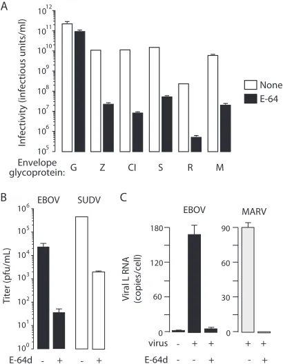
(Fig. 1A). We found that E-64 treatment of target cells reduced
infection by these viruses by
⬎
99.5% under conditions where
VSV G infection was reduced by
⬍
50% (Fig. 1A). These findings
were confirmed in studies of viruses bearing GPs lacking the
mucin-rich domain in GP1. To verify that cysteine proteases are
bona fide host factors, we tested the effect of E-64d on the growth
of EBOV-May, ebolavirus Sudan-Gulu, and Lake Victoria
mar-burgvirus Musoke in Vero cells. The production of new virus was
reduced by more than 99% in the presence of E-64d, as
deter-mined by quantitative RT-PCR (Fig. 1B and C). Taken together,
the results of these studies indicate that infection by EBOV-May,
CIEBOV, SUDV, RESTV, and MARV is dependent on endosomal
cysteine proteases sensitive to E-64.
Proteases target virus GP1.
Analysis of the amino acid
se-quences of GPs from EBOV-May, CIEBOV, SUDV, RESTV, and
MARV suggests that the domain structures of GP1 and GP2 are
conserved (data not shown), a conclusion that has been recently
confirmed for ebolavirus Sudan GP (11, 25). Biochemical studies
of EBOV-May indicate that endosomal cysteine proteases remove
the heavily glycosylated carboxyl-terminal “cap” domain and
ex-pose the N-terminal domain (7, 12, 19, 24, 25, 35) that is the ligand
for the essential host factor NPC1 (10). To determine if a
protease-sensitive carboxyl-terminal domain in GP1 is conserved among
ebolaviruses, we incubated VSV particles pseudotyped with GPs
from EBOV-May, CIEBOV, SUDV, and RESTV with
chymotryp-sin and analyzed the effect on virus infection. Chymotrypchymotryp-sin is a
serine endoprotease that faithfully mimics the action of cathepsin
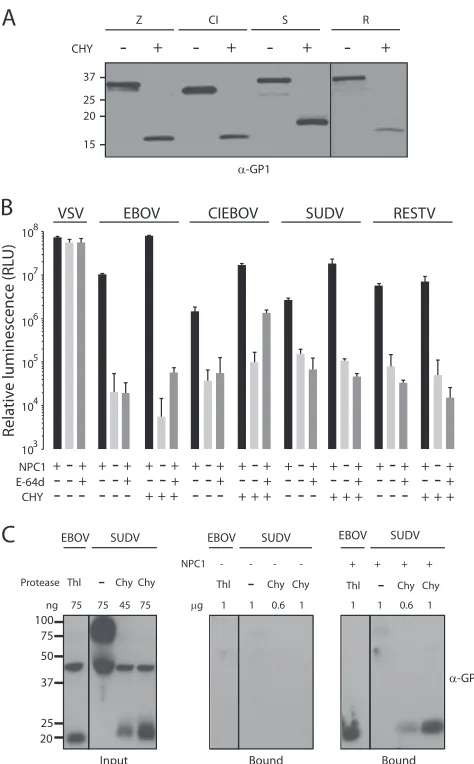
L (46). We observed that like chymotrypsin digestion of
EBOV-May, chymotrypsin digestion of CIEBOV, SUDV, and RESTV
re-moves the carboxyl-terminal domains of GP1 to create an
N-terminal 18- to 20-kDa fragment (Fig. 2A). We analyzed the
function of chymotrypsin-cleaved virus particles on CHO cells
and found that they are infectious and dependent on both NPC1
and cysteine protease activity (Fig. 2B). We next analyzed the
FIG 1Endosomal cysteine proteases are host factors for filoviruses. (A) Vero cells were incubated in the presence of E-64 (cysteine protease inhibitor, 300 M) or vehicle (1% DMSO) for 4 h prior to being challenged with VSV par-ticles encoding GFP (VSVGFP) and bearing GPs from EBOV-May (Z),CIEBOV (CI), SUDV (S), RESTV (R), MARV (M), or VSV (G). After 24 h, infectivity (IU/ml) was determined by manually counting GFP-positive cells using fluorescence microscopy. Data are means⫾standard deviations (SD;
n⫽3). Shown is a representative of four independent experiments. (B) Vero cells were incubated in the presence of E-64d (cysteine protease inhibitor, 300 M) or vehicle for 4 h prior to being challenged with the filovirus EBOV-May or SUDV (MOI, 0.1). After 3 days, the titer was determined by quantitative RT-PCR. Data are means of three wells⫾SD (n⫽3). (C) Vero cells were incubated in the presence of E-64d (300M) or vehicle for 4 h prior to being challenged with the filovirus EBOV or MARV (MOI, 0.2). After 72 h, copies of the viral L gene per cell were determined by quantitative RT-PCR. Data are means of three wells⫾SD (n⫽3).
on November 7, 2019 by guest
http://jvi.asm.org/
[image:3.585.319.524.67.331.2]binding of ebolavirus Sudan GP to NPC1 membranes. The source
of GP is an ectodomain trimer in which the transmembrane
do-main is replaced with a GCN4 trimerization dodo-main (SUDV
⌬TM).
We incubated uncleaved and chymotrypsin-cleaved SUDV
⌬TMGP with purified LE/LY membranes from knockout and
NPC1-expressing CHO cells. We found that, like that of EBOV-May
⌬TMGP, SUDV
⌬TMGP binding to membranes is dependent on the
cleavage of GP1 and expression of NPC1 (Fig. 2C). Although more
work is needed, these findings suggest that at least one function of
endosomal cysteine proteases in filovirus infection is to remove
the carboxyl-terminal domain of GP1 and expose the NPC1
bind-ing site.
Requirement for cathepsin B is not conserved.
EBOV-May
infection is strongly dependent on the endosomal cysteine
pro-tease cathepsin B (7, 35). To test the hypothesis that cathepsin B
activity is required for infection by other filoviruses, we measured
the VSV GP-dependent infection of Vero cells treated with the
selective cathepsin B inhibitor CA074. We confirmed that
GP-dependent EBOV-May infection is reduced as a function of the
concentration of CA074 (0 to 80
M) and is closely correlated
with cathepsin B activity (Fig. 3A and B). We observed that like
that of EBOV-May, CIEBOV GP-mediated infection is also closely
correlated with cathepsin B activity. However, SUDV GP infection
was not as sensitive to CA074 inhibition as EBOV-May or
CIEBOV GP and neither RESTV nor MARV GP-mediated
infec-tion was significantly reduced when cathepsin B activity was
in-hibited (Fig. 3A and B).
As an independent test of the role of cathepsin B in infection,
we studied MEF cells derived from cathepsin B knockout
(cathep-sin B
⫺/⫺) mice. The pattern of infection of cathepsin B
⫺/⫺MEF
cells by filovirus GPs is closely correlated with the pattern of
infection of Vero cells treated with CA074: EBOV-May and
CIEBOV GP is 1%, SUDV 7%, RESTV 25 to 40%, and MARV 65
to 99% of infection of wild-type MEF cells (Fig. 3C and D). The
introduction of an expression plasmid encoding cathepsin B into
cathepsin B
⫺/⫺MEF cells enhanced infection by each of the
vi-ruses to 90 to 150% of the infection of wild-type MEF cells (Fig.
3D), thus confirming that the defect in infection is due to
cathep-sin B deficiency. These findings demonstrate that the dependence
of EBOV-May and CIEBOV on cathepsin B is not a conserved
property of all filoviruses.
Cathepsin L.
Cathepsin L is an E-64-sensitive endopeptidase
that is expressed in cells expressing cathepsin B (15, 17, 32, 40, 42,
43). To investigate the role of cathepsin L in infection by cathepsin
B-dependent and -independent viruses, we studied the infection
of Vero cells treated with the inhibitor FYdmk. At low
concentra-tions where cathepsin L, but not cathepsin B, activity is blocked
(
⬍
1
M), there was no effect of FYdmk on the infection of
ebolavirus- or marburgvirus-pseudotyped particles (Fig. 4A and
B). However, when the concentration of FYdmk exceeded
⬎
1
M
and cathepsins B, and likely other endosomal cysteine proteases,
are also inhibited, infection by each virus was reduced, consistent
with the results of studies using the general cysteine protease
in-hibitor E-64. These results indicate that cathepsin L is not essential
for either the cathepsin B-dependent or -independent viruses.
In cathepsin B
⫺/⫺MEF cells, overexpression of cathepsin L
enhanced infection by SUDV, RESTV, and MARV but was unable
to overcome the defect imposed by loss of cathepsin B activity for
EBOV-May or CIEBOV (Fig. 3D). To determine if cathepsin L is
able to support filovirus infection in the absence of cathepsin B, we
used fibroblasts obtained from cathepsin B
⫺/⫺cathepsin L
⫺/⫺mice. In our initial experiments, we found that infection of these
cells by CIEBOV, SUDV, and RESTV closely corresponds to
in-fection of cathepsin B
⫺/⫺MEF cells and CA074-treated Vero cells
(Fig. 4C). However, unlike that of cathepsin B
⫺/⫺MEF cells and
Vero cells treated with a low dose of FYdmk, MARV infection of
cathepsin B
⫺/⫺cathepsin L
⫺/⫺cells was reduced by
⬎
95%. The
transfection efficiency of cathepsin B
⫺/⫺cathepsin L
⫺/⫺MEF
cells was too low to assess the role of cathepsin L and B
overex-pression using VSVGFP
particles. To circumvent this limitation,
FIG 2Function of protease-cleaved ebolavirus GPs. (A) VSV particles bearingGPs from EBOV-May (Z), CIEBOV (CI), SUDV (S), and RESTV (R) were incubated with chymotrypsin (CHY) or reaction buffer alone for 1 h. Virus particles were deglycosylated with PNGase F and analyzed by immunoblot assay using rabbit anti-GP1 antibody. (B) Untreated CHO cells expressing NPC1 or treated with E-64d (300M) and NPC⫺/⫺CHO cells were chal-lenged with VSV particles encoding luciferase (VSVluc) and bearing GPs from
uncleaved or chymotrypsin-cleaved⌬Muc GP from EBOV-May, CIEBOV, SUDV, RESTV, or VSV. After 16 h, infection was measured in RLU. Data are means⫾SD (n⫽3). (C) SUDV⌬TMGP was cleaved with chymotrypsin, and
binding to LE/LY membranes from CHO cells expressing NPC1 (right panel) or lacking NPC1 (middle panel) was determined as described in Materials and Methods. Bound proteins were analyzed by immunoblot assay for GP1. Un-cleaved SUDV⌬TM GP and thermolysin (Thl)-cleaved EBOV-May⌬TM GP
were included as controls. Input proteins are shown in the left panel.
on November 7, 2019 by guest
http://jvi.asm.org/
[image:4.585.43.280.63.445.2]we utilized pseudotyped VSV
lucparticles at an MOI of
⬎
1, which
enhances the sensitivity of the measurement of virus infection. As
expected, the expression of cathepsin B was markedly superior to
the expression of cathepsin L in supporting infection by
EBOV-May and CIEBOV (cathepsin B, 9- to 30-fold; cathepsin L,
⬍
3-fold). Remarkably, cathepsin B-dependent infection was
mark-edly increased when cathepsin L was also expressed (Fig. 4D).
These findings are consistent with the results of previous studies of
EBOV-May (7) and indicate that expression of cathepsin L is not a
substitute for the essential role of cathepsin B in EBOV-May and
CIEBOV infections. In contrast, expression of either cathepsin L
or cathepsin B enhanced the RESTV infection of cathepsin B
⫺/⫺cathepsin L
⫺/⫺MEF cells. A consistent observation was that the
response of SUDV to overexpression of cathepsin L and/or
ca-thepsin B was not as robust as that of the other viruses. In contrast
to the studies of the ebolaviruses, cathepsin L (90-fold) was much
more active than cathepsin B (2.5-fold) in supporting MARV
in-fection of these cells (Fig. 4D). Taken together, these findings
identify marked virus-specific variation in the effect of cathepsin L
expression on filovirus infection.
Mapping of GP determinants of cathepsin B.
EBOV-May and
RESTV are closely related but differ in dependence on cathepsin B
and therefore provided an opportunity to map the virus
determi-nants of the cathepsin B requirement. To this end, we analyzed
virus particles bearing chimeric GPs created by reciprocal
ex-change of portions of the EBOV-May and RESTV GPs. In our
initial experiment, the effect of exchanging GP1 and GP2 was
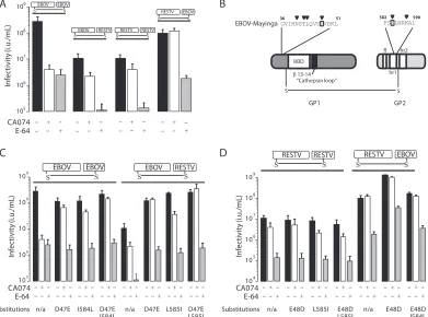
examined. We observed that the titer of EBOV-GP1/RESTV-GP2
particles on Vero cells was similar to that of RESTV and the titer of
RESTV-GP1/GP2 particles was similar to that of
EBOV-May, indicating that the chimeric GPs were functional (Fig. 5A).
We observed that neither of the virus particles expressing chimeric
GP EBOV/RESTV or RESTV/EBOV were as sensitive as
EBOV-May particles to inhibition by CA074 as to inhibition by E-64.
These findings indicate that the determinants of cathepsin B
de-pendence are not localized exclusively within either GP1 or GP2. A
previous study of EBOV-May-derived viruses selected for
resis-tance to CA074 suggested an explanation. This study showed that
single amino acid changes in residues clustered either near the N
terminus of GP1 (N40K/S/T, T42A, L43F, or D47V) or in the hr1
segment of GP2 (I584F or K588R) conferred resistance to CA074
(46). Using these findings as a guide, we noted that RESTV GP
differs from EBOV-May GP at residue 47 (D
¡
E) in GP1 and
residue 584 (I
¡
L) in GP2 (Fig. 5B). To determine if these subtle
changes mediate the difference in the behavior of these viruses, we
exchanged these residues in EBOV-May and RESTV and tested
the effect on sensitivity to CA074. We found that substitution of
glutamic acid for D47 or leucine for I584 alone or in combination
FIG 3Cathepsin (Cat) B is not an essential host factor for all filoviruses. (A, B) Effect of cathepsin B selective inhibitor CA074 on cathepsin B and L activities (A) and on infectivity (B) by VSVGFPparticles bearing filovirus GPs. Vero cells were incubated in the presence of increasing concentrations of CA074 (0 to 80M)or vehicle for 4 h prior to cell lysis or the addition of VSVGFPparticles bearing GPs from MARV (M) or⌬Muc GP from EBOV-May (Z), CIEBOV (CI), SUDV
(S), or RESTV (R). (A) Cathepsin B and L activities were determined by fluorogenic substrates (n⫽2). (B) After 24 h, GFP-positive cells were manually counted using fluorescence microscopy. Infectivities from each of two replicates are shown and are representative of four independent experiments. Data presented were determined as follows: (infection [IU/ml] of CA074-treated cells)/(infection of vehicle-treated cells)⫻100%. (C) Wild-type and cathepsin B-deficient (cathepsin B⫺/⫺cathepsin L⫹/⫹) MEF cells were infected with VSV
GFPparticles bearing GP from MARV (M) or⌬Muc GP from EBOV-May (Z), CIEBOV (CI), SUDV (S),
or RESTV (R) or VSV (G). After 24 h, infectivity was determined as described for panel B. Data presented were determined as follows: (infection [IU/ml] of cathepsin B-deficient cells)/(infection of wild-type MEF cells)⫻100%. Data are means⫾SD (n⫽3). Shown is a representative of three independent experiments. (D) Wild-type and cathepsin B-deficient MEF cells were transfected with expression plasmids encoding mouse cathepsin B, mouse cathepsin L, or a sham plasmid. After 24 h, cells were exposed to VSVGFPparticles as described above. After 24 h, the percentage of cells infected was determined using flow
cytometry. Data presented were determined as follows: (percent infection of transfected cathepsin B-deficient MEF cells)/(percent infection of wild-type MEF cells)⫻100%. Data are means⫾SD (n⫽3). Shown is a representative of three independent experiments.
on November 7, 2019 by guest
http://jvi.asm.org/
[image:5.585.138.451.67.311.2]conferred resistance to CA074 on EBOV-May. However, the
in-troduction of the reciprocal changes E48D and/or L585I into
RESTV GP did not confer a requirement for cathepsin B (Fig. 5C
and D). These findings indicate that D47 and I584 are necessary
for EBOV-May but not sufficient for RESTV to depend on
cathep-sin B as a host factor for the infection of Vero cells.
Sequence analysis predicts cathepsin B dependence.
The
dis-covery that the cathepsin B dependence of EBOV-May maps to
residues D47 and I584 suggested the possibility that these residues
might also predict the protease requirements of other filoviruses.
Indeed, we noted that D47 and I584 are conserved in the GPs of
EBOV-May and CIEBOV, which are cathepsin B dependent, but
not in those of SUDV (E47) and MARV (N47 and L584), which
are not. A search of the NCBI database identified 27 partial and
complete sequences of GPs obtained from independent isolates
classified as EBOV. Only one of the virus isolates, EBOV-1995
(GenBank accession no.
ID-AY354458
), differed in the cathepsin
B-determining residues. Excluding the mucin regions, which are
deleted from the GPs in this analysis, the GPs of EBOV-May and
EBOV-1995 differ only at residues 47 (D47E) and 544 (I544T),
which had not been identified in previous studies of cathepsin B
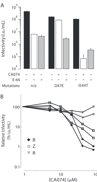
requirements (46). We prepared and analyzed VSV EBOV-May
GP-derived particles containing E47 or T544 GP. As expected,
these viruses are highly infectious and sensitive to E-64. However,
inhibition of cathepsin B by CA074 reduced the titer of T544 GP
particles by
⬎
99% under conditions where the titer of E47 GP
particles was minimally changed (Fig. 6A). Thus, escape
from dependence on cathepsin B was closely correlated with
E47 and not T544. Since we began these investigations, an
out-break of hemorrhagic fever occurred in Uganda due to a
filo-virus that is sufficiently different from EBOV, CIEBOV,
RESTV, and MARV to be classified as Bundibugyo (BDBV)
(39). Analysis of the BDBV sequence revealed the presence of
D47 and I584, thus predicting that BDBV is cathepsin B
depen-dent. Indeed, we found that infection by VSV BDBV GP
parti-cles was inhibited by increasing concentrations of CA074 (0 to
80
M) in a pattern closely correlated with that of EBOV-May
(Fig. 6B). In addition, we found that infection by BDBV
pseu-dotypes was blocked by the treatment of cells with E-64 and in
ca-thepsin B
⫺/⫺MEF cells (data not shown). Thus, in our limited cohort
FIG 4Cathepsin (Cat) L is a host factor for Reston ebolavirus and Marburg virus. (A, B) Effect of the inhibitor FYdmk on cathepsin L and B activities (A) and on the infectivity (B) of VSVGFPparticles bearing filovirus GPs. Vero cells were incubated in the presence of increasing concentrations of FYdmk (0 to 10M)or vehicle for 4 h prior to cell lysis or the addition of VSVGFPparticles bearing GP from MARV (M) or⌬Muc GP from EBOV-May (Z), CIEBOV(CI), SUDV(S),
or RESTV (R). (A) Cathepsin B and L activities were determined by fluorogenic substrates (n⫽2). (B) After 24 h, GFP-positive cells were manually counted using fluorescence microscopy. Infectivities from two replicates are shown and are representative of four independent experiments. Data presented were determined as follows: (infection [IU/ml] of FYdmk-treated cells)/(infection of vehicle-treated cells)⫻100%. (C) Wild-type and cathepsin B/cathepsin L-deficient (cathep-sin B⫺/⫺cathepsin L⫺/⫺) MEF cells were infected with VSV
GFPparticles bearing GP from MARV (M) or⌬Muc GP from EBOV-May (Z), CIEBOV(CI),
SUDV(S), or RESTV (R) or with VSV (G). Infectivity was determined as described in the legend to Fig. 3C. Data are means⫾SD (n⫽3). Shown is a representative of three independent experiments. (D) Cathepsin B/cathepsin L-deficient MEF cells were transfected with expression plasmids encoding mouse cathepsin B, mouse cathepsin L, both cathepsins B and L, or the vector alone. After 2 days, cells were exposed to VSVlucbearing GP from MARV (M) or⌬Muc
GP from EBOV-May (Z), CIEBOV(CI), SUDV(S), or RESTV(R) or to VSV (G). Twenty-four hours later, infectivity was determined by measuring the RLU in cell lysates. Data presented were determined as follows: virus-encoded RLU in cathepsin-transfected cells/RLU in vector-transfected cells. Data are means⫾SD (n⫽6). Shown is a representative of four independent experiments.
on November 7, 2019 by guest
http://jvi.asm.org/
[image:6.585.95.492.65.354.2]of filoviruses analyzed in this study, the presence of D47 correlated
with dependence on cathepsin B for infection.
DISCUSSION
Over the past 6 years, significant progress has been made in
un-derstanding how filoviruses gain entry into cells and a model of
infection based on this progress has been proposed (10).
EBOV-May particles attach to lectins on the cell surface and are taken up
by macropinocytosis into vesicles that are transported to
LE/LY-containing endosomal cysteine proteases and NPC1 (5, 10, 20, 31,
32, 34, 43). One function of endosomal cysteine proteases is to
cleave the carboxyl-terminal “cap” region of GP1 from the
mushroom-shaped GP protruding from the virus membrane and
expose the stalk containing the protease-resistant N-terminal
do-main in GP1 that is the ligand for the NPC1 protein (4, 10–12, 19,
24, 25, 28). A second function may be to biochemically destabilize
the GP, thereby sensitizing it to triggering for viral membrane
fusion (3, 46). Further studies are needed to determine the roles of
NPC1 (5, 10) and additional events, including further cleavage of
GP1 in this process (3, 11, 21, 35, 46).
In this report, we provide evidence that key aspects of this
scheme are conserved among other filoviruses. The new findings
show that as in EBOV-May, the carboxyl-terminal domain of
SUDV GP1 is a substrate for proteolytic cleavage, that cleaved
particles are infectious and NPC1 dependent, and that the
protease-resistant N-terminal domain of GP1 binds to purified
LE/LY membranes in an NPC1-dependent manner. These
find-ings are consistent with the recent report showing that the domain
organization of EBOV-May is conserved in SUDV (11).
More-over, we find that the sensitivity of the C-terminal domain of GP1
to cleavage and the dependence of cleaved particles on NPC1 is
conserved among other ebolaviruses. Consistent with this view,
we find that isolates from each species of
Filoviridae
are E-64
sen-sitive, including the growth of EBOV-May, SUDV, and MARV.
While more work needs to be done, including studies of other
proteases and MARV, the results of the experiments described in
this report, coupled with alignment of primary amino acid
se-quences of GPs which indicate that the domain structure is likely
to be conserved (data not shown), suggest that cleavage and
bind-ing are key steps in the filovirus entry pathway. In this model,
endosomal cysteine proteases are required for the efficient
re-moval of the carboxyl-terminal domain of GP1 to expose the
NPC1 binding domain. Endosomal cysteine proteases may
medi-ate the additional steps necessary for orderly deployment of the
FIG 5Comparison of cathepsin B requirements for EBOV and RESTV. (A) Virus determinants of cathepsin B dependence. Vero cells were incubated in the presence of CA074 (80M), E-64 (300M), or the vehicle (1% DMSO) for 3 h prior to the addition of VSVGFPparticles bearing⌬Muc EBOV-May GP, RESTVGP, EBOV-May GP1/RESTV GP2, or RESTV GP1/EBOV-May GP2. Infectivity was determined as described in the legend to Fig. 1A. Data are means⫾SD (n⫽
3). Shown are representatives of three independent experiments. (B) Schematic of EBOV-May GP and locations of amino acid residues previously shown to mediate resistance to CA074. Inverted triangles identify residues that mediate resistance. Boxes identify differences in amino acids between EBOV-May and RESTV. The positions of the receptor-binding domain (RBD), the-13-14 disordered loop, the fusion loop (fl), and heptad repeats 1 and 2 (hr1, hr2) are indicated. Residue numbering relative to EBOV GP. (C and D) Analysis of the effects of D47E and I584L on cathepsin B dependence. The effects of reciprocal substitutions of residues (D47/E48 and I584/L585) that differ between EBOV-May and RESTV GP were measured in native and chimeric GPs from panel A. Infectivity was determined as described in the legend to Fig. 1A. Data are means⫾SD (n⫽3). Shown are representatives of three independent experiments. n/a, not applicable.
on November 7, 2019 by guest
http://jvi.asm.org/
[image:7.585.101.492.68.358.2]virus membrane fusion activity (21, 35, 46). Indeed, two recent
reports suggest that cysteine protease cleavage of the

-13-14 loop
in GP1 promotes release of the GP2 fusion peptide (3, 11). The
studies in this report provide the basis for future studies to
com-pare the virus requirements for specific proteases to cleave GP1, to
bind to NPC1, and to release the GP2 fusion peptide.
The endosomal cysteine proteases are a family of 11
acid-dependent proteases that reside in LE/LY (15, 17, 32, 40–43). Little
is known about the roles individual members of this family play
during filovirus infection. Previously, we showed that EBOV-May
requires cathepsin B, but not cathepsin L, for entry (7). We
con-firm this finding and show that CIEBOV has the same
require-ment. Additional studies are needed to determine if the virus’s
sensitivity to the absence of cathepsin B activity is due to a specific
requirement for the double-chain isoform of cathepsin B (36).
Although cathepsin L is not essential for any of the filoviruses
studied, we found that it works in concert with cathepsin B to
enhance the infection of EBOV-May, CIEBOV, and RESTV.
Fur-thermore, cathepsin L activity is required for MARV infection of
MEF cells but not Vero cells, suggesting that the role of cathepsin
L may be shared by other cysteine proteases with endopeptidase
activity. Remarkably, RESTV infection is sensitive to E-64 but is
not sensitive to the loss of cathepsins B and L. This suggests that
one or more additional E-64-sensitive proteases can support
RESTV infection.
The substrate specificity of endosomal cysteine proteases is
governed largely by the accessibility of the polypeptide chain to
the active site of the protease and not by a strong preference for
specific sequence motifs (32, 41, 42). Thus, one consequence of
the extensive variation in the sequence of the carboxyl-terminal
domain of GP1 is that it may alter the repertoire of cysteine
pro-teases that are able to cleave GP1. Therefore, the presence of
mul-tiple proteases with overlapping substrate preferences in late
en-dosomes and lysosomes of host cells may provide for redundancy
in the conditions for cleavage of GP and might explain the
virus-specific differences in dependence on cathepsins B and L observed
in our studies. One advantage of this scheme may be that effective
cleavage of the GP1 cap and/or fusion peptide release is
main-tained in the presence of selective pressure from host immune
recognition for sequence diversification, analogous to the
func-tion of variable loops in HIV gp120 (2, 18). In this model,
adap-tation to loss of cathepsin B activity by a change to a single amino
acid residue (i.e., D47 or I584) may provide a means for a rapid
response to changes in endosomal cysteine protease expression
between hosts or cell types. Sequence polymorphisms that
specif-ically predict host factor preference have been identified in other
virus envelope GPs, including SARS GP N479K interactions with
human or civet ACE2 receptor, influenza virus HA1 interactions
with
␣
-2 glycan linkage in human or
␣
-2-6 glycan linkage in avian
sialic acid, and HIV gp120 binding to receptor CXCR4 and/or
CCR5 (2, 26, 33, 38). Analysis of the filoviruses identified in future
outbreaks will provide further tests of the utility of using the D47/
I584 polymorphisms in GP in determining virus preference for
host endosomal cysteine proteases.
ACKNOWLEDGMENTS
This work was supported by grants U54 AI057159 and R01 CA104266 to J.C. and PIDS-Sanofi-Pasteur fellowship K12-HD052896 and 5K08AI079381 to J.M. K.C. was supported by a postdoctoral fellowship from the New England Research Center of Excellence in Biodefense and Emerging Infectious Dis-eases (NERCE/BEID). C.F. was supported by the Postgraduate Research Par-ticipation Program at the U.S. Army Medical Research and Material Com-mand administered by the Oak Ridge Institute for Science and Education through an interagency agreement between the U.S. Department of Energy and USAMRMC. M.C. was supported by the Fonds de la Recherche en Sant é du Quebéc.
We thank Scott Aoki, Anna Bruchez, Brenna Hill, and Daniel Douek for assistance.
The opinions, interpretations, conclusions, and recommendations in this report are ours and are not necessarily endorsed by the U.S. ment of Defense, the U.S. Department of the Army, or the U.S. Depart-ment of Health and Human Services.
REFERENCES
1.Alvarez CP, et al.2002. C-type lectins DC-SIGN and L-SIGN mediate cellular entry by Ebola virus incisand intrans. J. Virol.76:6841– 6844. 2.Arthos J, et al.1989. Identification of the residues in human CD4 critical
for binding HIV. Cell57:469 – 481.
FIG 6Analysis of cathepsin B dependence of EBOV-1995 and BDBV. (A) VSVGFPparticles pseudotyped with GP from EBOV-May containing E47 or
T544 from EBOV-1995 were prepared, and dependence on cathepsin B was analyzed. Vero cells were incubated in the presence of CA074 (80M), E-64 (300M), or the vehicle for 3 h prior to the addition of VSVGFPbearing⌬Muc
GP from EBOV-May D47E or I544T or wild-type EBOV-May GP. Infection was measured as described in the legend to Fig. 1A. Data are means⫾SD (n⫽
3) Shown is a representative of three independent experiments. (B) Effect of cathepsin B selective inhibitor CA074 on infection by BDBV GP. Vero cells were incubated in the presence of increasing concentrations of CA074 (0 to 80 M) or the vehicle for 3 h prior to the addition of VSVlucparticles bearing
⌬Muc GP from EBOV-May (Z), BDBV (B), or RESTV (R). After 6 h, cells were lysed and relative luminescence (RLU) was measured. Data are presented as follows: (virus-encoded luciferase activity of cells treated with CA074)/(lucif-erase activity of cells treated with vehicle)⫻100%. Shown are representatives of three independent experiments. n/a, not applicable.
on November 7, 2019 by guest
http://jvi.asm.org/
[image:8.585.78.242.62.370.2]3.Brecher M, et al.2012. Cathepsin cleavage potentiates the Ebola virus glycoprotein to undergo a subsequent fusion-relevant conformational change. J. Virol.86:364 –372.
4.Brindley M, et al.2007. Ebola virus glycoprotein 1: identification of residues important for binding and postbinding events. J. Virol.81:7702– 7709.
5.Carette JE, et al.2011. Ebola virus entry requires the cholesterol trans-porter Niemann-Pick C1. Nature477:340 –343.
6.Centers for Disease Control and Prevention.2001. Outbreak of Ebola hemorrhagic fever Uganda, August 2000-January 2001. MMWR Morb. Mortal. Wkly. Rep.50:73–77.
7.Chandran K, Sullivan NJ, Felbor U, Whelan SP, Cunningham JM.2005. Endosomal proteolysis of the Ebola virus glycoprotein is necessary for infection. Science308:1643–1645.
8.Colebunders R, Borchert M.2000. Ebola haemorrhagic fever—a review. J. Infect.40:16 –20.
9.Colebunders R, et al.2007. Marburg hemorrhagic fever in Durba and Watsa, Democratic Republic of the Congo: clinical documentation, fea-tures of illness, and treatment. J. Infect. Dis.196(Suppl. 2):S148 –S153. 10. Côté M, et al.2011. Small molecule inhibitors reveal Niemann-Pick C1 is
essential for Ebola virus infection. Nature477:344 –348. (Letter.) 11. Dias J, et al.2011. A shared structural solution for neutralizing
ebolavi-ruses. Nat. Struct. Mol. Biol.18:1424 –1427.
12. Dube D, et al.2009. The primed ebolavirus glycoprotein (19-kilodalton GP1,2): sequence and residues critical for host cell binding. J. Virol.83: 2883–2891.
13. Dube D, et al.2010. Cell adhesion-dependent membrane trafficking of a binding partner for the ebolavirus glycoprotein is a determinant of viral entry. Proc. Natl. Acad. Sci. U. S. A.107:16637–16642.
14. Fabozzi G, Nabel C, Dolan M, Sullivan N.2011. Ebolavirus proteins suppress the effects of small interfering RNA by direct interaction with the mammalian RNA interference pathway. J. Virol.85:2512–2523. 15. Fujishima A, et al.1997. The crystal structure of human cathepsin L
complexed with E-64. FEBS Lett.407:47–50.
16. Geisbert TW, Jahrling PB.2004. Exotic emerging viral diseases: progress and challenges. Nat. Med.10:S110 –S121.
17. Guncar G, Pungercic G, Klemencic I, Turk V, Turk D.1999. Crystal structure of MHC class II-associated p41 Ii fragment bound to cathepsin L reveals the structural basis for differentiation between cathepsins L and S. EMBO J.18:793– 803.
18. Harrison SC.2008. Viral membrane fusion. Nat. Struct. Mol. Biol.15: 690 – 698.
19. Hood CL, et al.2010. Biochemical and structural characterization of cathepsin L-processed Ebola virus glycoprotein: implications for viral en-try and immunogenicity. J. Virol.84:2972–2982.
20. Hunt CL, Kolokoltsov AA, Davey RA, Maury W. 2011. The Tyro3 receptor kinase Axl enhances macropinocytosis of Zaire ebolavirus. J. Vi-rol.85:334 –347.
21. Kaletsky R, Simmons G, Bates P.2007. Proteolysis of the Ebola virus glycoproteins enhances virus binding and infectivity. J. Virol.81:13378 – 13384.
22. Kondratowicz AS, et al.2011. T-cell immunoglobulin and mucin domain 1 (TIM-1) is a receptor for Zaire Ebolavirus and Lake Victoria Marburg-virus. Proc. Natl. Acad. Sci. U. S. A.108:8426 – 8431.
23. Kuhn JH, et al.2010. Proposal for a revised taxonomy of the family
Filoviridae: classification, names of taxa and viruses, and virus abbrevia-tions. Arch. Virol.155:2083–2103.
24. Kuhn JH, et al.2006. Conserved receptor-binding domains of Lake Vic-toria marburgvirus and Zaire ebolavirus bind a common receptor. J. Biol. Chem.281:15951–15958.
25. Lee JE, et al.2008. Structure of the Ebola virus glycoprotein bound to an antibody from a human survivor. Nature454:177–182.
26. Li F, Li W, Farzan M, Harrison SC.2005. Structure of SARS coronavirus spike receptor-binding domain complexed with receptor. Science309: 1864 –1868.
27. Lin G, et al.2003. Differential N-linked glycosylation of human immu-nodeficiency virus and Ebola virus envelope glycoproteins modulates in-teractions with DC-SIGN and DC-SIGNR. J. Virol.77:1337–1346. 28. Manicassamy B, Wang J, Jiang H, Rong L.2005. Comprehensive
Anal-ysis of ebola virus GP1 in viral entry. J. Virol.79:4793– 4805.
29. Marzi A, et al.2006. The signal peptide of the ebolavirus glycoprotein influences interaction with the cellular lectins DC-SIGN and DC-SIGNR. J. Virol.80:6305– 6317.
30. Mulherkar N, Raaben M, de la Torre JC, Whelan SP, Chandran K.2011. The Ebola virus glycoprotein mediates entry via a non-classical dynamin-dependent macropinocytic pathway. Virology419:72– 83.
31. Nanbo A, et al.2010. Ebolavirus is internalized into host cells via mac-ropinocytosis in a viral glycoprotein-dependent manner. PLoS Pathog.
6(9):e1001121.
32. Otto H-H, Schirmeister T.1997. Cysteine proteases and their inhibitors. Chem. Rev.97:133–172.
33. Ribeiro RM, Hazenberg MD, Perelson AS, Davenport MP.2006. Naïve and memory cell turnover as drivers of CCR5-to-CXCR4 tropism switch in human immunodeficiency virus type 1: implications for therapy. J. Virol.80:802– 809.
34. Saeed MF, Kolokoltsov A, Albrecht T, Davey R.2010. Cellular entry of Ebola virus involves uptake by a macropinocytosis-like mechanism and subsequent trafficking through early and late endosomes. PLoS Pathog.
6:e1001110.
35. Schornberg K, et al.2006. Role of endosomal cathepsins in entry medi-ated by the Ebola virus glycoprotein. J. Virol.80:4174 – 4178.
36. Schornberg K, et al.2009.␣51-integrin controls ebolavirus entry by regulating endosomal cathepsins. Proc. Natl. Acad. Sci. U. S. A.106:8003– 8008.
37. Simmons G, et al.2003. DC-SIGN and DC-SIGNR bind ebola glycopro-teins and enhance infection of macrophages and endothelial cells. Virol-ogy305:115–123.
38. Stevens J, et al.2006. Structure and receptor specificity of hemagglutinin from an H5N1 influenza virus. Science312:404 – 410.
39. Towner JS, et al. 2008. Newly discovered ebola virus associated with hemorrhagic fever outbreak in Uganda. PLoS Pathog.4:e1000212. 40. Turk B, Turk D, Turk V.2000. Lysosomal cysteine proteases: more than
scavengers. Biochim. Biophys. Acta1477:98 –111.
41. Turk D, Podobnik M, Kuhelj R, Dolinar M, Turk V. 1996. Crystal structures of human procathepsin B at 3.2 and 3.3 Angstroms resolution reveal an interaction motif between a papain-like cysteine protease and its propeptide. FEBS Lett.384:211–214.
42. Turk D, Guncar G. 2003. Lysosomal cysteine proteases (cathepsins): promising drug targets. Acta Crystallogr. D Biol. Crystallogr.59(Pt. 2): 203–213.
43. Turk V, Turk B, Turk D.2001. Lysosomal cysteine proteases: facts and opportunities. EMBO J.20:4629 – 4633.
44. Vance JE, Peake KB.2011. Function of the Niemann-Pick type C proteins and their bypass by cyclodextrin. Curr. Opin. Lipidol.22:204 –209. 45. Weidmann M, Mühlberger E, Hufert F.2004. Rapid detection protocol
for filoviruses. J. Clin. Virol.30:94 –99.
46. Wong AC, Sandesara RG, Mulherkar N, Whelan SP, Chandran K.2010. A forward genetic strategy reveals destabilizing mutations in the Ebolavi-rus glycoprotein that alter its protease dependence during cell entry. J. Virol.84:163–175.
on November 7, 2019 by guest
http://jvi.asm.org/