0022-538X/09/$12.00 doi:10.1128/JVI.00638-09
Copyright © 2009, American Society for Microbiology. All Rights Reserved.
Detection of Novel Sequences Related to African Swine Fever Virus
in Human Serum and Sewage
䌤
†
Joy Loh,
1Guoyan Zhao,
1Rachel M. Presti,
2Lori R. Holtz,
3Stacy R. Finkbeiner,
1Lindsay Droit,
1Zoilmar Villasana,
1Collin Todd,
1James M. Pipas,
4Byron Calgua,
5Rosina Girones,
5David Wang,
1and Herbert W. Virgin
1*
Departments of Pathology & Immunology and Molecular Microbiology,1Department of Medicine,2and Department of Pediatrics,3
Washington University School of Medicine, St. Louis, Missouri; Department of Biological Sciences, University of Pittsburgh, Pittsburgh, Pennsylvania4; and Department of Microbiology, Faculty of Biology, University of Barcelona, Barcelona, Spain5
Received 27 March 2009/Accepted 25 September 2009
The familyAsfarviridaecontains only a single virus species, African swine fever virus (ASFV). ASFV is a viral agent with significant economic impact due to its devastating effects on populations of domesticated pigs during outbreaks but has not been reported to infect humans. We report here the discovery of novel viral sequences in human serum and sewage which are clearly related to the asfarvirus family but highly divergent from ASFV. Detection of these sequences suggests that greater genetic diversity may exist among asfarviruses than previously thought and raises the possibility that human infection by asfarviruses may occur.
The family Asfarviridae contains a single double-stranded DNA (dsDNA) virus called African swine fever virus (ASFV), which is thought to have evolved from an ancestral virus com-mon to the nucleocytoplasmic large DNA viruses including poxviruses, iridoviruses, and phycodnaviruses (10, 11). ASFV infects ticks and swine but has not been reported to infect humans. ASFV infection of wild swine typically causes persis-tent infection with few symptoms (9, 17, 24, 25), but domesti-cated pigs can develop severe disease including acute hemor-rhagic fever with nearly 100% mortality. As there is no vaccine and disease is contained by animal quarantine and slaughter, ASFV outbreaks can decimate pig populations and have sig-nificant economic impact. A 2007 outbreak in the former So-viet republic of Georgia resulted in the death and slaughter of over 80,000 pigs (20).
ASFV is endemic in sub-Saharan Africa but has also been introduced to countries in Europe, South America, and the Caribbean (26). Characterization of various ASFV isolates has led to the identification of 22 genotypes based on sequence variation in the portion of the B646L gene encoding the C terminus of the major capsid protein (2, 4, 14). Within this segment of B646L, approximately 14% of the nucleotide sites are variable among the ASFV isolates studied (2, 4). The ASFV genome has been completely sequenced for the Vero cell-adapted BA71V strain (29) and several wild isolates (5). Like many other large dsDNA viruses, ASFV contains open reading frames with homology to cellular genes involved in DNA replication, transcription, repair, and protein modifica-tion (29). ASFV also has an array of open reading frames with
potential function in modulating host cell function or immune response (7).
We report here the discovery of novel viral sequences in human serum from the Middle East and in sewage from Spain that have clear similarity to ASFV genes. Of the 36 sequences identified, 29 did not have significant overlap or nucleotide identity with any of the other sequences. These 29 sequences are similar to 18 different ASFV genes, with some sequences matching to different regions within the same ASFV genes. Sequence and phylogenetic analyses indicate that the novel viral sequences are most closely related to the asfarvirus family but are highly divergent from known ASFV strains. We there-fore hypothesize that these viral sequences are derived from at least one novel virus in theAsfarviridaefamily, which we refer to herein as ASFV-like virus (ASFLV).
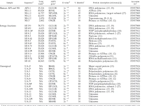
Discovery of novel asfarvirus-related viral sequences. We analyzed total nucleic acid extracted from human serum sam-ples by 454 sequencing as an approach to identifying potential novel human pathogens. Serum samples were collected from patients with acute febrile illness (AFI) and from healthy vol-unteers (normal serum [NS]) in the Middle East between 2002 and 2005 and stripped of identifying information before anal-ysis to protect patient confidentiality. Total nucleic acid was extracted from 199 AFI and 200 NS samples and reverse tran-scribed to enable detection of both RNA and DNA viruses. Each sample was then amplified by sequence-independent PCR using a primer that incorporates a 6-nucleotide barcode unique to that sample. Amplicons from multiple samples were pooled and subjected to 454 pyrosequencing. Additional de-tails on serum sample processing and sequence data analysis are provided in the “Supplemental methods” section of the supplemental material. From one AFI sample and three NS samples, we identified six novel viral sequences with no signif-icant nucleotide similarity to known viruses but whose trans-lated sequences had detectable sequence identity to several ASFV proteins (Table 1), as determined using tBLASTx (1).
ASFV-related sequences were also found in sewage col-lected from an urban wastewater treatment plant in Barcelona,
* Corresponding author. Mailing address: Department of Pathology & Immunology, Washington University School of Medicine, 660 S. Euclid Ave., Box 8118, St. Louis, MO 63110. Phone: (314) 362-9223. Fax: (314) 362-4096. E-mail: virgin@wustl.edu.
† Supplemental material for this article may be found at http://jvi .asm.org/.
䌤Published ahead of print on 7 October 2009.
13019
on November 8, 2019 by guest
http://jvi.asm.org/
Spain. Briefly, viral particles from the sewage samples were concentrated and separated into fractions by cesium chloride equilibrium gradient centrifugation. Fractions with high con-centrations of virus, as determined by quantitative PCR for human adenovirus, were treated first with DNase I to degrade nonviral DNA not protected by a viral capsid and then with a lysis buffer to disrupt viral capsids and release viral nucleic acids. Total nucleic acid was extracted from these fractions and was reverse transcribed and PCR amplified prior to 454 se-quencing as described above. Additional details on the pro-cessing of sewage samples are provided in the “Supplemental methods” section of the supplemental material. Fifteen ASFV-related sequences were identified in five sewage fractions (SF). We also identified an additional 15 ASFV-related sequences
from our sequencing runs which could not be assigned to specific samples due to lack of a perfect sequence match with the barcode sequence. All of these unassignable sequences were identified in sequencing runs containing multiple samples including the sewage fractions which were found to contain ASFV-related sequences. We did not detect ASFV-related sequences in the other samples in these sequencing runs and therefore speculate that these unassignable sequences are de-rived from the sewage samples within these runs that contained ASFV-like sequences. However, as we cannot verify the spe-cific sample source of these unassignable sequences, they are reported here as unassigned (UA) sequences.
A total of 36 novel ASFV-related sequences were identified, all of whose best-scoring tBLASTx matches were to the
respec-TABLE 1. Novel viral sequences with similarity to ASFV
Sample Sequencea Total
readsb
ASFV
genec E valued % Identitye Protein description (reference关s兴)
Accession no.f
Human AFI and NS AFI-1 23,224 G1211R 1e⫺10 64 DNA polymerase (15, 19) FJ957903
NS-1.1 83,028 B354L 3e⫺11 42 ATPase (10) FJ957904
NS-1.2 14,737 NP1450L 2e⫺13 45 RNA polymerase, largest subunit (27) FJ957905
NS-2.1 42,635 M1249L 2e⫺10 36 Unknown FJ957906
NS-2.2 3,870 P1192R 5e⫺12 37 Topoisomerase II (3, 8) FJ957907
NS-3 2,602 C962R 7e⫺12 38 Primase or ATPase (10, 12) FJ957908
Sewage fractions SF-3g† 78,573 G1211R 2e⫺26 51 DNA polymerase (15, 19) FJ957909
SF-6g† 6,633 G1211R 1e⫺25 51 DNA polymerase (15, 19) FJ957910
SF-8.1# 33,028 D250R 4e⫺14 60 NTPhpyrophosphohydrolase (10) FJ957911
SF-8.2 33,028 EP1242L 1e⫺19 45 RNA polymerase, subunit 2 (27) FJ957912
SF-8.3 33,028 EP364R 5e⫺9 39 Nuclease (11) FJ957913
SF-8.4§ 33,028 EP424R 5e⫺9 58 RNA methyltransferase (11) FJ957914
SF-8.5 33,028 F1055L 3e⫺18 32 Helicase (23) FJ957915
SF-8.6g 33,028 F1055L 6e⫺8 50 Helicase (23) FJ957916
SF-8.7† 33,028 G1211R 5e⫺23 49 DNA polymerase (15, 19) FJ957917
SF-8.8 33,028 G1340L 1e⫺8 27 Unknown FJ957918
SF-8.9¶ 33,028 H339R 5e⫺13 44 Unknown FJ957919
SF-9.1ⴱ 13,968 C962R 4e⫺10 40 Primase or ATPase (10, 12) FJ957920
SF-9.2 13,968 CP2475L 5e⫺10 41 220-kDa polyprotein (21) FJ957921
SF-9.3§ 78,307 EP424R 2e⫺11 44 RNA methyltransferase (11) FJ957922
SF-10 16,843 C475L 9e⫺14 46 Polyadenylate polymerase (6) FJ957923
Unassigned UA.1g NA B646L 1e⫺11 44 Major capsid protein (13) FJ957924
UA.2g NA B962L 7e⫺37 50 Helicase (28) FJ957925
UA.3 NA C475L 1e⫺10 31 Polyadenylate polymerase (6) FJ957926
UA.4 NA C475L 8e⫺9 37 Polyadenylate polymerase (6) FJ957927
UA.5 NA C962R 6e⫺38 38 Primase or ATPase (10, 12) FJ957928
UA.6ⴱ NA C962R 1e⫺17 32 Primase or ATPase (10, 12) FJ957929
UA.7g NA CP2475L 2e⫺15 30 220-kDa polyprotein (21) FJ957930
UA.8# NA D250R 5e⫺14 60 NTP pyrophosphohydrolase (10) FJ957931
UA.9g NA EP1242L 1e⫺20 44 RNA polymerase, subunit 2 (27) FJ957932
UA.10g† NA G1211R 2e⫺26 51 DNA polymerase (15, 19) FJ957933
UA.11 NA G1211R 1e⫺15 41 DNA polymerase (15, 19) FJ957934
UA.12 NA G1340L 6e⫺15 32 Unknown FJ957935
UA.13 NA G1340L 1e⫺28 50 Unknown FJ957936
UA.14¶ NA H339R 8e⫺12 35 Unknown FJ957937
UA.15 NA M448R 2e⫺9 34 Unknown FJ957938
aSets of sequences whose members were similar to the same regions of the ASFV genome and had significant overlap and nucleotide sequence identity with each
other are indicated by the same symbols (†, #, §, *, or ¶).
bThe total number of reads generated from each sample is given for each sequence whose parent sample is known. For samples that were sequenced in more than
one sequencing run, the total number of reads for the run in which the indicated novel sequence was identified is given. NA, not applicable.
cASFV genes to which the novel viral sequences have sequence similarity, as determined using tBLASTx (1). ASFV genes are listed using nomenclature for the
BA71V strain (29).
dE value for tBLASTx comparison of the novel viral sequences to the ASFV BA71V complete genome (29).
eAmino acid identity between the novel viral sequences and corresponding sequences in the ASFV BA71V strain within the region of similarity identified using
tBLASTx.
fGenBank accession numbers.
gSequence was assembled from two or more overlapping reads. hNTP, nucleoside triphosphate.
13020 NOTES J. VIROL.
on November 8, 2019 by guest
http://jvi.asm.org/
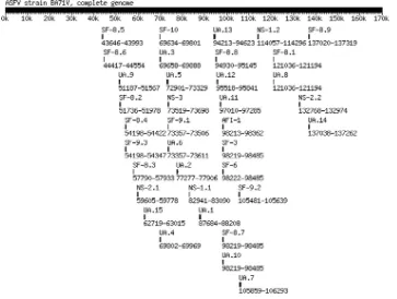
[image:2.585.44.540.81.453.2]tive ASFV genes from various ASFV isolates (not shown). These sequences are listed in Table 1 and were positionally mapped to the complete genome for the ASFV BA71V strain (Fig. 1) (29). These novel viral sequences were similar not only to ASFV genes such as the DNA polymerase and RNA poly-merase genes, which are also conserved in other large dsDNA virus families (10), but also to multiple ASFV genes including EP364R and M448R, which do not have significant similarity to genes in other viral families and are therefore, to date, specific to ASFV.
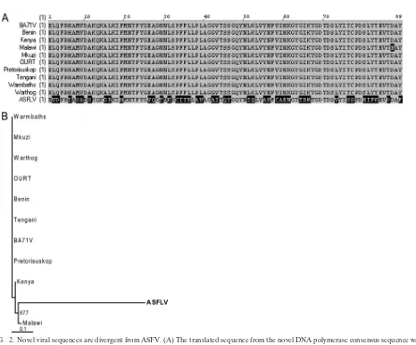
Novel asfarvirus-related sequences are highly divergent from ASFV. The low amino acid identity between the novel viral sequences and the corresponding ASFV proteins (Table 1) suggested that these sequences may belong to a genetically distinct virus rather than to a new isolate of ASFV. We there-fore performed multiple sequence alignments to compare the DNA polymerase-like sequences from our samples to the cor-responding sequences from ASFV isolates. Sequences SF-3, SF-6, SF-8.7, and UA.10 all mapped to the same region of ASFV DNA polymerase (Fig. 1) and were nearly identical in nucleotide sequence where they overlapped (see Fig. S1 in the supplemental material). These sequences formed a 575-nucle-otide consensus sequence, whose translated sequence had 51% amino acid identity with ASFV DNA polymerase when com-pared by tBLASTx (E value of 2e⫺26). Alignment of the
re-gions of similarity between our novel DNA polymerase se-quence and the corresponding ASFV sese-quences showed that the ASFV sequences were highly conserved with each other, but the sequence from our samples was divergent except for small blocks of amino acids which were conserved with ASFV
sequences (Fig. 2A). The same result was obtained using the NS-1.2 and NS-2.2 sequences, which are similar to ASFV RNA polymerase and topoisomerase II, respectively (see Fig. S2A and B in the supplemental material). We also performed phy-logenetic analysis using sequence SF-8.3, with similarity to the EP364R gene, which appears to be ASFV specific. Sequences from ASFV isolates were again found to be highly conserved with each other, whereas the novel viral sequence was diver-gent (Fig. 2B). The same result was obtained using the UA.15 sequence, with similarity to another ASFV-specific gene, M448R (see Fig. S2C in the supplemental material).
Novel viral sequences are most closely related to the asfar-virus family.As viruses in multiple dsDNA viral families en-code homologs of DNA polymerase, RNA polymerase, and topoisomerase II, we compared those sequences from our sam-ples to corresponding ones from ASFV isolates as well as other dsDNA viruses from families includingPoxviridae,Iridoviridae,
Phycodnaviridae, Herpesviridae, and Ascoviridae. We also in-cluded in our analyses several sequences that had the most significant E values aside from those for ASFV strains when the novel DNA and RNA polymerase sequences were queried against the nucleotide database by tBLASTx. The novel topo-isomerase II sequence did not have any hits with significant E values other than those for ASFV strains.
[image:3.585.109.472.69.343.2]We found that our translated DNA polymerase consensus sequence formed a separate branch which was most closely related to the cluster containing the ASFV isolates (Fig. 3A). The same result was observed for the RNA polymerase (Fig. 3B) (NS-1.2) and topoisomerase II (Fig. 3C) (NS-2.2) se-quences. For all three proteins, grouping of the various dsDNA
FIG. 1. Novel viral sequences with similarity to ASFV genes. Novel viral sequences were positionally mapped to the complete genome of the ASFV BA71V strain (29) based on the similarity of the associated amino acid sequences to ASFV proteins. Each novel viral sequence is represented by a black bar, and the ASFV genomic nucleotide position to which it is similar is indicated below the bar.
on November 8, 2019 by guest
http://jvi.asm.org/
FIG. 2. Novel viral sequences are divergent from ASFV. (A) The translated sequence from the novel DNA polymerase consensus sequence was aligned to corresponding sequences from various ASFV isolates using AlignX (VectorNTI suite; Invitrogen). Residues which are highly conserved are shaded in gray, and those which are divergent are in black. (B) Phylogenetic analysis of the translated SF-8.3 sequence and corresponding sequences from the EP364R genes of various ASFV isolates was performed using the neighbor-joining method with 1,000 bootstrap replicates. Bootstrap values over 65% are shown. Sequences were aligned using ClustalX (2.0), and phylogenetic trees were visualized using TreeView (16). GenBank accession numbers for ASFV sequences are provided in the supplemental material. Abbreviations: Benin, Benin 97/1 (5); Kenya, Kenya 1950; Malawi, Malawi Lil-20/1 1983; Mkuzi, Mkuzi 1979; OURT, OURT 88/3 (5); Pretorisuskop, Pretorisuskop/96/4; Tengani, Tengani 62.
FIG. 3. Phylogenetic analysis of novel viral sequences. Translated novel viral sequences similar to ASFV (A) DNA polymerase, (B) RNA polymerase, and (C) topoisomerase II were compared to corresponding sequences from dsDNA viruses and high-scoring nonviral BLAST matches as described in the legend to Fig. 2. Sequences are shown in color as follows: asfarviruses, red; mimivirus, brown; poxviruses, purple; herpesviruses, gray; phycodnaviruses, green; ascoviruses, orange; iridoviruses, blue; nonviral BLAST matches, black. Bootstrap values over 65% are shown. In panel A, virus intrafamily subclassifications are also shown where applicable. For panels B and C, virus families for which the corresponding RNA polymerase and topoisomerase II sequences could not be identified by BLAST were omitted from the analyses. GenBank accession numbers for the sequences analyzed are provided in the “Supplemental methods” section of the supplemental material. Abbreviations: AmEPV,Amsacta mooreientomopoxvirus; APMV,Acanthamoeba polyphagamimivirus; ATCV1,Acanthocystis turfaceachlorella virus 1; CIV, Chilo iridescent virus; CVM1, chlorella virus Marburg 1; D. autotrophicum,Desulfobacterium autotrophicum; DpAV4,Diadromus pulchellusascovirus 4; EBV, Epstein-Barr virus; EhV86,Emiliania huxleyivirus isolate 86; ESV,Ectocarpus siliculosusvirus; FsV158,Feldmanniaspecies virus isolate 158; G. anomala,
Glugea anomala; H. andersenii, Hemiselmis andersenii; H. butylicus,Hyperthermus butylicus; HCMV, human cytomegalovirus; HHV6, human herpesvirus 6; HSV1, herpes simplex virus type 1; HSV2, herpes simplex virus type 2; HvAV3,Heliothis virescensascovirus 3; I. scapularis,Ixodes scapularis; ISKNV, infectious spleen and kidney necrosis virus; K. JI2008,Kabatana sp. strain JI2008; KSHV, Kaposi’s sarcoma-associated herpesvirus; L. acerinae,Loma acerinae; LDV, lymphocystis disease virus; LSDV, lumpy skin disease virus; M. labreanum,Methanocorpusculum labreanum; M. marisnigri,Methanoculleus marisnigri; MIV, mosquito (Aedes taeniorhynchus) iridescent virus; MsEPV,Melanoplus sanguinipes
entomopoxvirus; OSGIV, orange-spotted grouper iridovirus; OsV5,Ostreococcusvirus 5; PBCV1, Paramecium bursariachlorella virus 1; S. islandicus, “Sulfolobus islandicus”; SfAV1,Spodoptera frugiperdaascovirus 1; SGIV, Singapore grouper iridovirus; TFV, tiger frog virus; TnAV2c,
Trichoplusia niascovirus 2c; VZV, varicella-zoster virus; YMTV, Yaba monkey tumor virus. Other abbreviations are as defined for Fig. 2.
13022 NOTES J. VIROL.
on November 8, 2019 by guest
http://jvi.asm.org/
on November 8, 2019 by guest
http://jvi.asm.org/
viruses was consistent with their classifications and with previ-ously published studies using other sets of conserved genes (10, 18, 22).
As several additional novel sequences from our samples are similar to other ASFV genes that have also been reported to have homologs in other dsDNA virus families (10, 11), we performed similar phylogenetic analyses on these sequences. We found that 10 of the 11 sequences with similarity to ASFV gene B354L, C962R, D250R, EP1242L, B646L, or B962L were most closely related to, but distinct from, ASFV sequences (not shown), consistent with our results for the DNA polymer-ase, RNA polymerpolymer-ase, and topoisomerase II sequences. The exception was sequence SF-9.1, which is similar to ASFV C962R but did not group with sequences from any of the dsDNA families in the phylogenetic analysis. SF-9.1 is 99% identical in nucleotide sequence over a 238-nucleotide overlap with sequence UA.6 (not shown), which is also similar to C962R. Two of the three nucleotide changes in SF-9.1 are insertions or deletions resulting in frameshifts that significantly altered portions of the SF-9.1 translated sequence relative to that of UA.6. It is therefore likely that the results of phyloge-netic analysis for SF-9.1 differed from those for UA.6 because the frameshifts decreased the overall similarity of the SF-9.1 translated sequence to ASFV.
Discussion. The detection in this study of multiple viral sequences that are clearly related to, but phylogenetically dis-tinct from, ASFV suggests that at least one additional member of the familyAsfarviridaeexists. The facts that these sequences were identified in multiple 454 sequencing runs over a period of approximately 7 months and that the first sequences iden-tified, NS-1.2 and NS-2.2, were not detected in subsequent sequencing runs of different samples strongly argue against the possibility that the detection of ASFV-like sequences in these samples is due to cross-contamination.
Since the ASFV-like sequence fragments were identified from two types of samples from different geographic regions, it is possible that these sequences are derived from more than one virus in this family. In support of this possibility, the trans-lated SF-10, UA.3, and UA.4 sequences aligned to the same region in C475L of ASFV but were significantly different from each other in nucleotide sequence. An alignment of the trans-lated SF-10, UA.3, and UA.4 sequences with the correspond-ing segment of ASFV C475L showed only 28% identity be-tween UA.3 and SF-10 and 71% identity bebe-tween UA.3 and UA.4 in their respective regions of overlap (see Fig. S3 in the supplemental material). This suggests the possibility that our samples contained more than one asfarvirus-related virus, al-though this cannot be conclusively determined since the re-gions of overlap between these sequences are short.
Although ASFV is not known to infect humans even where the virus is endemic, identification of ASFV-like sequences in serum from multiple human patients suggests that human in-fection may occur. Further studies are under way to prospec-tively screen patient samples by PCR for the presence of ASFV-like sequences using primers to the sequences reported here in order to assess prevalence and geographic distribution. These studies, in combination with serological analyses, will be required to determine whether the ASFV-like virus is, in fact, a human virus and whether it is associated with human disease. The finding of ASFV-like sequences in sewage from Spain
indicates that they are not geographically limited to the Middle East, where the human patient specimens were obtained. Al-though it is unclear whether the source of the ASFV-like sequences in sewage is human or animal, this also suggests the virus may be fecally shed and that screening of stool in addition to serum may be informative. Identification of additional sam-ples containing ASFV-like sequences will be important for the key future goals of obtaining more sequence from the viral genome, determining if the virus can be cultured, and estab-lishing a small-animal model of infection.
Nucleotide sequence accession numbers.The sequences ob-tained in this study have been assigned GenBank accession numbers FJ957903 to FJ957938.
We thank Guillermo Pimentel and Erica Dueger for supplying crit-ical reagents, contributing to discussions of this work, and providing feedback on the manuscript.
This work was supported in part by National Institutes of Health grant U54 AI057160 to the Midwest Regional Center of Excellence for Biodefense and Emerging Infectious Diseases Research.
REFERENCES
1.Altschul, S. F., T. L. Madden, A. A. Schaffer, J. Zhang, Z. Zhang, W. Miller, and D. J. Lipman.1997. Gapped BLAST and PSI-BLAST: a new generation of protein database search programs. Nucleic Acids Res.25:3389–3402. 2.Bastos, A. D., M. L. Penrith, C. Cruciere, J. L. Edrich, G. Hutchings, F.
Roger, E. Couacy-Hymann, and R. Thomson.2003. Genotyping field strains of African swine fever virus by partial p72 gene characterisation. Arch. Virol.
148:693–706.
3.Baylis, S. A., L. K. Dixon, S. Vydelingum, and G. L. Smith.1992. African swine fever virus encodes a gene with extensive homology to type II DNA topoisomerases. J. Mol. Biol.228:1003–1010.
4.Boshoff, C. I., A. D. Bastos, L. J. Gerber, and W. Vosloo.2007. Genetic characterisation of African swine fever viruses from outbreaks in southern Africa (1973–1999). Vet. Microbiol.121:45–55.
5.Chapman, D. A., V. Tcherepanov, C. Upton, and L. K. Dixon.2008. Com-parison of the genome sequences of non-pathogenic and pathogenic African swine fever virus isolates. J. Gen. Virol.89:397–408.
6.Claverie, J. M., C. Abergel, and H. Ogata. 2009. Mimivirus. Curr. Top. Microbiol. Immunol.328:89–121.
7.Dixon, L. K., C. C. Abrams, G. Bowick, L. C. Goatley, P. C. Kay-Jackson, D. Chapman, E. Liverani, R. Nix, R. Silk, and F. Zhang.2004. African swine fever virus proteins involved in evading host defence systems. Vet. Immunol. Immunopathol.100:117–134.
8.Garcia-Beato, R., J. M. Freije, C. Lopez-Otin, R. Blasco, E. Vinuela, and M. L. Salas.1992. A gene homologous to topoisomerase II in African swine fever virus. Virology188:938–947.
9.Heuschele, W. P., and L. Coggins.1969. Epizootiology of African swine fever virus in warthogs. Bull. Epizoot. Dis. Afr.17:179–183.
10.Iyer, L. M., L. Aravind, and E. V. Koonin.2001. Common origin of four diverse families of large eukaryotic DNA viruses. J. Virol.75:11720–11734. 11.Iyer, L. M., S. Balaji, E. V. Koonin, and L. Aravind.2006. Evolutionary genomics of nucleo-cytoplasmic large DNA viruses. Virus Res.117:156–184. 12.Iyer, L. M., E. V. Koonin, D. D. Leipe, and L. Aravind.2005. Origin and evolution of the archaeo-eukaryotic primase superfamily and related palm-domain proteins: structural insights and new members. Nucleic Acids Res.
33:3875–3896.
13.Lopez-Otin, C., J. M. Freije, F. Parra, E. Mendez, and E. Vinuela.1990. Mapping and sequence of the gene coding for protein p72, the major capsid protein of African swine fever virus. Virology175:477–484.
14.Lubisi, B. A., A. D. Bastos, R. M. Dwarka, and W. Vosloo.2005. Molecular epidemiology of African swine fever in East Africa. Arch. Virol.150:2439– 2452.
15.Martins, A., G. Ribeiro, M. I. Marques, and J. V. Costa.1994. Genetic identification and nucleotide sequence of the DNA polymerase gene of African swine fever virus. Nucleic Acids Res.22:208–213.
16.Page, R. D. 2002. Visualizing phylogenetic trees using TreeView. Curr. Protoc. Bioinformatics, chapter 6, unit 6.2.
17.Parker, J., W. Plowright, and M. A. Pierce.1969. The epizootiology of African swine fever in Africa. Vet. Rec.85:668–674.
18.Raoult, D., S. Audic, C. Robert, C. Abergel, P. Renesto, H. Ogata, S. B. La, M. Suzan, and J. M. Claverie.2004. The 1.2-megabase genome sequence of Mimivirus. Science306:1344–1350.
19.Rodriguez, J. M., R. J. Yanez, J. F. Rodriguez, E. Vinuela, and M. L. Salas.
1993. The DNA polymerase-encoding gene of African swine fever virus: sequence and transcriptional analysis. Gene136:103–110.
13024 NOTES J. VIROL.
on November 8, 2019 by guest
http://jvi.asm.org/
20.Rowlands, R. J., V. Michaud, L. Heath, G. Hutchings, C. Oura, W. Vosloo, R. Dwarka, T. Onashvili, E. Albina, and L. K. Dixon.2008. African swine fever virus isolate, Georgia, 2007. Emerg. Infect. Dis.14:1870–1874. 21.Simon-Mateo, C., G. Andres, and E. Vinuela.1993. Polyprotein processing in
African swine fever virus: a novel gene expression strategy for a DNA virus. EMBO J.12:2977–2987.
22.Stasiak, K., S. Renault, M. V. Demattei, Y. Bigot, and B. A. Federici.2003. Evidence for the evolution of ascoviruses from iridoviruses. J. Gen. Virol.
84:2999–3009.
23.Sussman, M. D., Z. Lu, G. F. Kutish, C. A. Afonso, and D. L. Rock.1993. The identification of an African swine fever gene with conserved helicase motifs and a striking homology to herpes virus origin binding protein, UL9. Nucleic Acids Res.21:2254.
24.Thomson, G. R.1985. The epidemiology of African swine fever: the role of free-living hosts in Africa. Onderstepoort J. Vet. Res.52:201–209. 25.Thomson, G. R., M. D. Gainaru, and A. F. Van Dellen.1980. Experimental
infection of warthogs (Phacochoerus aethiopicus) with African swine fever virus. Onderstepoort J. Vet. Res.47:19–22.
26.Vinuela, E.1985. African swine fever virus. Curr. Top. Microbiol. Immunol.
116:151–170.
27.Yanez, R. J., M. Boursnell, M. L. Nogal, L. Yuste, and E. Vinuela.1993. African swine fever virus encodes two genes which share significant homol-ogy with the two largest subunits of DNA-dependent RNA polymerases 4. Nucleic Acids Res.21:2423–2427.
28.Yanez, R. J., J. M. Rodriguez, M. Boursnell, J. F. Rodriguez, and E. Vinuela.
1993. Two putative African swine fever virus helicases similar to yeast ‘DEAH’ pre-mRNA processing proteins and vaccinia virus ATPases D11L and D6R. Gene134:161–174.
29.Yanez, R. J., J. M. Rodriguez, M. L. Nogal, L. Yuste, C. Enriquez, J. F. Rodriguez, and E. Vinuela.1995. Analysis of the complete nucleotide se-quence of African swine fever virus. Virology208:249–278.


