R E S E A R C H
Open Access
Genetic polymorphism and natural selection of
Duffy binding protein of
Plasmodium vivax
Myanmar isolates
Hye-Lim Ju
1†, Jung-Mi Kang
1†, Sung-Ung Moon
2, Jung-Yeon Kim
3, Hyeong-Woo Lee
4, Khin Lin
5, Woon-Mok Sohn
1,
Jin-Soo Lee
6, Tong-Soo Kim
7*and Byoung-Kuk Na
1*Abstract
Background:Plasmodium vivax Duffy binding protein (PvDBP) plays an essential role in erythrocyte invasion and a potential asexual blood stage vaccine candidate antigen againstP. vivax. The polymorphic nature of PvDBP, particularly amino terminal cysteine-rich region (PvDBPII), represents a major impediment to the successful design of a protective vaccine against vivax malaria. In this study, the genetic polymorphism and natural selection at PvDBPII among MyanmarP. vivaxisolates were analysed.
Methods:Fifty-fourP. vivax infected blood samples collected from patients in Myanmar were used. The region flanking PvDBPII was amplified by PCR, cloned intoEscherichia coli, and sequenced. The polymorphic characters and natural selection of the region were analysed using the DnaSP and MEGA4 programs.
Results:Thirty-two point mutations (28 non-synonymous and four synonymous mutations) were identified in PvDBPII among the MyanmarP. vivax isolates. Sequence analyses revealed that 12 different PvDBPII haplotypes were identified in MyanmarP. vivaxisolates and that the region has evolved under positive natural selection. High selective pressure preferentially acted on regions identified as B- and T-cell epitopes of PvDBPII. Recombination may also be played a role in the resulting genetic diversity of PvDBPII.
Conclusions:PvDBPII of MyanmarP. vivaxisolates displays a high level of genetic polymorphism and is under selective pressure. MyanmarP. vivax isolates share distinct types of PvDBPII alleles that are different from those of other geographical areas. These results will be useful for understanding the nature of theP. vivaxpopulation in Myanmar and for development of PvDBPII-based vaccine.
Keywords:Plasmodium vivax, Duffy binding protein, Myanmar
Background
Plasmodium vivaxDuffy binding protein (PvDBP) is one of the erythrocyte-binding proteins, which belongs to the large erythrocyte binding protein family [1]. PvDBP is expressed on the merozoite of P. vivax and plays an essential role in erythrocyte invasion of the parasite by mediating irreversible binding with its corresponding receptor, the duffy antigen receptor for chemokines
(DARC), on the surface of erythrocytes [1-4]. Similar to other plasmodial proteins known to participate in such processes, PvDBP is suggested to be an important vac-cine candidate antigen, because it elicits strong immune responses in humans [5,6]. Experimental evidences that antibodies against PvDBP inhibit the interaction of this protein with DARCin vitro and block the invasion of P. vivax into human erythrocytes also support the notion that this protein is a potential asexual blood stage vaccine candidate antigen againstP. vivax [7-9].
PvDBP is divided into seven regions (regions I-VII), and the amino terminal cysteine-rich region, region II (PvDBPII), contains the central binding motifs necessary for adherence to DARC [10-12]. Critical binding motifs * Correspondence: tongsookim@inha.ac.kr; bkna@gnu.ac.kr
†Contributed equally 1
Department of Parasitology and Institute of Health Sciences, Gyeongsang National University School of Medicine, Jinju 660-751, Korea
7
Department of Parasitology and Inha Research Institute for Medical Sciences, Inha University School of Medicine, Incheon 400-712, Korea Full list of author information is available at the end of the article
in PvDBPII have been mapped to a 170 amino acid stretch (amino acids 291-460), which includes cysteines 5-8 [11,12]. PvDBPII shows the highest genetic diversity compared to the remaining PvDBP regions and appears to be under strong selective pressure [13,14]. Analysis of genetic variation of PvDBPII amongP. vivaxfield isolates from different geographical regions, including Brazil, Colombia, South Korea, Papua New Guinea, Thailand, showed that the PvDBPII is highly polymorphic, but the cysteine residues are conserved within and between P. vivaxpopulations from different geographic regions [14-21]. Although it has been suggested that these poly-morphisms do not significantly alter host-parasite bind-ing [17,22], some of them alter immune recognition of PvDBP [23] and most of the PvDBP-specific antibodies detected in infected individuals recognize PvDBPII, rather than other PvDBP regions [7,15]. Consequently, the polymorphic nature of PvDBP, particularly PvDBPII, represents a major impediment to the successful design of a protective vaccine against vivax malaria [14]. There-fore, understanding the nature and genetic polymorph-ism in PvDBPII amongP. vivax isolates from distinct geographic areas, particularly where a large proportion of P. vivaxinfections occurs, is important for the rational design of vaccines against vivax malaria.
In this study, the genetic polymorphism and natural selection of PvDBPII amongP. vivaxisolates from Myan-mar were analysed. These results suggest that excessive polymorphism of PvDBPII is found in the filed isolates of P. vivaxin Myanmar.
Methods
Blood samples and DNA preparation
The 54 blood samples used in this study were collected from patients who were infected withPlasmodium vivax at Wet-Won Station Hospital, Pyin Oo Lwin township, Mandalay Division, Myanmar between 2004 and 2006 [24]. The confirmation ofP. vivaxinfection was performed by microscopic examination of thin and thick blood smears and polymerase chain reaction [24]. Informed con-sent was obtained from all individuals participating in this study before blood sampling under the protocol approved by the Department of Health, The Union of Myanmar, and the Ethics Committee of the Centers for Disease Con-trol and Prevention, Korea. Genomic DNA was extracted from 200μl of whole blood using a QIAamp Blood kit (Qiagen, Valencia, CA, USA), according to the manufac-turer’s instruction.
Amplification and sequencing analysis of PvDBPII
Amplification of the PvDBPII region was performed by the polymerase chain reaction (PCR) using the specific primers, PvDBPII F: 5’ -ACCACGATCTCTAGTGC-TATTATA-3’and PvDBPII R: 5’-ATTTGTCACAACTT
CCTGAGTATT-3’. The amplification reaction was per-formed using the following thermal cycling profile: 94°C for 5 min, 30 cycles at 94°C for 1 min, 50°C for 1 min, and 72°C for 1 min, followed by a 72°C extension for 10 min. Ex Taq DNA polymerase (Takara, Otsu, Japan) was used in all PCR reactions to prevent any possible nucleo-tide mis-incorporation. The PCR product was analysed on a 1.2% agarose gel, gel-purified, and ligated into the T&A cloning vector (Real Biotech Cooperation, Banqiaa City, Taiwan). Each ligation mixture was transformed into Escherichia coliDH5acompetent cells, and positive clones were screened for the presence of the plasmid with the appropriate insert. The sequencing reaction was per-formed using the BigDye Terminator Cycle Sequencing Ready Reaction kit in an ABI 377 automatic DNA sequen-cer (Applied Biosystems, Foster City, CA, USA). To verify the sequences, sequence analysis was performed by analys-ing at least two clones from each isolate.
Sequence and phylogenetic analyses
Nucleotide and deduced amino acid sequences were ana-lysed using the EditSeq program and Clustal in the Megalign program, a multiple alignment program of the DNASTAR package (DNASTAR, Madison, WI, USA). The phylogeny tree was constructed using the neighbour-joining method with MEGA4 version 4.0 [25]. Bootstrap proportions were used to assess the robustness of the tree with 1,000 bootstrap replications. The sequences reported here have been deposited in the GenBank database under the accession numbers JN255576-JN255587.
DNA sequence polymorphism analysis
DNA sequence polymorphism analysis was performed on 54 Myanmar PvDBPII sequences. The number of segregat-ing sites (S), haplotype diversity (Hd), nucleotide diversity (π), and average number of pairwise nucleotide differences within the population (K) were calculated using the DnaSP ver. 5.10.00 package [26]. Theπwas also calculated on a sliding window of 100 bases, with a step size of 25 bp to estimate the stepwise diversity across PvDBPII. The rates of synonymous (Ks) and non-synonymous (Kn) sub-stitutions were estimated and compared by the Z-test (P < 0.05) in MEGA4 program [25] using the Nei and Gojo-bori’s method [27] with the Jukes and Cantor correction. Tajima’s D test [28] and Fu and Li’s D and F tests using P. knowlesiPvDBPII as an outgroup [29] were performed on DnaSP 5.10.00 to test the neutral theory of evolution.
Analysis of polymorphism associated with B- and T- cell epitopes
regions [30] in PvDBPII was examined. Polymorphism of each region was analysed by DnaSP ver. 5.10.00 [26], as described above.
Recombination parameters and linkage disequilibrium
The recombination parameter (R), which included the effective population size and probability of recombination between adjacent nucleotides per generation, and the minimum number of recombination events (Rm) were measured using DnaSP ver. 5.10.00 [26]. Linkage disequili-brium (LD) between different polymorphic sites was com-puted in terms of the R2index.
Results
Genetic polymorphisms and amino acid changes
The region corresponding to PvDBPII was amplified from the 54 MyanmarP. vivaxisolates by PCR. Each amplified product was cloned into the T&A cloning vector and sequenced in both directions. To verify the sequences, sequencing reactions were performed for at least two plas-mid clones for each gene. No size polymorphism was found between the sequences. Analysis and comparison of the sequences against the Sal I (DQ156512) as a reference sequence at the nucleotide level showed that point muta-tions occurred at 32 posimuta-tions among the Myanmar iso-lates. Twelve of these 32 mutations occurred at the first base of the codon, 10 at the second base and 10 at the third base of the codon, resulting in significant amino acid changes (28 non-synonymous and four synonymous muta-tions) across the PvDBPII among Myanmar isolates. Most of the non-synonymous polymorphisms were non-conser-vative, resulting in changes in the physico-chemical family of the respective amino acid. A sequence analysis of the deduced amino acid sequences classified them into 12 dif-ferent haplotypes (haplotypes 1-12) with amino acid changes at 28 positions, in which 1 showed a trimorphic polymorphism (position 386) and the others were dimorphic (Figure 1A). Seventeen of the 28 changes were previously reported, whereas the remaining 11 changes (I310L, F344S, R391H, K455I, K473R, C477G, R490K, D528G, V533M, K541T, and A545V) were new changes that have not been reported previously. Haplotype 3 was predominant (n= 16, 29.6%) among the Myanmar isolates. High frequencies of variant amino acids (> 50%), com-pared to the Sal I sequence, were found for L333F (27/54, 50.0%), D384G (46/54, 85.2%), R390H (34/54, 63.0%), L424I (45/54, 83.3%), W437R (33/54, 61.1%), and I503K (42/54, 77.7%) (Figure 1B). A BLAST search was con-ducted in the GenBank database to compare these haplo-types with the previously identified PvDBPII sequences. Comparison of the 12 haplotypes found in the Myanmar P. vivaxisolates with the GenBank sequences revealed that 10 of them (except haplotypes 1 and 8) were novel. Phylogenetic analysis revealed that the 12 Myanmar
haplotypes were widely distributed among different iso-lates from distinct geographic regions (Figure 2). Compari-son of the most common variants in PvDBPII among presently studiedP. vivaxpopulations revealed that Myan-mar isolates showed a pattern similar to Thailand isolates [20] but one that differed from Papua New Guinean, Colombian, Brazilian, Iranian, and Sri Lankan isolates [19,21,31] (Table 1). Although the Myanmar isolates showed similar amino acid changes compared to Thailand isolates, nine variants found in the Thailand isolates (R268S, S351C, I367T, S398T, T404R, Q433K, R436T, N507H, and T513K) were not identified in the Myanmar isolates. Meanwhile, 11 variations (I310L, F343S, R391H, K455I, K473R, C477G, R490K, D528G, V533M, K541T, and A545V) found in the Myanmar isolates did not occur in the Thailand isolates.
Nucleotide diversity and natural selection of PvDBPII
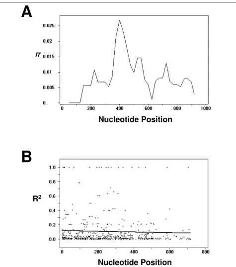
DNA sequence analyses were conducted to determine nucleotide diversity and genetic differentiation at PvDBPII among the Myanmar P. vivax isolates. The average number of pairwise nucleotide differences (K) for the 963 bp of the PvDBPII region was 7.851 (Table 2). The overall haplotype diversity (Hd) and nucleotide diversity (π) for all 54 sequences were 0.875 ± 0.029 and 0.0079 ± 0.0004, respectively (Table 2). Further analysis, with a sliding window plot (window length 100 bp, step size 25 bp) using the DnaSP package, revealed that π diversity ranged from 0 to 0.027. The two highest peaks of nucleotide diversity within the PvDBPII region were identified between nucleotide positions 370-580 (Figure 3A). To determine whether natural selection contributed to generation of this diversity in PvDBPII within the Myanmar P. vivax population, the rate of non-synon-ymous (Kn) to synonymous substitutions (Ks) was esti-mated using the Nei and Gojobori’s method [27], as implemented in the MEGA4 program [25]. The stan-dard error was determined by 1,000 bootstrap replica-tions. The rate of non-synonymous substitutions (Kn) (0.00917) exceeded the rate of synonymous substitutions (Ks) (0.00278). The Kn/Ks ratio was 3.299 (Table 2). These results suggest that positive natural selection may be occurring in PvDBPII and favoring fixed amino acid replacement in certain areas of the protein. The Taji-ma’s D statistics was 0.264 (P > 0.1), indicating a decrease in population size and/or balancing selection. The Fu and Li’s D and F values were 1.867 (P < 0.02) and 1.535 (0.1 > P > 0.05), respectively.
Polymorphisms associated with B- and T-cell epitopes
predicted B- and T-cell epitopes and MHC binding regions were polymorphic, and most had a higher rate of non-synonymous mutations (Kn) than that of synonymous mutations (Ks) with the exceptions being the regions within peptides 13 and Ic (Table 3). High levels of nucleo-tide diversity were particularly found in pepnucleo-tides 48 and Ia, which contained spatially proximate polymorphic resi-dues at positions 417, 419, and 427 and positions 385, 386, 390, and 391, respectively, and they showed a clear posi-tive natural selection signature with posiposi-tive Tajima’s D values. In contrast, three peptides (28, 40 and 54), which did not contain known epitopes [14], had lower nucleotide diversity. These results are consistent with the hypothesis that natural selection acts on epitopes in PvDBPII and is related to diversity of PvDBPII [14,30].
Recombination
The minimum number of recombination events between adjacent polymorphic sites (Rm) was 7, whereas R between adjacent sites (Ra) and per gene (Rb) was 0.0314 and 30.2, respectively. The high value of the recombination para-meters indicated that high meiotic recombination may occur between sites, resulting in genetic diversity in the PvDBPII. The LD index R2for the Myanmar population also declined across the analysed region, suggesting that intragenic recombination may also be contributing the increased diversity at PvDBPII (Figure 3B).
Discussion
Malaria is endemic or hypoendemic in Myanmar and is characterized by the occurrence of all four
human-A
B
F306
L
R308S
Frequenc
y
(
%
)
I3
10L
L33
3F
F344S
K371E
N375D
D384G
E385K
K386N
K386
Q
R390H
R391H
N417K
I4
19M
L42
4I
W
43
7R
K455
I
K473R
P475A
C477G
Q48
6E
R490K
I5
03K
D528G
V533M
K541T
A545V
0
20
40
60
80
100
F
F
F
R
R
R
F
F
S
R
R
F
F
F
S
S
R
F
F
F
R
R
R
Haplotype 1
Haplotype 2
Haplotype 3
Haplotype 4
Haplotype 5
Haplotype 6
Haplotype 7
Haplotype 8
Haplotype 9
Haplotype 10
Haplotype 11
Haplotype 12
306
308
310
333
344
371
375
384
385
386
390
391
417
419
424
437
455
473
475
486
490
503
528
533
541
L
I
I
L
L
L
I
I
L
L
F
I
L
I
F
F
F
I
I
I
F
F
F
I
F
F
F
K
K
K
F
F
K
E
K
F
F
F
K
K
K
F
S
F
E
E
E
F
N
N
N
D
G
G
N
N
G
G
D
D
D
N
G
G
G
N
N
N
G
G
G
N
E
E
E
K
K
K
E
E
K
K
K
K
K
K
N
N
N
K
K
E
N
N
Q
E
R
H
H
R
R
R
R
R
R
H
R
H
H
H
R
R
R
R
R
R
R
R
R
H
N
N
N
I
I
I
N
N
M
I
I
K
K
K
I
I
I
K
K
K
I
I
I
N
L
L
I
W
W
W
I
I
R
R
R
I
I
I
R
R
R
I
I
I
R
R
R
L
K
K
K
K
K
K
K
K
K
K
K
K
K
K
R
K
R
K
K
I
K
K
K
K
P
P
P
P
A
P
P
P
P
P
P
P
Q
E
Q
R
K
R
Q
Q
R
R
R
Q
Q
Q
R
R
R
Q
Q
Q
R
R
R
Q
I
K
D
D
D
K
K
D
D
D
K
K
K
D
D
D
I
I
K
D
D
G
K
V
V
M
K
K
K
V
V
K
K
K
V
V
V
K
K
K
V
V
V
K
K
T
V
A
A
A
2
3
16
A
A
4
2
6
A
A
A
6
2
3
V
A
A
2
5
3
A
Total
F R I L F K N D E K R R N I L W K K P
Q R I D V K
[image:4.595.60.538.87.490.2]Sal I
I
A
545
C
C
C
C
C
C
C
C
C
C
G
C
C
477
I
Figure 1Sequence polymorphism of PvDBPII inP. vivaxMyanmar isolates. (A) Amino acid changes. Polymorphic amino acids are listed for
Iran 5 (ACJ54191)
South Korea 02-13 (AAZ81528) South Korea 97-14 (AAF25489)
South Korea 96-8 (AAF25486) Colombia V6 (AAC47179)
Haplotype 6*
Thailand 51 (ABR14008) Iran 8 (ACJ54194)
Colombia V9 ( AAC47181) Iran 7 (ACJ54193)
India 03-1 (AAZ81527) Sal I (XP_001608387) Iran 14 (ACJ54195)
Haplotype 1
Colombia T1524 (AAC47183) Thailand 3 (ABQ10597)
Colombia T3067 (AAC47187) Thailand 13 (ABR13992)
Iran 6 (ACJ54192)
Haplotype 2*
Colombia 1 (AAZ81526) Thailand 64 (ABR08404)
Haplotype 3*
Thailand 46 (ABR14003)
Haplotype 4*
PNG 7 (AAG53620) PNG 23 (AAG53624) Thailand 300 (ABR08410)
PNG 183 (AAG53627) PNG 185 (AAG30850)
PNG 194 (AAG53626) India CH51 (ACN69883) Iran 3 (ACJ54189)
Bangladesh 01-8 (ABA39301) Indonesia 19 (ACB42429)
South Korea 02-25 (AAZ81536)
Haplotype 5*
India ND38 (ACN69931)
Haplotype 12*
Iran 4 (ACJ54190)
South Korea 97-11 (AAF25487) South Korea 96-50 (AAF25483) South Korea 97-13 (AAF25488) Colombia V5 (AAC47178)
India 7 (ACB42428) Indonesia 7 (AAZ81534) Iran 1 (ACJ54187) Iran 2 (ACJ54188)
Brazil 1 (ACB42426)
Haplotype 10*
Haplotype 11*
Thailand 41 (ABR13999) India NE39 (ACN69947) Belem (AAC47191)
Colombia V4 (AAC47177) India M44 (ACN69910)
Belem (ACB42425) Honduras 3 (AAZ81530)
India M11 (ACN69899)
India ND2 (ACN69916) Vietnam 4 (AAZ81531)
Haplotype 9*
Thailand 43 (ABR14000) Thailand 27 (ABR13997)
Haplotype 7*
Palo Alto 1 (ACD76802)
Haplotype 8
Palo Alto 6 (ACD76807) Palo Alto 3 (ACD76804) Palo Alto 2 (ACD76803) Palo Alto 4 (ACD76805) Palo Alto 7 (ACD76808)
69
64 86
65 44
27 13 15 33 40 41 22
85
93
35 50 29 75
73 55
40 60
56
66
17 9
14 12
6
5 35
53
65 57
4
14
29 7
24
16 49 40
40
0 13 15
0 4
56
3
0
23 32 21
27
16
3
1
4
1
39 22 0
16 0
39
[image:5.595.53.552.89.692.2]0.005
Figure 2Phylogenetic analysis. The phylogenetic tree for the 12 haplotypes of PvDBPII was constructed with a neighbor-joining method
infecting Plasmodiumspecies [32]. Although morbidity and mortality rates due to malaria have been declining gradually in recent years, Myanmar still contributes to approximately 60% of malaria deaths in the Southeast Asia [32]. Genetic polymorphisms in the circumsporo-zoite protein, merocircumsporo-zoite surface protein-1 (MSP-1), MSP-3a, and apical membrane antigen-1 (AMA-1) of P. vivax Myanmar isolates were analysed previously [24,33,34]. As expected, they showed high levels of genetic polymorphisms, but information on the nature and extent of population diversity within malaria para-sites in Myanmar is still limited. In this study, genetic polymorphism and natural selection of PvDBPII in MyanmarP. vivaxisolates were analysed to expand our knowledge on population diversity of the parasite in the country.
A total of 54 PvDBPII sequences were obtained from MyanmarP. vivaxisolates. The sequence analysis revealed that the 54 sequences were classified into 12 different hap-lotypes. A GenBank search for each haplotype revealed that two haplotypes were identical to at least one pre-viously reported PvDBPII sequence found in other regions of the world, whereas the other 10 haplotypes were novel and have not been reported so far. The sequence analysis revealed that a total of 32 point mutations was identified, which resulted in significant amino acid changes (28 non-synonymous and four non-synonymous changes) through the PvDBPII in Myanmar isolates. Seventeen of the 28 non-synonymous changes were previously reported, whereas the other 11 changes (I310L, F344S, R391H, K455I, K473R, C477G, R490K, D528G, V533M, K541T, and A545V) were unique to Myanmar isolates, which did not
hitherto identified. The two highest peaks of nucleotide diversity within PvDBPII in the MyanmarP. vivaxisolates were identified between C5 and C7, which was consistent with previous observations [14,15,17]. Interestingly, a unique amino acid change (C477G caused by TGT to GGT) was identified in three Myanmar isolates. It is well known that the cysteine residues within PvDBPII are well conserved within and betweenP. vivaxpopulations from different geographic regions studies [14-21,31]. It is not certain the nature of this amino acid change. It can be assumed that this mutation may resulted from a PCR or sequencing error, but to eliminate any possible errors, high quality Taq polymerase with a proof-reading function was used in all amplification processes and sequencing reactions were performed on at least two individual clones for each gene in both directions. But, considering the small number of isolates used in this study, further ana-lyses with a large number of isolates is necessary to con-firm this change in the MyanmarP. vivaxisolates. Taken together, the results of this study suggest that PvDBPII in the Myanmar isolates showed a high level of genetic poly-morphism with a nucleotide diversity (π) of 0.0079 ± 0.0004.
[image:6.595.54.540.649.712.2]Some polymorphic residues in PvDBPII occurred in only one population or geographic region, but common variant amino acids (K371E, D384G, E385K, K386N, N417K, L424I, W437R and I503K) are found in global isolates [19-21,31]. A high frequency (> 50%) of D384G (85.2%), R390H (63.0%), L424I (83.3%), W437R (61.1%), and I503K (77.7%) residues were found in Myanmar isolates com-pared to that in the Sal I sequence. This was highly similar to a report on Thailand isolates (D384G, 76.7%; R390H, Table 1 Frequencies of the most common variant amino acids in PvDBPII
F306L R308S D384G K386N K386Q N417K L424I W437R S447K I503K
Myanmar 7.4 22.2 85.2 33.3 5.6 38.9 83.3 61.1 0 77.8
Thailanda 6.7 26.7 76.7 40.0 3.0 36.6 86.7 63.3 0 56.7
Iranb 5.3 6.6 61.3 6.6 0 44.0 50.6 45.3 0 70.6
Sri Lankac 7.0 13.0 94.0 20.0 0 36.0 49.0 37.0 0 55.0
Papua New Guinead 0 67.0 66.0 8.0 11.0 23.0 34.0 26.0 59.0 29.0
Colombiad 0 0 59.0 23.0 0 47.0 47.0 18.0 0 12.0
Brazild 0 12.5 85.0 12.5 0 27.5 32.5 27.5 0 55.0
a
[20];b
[21];c
[31];d
[19]
The first letter represents the amino acid in that position in Sal I sequence, and the other letter represents the substituted amino acid
Table 2 Estimates of DNA sequence polymorphism and tests of neutrality at PvDBPII among Myanmar isolates
Total no.
of isolates
Segregating sites (S)
Singleton variable sites
Parsimony informative sites
Total no. of mutations
K H Hd ± SD π± SD Kn Ks Tajima’s
54 32 0 32 32 7.581 12 0.875 ±
0.029
0.0079 ± 0.0004
0.00917 0.00278 0.264 (P > 0.1)
56.7%; L424I, 86.7%; W437R, 63.3%; I503K, 56.7%) [20] but differed from previous studies showing R308S (67%), D384G (66%), and S447K (59%) in Papua New Guinean isolates; D384G (59%) in Colombian isolates; D384G
(85%) and I503K (55%) in Brazilian isolates; D384G (61.3%) and I503K (70.6%) in Iranian isolates; and D384G (94%) and I503K (55%) in Sri Lankan isolates [19,21,31]. F306L, which has only been reported from Asian malaria
Nucleotide Position
˭
˭
˭
˭
A
R
2
B
[image:7.595.61.537.85.623.2]Nucleotide Position
Figure 3Natural selection of PvDBPII. (A) Sliding window plot of nucleotide diversity per site (π) comparing the level of genetic diversity at
Table 3 Polymorphism observed in the each epitope sequence Epitope name Epitopa Segregating sites (S) Singleto
n variable sites
Parsimony informative sites
Total no. of mutations
K H Hd ± SD π± SD Kn Ks Tajima’s D
5 T/B 3 0 3 3 0.565 4 0.383 ± 0.079 0.013 ± 0.003 0.016 0 -0.289 (P > 0.1)
13 T 0 0 0 0 0 1 0 0 0 0 0
16 T/B 1 0 1 1 0.509 2 0.509 ± 0.013 0.011 ± 0.000 0.015 0 1.741 (P > 0.05)
18 B 1 0 1 1 0.509 2 0.509 ± 0.013 0.011 ± 0.000 0.015 0 1.741 (P > 0.05)
20 T/B 1 0 1 1 0.509 2 0.509 ± 0.013 0.011 ± 0.000 0.014 0 1.741 (P > 0.05)
28 None 0 0 0 0 0 1 0 0 0 0 0
40 None 3 0 3 3 0.867 3 0.542 ± 0.059 0.019 ± 0.003 0.018 0.025 0.642 (P > 0.1)
48 B 5 0 5 5 1.560 6 0.748 ± 0.040 0.035 ± 0.003 0.041 0 1.007 (P > 0.1)
54 None 0 0 0 0 0 1 0 0 0 0 0
66 T 1 0 1 1 0.283 2 0.283 ± 0.068 0.006 ± 0.002 0.008 0 0.382 (P > 0.1)
78 B 1 0 1 1 0.107 2 0.107 ± 0.055 0.002 ± 0.001 0.003 0 -0.675 (P > 0.1)
Ia MHCIa 3 0 3 3 0.907 4 0.649 ± 0.037 0.034 ± 0.004 0.040 0 0.767 (P > 0.1)
Ib MHCIb 2 0 2 2 0.214 2 0.107 ± 0.055 0.008 ± 0.004 0.010 0 -0.898 (P > 0.1)
Ic MHCIc 2 0 2 2 0.492 3 0.458 ± 0.066 0.012 ± 0.002 0.011 0.017 0.212 (P > 0.1)
IIa MHCIIa 2 0 2 2 0.557 3 0.511 ± 0.043 0.019 ± 0.002 0.021 0.011 0.471 (P > 0.1)
IIb MHCIIb 2 0 2 2 0.423 3 0.297 ± 0.076 0.016 ± 0.004 0.019 0 -0.064 (P > 0.1)
K, average number of pairwise nucleotide differences; H, number of haplotypes; Hd, haplotype diversity;π, observed average pairwise nucleotide diversity;Kn, rate of non-synonymous mutations;Ks, rate of synonymous mutations.a
B, B-cell epitope; T, T-cell epitope; none, no epitope identified
Journal
2012,
11
:60
al.com/conten
t/11/1/60
Page
8
of
endemic areas, including Thailand [20], Iran [21], and Sri Lanka [31], was also identified in the Myanmar isolates. Although the Myanmar isolates showed similar amino acid changes compared to those in the Thailand isolates, 9 variants found in the Thailand isolates (R268S, S351C, I367T, S398T, T404R, Q433K, R436T, N507H, and T513K) were not identified in the Myanmar isolates. Meanwhile, 11 variations (I310L, F344S, R391H, K455I, K473R, C477G, R490K, D528G, V533M, K541T, and A545V) found in the Myanmar isolates did not occur in the Thailand isolates. These results indicate that the Myan-mar isolates are different from the Thailand isolates even though the two countries are very close geographically.
Although polymorphic residues were widely distributed throughout the PvDBPII sequence, polymorphisms at resi-dues 417, 437, and 503, either in single or in combination, can affect the efficacy of inhibitory antibodies against ery-throcyte binding [23,35]. As these residues compose an important discontinuous epitope in PvDBP, which might be the main target for inhibitory antibodies, these poly-morphisms could be subject to immune pressure responsi-ble for parasite escape from the host immune system. It has been confirmed that this strong positive selection pressure in PvDBPII promotes greater diversity [14,30]. The immune pressure drives the generation of new PvDBP variants that are still able to bind erythrocytes but become resistant to inhibitory antibodies, suggesting that this DBP region is under positive pressure at critical resi-dues and under negative pressure at the resiresi-dues involved in receptor recognition [22,23,35]. A low prevalence of variant N417K (38.9%) was observed among Myanmar iso-lates, but more than 50% of W437R (61.1%) and I503K (77.8%) were identified. Analyses of the combination of variants revealed that W437R-I503K occurred at a higher frequency (70.4%), whereas W437K and N417K-I503K occurred at frequencies of 38.9% and 25.9%, respec-tively. This result suggests that there is a strong associa-tion between W437R-I503K in PvDBPII in Myanmar P. vivaxisolates, but not between N417K with either W437R or I503K.
The rate of non-synonymous mutations (Kn) and that of synonymous mutations (Ks) is generally used as an indica-tor of the action of natural selection in most coding gene sequences [27]. Negative selection acting on coding genes can usually be identified when non-synonymous mutations are not advantageous, so the rate of synonymous muta-tions surpasses that of non-synonymous mutamuta-tions (Ks >Kn). Meanwhile, positive selection is acting on a gene, when non-synonymous mutations can be beneficial (e.g. to avoid the host immune response), and the rate of non-synonymous mutations exceed that of non-synonymous muta-tions (Kn> Ks). Previous studies on PvDBPII diversity indicate that the high rate of non-synonymous mutations (Kn) relative to that of synonymous mutations (Ks) reflects
positive selection pressure [14,31,36]. The positive value of Kn/Ks (3.299) for all 54 sequences suggest that PvDBPII in MyanmarP. vivaxisolates is under positive natural selec-tion. The positive values of Tajima’s D (0.2635, P > 0.10) and Fu and Li’s D (1.867, P < 0.02) and F statistics (1.535, 0.1 > P > 0.05) indicate that PvDBPII alleles occur at more intermediate frequencies than expected and that few alleles are rare or near fixation, which is consistent with the action of balancing selection, which maintains allelic variation in a population. These results collectively sug-gested that strong balancing selection, probably by host immune selection pressure, occurs at PvDBPII in the Myanmar isolates.
Polymorphism in B- and T-cell epitopes of parasite anti-gens may well enable parasites to escape host immune responses, as a polymorphism in the epitopes can up or down regulate T-cell responses to the index peptide or completely arrest an immune response, assisting escape of the parasite from the host immune system [37]. The high degree of nucleotide diversity and high rates of non-synonymous to non-synonymous mutations are observed in known or predicted B- and T-cell epitopes and MHC binding regions of PvDBPII in the Myanmar isolates. Overall nucleotide diversity values for these epitopes and regions were greater than those for the entire PvDBPII. In particular, high levels of nucleotide diversity were identi-fied in peptides 48 and Ia, which is comparable to Brazi-lian isolates [30]. Positive Tajima’s D values for these epitopes also suggested that positive natural selection pre-ferentially acted on the epitopes in PvDBPII in the Myan-mar isolates. These epitopes are predicted to be exposed to the surface of the PvDBP molecule [30]. The putative changes in protein structure may alter antibody binding efficacy of a particular epitope, thereby allowing escape from the host protective immune response [13,30].
Many factors may contribute to genetic diversity in malaria populations, including mutations, intragenic recombination, natural selection, gene flow between differ-ent regions, and population size. Although it remains con-troversial, it has been suggested that recombination also contributes to the diversity of PvDBPII [30,36]. The exis-tence of recombination events and the decline in the LD with increasing distance between nucleotide sites suggest that in addition to natural selection meiotic recombination may also contribute to maintain the diversity of PvDBPII among MyanmarP. vivaxisolates, as reported previously in Brazil, Colombia, and Sri Lanka [30,31].
Conclusion
recombination events maintained the diversity in the form of balancing selection. However, further studies using a larger number of isolates collected from different geogra-phical regions in Myanmar will be helpful to reveal the nationwide parasite heterogeneity and the implementation of malarial control programmes in Myanmar.
Acknowledgements
We are grateful to all blood donors and the staffs at Department of Medical Research (Upper Myanmar). This work was supported by a grant of the Korea Centers for Disease Control and Prevention (2009-E54004-00) and the National Research Foundation of Korea (NRF) grant funded by the Korea government (MEST) (2011-0028135).
Author details
1Department of Parasitology and Institute of Health Sciences, Gyeongsang
National University School of Medicine, Jinju 660-751, Korea.2Department of Anatomy, Yonsei University College of Medicine, Seoul 120-752, Korea. 3
Division of Malaria and Parasitic Diseases, National Institute of Health, Korea Centers for Disease Control and Prevention, Osong 122-701, Korea. 4
Department of Pathology, University of Florida, J-566, 1600 SW Archer Road, Gainesville, FL 32610, USA.5Department of Health, Vector Borne Diseases Control Project, 36 Theinbyu Road, Mandalay, Myanmar.6Department of Internal Medicine and Inha Research Institute for Medical Sciences, Inha University School of Medicine, Incheon 400-712, Korea.7Department of Parasitology and Inha Research Institute for Medical Sciences, Inha University School of Medicine, Incheon 400-712, Korea.
Authors’contributions
HLJ and JMK performed all the experiments and analysed the sequence data. SUM, JYK, HWL, KL, and BKN collected the blood samples. SUM performed sequence and phylogenetic analyses. BKN and TSK designed the study and supervised the study process. BKN wrote the paper. TSK, WMS, and JSL assisted in writing and editing the manuscript. All authors read and approved the final manuscript.
Competing interests
The authors declare that they have no competing interests.
Received: 24 October 2011 Accepted: 1 March 2012 Published: 1 March 2012
References
1. Adams JH, Sim BK, Dolan SA, Fang X, Kaslow DC, Miller LH:A family of erythrocyte binding proteins of malaria parasites.Proc Natl Acad Sci USA
1992,89:7085-7089.
2. Barnwell JW, Nichols ME, Rubinstein P:In vitro evaluation of the role of the Duffy blood group in erythrocyte invasion byPlasmodium vivax.
J Exp Med1989,169:1795-1802.
3. Wertheimer SP, Barnwell JW:Plasmodium vivaxinteraction with the human Duffy blood group glycoprotein: identification of a parasite receptor-like protein.Exp Parasitol1989,69:340-350.
4. Horuk R, Chitnis C, Darbonne W, Colby T, Rybicki A, Hadley T, Miller L:A receptor for the malarial parasitePlasmodium vivax: the erythrocyte chemokine receptor.Science1993,261:1182-1184.
5. Michon PA, Arevalo-Herrera M, Fraser T, Herrera S, Adams JH:Serologic responses to recombinantPlasmodium vivaxDuffy binding protein in a Colombian village.Am J Trop Med Hyg1998,59:597-599.
6. Xainli J, Baisor M, Kastens W, Bockarie M, Adams JH, King CL: Age-dependent cellular immune responses toPlasmodium vivaxDuffy binding protein in humans.J Immunol2002,169:3200-3207. 7. Michon P, Fraser T, Adams J:Naturally acquired and vaccine-elicited
antibodies block erythrocyte cytoadherence of thePlasmodium vivax
Duffy binding protein.Infect Immun2000,68:3164-3171. 8. Grimberg B, Udomsangpetch R, Xainli J, McHenry A, Panichakul T,
Sattabongkot J, Cui L, Bockarie M, Chitnis C, Adams J, Zimmerman PA, King CL:Plasmodium vivaxinvasion of human erythrocytes inhibited by
antibodies directed against the Duffy binding protein.PLoS Med2007,4: e337.
9. Cerávolo I, Souza-Silva F, Fontes C, Braga E, Madureira A, Krettli A, Souza J, Brito C, Adams J, Carvalho L:Inhibitory properties of the antibody response toPlasmodium vivaxDuffy binding protein in an area with unstable malaria transmission.Scand J Immunol2008,67:270-278. 10. Chitnis CE, Miller LH:Identification of the erythrocytes binding domains
ofPlasmodiumvivaxandPlasmodium knowlesiproteins involved in erythrocyte invasion.J Exp Med1994,180:497-506.
11. Chitnis CE, Chaudhuri A, Horuk R, Pogo AO, Miller LH:The domain on the Duffy blood group antigen for binding.J Exp Med1996,184:1531-1536. 12. Ranjan A, Chitnis CE:Mapping regions containing binding residues within
functional domains ofPlasmodium vivaxandPlasmodium knowlesi
erythrocyte-binding proteins.Proc Natl Acad Sci USA1999,96:14067-14072. 13. Cole-Tobian J, Cortes A, Baisor M, Kastens W, Xainli J, Bockarie M, Adams JH, King CL:Age-acquired immunity to aPlasmodium vivaxinvasion ligand, the Duffy binding protein.J Infect Dis2002,186:531-539.
14. Cole-Tobian J, King CL:Diversity and natural selection inPlasmodium vivaxDuffy binding protein gene.Mol Biochem Parasitol2003, 127:121-132.
15. Tsuboi T, Kappe SHI, Alyaman F, Prickett MD, Alpers M, Adams JH:Natural variation within the principal adhesion domain of thePlasmodium vivax
Duffy binding-protein.Infect Immun1994,62:5581-5586.
16. Ampudia E, Patarroyo M, Patarroyo M, Murillo L:Genetic polymorphism of the Duffy receptor binding domain ofPlasmodium vivaxin Colombian wild isolates.Mol Biochem Parasitol1996,78:269-272.
17. Xainli J, Adams J, King C:The erythrocyte binding motif ofPlasmodium vivaxduffy binding protein is highly polymorphic and functionally conserved in isolates from Papua New Guinea.Mol Biochem Parasitol
2000,111:253-260.
18. Kho W, Chung J, Sim E, Kim D, Chung W:Analysis of polymorphic regions ofPlasmodium vivaxDuffy binding protein of Korean isolates.Korean J Parasitol2001,39:143-150.
19. Sousa T, Cerávolo I, Fernandes Fontes C, Couto A, Carvalho L, Brito C:The pattern of major polymorphisms in the Duffy binding protein ligand domain amongPlasmodium vivaxisolates from the Brazilian Amazon area.Mol Biochem Parasitol2006,146:251-254.
20. Gosi P, Khusmith S, Khalambaheti T, Lanar D, Schaecher K, Fukuda M, Miller S:Polymorphism patterns in Duffy-binding protein among Thai
Plasmodium vivaxisolates.Malar J2008,7:112.
21. Babaeekho L, Zakeri S, Djadid ND:Genetic mapping of the duffy binding protein (DBP) ligand domain ofPlasmodium vivaxfrom unstable malaria region in the middle east.AmJTrop Med Hyg2009,80:112-118.
22. VanBuskirk K, Sevova E, Adams J:Conserved residues in thePlasmodium vivaxDuffy-binding protein ligand domain are critical for erythrocyte receptor recognition.Proc Natl Acad Sci USA2004,101:15754-15759. 23. VanBuskirk KM, Cole-Tobian JL, Baisor M, Sevova ES, Bockarie M, King CL,
Adams JH:Antigenic drift in the ligand domain ofPlasmodium vivax
Duffy binding protein confers resistance to inhibitory antibodies.J Infect Dis2004,190:1556-1562.
24. Moon SU, Na BK, Kang JM, Kim JY, Cho SH, Park YK, Sohn WM, Lin K, Kim TS:Genetic polymorphism and effect of natural selection at domain I of apical membrane antigen-1 (AMA-1) inPlasmodium vivaxisolates from Myanmar.Acta Trop2010,114:71-75.
25. Tamura K, Dudley J, Nei M, Kumar S:MEGA4: Molecular Evolutionary Genetics Analysis (MEGA) software version 4.0.Mol Biol Evol2007, 24:1596-1599.
26. Librado P, Rozas J:DnaSP v5: a software for comprehensive analysis of DNA polymorphism data.Bioinformatics2009,25:1451-1452.
27. Nei M, Gojobori T:Simple methods for estimating the numbers of synonymous and nonsynonymous nucleotide substitutions.Mol Biol Evol
1986,3:418-426.
28. Tajima F:Statistical method for testing the neutral mutation hypothesis by DNA polymorphism.Genetics1989,123:585-595.
29. Fu YX, Li WH:Statistical tests of neutrality of mutations.Genetics1993, 133:693-709.
31. Premaratne PH, Aravinda BR, Escalante AA, Udagama PV:Genetic diversity ofPlasmodium vivaxDuffy Binding Protein II (PvDBPII) under unstable transmission and low intensity malaria in Sri Lanka.Infect Genet Evol
2011,11:1327-1339.
32. Lin K:Malaria control in Myanmar.InAsian Parasitology, Malaria in Asia.
Edited by: Kano S, Tongol-Rivera P. The federation of Asian Parasitologists; 2005:123-134.
33. Moon SU, Lee HW, Kim JY, Na BK, Cho SH, Lin K, Sohn WM, Kim TS:High frequency of genetic diversity ofPlasmodium vivaxfield isolates in Myanmar.Acta Trop2009,109:30-36.
34. Kim TS, Kim HH, Lee SS, Na BK, Lin K, Cho SH, Kang YJ, Kim DK, Sohn Y, Kim H, Lee HW:Prevalence ofPlasmodium vivaxVK210 and VK247 subtype in Myanmar.Malar J2010,9:195.
35. Hans D, Pattnaik P, Bhattacharyya A, Shakri A, Yazdani S, Sharma M, Choe H, Farzan M, Chitnis C:Mapping binding residues in thePlasmodium vivax
domain that binds Duffy antigen during red cell invasion.Mol Microbiol
2005,55:1423-1434.
36. Martinez P, Suarezi CF, Cardenas PP, Patarroyo MA:Plasmodium vivaxDuffy binding protein: a modular evolutionary proposal.Parasitology2004, 128:353-366.
37. Tanabe K, Escalante A, Sakihama N, Honda M, Arisue N, Horii T, Culleton R, Hayakawa T, Hashimoto T, Longacre S, Pathirana S, Handunnetti S, Kishino H:Recent independent evolution of msp1 polymorphism in
Plasmodium vivaxand related simian malaria parasites.Mol Biochem Parasitol2007,156:74-79.
doi:10.1186/1475-2875-11-60
Cite this article as:Juet al.:Genetic polymorphism and natural selection of Duffy binding protein ofPlasmodium vivaxMyanmar isolates.Malaria Journal201211:60.
Submit your next manuscript to BioMed Central and take full advantage of:
• Convenient online submission
• Thorough peer review
• No space constraints or color figure charges
• Immediate publication on acceptance
• Inclusion in PubMed, CAS, Scopus and Google Scholar
• Research which is freely available for redistribution