RESEARCH
Molecular monitoring of the diversity
of human pathogenic malaria species in blood
donations on Bioko Island, Equatorial Guinea
Tobias Schindler
1,2*, Tamy Robaina
3, Julian Sax
1,2, Jose Raso Bieri
4, Maximilian Mpina
1,2,5, Linda Gondwe
1,2,5,
Ludmila Acuche
4, Guillermo Garcia
6, Carlos Cortes
6, Carl Maas
7and Claudia Daubenberger
1,2*Abstract
Background: Malaria can be transmitted by blood transfusion from human to human and it is responsible for the majority of transfusion-transmitted infectious diseases worldwide. In sub-Saharan Africa, it had been estimated that almost a quarter of blood donations contain malaria parasites. Since rapid diagnostic tests and thick blood smear microscopy lack sensitivity for low density parasitaemia, particularly in asymptomatic adults, the most reliable method to assess the problem of transfusion-transmitted malaria are nucleic acid-based molecular approaches such as quanti-tative polymerase chain reaction. The study was undertaken to determine the prevalence of sub-microscopic malaria parasite infection among blood donors in Malabo, Equatorial Guinea.
Methods: Between July and August 2017, a total of 200 individual blood samples from blood donors at the Malabo Blood Bank were collected and screened by rapid diagnostic tests and thick blood smear microscopy. Retrospectively, the same samples were analysed for the presence of undetected, low-density malaria parasites using quantitative polymerase chain reaction.
Results: In comparison to 6.5% (13/200) by rapid diagnostic test and 2.0% (4/200) by microscopy, the proportion of
Plasmodium falciparum positive blood donations analysed by quantitative polymerase chain reaction was significantly higher (26%, 52/200). Densities of P. falciparum positive blood donations were ranging from 0.06 to 3707.0 parasites/ µL with 79.6% below 100 parasites/µL and therefore not detectable by non-molecular malaria diagnostic tests. qPCR based species identification revealed that P. falciparum was the dominating species responsible for 88.1% (52/59) of positive blood donations, followed by Plasmodium malariae (15.3%, 9/59) and Plasmodium ovale (3.4%, 2/59). Conclusions: This study confirms that in malaria endemic settings, sub-patent malaria infections among blood donors are prevalent. In blood collected from healthy donors living in Malabo, P. falciparum, P. malariae and P. ovale
parasites were identified. Currently widely used malaria diagnostic tools have missed more than 75% of P. falciparum
containing blood donations, demonstrating the value of quantitative polymerase chain reaction to reliably detect low density P. falciparum infections. Since the availability of molecular diagnostic methods in malaria endemic countries is still limited, the blood recipients living in malaria endemic countries should be treated following WHO recommendations.
Keywords: Transfusion-transmitted malaria, P. falciparum, P. malariae, P. ovale, qPCR
© The Author(s) 2019. This article is distributed under the terms of the Creative Commons Attribution 4.0 International License (http://creat iveco mmons .org/licen ses/by/4.0/), which permits unrestricted use, distribution, and reproduction in any medium, provided you give appropriate credit to the original author(s) and the source, provide a link to the Creative Commons license, and indicate if changes were made. The Creative Commons Public Domain Dedication waiver (http://creat iveco mmons .org/ publi cdoma in/zero/1.0/) applies to the data made available in this article, unless otherwise stated.
Open Access
*Correspondence: tobias.schindler@unibas.ch; claudia.daubenberger@swisstph.ch
1 Department of Medical Parasitology and Infection Biology, Swiss Tropical and Public Health Institute, Basel, Switzerland
Background
Malaria is a vector-borne parasitic tropical disease found in 55 countries globally [1]. Six Plasmodium species are known to infect humans including Plasmodium
falcipa-rum, Plasmodium vivax, Plasmodium malariae,
Plas-modium ovale curtisi, Plasmodium ovale wallikeri and
Plasmodium knowlesi [2]. Malaria is transmitted to the
human host through the bite of a female Anopheles mos-quito when the sporozoite stage is inoculated during the feeding. The parasite undergoes a pre-erythrocytic liver stage reproduction that is clinically silent and lasts 1 to 2 weeks before the release of merozoites into the blood-stream. These merozoites rapidly infect red blood cells, starting the asexual blood stage whereby the parasite undergoes cycles of asexual replication that include the ring, trophozoite, and schizont stages leading to regular fever episodes [2]. In adult human populations living in malaria endemic countries, the prevalence of sub-patent malaria infections is up to 80% [3] with certain parasite clones surviving in the human host more than 300 days [4].
Diagnosis of malaria is performed in blood samples collected from potentially infected humans. The gold standard for malaria diagnosis remains light microscopy of stained blood smears. Thick blood smears provide sensitivity and thin smears allow the identification of dif-ferent malaria species and the quantitation of infection [5]. Rapid diagnostic tests (RDTs) are widely used since they offer less dependence on the availability of labora-tory infrastructure, and can also be employed by inex-perienced health workers [6]. RDTs are often based on parasite-derived histidine-rich protein 2 (PfHRP2) for sensitive and specific detection of P. falciparum, and lac-tate dehydrogenase enzyme (LDH) or aldolase for a Pan
-Plasmodium detection of all human infective malaria
species [7]. The lower limit of detection (LOD) for microscopy is between 50 and 500 parasites/µL depend-ing on the expertise of the microscopist [5] and 100 para-site/µL for the PfHRP2 based P. falciparum RDTs [8]. Differential diagnosis of P. ovale, P. malariae, P. vivax and
P. knowlesi is hampered by the low density blood stage
infections and the difficulty in distinguishing these spe-cies based on morphology of trophozoites using micros-copy [9].
Nucleic acid amplification technologies (NATs) are more expensive and require advanced laboratory infra-structure but provide much better sensitivity. The parasites’ multi-copy 18S small subunit ribosomal deox-yribonucleic acid (rDNA) and/or its highly expressed ribonucleic acid (rRNA) is the most frequently used bio-marker for NATs [10]. Different methods for 18S rDNA/ rRNA detection are currently in use with a great varia-tion in sensitivity. Qualitative methods targeting 18S
rDNA, such as nested polymerase chain reaction (PCR) [11] and LAMP [12], reach LODs of 0.1–10 parasites/µL. The use of quantitative PCR (qPCR) [13], does not only allow absolute quantification of infections but also lowers the LOD to less than 1 parasite/µL. The use of Reverse Transcription quantitative PCR (RT-qPCR), amplifying total nucleic acids (RNA and DNA) of the 18S genes [14– 16], and qPCR assays targeting alternative multi-copy genomic sequences [17] further increase sensitivities of NATs. Apart from increased sensitivity of NATs, molecu-lar detection using PCR has improved species discrimi-nation compared to either microscopy or RDTs [18].
Screening for transfusion transmitted infections should follow the World Health Organization (WHO) recom-mendations which include searching for chronic infec-tious diseases including HIV-1, HIV-2, Hepatitis B, Hepatitis C and Treponema pallidum (syphillis) [19]. Based on the local epidemiological infectious disease situation, the WHO recommends that blood donations should also be screened for malaria [19, 20]. Malaria can be efficiently transmitted by blood transfusion from human to human and is undoubtedly responsible for the majority of transfusion transmitted diseases in the world [21]. The rate of transfusion-transmitted malaria (TTM) in malaria endemic sub-Saharan regions is estimated between 14 and 28% [22]. Several reports describe that P. falciparum [22], P. vivax [23], P. malariae [24], P. knowlesi
[25] and P. ovale [26] can be transmitted either through blood donations or solid organ transplantations.
Malaria species circulating in Equatorial Guinea include P. falciparum, P. malariae, P. ovale and P. vivax
[27–29]. The Bioko Island Malaria Control Project (BIMCP) started in 2004 with the aim to reduce malaria transmission and to control the burden of disease and has received substantial funding from the Government of Equatorial Guinea and private donors including Mara-thon EG Production Limited, Noble Energy, and Atlan-tic Methanol Production Company. The malaria control strategy of the BIMCP rests on vigorous vector control measures, effective case management, prevention of malaria during pregnancy, behavioural change commu-nications and regular monitoring and evaluation [30]. The BIMCP has had significant success in reducing P.
fal-ciparum parasite prevalence in the 2–14 years old
chil-dren and adolescent from 45% (95% CI 40–50%) in 2004 (baseline) to 12.1% (95% CI 11.2–13.3%) in 2016 based on annual Malaria Indicator Survey (MIS) data [30].
infections that cannot be detected by commonly used RDTs and microscopy. In addition, the presence of other TTM causing Plasmodium species was analysed using a newly developed single tube multiplex qPCR assay.
Methods
Study site and participant recruitment
The study was conducted at the Central Blood Bank in Malabo on Bioko Island. Bioko Island, located in West-central Africa and home of Equatorial Guinea’s capi-tal city Malabo, is 32 km from the coast of Cameroon. The approximately 250,000 people, who mainly reside in Malabo are at risk of malaria year-round [30]. Adults willing to donate blood and who are Hepatitis B, Hepa-titis C, and HIV negative were eligible for blood dona-tion. Microscopy and RDT (CareStart™ Malaria HRP2/ pLDH Pf/PAN Combo) were used for malaria screen-ing at the blood bank and the results were kept at the blood bank until the end of qPCR analysis. The Malabo Central Blood Bank processes around 100 donors per month; the majority being donors assisting friends and families during emergency situations, and the remain-der are donors that voluntarily donate blood on a regular basis. The blood bank is run by a public–private coopera-tion between the Ministry of Health and Social Welfare, the University of Valencia Hospital, and funded by the AGEM-GUINEA company.
Sample collection for molecular diagnostics
Between July and August 2017, a total of 200 individual blood donations from routine visitors were selected to conduct this study. 1 mL of whole blood was collected in EDTA tubes as part of the usual blood donation pro-cedures in the blood bank. The blood was immediately used for microscopy and RDT, with the remaining blood immediately stored at − 20 °C. The samples were trans-ported in a cooling box to the laboratory of the Equa-torial Guinea Malaria Vaccine Initiative (EGMVI) in Malabo. The laboratory infrastructure of the EGMVI, located on the premises of the La Paz Hospital in Malabo, which conducted the first clinical trial in the history of the country [31], was used for qPCR analysis of the blood donations.
The 200 blood samples, from healthy, Hepatitis B, Hepatitis C, and HIV-1 and HIV-2 negative blood donors were analysed for the presence of possibly undetected, low-level malaria parasites using high-sensitive qPCR assays. DNA extraction was done manually from 180 µL whole blood using Quick-DNA Miniprep kits (Zymo Research, Irvine, USA) following manufactures’ guide-lines and DNA was eluted with 50 µL Elution Buffer. DNA samples were kept at − 20 °C prior to qPCR analysis.
qPCR for Plasmodium screening and quantification (PlasQ assay)
Plasmodium spp. and P. falciparum parasites were
quan-tified using the PlasQ assay. This assay consists of two independent Plasmodium targets combined in a multi-plex assay. The Pan-Plasmodium 18S rDNA sequence (Pspp18S) [13, 32], and the P. falciparum-specific acidic terminal sequence of the var genes (PfvarATS) [17] were used for detection and quantification of parasites. The human RNaseP (HsRNaseP) gene served as an internal control to assess the quality of DNA extraction and qPCR amplification.
Amplification and qPCR measurements were per-formed using the Bio-Rad CFX96 Real-Time PCR System (Bio-Rad Laboratories, California, USA). The thermal profile used for PlasQ qPCR is as follows: 60 s at 95 °C; 45 cycles of 15 s at 95 °C and 45 s at 57 °C. Each reac-tion contained 2 µL DNA and 8 µL reacreac-tion master mix containing 1× Luna Universal Probe qPCR Master Mix (New England Biolabs, Ipswich, USA) and 1× PlasQ Primer Mix (Additional file 1). All qPCR assays were run in duplicates with appropriate controls including Non-Template Control [NTC] and P. falciparum 3D7 DNA as positive control [PC]. The mean Cq of the two repli-cates was reported and in case of one qPCR replicate interpreted as malaria positive and the other replicate negative, then the assay had to be repeated to arrive at an unequivocal result.
Using the first WHO International Standard for P.
fal-ciparum DNA Nucleic Acid Amplification Techniques
(NIBSC code: 04/176), a serial dilution ranging from 0.05 to 10,000 parasites/µL was prepared and used for quan-tification of P. falciparum. The WHO standards were run as duplicates in two out of the total 13 qPCR runs conducted.
qPCR for Plasmodium species identification (PlasID assay)
Several published [17, 33–37] and unpublished qPCR assays, detecting P. falciparum, P. malariae, P. ovale, P.
vivax and P. knowlesi were evaluated and the best
per-forming primer and probes for each Plasmodium spe-cies were combined into a new multiplex assay (PlasID). The primers and probe combinations used for this novel pentaplex malaria qPCR assay is provided in Additional file 1.
(Solis Bio Dyne, Tartu, Estonia) and 1× PlasID Primer Mix (Additional file 1). All qPCR assays were run in duplicates with appropriate controls including Non-Tem-plate Control [NTC] and DNA of six Plasmodium species as positive controls [PC]. DNA controls for P. falciparum,
P. knowlesi, P. malariae, P. vivax, P. ovale curtisi and P.
ovale wallikeri were used to evaluate species specificity of
the PlasID assay. The positive controls were reconfirmed by two commercial qPCR assay (GeneFinder™ Malaria RealAmp Pf/Pv/Pk and Pf/Pm/Po). Samples with a Cq value less than 45 for any of the five Plasmodium targets were considered positive for the corresponding Plasmo-dium species.
Data management and analysis
Geneious version 8.1.9 (Biomatters Ltd, Auckland, New Zealand) was used for sequence alignments and oligo-nucleotide designs. Cq values were obtained from the Bio-Rad CFX96 Manager 3.1 software (Bio-Rad Labora-tories, California, USA) and transferred to a Microsoft Access based in-house database designed for storage and analysis of qPCR data. Cq values were transformed to parasite densities and linked to patient’s metadata col-lected at the blood bank. Categorical variables were com-pared by Fisher’s exact test and continuous variables by Mann–Whitney using GraphPad Prism version 7.00 for
Windows (GraphPad Software, La Jolla, USA). P val-ues < 0.05 were considered to be significant.
Results
Development and implementation of multiplex qPCR assays for malaria screening
Two independent qPCR assays were systematically used for the screening and identification of malaria parasites. The use of two consecutive qPCR assays maximizes the amount of information generated by the analysis. The first assay (PlasQ), applied to all samples, was designed to identify and quantify P. falciparum and/or non-
falci-parum species. Additionally, the internal control serves
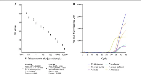
as a general control for quality and performance of the DNA extraction and qPCR reaction. The performance of the PlasQ assay is shown in Fig. 1a. For both targets, PfvarATS and Pspp18S, the Cq values for different par-asite densities, ranging from 0.05 to 10,000 parpar-asites/ µL were obtained (Pearson r − 0.9969 and − 0.9968, respectively). Similar qPCR efficiencies for both targets were observed (87.6% and 89.7%). The PfvarATS target did detect two additional samples carrying low parasite densities (0.1 and 0.05 parasites/µL) resulting in 10 times lower LOD compared to the Pspp18S target. The second assay (PlasID), which is applied to all Plasmodium spp. positive samples, was designed for rapid identification
20 25 30 35 40 45
P. falciparum density [parasites/µL]
Cq valu
e
PfvarATS slope -3.575 ± 0.08882 y-intercept 34.81 ± 0.1938 qPCR efficiency 87.6% LOD 0.05 Pf/µL Pearson r -0.9969
Pspp18S slope -3.637 ± 0.103 y-intercept 35.53 ± 0.2403 qPCR efficiency 89.7% LOD 0.5 Pf/µL Pearson r -0.9968
0.01 0.1 1 10 100 1000 10000 00 5 10 15 20 25 30 35 40 45 1000
2000 3000 4000
Cycle
Relative Fluoroscence Unit
P. falciparum P. malariae
P. ovale curtisi P. ovale wallikeri
P. vivax P. knowlesi
a b
[image:4.595.60.537.427.677.2]of five different malaria causing Plasmodium species. Performance of the PlasID assay was assessed using well-defined clinical samples as references and this novel assay’s ability of identifying P. falciparum, P. malariae, P. ovale curtisi, P. ovale wallikeri, P. vivax and P. knowlesi is demonstrated in Fig. 1b.
Baseline characteristics of blood donors attended Malabo Central Blood Bank
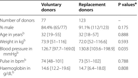
Blood donor characteristics are given in Table 1. All donors were Equato-Guineans living in Malabo on Bioko Island except two persons, one originating from neigh-bouring Douala, Cameroon and the other from the United States. The majority were male (88.5%) with a median age of 32 years, ranging from 18 to 57 years. Sev-enty-seven donations were from voluntary blood donors and 123 donations were from replacement donors, who donate blood when required by a member of the patient’s family or community (Table 1). Except for the blood pres-sure, no significant difference in relation to gender, age, weight, pulse and anaemia status between these two donor groups were observed.
Prevalence of malaria positive blood donations
The flow diagram depicting the sequential malaria diag-nostic tools applied to the blood donations is summa-rized in Fig. 2. In the Malabo blood bank, all samples were screened using thick blood smear microscopy and RDT. Microscopy identified four P. falciparum posi-tive donations, while RDTs detected 13 malaria posiposi-tive donations. Three donations were positive for both anti-gens, the P. falciparum specific HRP2 and the Plasmo-dium spp. LDH antigens. The remaining ten donations were positive for HRP2 only. Microscopy and RDT posi-tive blood donations were not considered for donation
and were included blinded into this study sample col-lection on purpose to test the performance of the PlasQ assay.
Upon arrival at the EGMVI laboratory all samples were screened with the first multiplex qPCR assay (PlasQ), targeting P. falciparum (PfvarATS), Plasmodium spp. (Pspp18S) and the human RNAseP gene (as an internal control). All 200 samples amplified the internal control with Cq < 28 and were therefore eligible for further analy-sis. Over 70% (n = 141) of blood donations were negative for any malaria species. Forty-six donations were positive for both Plasmodium targets, while six and seven dona-tions were positive only for P. falciparum or Plasmodium
spp., respectively. All samples (n = 53) with a positive amplification of the Pspp18S target were analysed with the species identification assay (PlasID). Apart from con-firming P. falciparum (n = 33) cases, we also found cases of P. malariae (n = 9) and P. ovale (n = 2). Amongst the 200 blood samples analysed, no P. vivax or P. knowlesi
was detected.
The assay identified P. malariae or P. ovale in all seven samples which were positive for Plasmodium spp. and negative for P. falciparum, highlighting the assays abil-ity to identify non-falciparum species. Compared to the PlasQ assay, a reduced sensitivity (71.7%, 33/46) for P.
fal-ciparum detection using the PlasID assay was observed.
The 13 P. falciparum positive samples missed by the PlasID assay had moderate parasitaemia (median of 36.9 with IQR of 1.3–380.0 parasites/µL) and the five sam-ples with the highest parasitaemia were positive by RDT. None of these samples were positive for any other non-P.
falciparum species.
An additional 29 malaria negative blood samples were also run with the PlasID assay to test specificity of the assay. No false positive Plasmodium spp. cases were detected, resulting in 100% specificity.
Notably, none of the non-falciparum species had been identified by microscopy or RDT. Pan-positive RDTs, which detected the Plasmodium spp. LDH, were rather associated with higher P. falciparum density than with the detection of non-falciparum malaria parasite species. The three HRP2 and LDH positive RDTs had a geometric mean of 916.3 parasites/µL (range 244.1–3707.0) while the nine HRP2 only positive RDTs had a geometric mean of 106.0 parasites/µL (range 11.2–543.3) (Table 2).
In summary, 59 blood donations were positive for at least one malaria species (29.5%). Plasmodium falcipa-rum was the dominating species responsible for 88.1% of positive blood donations, followed by P. malariae
(15.3%) and P. ovale (3.4%). Mixed species infections were found in 6.8% (4/59) of the malaria positive blood donations. One blood donation carried a P. malariae
[image:5.595.57.291.563.696.2]and P. ovale co-infection and three donations contained
Table 1 Comparison of health characteristics between voluntary and replacement donors
a Variables were compared by Fisher’s exact test for categorical variables or Mann–Whitney test for continuous variables
b Values expressed as medians with ranges
Voluntary
donors Replacement donors P values
a
Number of donors 77 123 –
% male 84.4% (65/77) 91.1% (112/123) 0.175 Age in yearsb 32 [19–55] 32 [18–57] 0.888
Weight in kgb 73.9 [51–116] 72.0 [52–116.6] 0.593
Blood pressure in
mmHgb 126.7 [97.7–169.0] 130.8 [103.6–198.9] 0.035
Pulse in bpmb 74 [48–101] 73 [51–102] 0.788
Haemoglobin in
a combination of P. falciparum and P. malariae. The observed co-infection proportion between P. falcipa-rum and P. malariae was slightly higher than expected (1.5% versus 1.2%, P = 0.037).
Plasmodium falciparum densities in malaria positive blood donations
Data obtained from the PlasQ assay was used to quan-tify P. falciparum positive blood donations. Para-site densities calculated based on both targets show a high correlation (Spearman r 0.894, Fig. 3). Identified
non-falciparum species are indicated with green dots.
Quantification based on the Pspp18S assay revealed, that the parasitaemia of these non-falciparum species is around or below 10 parasites/µL which was sup-ported by the high Cq values obtained from the PlasID assay. The geometric mean Cq value of the nine P.
malariae positive samples was 36.9 (range 35.3–39.0)
and the two positive P. ovale samples had Cq values of 37.9 and 39.8.
A strong association between the diagnostic tests and the parasitaemia in P. falciparum positive blood
Samples positive for Pf (n=33), Pm (n=9), Po (n=2),
Pv (n=0), Pk (n=0)
Samples collected at Blood bank in Malabo
n=200
Samples positive for Pf only
n=6
Samples positive for Pf andPspp
n=46
Samples positive for Pspp only
n=7
Samples negative for Pf andPspp
n=141 Screening and quantification
of PfandPspp by PlasQ assay (n=200)
Microscopy and RDT screening at Blood Bank
Identification of Plasmodium
species by PlasID assay (n=53)
Microscopy positive
Pf=4
RDT positive Pf=10 Pan/Pf=3
Fig. 2 Overview of blood sample analysis. Blood donations were systematically screened for the presence of malaria parasites by microscopy, RDT and qPCR assays
Table 2 Parasitaemia of blood donations detected with different diagnostic methods
a Quartiles (Median, 25th and 75th percentile)
b Geometric mean and 95% confidence interval
c Pf- and Pf/Pan-RDT results combined
d Pf-RDT results only
e Pf/Pan-RDT results only
Diagnostic tool Median/IQR (parasites/µL)a Geometric mean (parasites/µL)b Range (parasites/µL)
qPCR-PfvarATS 4.6 [0.8–49.0] 5.6 [2.6–12.1] 0.06–3707.0
qPCR-Pspp18S 7.8 [1.3–65.1] 9.5 [4.5–19.8] 0.07–3707.0
RDT combinedc 134.6 [65.1–536.5] 181.7 [68.98–478.7] 11.16–3707.0
HRP2-RDTd 112.5 [52.2–331.2] 106.0 [42.4–265.1] 11.16–3707.0
HRP2/LDH-RDTe 850.4 [244.1–3707.0] 916.3 [31.11–26990.0] 244.1–3707.0
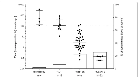
[image:6.595.58.537.87.375.2] [image:6.595.63.540.441.535.2]donations was observed. Non-cumulative parasite den-sity data grouped by diagnostic test is shown in Fig. 4 (scatter plot, left y-axis).
In Table 2, the parasite densities grouped by diagnostic tool are summarized. Densities of P. falciparum positive blood donations ranged from 0.06 to 3707.0 parasites/µL with a median of 4.6 parasites/µL (IQR 0.8–49.0), which is lower than the LOD for conventional diagnostic tests. Only samples with the highest parasitaemia were detect-able by microscopy (381.1 [145.4–2909.0] parasites/ µL) and RDTs (134.6 [65.1–536.5] parasites/µL), while the two qPCR assays detected additional infections with lower parasite densities. The median parasitaemia was 7.8 parasites/µL (IQR 1.3–65.1) for the Pspp18S target and 4.6 parasites/µL (IQR 0.8–49.0) for the PfvarATS tar-get (Table 2).
Performance of diagnostic methods for P. falciparum
detection in blood donations
In total, 48 previously undetected P. falciparum infec-tions were identified by conducting the varATS based qPCR analysis compared to microscopy, increasing the proportion of infected blood donations from 2.0 to
0.1 1 10 100 1000 10000 0.1
1 10 100 1000 10000
Parasitemia by PfvarATS [0.05 + parasites/µL]
Parasitemia by Pspp18S [0.05+parasites/
µ
L]
Fig. 3 Parasite densities of positive blood donations obtained from qPCR assays. Strong correlation between the two targets is observed (Spearman r 0.894). The green dots highlight samples containing non-falciparum malaria species. An offset of 0.05 parasites/µL was used
Microscopy n=4
RDT n=13
Pspp18S n=46
PfvarATS n=52
0.01 0.1 1 10 100 1000 10000
0 20 40 60 80 100
P.falciparum
parasitemia[parasites/
µ
L]
% of contaminated blood donations
Fig. 4 Association between parasitaemia and sensitivity of diagnostic methods applied in this study. Median and interquartile ranges of
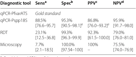
[image:7.595.57.290.86.304.2] [image:7.595.58.541.386.656.2]26.0%. The detection rate for RDTs and 18S based qPCR were 6.5% and 23.0%, respectively (Fig. 4, bar plot, right y-axis). This increased detection rate of the qPCR assays is also reflected in analytical sensitivities of the diagnostic tests. Using the PfvarATS qPCR results as the gold stand-ard for P. falciparum detection, Pspp18S qPCR (88.5%), RDT (23.1%) and microscopy (7.7%) have all reduced sen-sitivities. Specificities were for all tests above 95%. One RDT positive sample could not be confirmed by qPCR, while the specificity for Pspp18S was reduced because of
non-falciparum malaria parasite species, which are also
detected (Table 3).
Identification of risk factors for malaria positivity
Malaria positive and negative donors were stratified according to the differences in the questionnaire that all blood donors must fill before proceeding to blood dona-tion (Addidona-tional file 2). The major risk factor for being a malaria positive blood donor was reporting fever or malaria during the past 3 weeks (Additional file 2). When comparing the anthropometric measurements between malaria positive and negative donors, marginal differ-ences in age and weight become apparent (Table 4). An increased rate of positive blood donations in replace-ment donors (29.3%) compared to voluntary donors (20.8%) was observed, as well as an increased parasitae-mia among replacement donors (8.9 versus 2.1 parasites/ µL). However, these differences were not statistically significant.
Discussion
This study, conducted in Equatorial Guinea, extends pre-vious observations of presence of TTM in sub-Saharan Africa, and demonstrates the value of NAT approaches to reliably detect sub-patent P. falciparum and non- falcipa-rum malaria parasites including P. malariae and P. ovale.
Apart from P. falciparum, other malaria causing spe-cies such as P. vivax, P. malariae, and P. ovale have been described to circulate on Bioko Island—similar to neigh-bouring West African countries [27, 28]. Transfusion of any of these malaria species during blood donations have the potential to cause severe disease in recipients. In sub-Saharan Africa, those most at risk are anaemic infants and children, pregnant women, or women who suffer from high blood losses while giving birth [25, 38, 39]. Although, there is a high frequency of TTM in malaria endemic regions, the semi-immunity of recipients may largely control parasite replication and therefore clinical outcome [40].
In malaria non-endemic countries, TTM needs also be taken into account to ensure safe blood donations [41]. Here, blood the recipients are mainly malaria naïve and the immunological control of potentially transfused para-sites is non-existent resulting in in-creased risk of severe malaria disease. P. falciparum parasites have been found to survive in blood donations for up to 14 days limiting the potential to neutralize malaria during blood donation processing and storage [42].
This study demonstrates the challenges blood banks in malaria endemic countries encounter. The blood donors at the Malabo blood bank were mainly male adults within an age range of 18–57 years. Because of their pre-exist-ing immunity, blood donors of that age tend to have low levels of parasites without exhibiting clinical symptoms. Ninety-seven percent (97.0%) reported to be in good health at the time of blood donations. Apart from self-reported malaria and/or fever episodes within the last 3 weeks, no other risk factor had been identified based on the provided questionnaire. Since only about 3% (6/198) had reported to have malaria and/or fever episodes, the use of this risk factor to pre-select blood donors would not be very efficient. Deferral of about a quarter of blood Table 3 Analytical comparison of methods deployed for P.
falciparum detection
a Sensitivity and 95% confidence interval
b Specificity and 95% confidence interval
c Positive predictive value and 95% confidence interval
d Negative predictive value and 95% confidence interval
e Specificity and PPV of Pspp18S qPCR assay is reduced because non-falciparum Plasmodium species are also detected
Diagnostic tool Sensa Specb PPVc NPVd
qPCR-PfvarATS Gold standard
[image:8.595.306.539.114.236.2] [image:8.595.56.291.558.657.2]qPCR-Pspp18S 88.5%
[76.6–95.7] 95.3%[90.5–98.1]e 86.8%[76.0–93.2]e 95.9%[91.7–98.0]
RDT 23.1%
[12.5–36.8] 99.3%[96.3–99.9] 92.3%[61.5–100.0] 79.0%[76.0–81.0] Microscopy 7.7%
[2.1–18.5] 100.0%[97.54–100] 100%– 75.5%[74.0–76.9]
Table 4 Anthropometric differences in P. falciparum positive versus P. falciparum negative blood donors
a Fisher’s exact test for categorical variables or Mann–Whitney test for continuous variables was applied
b Values expressed as medians with ranges
Negative donors Positive donors P valuesa
Number of donors 148 52 –
% male 86.5% (128/148) 94.2% (49/52) 0.205 Age in yearsb 33 [18–57] 29 [20–55] 0.013
Weight in kgb 74.6 [51–116.6] 69.2 [53–110] 0.025
Blood pressure in
mmHgb 128.7 [97.7–198.9] 128.3 [100.6–156.9] 0.326
Pulse in bpmb 74 [48–102] 72 [51–96] 0.519
Haemoglobin in
donors that are sub-patent malaria carriers in a region where blood supply shortage is eminent could be even more damaging to public health.
Conventional diagnostic tests are important tools, since they are affordable and can be applied immedi-ately at blood banks, but lack in sensitivity limits their usefulness. Microscopy is still an important tool for malaria detection since it allows for species identifica-tion and quantificaidentifica-tion. The detecidentifica-tion limit of an expert microscopy is close to 20–50 parasites/µL, but for non-experienced microscopists, it is up to tenfold higher (i.e. 500 parasites/µL) [5]. Currently available RDTs are based on detection of circulating, parasite derived HRP2, aldolase or lactate dehydrogenase in serum or whole blood samples. These RDTs have the ability to detect Plasmodium spp. infections with a LOD between 100 and 1000 parasites/µL which is significantly higher when compared to NATs [43]. In addition, there is cur-rently no single RDT available that could distinguish all five malaria species infections posing a clear limitation to comprehensive malaria infection status monitoring [44, 45]. Infections with P. malariae [46], P. ovale [47] and P.
vivax [48] are usually characterized by low parasitaemia
levels, rendering these infections largely undetectable by using currently available RDT technologies [49].
In this study, more than three-quarter of P. falcipa-rum and all non-falciparum infections were missed by the conventional diagnostic tools. A suitable screening method for malaria detection in blood units must have high sensitivity, high negative predictive value, must include all Plasmodium species, and should be cost effi-cient. Such a test must enable a reduction of TTM risk, as well as that of falsely deferred blood donors found to have false positive results.
The PlasQ malaria screening assay applied in this study meets some of these requirements. With a LOD of 0.05 parasites/µL, PlasQ has high sensitivity for P.
falci-parum and includes all other relevant Plasmodium
spe-cies. This assay was extensively used during controlled human malaria infections in Tanzanian and Equatorial Guinean volunteers and no issues with specificity were observed (unpublished). As a limitation, qPCR analysis requires an advanced laboratory infrastructure, trained personnel and the costs for reagents and consumables for this assay is about USD 5, which is higher than the costs for RDT or microscopy-based malaria diagnosis.
This study was used to obtain more information about
non-falciparum malaria species circulating in Malabo.
Therefore a novel multiplex qPCR assay for parallel malaria parasite identification was developed. Using the PlasID assay, the main five human infectious malaria spe-cies can be detected qualitatively in a multiplex assay rendering it highly cost efficient since only one DNA
extraction and qPCR reaction needs to be run. This novel malaria species identification assay should be used in combination with the PlasQ assay, based on the finding that in the PlasID assay, a reduced sensitivity for P.
fal-ciparum is observed. Further development of diagnostic
assays for the detection of P. malariae and P. ovale, should include the ability of species-specific quantification. Esti-mations based on Pspp18S target of the PlasQ assay, indi-cate that P. malariae and P. ovale single infections had a geometric mean density of about 0.6 parasites/µL, which is more than 14 times lower than P. falciparum single infections (8.8 parasites/µL).
Educating the general public in malaria endemic regions via TV, radio, and pamphlet about the existence of TTM and its potential health impact might help to raise the awareness of this malaria transmission route, both in potential blood donors and medical and nursing staff involved in blood transfusions [50]. This study con-firms prior reports that replacement blood donors are particularly prone to harbour sub-patent malaria para-sites [51]; hence this sub-population of donors are opti-mal for such awareness campaigns regarding potential TTM when donating blood for a family or friend. In addi-tion to becoming aware of the potential TTM, medical and nursing staff should also use anti-malarial interven-tions when the potential for TTM is high and the malaria diagnostic infrastructure is limited. Ultimately, if the goal of malaria elimination is pursued vigorously, tackling TTM in sub-Saharan Africa needs to be included into the malaria elimination agenda.
Conclusions
More than one quarter of blood donations from healthy donors living in Malabo were infected with malaria para-sites. Three species, including P. falciparum, P. malariae
and P. ovale were detected using two different qPCR
assays. The majority of these infections were not identi-fied by the currently widely used malaria diagnostic tools such as RDTs and microscopy. Since the availability of molecular diagnostic methods in malaria endemic coun-tries is still limited, the blood recipients living in malaria endemic countries should be treated following WHO recommendations.
Additional files
Additional file 1. Oligos used in this study.
Abbreviations
RDT: rapid diagnostic test; HRP2: histidine rich protein 2; LDH: lactate dehydro-genase; LOD: limit of detection; NAT: nucleic acid amplification technology; qPCR: quantitative polymerase chain reaction; TTM: transfusion-transmitted malaria; EGMVI: Equatorial Guinea Malaria Vaccine Initiative; BIMCP: Bioko Island Malaria Control Project; Pspp18S: Pan-Plasmodium 18S rDNA sequence; PfvarATS: P. falciparum-specific acidic terminal sequence of the var genes.
Authors’ contributions
Study concept and design: TS, TR, CM, CD. Acquisition of data: TS, TR, JS, LG, LA. Analyses and interpretation of data: TS, TR, MM, CD. Drafting the manuscript: TS, MM, CM, CD and all other authors reviewed the manuscript. Technical or material support: TR, JRB, GG, CC. Study supervision: MM, CM, CD. All authors read and approved the final manuscript.
Author details
1 Department of Medical Parasitology and Infection Biology, Swiss Tropical and Public Health Institute, Basel, Switzerland. 2 University of Basel, Basel, Swit-zerland. 3 Malabo Blood Bank, Malabo, Equatorial Guinea. 4 Equatorial Guinea Malaria Vaccine Initiative, Malabo, Equatorial Guinea. 5 Bagamoyo Research and Training Centre, Ifakara Health Institute, Bagamoyo, United Republic of Tanzania. 6 Medical Care Development International, Silver Spring, USA. 7 Marathon EG Production Ltd, Malabo, Equatorial Guinea.
Acknowledgements
We would like to thank the voluntary and replacement blood donors at the Malabo blood bank for their willingness to donate blood and to save lives. We highly appreciate the support of Paul Lansdell, PHE Malaria Reference Laboratory and Don van Schalkwyk, Dept. of Immunology & Infection, London School of Hygiene & Tropical Medicine who kindly provided the DNA controls for non-falciparum malaria species. We would also like to thank Kenneth Mbwanji and Lukman Aroworamimo from Inqaba Biotec who kindly provided the GeneFinder Malaria kits, which were used to confirm the positive controls in this study.
Competing interests
The authors declare that they have no competing interests.
Availability of data and materials
The datasets supporting the conclusions of this article are included within the article.
Consent for publication Not applicable.
Ethics approval and consent to participate
Approval for this study was given by the Ministry of Health and Social Welfare, Malabo, Bioko Norte, Republic of Equatorial Guinea. Study participants were assured of confidentiality and all data collected remained anonymous.
Funding
This study was supported by funds from the Equatorial Guinea Malaria Vaccine Initiative.
Publisher’s Note
Springer Nature remains neutral with regard to jurisdictional claims in pub-lished maps and institutional affiliations.
Received: 6 September 2018 Accepted: 8 January 2019
References
1. WHO. World malaria report 2017. Geneva: World Health Organization; 2017.
2. Ashley EA, Pyae Phyo A, Woodrow CJ. Malaria. Lancet. 2018;391:1608–21. 3. Nguyen T-N, von Seidlein L, Nguyen T-V, Truong P-N, Do Hung S, Pham
H-T, et al. The persistence and oscillations of submicroscopic Plasmodium
falciparum and Plasmodium vivax infections over time in Vietnam: an open cohort study. Lancet Infect Dis. 2018;18:565–72.
4. Bousema T, Okell L, Felger I, Drakeley C. Asymptomatic malaria infections: detectability, transmissibility and public health relevance. Nat Rev Micro-biol. 2014;12:833–40.
5. Moody A. Rapid diagnostic tests for malaria parasites. Clin Microbiol Rev. 2002;15:66–78.
6. Boyce MR, O’Meara WP. Use of malaria RDTs in various health contexts across sub-Saharan Africa: a systematic review. BMC Public Health. 2017;17:470.
7. Mouatcho JC, Goldring JPD. Malaria rapid diagnostic tests: challenges and prospects. J Med Microbiol. 2013;62:1491–505.
8. Hendriksen ICE, Mtove G, Pedro AJ, Gomes E, Silamut K, Lee SJ, et al. Evaluation of a PfHRP2 and a pLDH-based rapid diagnostic test for the diagnosis of severe malaria in 2 populations of African children. Clin Infect Dis. 2011;52:1100–7.
9. Wongsrichanalai C, Barcus MJ, Muth S, Sutamihardja A, Wernsdorfer WH. A review of malaria diagnostic tools: microscopy and rapid diagnostic test (RDT). Am J Trop Med Hyg. 2007;77(Suppl. 6):119–27.
10. Srisutham S, Saralamba N, Malleret B, Rénia L, Dondorp AM, Imwong M. Four human Plasmodium species quantification using droplet digital PCR. PLoS ONE. 2017;12:e0175771.
11. Chew CH, Lim YAL, Lee PC, Mahmud R, Chua KH. Hexaplex PCR detection system for identification of five human Plasmodium species with an internal control. J Clin Microbiol. 2012;50:4012–9.
12. Hopkins H, Gonzalez IJ, Polley SD, Angutoko P, Ategeka J, Asiimwe C, et al. Highly sensitive detection of malaria parasitemia in a malaria-endemic setting: performance of a new loop-mediated isothermal amplification kit in a remote clinic in Uganda. J Infect Dis. 2013;208:645–52. 13. Kamau E, Alemayehu S, Feghali KC, Saunders D, Ockenhouse CF.
Multi-plex qPCR for detection and absolute quantification of malaria. PLoS ONE. 2013;8:e71539.
14. Kamau E, Tolbert LS, Kortepeter L, Pratt M, Nyakoe N, Muringo L, et al. Development of a highly sensitive genus-specific quantitative reverse transcriptase real-time PCR assay for detection and quantitation of Plasmodium by amplifying RNA and DNA of the 18S rRNA genes. J Clin Microbiol. 2011;49:2946–53.
15. Murphy SC, Prentice JL, Williamson K, Wallis CK, Fang FC, Fried M, et al. Real-time quantitative reverse transcription PCR for monitoring of blood-stage Plasmodium falciparum infections in malaria human challenge trials. Am J Trop Med Hyg. 2012;86:383–94.
16. Adams M, Joshi SN, Mbambo G, Mu AZ, Roemmich SM, Shrestha B, et al. An ultrasensitive reverse transcription polymerase chain reaction assay to detect asymptomatic low-density Plasmodium falciparum and
Plasmodium vivax infections in small volume blood samples. Malar J. 2015;14:520.
17. Hofmann N, Mwingira F, Shekalaghe S, Robinson LJ, Mueller I, Felger I. Ultra-sensitive detection of Plasmodium falciparum by amplification of multi-copy subtelomeric targets. PLoS Med. 2015;12:e1001788. 18. Mangold KA, Manson RU, Koay ESC, Stephens L, Regner M, Thomson RBJ,
et al. Real-time PCR for detection and identification of Plasmodium spp. J Clin Microbiol. 2005;43:2435–40.
19. WHO. Screening donated blood for transfusion–transmissible infections: recommendation. Geneva: World Health Organization; 2010.
20. WHO. Blood donor selection: guidelines on assessing donor suitability for blood donation. Geneva: World Health Organization; 2012.
21. Freimanis G, Sedegah M, Owusu-Ofori S, Kumar S, Allain J-P. Investigat-ing the prevalence of transfusion transmission of Plasmodium within a hyperendemic blood donation system. Transfusion. 2013;53:1429–41. 22. Owusu-Ofori AK, Betson M, Parry CM, Stothard JR, Bates I. Transfusion
transmitted malaria in Ghana. Clin Infect Dis. 2013;56:1735–41. 23. Lacerda MVG, Monteiro WM, Alexandre MAA, Alho RRM, Kiesslich D, Fraiji
NA. We need to talk more about transfusion-transmitted malaria in Plas-modium vivax endemic areas. Rev Bras Hematol Hemoter. 2014;36:385–7. 24. Scuracchio P, Vieira SD, Dourado DA, Bueno LM, Colella R, Ramos-Sanchez
EM, et al. Transfusion-transmitted malaria: case report of asymptomatic donor harboring Plasmodium malariae. Rev Instit Med Trop de São Paulo. 2011;53:55–9.
•fast, convenient online submission
•
thorough peer review by experienced researchers in your field
• rapid publication on acceptance
• support for research data, including large and complex data types
•
gold Open Access which fosters wider collaboration and increased citations maximum visibility for your research: over 100M website views per year
•
At BMC, research is always in progress.
Learn more biomedcentral.com/submissions
Ready to submit your research? Choose BMC and benefit from:
splenectomized patient with beta-thalassaemia major in Sabah, Malaysia: a case report. Malar J. 2016;15:357.
26. Martin-Davila P, Norman F, Fortun-Abete J, Piris M, Lovatti R, Rubio JM, et al. Donor-derived multiorgan transmission of mixed P. malariae and
P. ovale infection: impact of globalization on post-transplant infections. Transpl Infect Dis. 2018;20:e12938.
27. Guerra-Neira A, Rubio JM, Royo JR, Ortega JC, Auñón AS, Diaz PB, et al. Plasmodium diversity in non-malaria individuals from the Bioko Island in Equatorial Guinea (West Central-Africa). Int J Health Geogr. 2006;5:27. 28. Mendes C. Duffy negative antigen is no longer a barrier to Plasmodium
vivax—molecular evidences from the African West Coast (Angola and Equatorial Guinea). PLoS Negl Trop Dis. 2011;5:e1192.
29. Berzosa PJ, García ML, Micó M, Edú M, Rubio JM, Alvar J, et al. Semi-nested, multiplex polymerase chain reaction for detection of human malaria parasites and evidence of Plasmodium vivax infection in Equato-rial Guinea. Am J Trop Med Hyg. 1999;60:183–7.
30. Cook J, Hergott D, Phiri W, Rivas MR, Bradley J, Segura L, et al. Trends in parasite prevalence following 13 years of malaria interventions on Bioko island, Equatorial Guinea: 2004–2016. Malar J. 2018;17:62.
31. Olotu A, Urbano V, Hamad A, Eka M, Chemba M, Nyakarungu E, et al. Advancing global health through development and clinical trials partner-ships: a randomized, placebo-controlled, double-blind assessment of safety, tolerability, and immunogenicity of PfSPZ vaccine for malaria in healthy Equatoguinean men. Am J Trop Med Hyg. 2018;98:308–18. 32. Kamau E, Alemayehu S, Feghali KC, Komisar J, Regules J, Cowden J, et al.
Measurement of parasitological data by quantitative real-time PCR from controlled human malaria infection trials at the Walter Reed Army Insti-tute of Research. Malar J. 2014;13:288.
33. Cnops L, Jacobs J, Van Esbroeck M. Validation of a four-primer real-time PCR as a diagnostic tool for single and mixed Plasmodium infections. Clin Microbiol Infect. 2011;17:1101–7.
34. Phuong M, Lau R, Ralevski F, Boggild AK. Sequence-based optimization of a quantitative real-time PCR assay for detection of Plasmodium ovale and
Plasmodium malariae. J Clin Microbiol. 2014;52:1068–73.
35. Reller ME, Chen WH, Dalton J, Lichay MA, Dumler JS. Multiplex 5′ nuclease quantitative real-time PCR for clinical diagnosis of malaria and species-level identification and epidemiologic evaluation of malaria-causing par-asites, including Plasmodium knowlesi. J Clin Microbiol. 2013;51:2931–8. 36. Miller RH, Obuya CO, Wanja EW, Ogutu B, Waitumbi J, Luckhart S, et al.
Characterization of Plasmodium ovale curtisi and P. ovale wallikeri in West-ern Kenya utilizing a novel species-specific real-time PCR assay. PLoS Negl Trop Dis. 2015;9:e0003469.
37. Divis PCS, Shokoples SE, Singh B, Yanow SK. A TaqMan real-time PCR assay for the detection and quantitation of Plasmodium knowlesi. Malar J. 2010;9:344.
38. Allain JP, Owusu-Ofori AK, Assennato SM, Marschner S, Goodrich RP, Owusu-Ofori S. Effect of Plasmodium inactivation in whole blood on the incidence of blood transfusion-transmitted malaria in endemic regions: the African Investigation of the Mirasol System (AIMS) randomised con-trolled trial. Lancet. 2016;387:1753–61.
39. Guindo A, Touré BA, Guindo P, Baraika MA, Sarro YS, Fané B, et al. Transmission of Plasmodium falciparum by red blood cell transfusions in the management of sickle cell disease patients in Mali. Transfus Med. 2016;26:153–5.
40. Allain J-P, Assennato SM, Osei EN, Owusu-Ofori AK, Marschner S, Goodrich RP, et al. Characterization of posttransfusion Plasmodium falciparum
infection in semi-immune nonparasitemic patients. Transfusion. 2016;56:2374–83.
41. Verra F, Angheben A, Martello E, Giorli G, Perandin F, Bisoffi Z. A systematic review of transfusion-transmitted malaria in non-endemic areas. Malar J. 2018;17:36.
42. Chattopadhyay R, Majam VF, Kumar S. Survival of Plasmodium falciparum
in human blood during refrigeration. Transfusion. 2011;51:630–5. 43. Das S, Jang IK, Barney B, Peck R, Rek JC, Arinaitwe E, et al. Performance of
a high-sensitivity rapid diagnostic test for Plasmodium falciparum malaria in asymptomatic individuals from Uganda and Myanmar and naive human challenge infections. Am J Trop Med Hyg. 2017;97:1540–50. 44. Maltha J, Gillet P, Jacobs J. Malaria rapid diagnostic tests in endemic
set-tings. Clin Microbiol Infect. 2013;19:399–407.
45. Lover AA, Baird JK, Gosling R, Price R. Malaria elimination: time to target all species. Am J Trop Med Hyg. 2018;99:17–23.
46. Collins WE, Jeffery GM. Plasmodium malariae: parasite and disease. Clin Microbiol Rev. 2007;20:579–92.
47. Collins WE, Jeffery GM. Plasmodium ovale: parasite and disease. Clin Microbiol Rev. 2005;18:570–81.
48. Howes RE, Battle KE, Mendis KN, Smith DL, Cibulskis RE, Baird JK, et al. Global epidemiology of Plasmodium vivax. Am J Trop Med Hyg. 2016;95(6 Suppl):15–34.
49. Maltha J, Gillet P, Jacobs J. Malaria rapid diagnostic tests in travel medi-cine. Clin Microbiol Infect. 2013;19:408–15.