PEARLS
The devil is in the details: Genomics of
transmissible cancers in Tasmanian devils
Andrew Storfer1*, Paul A. Hohenlohe2,3, Mark J. Margres1, Austin Patton1, Alexandra K. Fraik1, Matthew Lawrance1, Lauren E. Ricci1, Amanda R. Stahlke2,3, Hamish I. McCallum4, Menna E. Jones5
1 School of Biological Sciences, Washington State University, Pullman, Washington, United States of
America, 2 Department of Biological Sciences, University of Idaho, Moscow, Idaho, United States of America,
3 Institute for Bioinformatics and Evolutionary Studies, University of Idaho, Moscow, Idaho, United States of
America, 4 School of the Environment, Griffith University, Nathan, Australia, 5 School of Biological Sciences, University of Tasmania, Hobart, Australia
Cancer poses one of the greatest human health threats of our time. Fortunately, aside from a few rare cases of cancer transmission in immune-suppressed organ transplant recipients [1] or a small number of transmission events from mother to fetus [2], cancers are not spread from human to human. However, transmissible cancers have been detected in vertebrate and inver-tebrate animals, sometimes with devastating effects [3]. Four examples of transmissible cancers are now known: 1) canine transmissible venereal tumor (CTVT) in dogs [4], 2) a tumor in a laboratory population of Syrian hamsters that is no longer cultured [3], 3) infectious neoplasias in at least four species of bivalve mollusks [5,6], and 4) two independently derived transmissi-ble cancers (devil facial tumor disease [DFTD]) in Tasmanian devils [7–10] (Fig 1A and 1B). The etiologic agents of CTVT [4], the bivalve cancers [5], and DFTD [7] are the transplants (allografts) of the neoplastic cells themselves, but the etiologic agent is unknown for the ham-ster tumor.
The effects of these transmissible cancers on their respective host populations vary. CTVT is spread in dogs through sexual contact and is at least 11,000 years old, placing the timing of its origin close to that of the domestication of dogs [11]. Although genomic analyses of the tumor suggest evasion of multiple components of the dog immune system, dogs most com-monly survive and often show evidence of spontaneous tumor regression within a year of ini-tial diagnosis [11,12]. For the infectious bivalve neoplasias, which have existed for at least 40 years, population effects vary from enzootic infections with no noticeable effects on population sizes to evidence of a catastrophic population decline [6,13]. In Tasmanian devils (Fig 1A), the first infectious tumor discovered (DFT1;Fig 1B) has spread across approximately 95% of the geographic range of Tasmanian devils since 1996 (Fig 1C). DFTD is almost always fatal (Fig 1B), with>90% declines in infected localities and an overall species-wide decline exceeding 80% [14–16]. Transmission dynamics appear consistent with frequency dependence, with DFTD spread by biting during social interactions [15], resulting in predictions of extinction from standard epidemiological models [14]. Despite these predictions, long-infected devil populations persist at reduced densities, suggesting that individual-level variability in fecun-dity and tumor growth rate in infected individuals are key for understanding epidemiological dynamics [17]. Additionally, the origin of the second, independent lineage of DFTD (i.e., DFT2) [9,10] within 20 years of the discovery of DFT1 suggests that transmissible cancers may be a recurring part of the Tasmanian devils’ evolutionary history, without causing extinction [17].
a1111111111 a1111111111 a1111111111 a1111111111 a1111111111 OPEN ACCESS
Citation: Storfer A, Hohenlohe PA, Margres MJ,
Patton A, Fraik AK, Lawrance M, et al. (2018) The devil is in the details: Genomics of transmissible cancers in Tasmanian devils. PLoS Pathog 14(8): e1007098.https://doi.org/10.1371/journal. ppat.1007098
Editor: Katherine R. Spindler, University of
Michigan Medical School, UNITED STATES
Published: August 2, 2018
Copyright:©2018 Storfer et al. This is an open access article distributed under the terms of the Creative Commons Attribution License, which permits unrestricted use, distribution, and reproduction in any medium, provided the original author and source are credited.
Funding: This work was funded by NIH Grant
R01-GM126563 as part of the joint NIH-NSF-USDA Ecology and Evolution of Infectious Diseases program. URL:https://www.nsf.gov/funding/pgm_ summ.jsp?pims_id=5269. The funders had no role in study design, data collection and analysis, decision to publish, or preparation of the manuscript.
Competing interests: The authors have declared
The origin of Tasmanian devil facial tumor disease
Based on a transcriptomic analysis of DFT1, the progenitor tumor likely originated from a mutated Schwann cell (a type of peripheral nerve cell) in a female Tasmanian devil [8]. DFT2 is also likely to be of neuroectodermal origin, but DFT2 does not express periaxin (PRX), a Schwann cell marker present in DFT1 [10]. Both DFT1 and DFT2 likely evolved from Tasma-nian devils located in eastern Tasmania, with their genetic assignments consistent with their geographic origins (in the NE and SE, respectively) [10]. The gross morphology and histology of DFT2 are different from DFT1 [9,10]. For example, DFT1 is generally composed of pleo-morphic round cells in bundles, whereas DFT2 is typically characterized by pleopleo-morphic sheets of cells [9]. Moreover, DFT2 karyotypes have a Y chromosome, indicating that this tumor arose from a male devil and thus independently from DFT1 [9]. Although DFT1 and DFT2 originated in the last 21 years, no evidence has been found for viral origin, and results are inconsistent with tumor evolution coming from exposure to anthropogenic stressors, such as increased UV light [10].
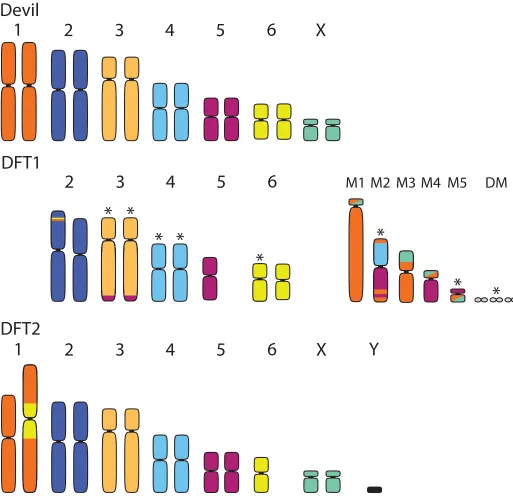
[image:2.612.144.573.76.249.2]Although the specific mechanism of how DFT1 or DFT2 became transmissible is still unknown, several lines of evidence suggest that chromosomal rearrangements, common to cancers, were critical [8,18]. Devils normally have six pairs of autosomes and one pair of sex chromosomes [7,19,20], but cytogenetic analyses show large-scale rearrangements in both DFT1 and DFT2 (Fig 2). To date, few isolates of DFT2 have been karyotyped, but DFT1 has abnormalities associated with Chromosomes 1, 3, 4, 5, 6, and the X chromosome [19–21]. DFT1 is characterized by a series of marker chromosomes, including double minutes, that vary in size and number among different cytogenetic strains [10,21]. Notably, both tumors show rearrangement of Chromosome 1 (referred to as Chromosome 2 in [10]; seeFig 2) [20,21]. Some karyotypes of DFT1 show complete fragmentation of Chromosome 1, as well as fusion with parts of the X chromosome and Chromosome 5, whereas DFT2 has an insertion of one copy of Chromosome 6 in the pericentric region in Chromosome 1 (Fig 2). Comparisons with wallaby and opossum genomes suggest that Chromosome 1 has been prone to rearrange-ment among marsupials [20]; Chromosome 1 rearrangements are stable in DFT1, indicating their possible importance for tumor transmissibility and fitness [19,20]. Common to cancers, DFT1 and DFT2 may have arisen from a critical shortening of telomeres on different
Fig 1. A. Healthy adult Tasmanian devil. B. Adult Tasmanian devil infected with devil facial tumor disease. C. Map of Tasmania showing initial site of disease emergence (red dot). Curved lines show the disease front demarcated by the year devil facial tumor disease was detected in each area.Photo Credit:Alexandra Fraik.
chromosomes that led to these abnormal karyotypes [see10,19,20]. Telomeres are DNA–pro-tein complexes that protect against chromosomal degradation during cell replication [22]. When telomeres become critically short, the p53 pathway typically induces apoptosis (terminal cell cycle arrest), but evasion of this pathway is often associated with cancer progression [23].
Fig 2. Karyotypes of a normal female Tasmanian devil (top) with a representative karyotype of DFT1 (middle) and DFT2 (bottom). Karyotypes of the Tasmanian devil and DFT1 follow [19]; this arrangement differs from [10] in respect to Chromosomes 1 and 2, in which the positions are reversed. Colors in rearranged/ fragmented chromosomes indicate the chromosome from which the material originated in the normal devil karyotype. M1–M5 indicate marker chromosomes resulting from fragmentation of Chromosome 1 and fusion with Chromosomes 5 and 1 in DFT1. Asterisks indicate those chromosomes/markers that have been observed to vary among karyotyped strains of DFT1. DFT2 originated from a male devil and thus possesses a Y chromosome. While one copy of Chromosome 6 has been inserted into the pericentric region of Chromosome 1, the limited available karyotypes of DFT2 thus far suggest it is otherwise relatively undifferentiated from the normal devil karyotype. DFT, devil facial tumor.
At least 64 chromosomal rearrangements map to genic regions in DFT1, including a gene encoding a histone methyltransferase (EZH2) that is dysregulated in many cancers [10]. Addi-tional translocations include a homolog of a tumor suppressor gene (NF2) and a deletion of a tumor suppressor copy (LZTR1). Moreover, there is evidence of copy number increases of two genes (ERBB3andGALNT2) that are overexpressed in many cancers, including schwannomas [18,20]. While early mutations in DFT1 and DFT2 have no overlap, both tumors notably had hemizygous mutations that led to deletion of second copies of genes in the Hippo pathway (WWC
in DFT1 andMPDZin DFT2, respectively), implicated in several human cancer histotypes, partic-ularly Schwann cell cancers [10]. Although both tumors are largely diploid, DFT2 appears to have a simpler structure and fewer rearrangements (23 versus 64) as compared to DFT1 [10].
Tasmanian devil facial tumor disease: Genomics of susceptibility
For an allograft to avoid rejection from a new host, it must circumvent recognition by major histocompatibility complex (MHC) genes [24]. MHC Class I is generally responsible for tumor recognition via identification of cell surface proteins expressed as “nonself” on cancer cells [24]; transmissible cancer cells are indeed nonself, having originated in a different individual [3,6]. Ubiquitous susceptibility of Tasmanian devils to DFTD has been hypothesized to result from low devil genetic variability overall [25], likely due to at least two historical genetic bottle-necks [25,26]. Compared to other mammals, devils have particularly low genetic variability in the MHC Class I peptide-binding region implicated in tumor recognition [27]. However, MHC diversity is not linked to variation in disease susceptibility among individuals [28], and devils reject allografts in challenge experiments [29]. Instead, DFT1 appears to down-regulate its own MHC expression, as well as MHC expression in the devil [30]. Epigenetic down-regu-lation of MHC expression is common in human cancers [31], as well as being a salient feature in CTVT [12,18]. In addition to MHC evasion, there are at least several other mechanisms that underlie widespread transmissibility of DFTD yet to be discovered. Moreover, there is docu-mented variation in tumor susceptibility among devils, including rare docudocu-mented cases of tumor regression and immune response, which are discussed below.
Tasmanian devil facial tumor disease: Evolution of devils
Recent field studies also suggest evolution of DFTD resistance. For the first time, spontane-ous regression of tumors, a phenomenon rarely seen in human cancers without treatment, has been documented in devils [35]. A comparative genomic study of devils with tumor regression versus those that succumbed to the disease shows evidence that two devil candidate genes (TLL1andNBAS1) are involved in the regression process [35,36]. The two genes, plus a third (PAX3, which is not significant after genome-wide correction), are involved in stimulating angiogenesis in both normal growth and cancer metastases, perhaps increasing tumor vascu-larization to enable lymphocyte penetration. Indeed, Pye and colleagues [36], in the first study that demonstrates evidence of adaptive immune response to DFTD, show that one devil had prominent lymphocyte infiltration in its tumor. The same study shows presence of serum anti-bodies against DFTD cells in six of 52 devils tested, and that four of these six devils had histo-ries of tumor regression [36].
Tasmanian devil facial tumor disease: Evolution of the tumors
Cytogenetic analyses currently recognize four karyotypes of DFT1 [21] and show that genomic rearrangements are limited to particular cancer regions, suggesting at least some genomic sta-bility [19,20]. When compared to the Tasmanian devil reference genome, two strains of DFT1 collected from SE and north central Tasmania, respectively, accumulated between 15,000 and 17,000 single-nucleotide substitutions between them [18]. Assuming they share a common ancestor that emerged approximately 20 years ago, this mutation rate is higher than most human cancers (approximately 5,000) but lower than lung cancer or melanomas [18]. Evi-dence of within-host tumor variation is limited; of 20 devils with multiple tumors, only six individuals had tumors that were genetically distinguishable. A comparison of the mitochon-drial genomes of 104 DFT1 tumors from 69 devils across Tasmania showed limited among-host genetic variability as well, with 21 somatic variants detected [18].
Overall, there appears to be variation in fitness across different DFT1 lineages. Evidence for this comes from displacement of one tumor lineage by another in at least one area of Tasmania [16] but coexistence of multiple lineages in other areas [18]. Currently, DFT2 is still limited in its geographic distribution, and it is not yet known how fast the lineage is evolving, nor how its fitness or effects on devil fitness compare with DFT1. However, early evidence suggests that DFT2 primarily infects males (9/11 documented cases), possibly indicating relative differences in susceptibility between males and females [10].
Conclusion
Acknowledgments
We thank three anonymous reviewers for their comments that improved the quality of the manuscript.
References
1. Myron Kauffman H, McBride MA, Cherikh WS, Spain PC, Marks WH, Roza AM. Transplant tumor regis-try: donor related malignancies. Transplantation. 2002; 74(3):358–62. PMID:12177614
2. Tolar J, Neglia JP. Transplacental and other routes of cancer transmission between individuals. J Pediatr Hematol Oncol. 2003; 25(6):430–4. PMID:12794519
3. Ostrander EA, Davis BW, Ostrander GK. Transmissible tumors: Breaking the cancer paradigm. Trends Genet. 2016; 32(1):1–15.https://doi.org/10.1016/j.tig.2015.10.001PMID:26686413
4. Murgia C, Pritchard JK, Kim SY, Fassati A, Weiss RA. Clonal origin and evolution of a transmissible cancer. Cell. 2006; 126(3):477–87.https://doi.org/10.1016/j.cell.2006.05.051PMID:16901782 5. Metzger MJ, Reinisch C, Sherry J, Goff SP. Horizontal transmission of clonal cancer cells causes
leuke-mia in soft-shell clams. Cell. 2015; 161(2):255–63.https://doi.org/10.1016/j.cell.2015.02.042PMID:
25860608.
6. Metzger MJ, Goff SP. A sixth modality of infectious disease: Contagious cancer from devils to clams. PLoS Pathog. 12(10), e1005904.https://doi.org/10.1371/journal.ppat.1005904PMID:27788268 7. Pearse AM, Swift K. Allograft theory: transmission of devil facial-tumour disease. Nature. 2006; 439
(7076):549.https://doi.org/10.1038/439549aPMID:16452970
8. Murchison EP, Tovar C, Hsu A, Bender HS, Kheradpour P, Rebbeck C., et al. The Tasmanian devil transcriptome reveals Schwann cell origins of a clonally transmissible cancer. Science 2010; 327 (5961), 84–87.https://doi.org/10.1126/science.1180616PMID:20044575
9. Pye RJ, Pemberton D, Tovar C, Tubio JM, Dun KA, Fox S, et al. A second transmissible cancer in Tas-manian devils. Proc Natl Acad Sci USA. 2016; 113(2), 374–9.https://doi.org/10.1073/pnas.
1519691113PMID:26711993
10. Stammnitz MR, Coorens THH, Gorl K, Hayes D, Fu B, et al. The origins and vulnerabilities of two trans-missible cancers in Tasmanian devils. Cancer Cell; 2018 33(4):607–19.https://doi.org/10.1016/j.ccell. 2018.03.013PMID:29634948
11. Murchison EP, Wedge DC, Alexandrov LB, Fu B, Martincorena I, Ning Z, et al. Transmissible dog can-cer genome reveals the origin and history of an ancient cell lineage. Science. 2014; 343(6169):437–40.
https://doi.org/10.1126/science.1247167PMID:24458646
12. Decker B, Davis BW, Rimbault M, Long AH, Karlins E, Jagannathan V, et al. Comparison against 186 canid whole-genome sequences reveals survival strategies of an ancient clonally transmissible canine tumor. Genome Res. 2015; 25(11):1646–55.https://doi.org/10.1101/gr.190314.115PMID:26232412 13. Muttray A, Reinisch C, Miller J, Ernst W, Gillis P, Losier M, Sherry J. Haemocytic leukemia in Prince
Edward Island (PEI) soft shell clam (Mya arenaria): spatial distribution in agriculturally impacted estuar-ies. Sci Total Environ. 2012; 424,130–142.https://doi.org/10.1016/j.scitotenv.2012.02.029PMID:
22425172
14. McCallum H, Jones M, Hawkins C, Hamede R, Lachish S, Sinn DL, et al. Transmission dynamics of Tasmanian devil facial tumor disease may lead to disease-induced extinction. Ecology. 2009; 90 (12):3379–92. PMID:20120807
15. Hamede RK, McCallum H, Jones M. Biting injuries and transmission of Tasmanian devil facial tumour disease. J. Anim. Ecol. 2013; 82(1), 182–190.https://doi.org/10.1111/j.1365-2656.2012.02025.x
PMID:22943286
16. Hamede RK, Pearse AM, Swift K, Barmuta LA, Murchison EP, Jones ME Transmissible cancer in Tas-manian devils: localized lineage replacement and host population response. Proc. Roy. Soc. B 2015; 282(1814), 20151468.https://doi.org/10.1098/rspb.2015.1468PMID:26336167
17. Wells K, Hamede RK, Kerlin DH, Storfer A, Hohenlohe PA, Jones ME, McCallum HI Infection of the fit-test: Devil facial tumour disease has greatest effect on individuals with highest reproductive output. Ecol. Letts. 2017; 20(6): 770–778.https://doi.org/10.1111/ele.12776PMID:28489304
18. Murchison EP, Schulz-Trieglaff OB, Ning Z, Alexandrov LB, Bauer MJ, Fu B, et al. Genome sequencing and analysis of the Tasmanian devil and its transmissible cancer. Cell. 2012; 148(4):780–91.https:// doi.org/10.1016/j.cell.2011.11.065PMID:22341448
19. Taylor RL, Zhang Y, Scho¨ning JP, Deakin JE. Identification of candidate genes for devil facial tumour disease tumourigenesis. Sci. Report. 2017; 7(8761)https://doi.org/10.1038/s41598-017-08908-9
20. Deakin JE, Bender HS, Pearse AM, Rens W, O’Brien PC, Ferguson-Smith MA, et al. Genomic restruc-turing in the Tasmanian devil facial tumour: chromosome painting and gene mapping provide clues to evolution of a transmissible tumour. PLoS Genet. 2014; 8(2), e1002483.https://doi.org/10.1371/ journal.pgen.1002483PMID:22359511
21. Pearse AM, Swift K, Hodson P, Hua B, McCallum HI, Pyecroft S, et al. Evolution in a transmissible can-cer: a study of the chromosomal changes in devil facial tumor (DFT) as it spreads through the wild Tas-manian devil population. Cancer Genet. 2012; 205(3), 101–112.https://doi.org/10.1016/j.cancergen. 2011.12.001PMID:22469509
22. Blackburn EH. Structure and function of telomeres. 1991; Nature 350 (6319), 569–573.https://doi.org/ 10.1038/350569a0PMID:1708110
23. Ducray C, Pommier JP, Martins L, Boussin FD, Sabatier L. Telomere dynamics, end-to-end fusions and telomerase activation during the human fibroblast immortalization process. Oncogene 1999; 18(29), 4211–4223.https://doi.org/10.1038/sj.onc.1202797PMID:10435634
24. Garcia-Lora A, Algarra I, Garrido F. MHC class I antigens, immune surveillance, and tumor immune escape. J Cell Physiol. 2003; 195(3), 346–55.https://doi.org/10.1002/jcp.10290PMID:12704644 25. Miller W, Hayes VM, Ratan A, Petersen DC, Wittekindt NE, Miller J, et al. Genetic diversity and
popula-tion structure of the endangered marsupial Sarcophilus harrisii (Tasmanian devil). Proc. Natl. Acad. Sci USA 2011; 108(30), 12348–12353.https://doi.org/10.1073/pnas.1102838108PMID:21709235 26. Bru¨niche-Olsen A, Jones ME, Austin JJ, Burridge CP, Holland BR. Extensive population decline in the
Tasmanian devil predates European settlement and devil facial tumour disease Biology Letters 2014; 10 (11): 619.https://doi.org/10.1098/rsbl.2014.0619PMID:25376800
27. Siddle HV, Sanderson C, Belov K. Characterization of major histocompatibility complex class I and class II genes from the Tasmanian devil (Sarcophilus harrisii). Immunogenetics. 2007; 59(9):753–60.
https://doi.org/10.1007/s00251-007-0238-2PMID:17673996
28. Lane A, Cheng Y, Wright B, Hamede R, Levan L, Jones M, Ujvari B, Belov K. 2012. New Insights into the Role of MHC Diversity in Devil Facial Tumor Disease. PLoS ONE 7(6): e36955.https://doi.org/10. 1371/journal.pone.0036955PMID:22701561
29. Kreiss A, Cheng Y, Kimble F, Wells B, Donovan S, Belov K, et al. Allorecognition in the Tasmanian devil (Sarcophilus harrisii), an endangered marsupial species with limited genetic diversity. PLoS ONE. 2011; 6(7):e22402.https://doi.org/10.1371/journal.pone.0022402PMID:21811598
30. Siddle HV, Kreiss A, Tovar C, Yuen CK, Cheng Y, Belov K, et al. Reversible epigenetic down-regulation of MHC molecules by devil facial tumour disease illustrates immune escape by a contagious cancer. Proc Natl Acad Sci U S A. 2013; 110(13):5103–8.https://doi.org/10.1073/pnas.1219920110PMID:
23479617
31. Garrido F, Perea F, Bernal M, Sa´nchez-Palencia A, Aptsiauri N, Ruiz-Cabello F. The Escape of Cancer from T Cell-Mediated Immune Surveillance: HLA Class I Loss and Tumor Tissue Architecture. 2017; Vaccines 5(1), pii:E7.https://doi.org/10.3390/vaccines5010007PMID:28264447.
32. Epstein B, Jones M, Hamede R, Hendricks S, McCallum H, Murchison EP, et al. Rapid evolutionary response to a transmissible cancer in Tasmanian devils. Nat. Comms. 2016; 7, 12684.https://doi.org/ 10.1038/ncomms5084PMID:27575253
33. Margres M, Jones M, Epstein B, Comte S, Fox S, Fraik AK, et al. Large-effect loci affect survival and tumor growth in Tasmanian devils (Sarcophilus harrisii) infected with a transmissible cancer. Submitted to Mol. Ecol.
34. Fraik A, Margres M, Epstein B, Jones M, Hendricks S, Stahlke A, et al. Disease-driven selection swamps molecular signals of local adaptation to abiotic factors in Tasmanian devil (Sarcophilus harrisii) populations. Submitted to Mol. Ecol.
35. Wright B, Willet CE, Hamede R, Jones M, Belov K, Wade CM. Variants in the host genome may inhibit tumour growth in devil facial tumours: evidence from genome wide association. Sci Reports; 2017; 7: 423.https://doi.org/10.1038/s41598-017-00439-7PMID:28341828
36. Pye R, Hamede R, Siddle HV, Caldwell A, Knowles G, Swift K, et al. Demonstration of immune responses against devil facial tumour disease in wild Tasmanian devils. Biol Letts. 12:20150553.
https://doi.org/10.1098/rsbl.2016.0553PMID:28120799