This is a repository copy of Cryptic silver resistance is prevalent and readily activated in certain Gram-negative pathogens.
White Rose Research Online URL for this paper: http://eprints.whiterose.ac.uk/118958/
Version: Accepted Version
Article:
Elkrewi, E, Randall, CP orcid.org/0000-0002-9565-8387, Ooi, N et al. (2 more authors) (2017) Cryptic silver resistance is prevalent and readily activated in certain Gram-negative pathogens. Journal of Antimicrobial Chemotherapy, 72 (11). pp. 3043-3046. ISSN
0305-7453
https://doi.org/10.1093/jac/dkx258
(c) 2017, The Author. Published by Oxford University Press on behalf of the British Society for Antimicrobial Chemotherapy. All rights reserved. For Permissions, please email:
[email protected]. This is a pre-copyedited, author-produced PDF of an article accepted for publication in the Journal of Antimicrobial Chemotherapy following peer review. The version of record, 'Elkrewi, E, Randall, CP , Ooi, N, Cottrell, JL and O'Neill, AJ (2017). Cryptic silver resistance is prevalent and readily activated in certain Gram-negative pathogens. Journal of Antimicrobial Chemotherapy, 72 (11). pp. 3043-3046,' is available online at: https://doi.org/10.1093/jac/dkx258
[email protected] https://eprints.whiterose.ac.uk/
Reuse
Items deposited in White Rose Research Online are protected by copyright, with all rights reserved unless indicated otherwise. They may be downloaded and/or printed for private study, or other acts as permitted by national copyright laws. The publisher or other rights holders may allow further reproduction and re-use of the full text version. This is indicated by the licence information on the White Rose Research Online record for the item.
Takedown
If you consider content in White Rose Research Online to be in breach of UK law, please notify us by
Cryptic silver resistance is prevalent and readily activated in certain
Gram-negative pathogens
Elham ELKREWI#1, Christopher P. RANDALL#1, Nicola OOI1$, Jennifer L. COTTELL2+ and Alex J.
O NEILL1*
1Antimicrobial Research Centre and School of Molecular and Cellular Biology, University of
Leeds, Leeds, LS2 9JT, United Kingdom
2Department of Microbiology, Northampton General Hospital NHS trust, Cliftonville,
Northampton, NN1 5BD
$Current address: Redx Pharma, Alderley Park, Cheshire SK10 4TG, UK
+Current address: Micropathology Ltd, University of Warwick Science Park, Coventry, CV4 7EZ,
United Kingdom
Running title: Cryptic silver resistance in Gram-negative bacteria
*Corresponding author. Mailing address: Antimicrobial Research Centre and School of
Molecular and Cellular Biology, Faculty of Biological Sciences, University of Leeds, Leeds,
LS2 9JT, United Kingdom. Phone +44 (0)113 343 5600, Fax +44 (0)113 343 5638, E-mail:
Abstract
Objectives: To assess the prevalence of cryptic silver (Ag+) resistance amongst clinical isolates
of Gram-negative bacteria, and to examine how overt Ag+ resistance becomes activated in
such strains.
Methods: Established methods were used to determine the susceptibility of 444 recent clinical isolates to Ag+, and to evaluate the potential for overt Ag+ resistance to emerge from
these isolates by spontaneous mutation. The genetic basis for Ag+ resistance was investigated
using PCR amplification and DNA sequencing.
Results: None of the isolates tested displayed overt Ag+ resistance. However, upon silver
challenge, high-level Ag+ resistance (silver nitrate MIC >128 mg/L) was selected at high
frequency (10-7 to 10-8) in ~76% isolates of Enterobacter spp., ~58% isolates of Klebsiella spp.,
and ~0.7% isolates of E. coli. All strains in which Ag+ resistance could be selected harboured
the sil operon, with resistance in each case apparently resulting from activation of this system
as a consequence of a single missense mutation in silS. By contrast, Ag+ resistance could not
be selected in isolates lacking sil, which included all tested representatives of Acinetobacter
baumannii, Pseudomonas aeruginosa, Proteus spp and Citrobacter spp.
Conclusions: Whilst overt Ag+ resistance in Gram-negative pathogens is uncommon, cryptic
Ag+ resistance pertaining to the sil operon is prevalent and readily activated in particular
Introduction
The silver cation (Ag+) has a long history of use as an antimicrobial agent, and continues to be
deployed extensively in the healthcare setting for the prevention and treatment of bacterial
infection. 1, 2 In view of this extensive use, concerns have been raised over the potential for
Ag+ resistance to emerge in bacteria of clinical relevance and to thereby compromise its
therapeutic utility. 3, 4 Although there is no evidence to date of Ag+ resistance in Gram-positive
pathogens such as Staphylococcus aureus, 5 several studies have reported a low prevalence
of Ag+ resistance in Gram-negative pathogens. 6-9 In the latter, resistance to Ag+ results from
expression of the Sil system, which mediates resistance through a combination of Ag+
sequestration in the periplasm (by the SilE protein) and efflux (via the SilCFBA transporter). 4,
10 Here we report that, whilst resistance to Ag+ is indeed uncommon amongst Gram-negative
bacteria, a substantial proportion of some Gram-negative species harbour a cryptic sil operon
that can be activated by mutation to yield overt resistance.
Our decision to investigate the phenomenon of cryptic Ag+ resistance was prompted by
observations that collectively suggest that the sil operon is not ordinarily expressed in the
pathogens that harbour it, and that derepression of expression of this system is required for
overt Ag+ resistance to manifest. In particular, a number of reports have noted the presence
of genes encoding Sil-system components in Gram-negative species that are nonetheless
susceptible to Ag+. 4, 9, 11, 12 For example, in a recent study we described individual strains of
Klebsiella pneumoniae and Enterobacter cloacae that carry the sil operon but do not exhibit
phenotypic resistance. 4 In these strains, overt resistance could be readily activated via a single
missense mutation in silS, 4 a gene which encodes a putative sensor kinase that positively
regulates expression of the sil operon. 10 Furthermore, amongst clinical isolates exhibiting
overt resistance to Ag+, there is evidence to suggest that resistance is the result of activation
of a once-cryptic Sil system; expression of the sil operon on the first reported Ag+-resistance
plasmid (pMG101) was shown to be constitutively high compared with that found on
closely-related plasmids that do not confer Ag+- resistance. 13
To date, there have been no surveys into the prevalence of cryptic Ag+ resistance in clinical
isolates. Using a recent collection of Gram-negative pathogens isolated from patients from
group of bacteria. We also establish that cryptic resistance is prevalent in some genera; the
majority of Enterobacter spp. and Klebsiella spp. tested contained a cryptic Ag+ resistance
determinant, and these strains were able to evolve high-level Ag+ resistance via a single
Materials and Methods
Bacteria, culture conditions and susceptibility testing
Bacterial isolates used in this study (n=444; Table 1) were recovered from patients in the UK,
Ireland and the USA between 2012 and 2015. Bacteria were routinely cultured at 37oC using
Mueller Hinton agar (MHA) or broth (MHB). Susceptibility of bacteria to Ag+ (in the form of
silver nitrate [AgNO3; Sigma Aldrich, Dorset, UK]) was determined by agar dilution using a
validated method 5 based on the CLSI guidelines for antibacterial drug susceptibility testing.
14
Selection and characterization of Ag+ resistance
Selection of spontaneous mutants resistant to Ag+ was carried out essentially as described, 4,
15 and involved plating saturated cultures (>109 cfu/ml) onto MHA containing 128 mg
AgNO3/L. The frequency with which mutants resistant to Ag+ arose was determined using
standard methodology. 15
Detection of genes encoding the Sil system was achieved by colony PCR using GoTaq® Green
mastermix (Promega, Southampton UK) and oligonucleotide primers directed at highly
conserved regions within the sil operon that flank the silS gene (
-AGCGACTCCGCGCTAAAATA and -GGCTTCTGTTTGCTGCATGA [Eurofins Genomics,
Ebersberg, Germany]). The resultant PCR amplicons were purified using the QiaQuick kit
(Qiagen, Hilden, Germany) and subjected to DNA sequence determination (GeneWiz, Essex
UK) using the amplification primers above and two internal sequencing primers (
Results and discussion
From diverse sources, we assembled a collection of 444 clinical isolates comprising the most
significant Gram-negative genera associated with skin and soft tissue infection (Table 1), and
tested their susceptibility to Ag+. All isolates were found to be susceptible to 1-8 mg/L AgNO 3
(MIC50 of 4 mg/L, MIC90 of 8 mg/L). These values are similar to those seen for Ag+-susceptible
laboratory strains of E. coli (MICs of 4-8 mg/L 4) and are substantially lower than those
observed for known Ag+-resistant strains (MICs of >128 mg/L 4). Consequently, we consider
all of the isolates in this study to be Ag+ susceptible, and in the absence of clinical breakpoints
propose an epidemiological cut-off (ECOFF) 16 of 8 mg/L, when using the susceptibility
testing methodology employed here, to distinguish Gram-negative strains whose response to
Ag+ - A + resistance mechanism. Our data corroborate
previous reports that overt Ag+ resistance is rare amongst Gram-negative pathogens. 6, 9
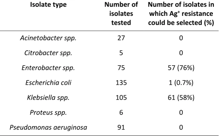
To detect cryptic Ag+ resistance, we screened all 444 isolates for their ability to yield resistant
colonies upon Ag+ challenge. Plating of saturated cultures onto agar containing AgNO
3 at 128
mg/L selected mutants exhibiting Ag+ resistance (MIC >128 mg/L) in ~76% isolates of
Enterobacter spp., ~58% isolates of Klebsiella spp., and ~0.7% isolates of E. coli (Table 1). The
frequency with which Ag+-resistant mutants arose was subsequently measured in a subset of
these strains (n=60). Mutation frequencies to Ag+ resistance in all 60 isolates were similar
(ranging from ~1.5 x 10⁻⁸ to ~5.2 x 10⁻⁷), and sufficiently high to imply that resistance was the
result of a single mutational event in each case. By contrast, resistance to Ag+ could not be
selected under these conditions in any of the other genera tested (Table 1).
To determine the genetic basis for Ag+ resistance in the mutants recovered, we proceeded on
the basis that the Ag+ resistance phenotype in these strains was likely the result of mutational
activation of a cryptic Sil system, and screened for the presence of the sil operon using PCR
to detect silS. In all 119 isolates in which spontaneous resistance to Ag+ could be selected, silS
was detected; by contrast, silS was not found in a cross-section of isolates (n=30) from which
Ag+ resistant mutants were not recovered. DNA sequence determination of the entire silS
gene from all 119 silS+ isolates, and from a single Ag+ resistant mutant recovered from each
isolate, revealed a single missense mutation in each resistant mutant by comparison with the
in all mutants selected is attributable to activation of the Sil system as a result of mutational
change in silS.
The SilS protein is the sensor histidine kinase (SHK) of a two-component regulatory system
that acts to positively regulate the sil operon via phosphorylation of the transcription factor,
SilR. 17 Though SilS is itself not well studied, SHK proteins as a class have been extensively
investigated, offering the opportunity to understand mutational gain of function in the SilS
protein by analogy. We employed Pfam 18 searches to predict the location and extent in SilS
of functional domains that are common to SHK proteins, and used this information to produce
a schematic of SilS onto which all of the substitutions identified in this study were mapped
(Figure 1). This analysis revealed that the substitutions leading to activation of SilS are
clustered within three functionally-distinct regions; the transmembrane domains, the
dimerization/histidine phosphotransfer (Dhp) domain and the catalytic/ATP-binding (CA)
domain (Figure 1). Substitutions identified in the latter two domains are located at similar
positions to those found in mutants of the SHKs EnvZ and PhoB that exhibit constitutive
activation as a consequence of being unable to dephosphorylate their cognate response
regulator, 19 suggesting that this is also the case for these mutant SilS proteins. By contrast,
the substitutions found in the transmembrane regions adjacent to the sensory domain may
prompt constitutive activation of the Sil system by uncoupling signalling from sensing. 20
In summary, within a large collection of recent Gram-negative clinical isolates, no overt
resistance to Ag+ was detected. Nevertheless, amongst Klebsiella and Enterobacter spp.,
high-level (MIC >128 mg/L) Ag+ resistance was readily selected in the majority of isolates, with
resistance arising in all cases apparently as a consequence of a single mutation in silS that led
to activation of the Sil system. The high frequencies (c. 10-8) with which Ag+ resistance arose
in these isolates is comparable to that seen for antibiotics that act upon a single cellular
target, agents that are generally considered unsuitable for monotherapy owing to resistance
liabilities and the consequent likelihood that resistance will arise during treatment. 21 Thus,
we consider that cryptic resistance has the potential to compromise the efficacy of Ag+-based
therapeutics, and suggest that it would be of benefit for clinicians to screen patients for the
presence of strains harbouring cryptic Ag+ resistance determinants prior to deployment of
such agents. We recommend the use of the simple screening approach for cryptic Ag+
onto agar containing suprainhibitory concentrations of Ag+, and noting the growth of resistant
colonies following overnight incubation.
Funding
This work was supported through sponsorship of EE by the Libyan Ministry of Higher
Education.
Transparency declaration
References
1. Edwards-Jones V. The benefits of silver in hygiene, personal care and healthcare. Lett Appl Microbiol 2009; 49: 147-52.
2. Silver S, Phung le T, Silver G. Silver as biocides in burn and wound dressings and bacterial resistance to silver compounds. J Ind Microbiol Biotechnol 2006; 33: 627-34.
3. Chopra I. The increasing use of silver-based products as antimicrobial agents: a useful development or a cause for concern? J Antimicrob Chemother 2007; 59: 587-90.
4. Randall CP, Gupta A, Jackson N et al. Silver resistance in Gram-negative bacteria: a dissection of endogenous and exogenous mechanisms. J Antimicrob Chemother 2015; 70: 1037-46.
5. Randall CP, Oyama LB, Bostock JM et al. The silver cation (Ag+): antistaphylococcal activity, mode of action and resistance studies. J Antimicrob Chemother 2013; 68: 131-8 6. Ip M, Lui SL, Chau SS et al. The prevalence of resistance to silver in a Burns unit. J Hosp Infect 2006; 63: 342-4.
7. Sutterlin S, Tano E, Bergsten A et al. Effects of silver-based wound dressings on the bacterial flora in chronic leg ulcers and its susceptibility in vitro to silver. Acta dermato-venereologica 2012; 92: 34-9.
8. Davis IJ, Richards H, Mullany P. Isolation of silver- and antibiotic-resistant Enterobacter cloacae from teeth. Oral microbiology and immunology 2005; 20: 191-4.
9. Finley PJ, Norton R, Austin C et al. Unprecedented Silver Resistance in Clinically Isolated Enterobacteriaceae: Major Implications for Burn and Wound Management. Antimicrob Agents Chemother 2015; 59: 4734-41.
10. Gupta A, Matsui K, Lo JF et al. Molecular basis for resistance to silver cations in Salmonella. Nat Med 1999; 5: 183-8.
11. Kremer AN, Hoffmann H. Subtractive hybridization yields a silver resistance determinant unique to nosocomial pathogens in the Enterobacter cloacae complex. J Clin Microbiol 2012; 50: 3249-57.
12. Sutterlin S, Edquist P, Sandegren L et al. Silver resistance genes are overrepresented among Escherichia coli isolates with CTX-M production. Appl Environ Microbiol 2014; 80: 6863-9.
13. Gupta A, Phung LT, Taylor DE et al. Diversity of silver resistance genes in IncH incompatibility group plasmids. Microbiology 2001; 147: 3393-402.
14. Clinical and Laboratory Standards Institute. Methods for dilution antimicrobial susceptibility tests for bacteria that grow aerobically Ninth Edition: Approved Standard M07-A9. CLSI, Wayne, PA, USA, 2012
15. O'Neill AJ, Cove JH, Chopra I. Mutation frequencies for resistance to fusidic acid and rifampicin in Staphylococcus aureus. J Antimicrob Chemother 2001; 47: 647-50.
16. Kahlmeter G, Brown DF, Goldstein FW et al. European harmonization of MIC breakpoints for antimicrobial susceptibility testing of bacteria. J Antimicrob Chemother 2003;
52: 145-8.
17. Silver S. Bacterial silver resistance: molecular biology and uses and misuses of silver compounds. FEMS Microbiol Rev 2003; 27: 341-53.
18. Finn RD, Coggill P, Eberhardt RY et al. The Pfam protein families database: towards a more sustainable future. Nucleic acids research 2016; 44: D279-85.
20. Hsing W, Russo FD, Bernd KK et al. Mutations that alter the kinase and phosphatase activities of the two-component sensor EnvZ. J Bacteriol 1998; 180: 4538-46.
Table 1. Prevalence of cryptic Ag+ resistance amongst the isolates analysed in this study.
Isolate type Number of isolates
tested
Number of isolates in which Ag+ resistance
could be selected (%)
Acinetobacter spp. 27 0
Citrobacter spp. 5 0
Enterobacter spp. 75 57 (76%)
Escherichia coli 135 1 (0.7%)
Klebsiella spp. 105 61 (58%)
Proteus spp. 6 0
Pseudomonas aeruginosa 91 0
Figure 1. Schematic of the SilS protein showing the predicted domain architecture by comparison with other sensor histidine kinase proteins, and indicating the sites of amino acid substitutions that activate the cryptic Sil system. The amino acid substitutions shown were all detected in single mutants, with the exception of A13V and G210E that were found in
[image:12.595.81.518.385.500.2]