This is a repository copy of
Nonequivalence of second sphere "noncatalytic" residues in
pentaerythritol tetranitrate reductase in relation to local dynamics linked to H-transfer in
reactions with NADH and NADPH coenzymes
.
White Rose Research Online URL for this paper:
http://eprints.whiterose.ac.uk/142170/
Version: Published Version
Article:
Iorgu, A.I., Baxter, N.J., Cliff, M.J. et al. (4 more authors) (2018) Nonequivalence of second
sphere "noncatalytic" residues in pentaerythritol tetranitrate reductase in relation to local
dynamics linked to H-transfer in reactions with NADH and NADPH coenzymes. ACS
Catalysis, 8 (12). pp. 11589-11599. ISSN 2155-5435
https://doi.org/10.1021/acscatal.8b02810
eprints@whiterose.ac.uk https://eprints.whiterose.ac.uk/
Reuse
This article is distributed under the terms of the Creative Commons Attribution (CC BY) licence. This licence allows you to distribute, remix, tweak, and build upon the work, even commercially, as long as you credit the authors for the original work. More information and the full terms of the licence here:
https://creativecommons.org/licenses/
Takedown
If you consider content in White Rose Research Online to be in breach of UK law, please notify us by
Nonequivalence of Second Sphere
“
Noncatalytic
”
Residues in
Pentaerythritol Tetranitrate Reductase in Relation to Local
Dynamics Linked to H
‑
Transfer in Reactions with NADH and NADPH
Coenzymes
Andreea I. Iorgu,
†Nicola J. Baxter,
†,⧧Matthew J. Cli
ff
,
†Colin Levy,
†Jonathan P. Waltho,
†,⧧Sam Hay,
†and Nigel S. Scrutton
*
,††
Manchester Institute of Biotechnology and School of Chemistry, Faculty of Science and Engineering, The University of Manchester, 131 Princess Street, Manchester M1 7DN, United Kingdom
⧧
Krebs Institute for Biomolecular Research, Department of Molecular Biology and Biotechnology, The University of Sheffield, Firth
Court, Western Bank, Sheffield S10 2TN, United Kingdom
*
S Supporting InformationABSTRACT: Many enzymes that catalyze hydride transfer reactions work via a mechanism dominated by quantum mechanical tunneling. The involvement of fast vibrational modes of the reactive complex is often inferred in these reactions, as in the case of the NAD(P)H-dependent pentaerythritol tetranitrate reductase (PETNR). Herein, we interrogated the H-transfer mechanism in PETNR by designing conservative (L25I and I107L) and side chain shortening (L25A and I107A) PETNR variants and using a combination of experimental approaches (stopped-flow rapid kinetics, X-ray crystallography, isotope/
temperature dependence studies of H-transfer and NMR spectroscopy). X-ray data show subtle changes in the local environment of the targeted
side chains but no major structural perturbation caused by mutagenesis of these two second sphere active site residues. However, temperature dependence studies of H-transfer revealed a coenzyme-specific and complex thermodynamic equilibrium
between different reactive configurations in PETNR−coenzyme complexes. Wefind that mutagenesis of these second sphere
“noncatalytic”residues affects differently the reactivity of PETNR with NADPH and NADH coenzymes. We attribute this to
subtle, dynamic structural changes in the PETNR active site, the effects of which impact differently in the nonequivalent reactive
geometries of PETNR−NADH and PETNR−NADPH complexes. This inference is confirmed through changes observed in the
NMR chemical shift data for PETNR complexes with unreactive 1,4,5,6-tetrahydro-NAD(P) analogues. We show that H-transfer rates can (to some extent) be buffered through entropy−enthalpy compensation, but that use of integrated experimental tools reveals hidden complexities that implicate a role for dynamics in this relatively simple H-transfer reaction. Similar approaches are likely to be informative in other enzymes to understand the relative importance of (distal) hydrophobic side chains and dynamics in controlling the rates of enzymatic H-transfer.
KEYWORDS: pentaerythritol tetranitrate reductase, kinetic isotope effects, hydride transfer, protein dynamics, protein NMR, protein crystallography
■
INTRODUCTIONUnderstanding the physical basis of enzyme catalysis is important from a fundamental point of view and also to drive applications in contemporary areas of research, such as biotechnology and synthetic biology. The development of sustainable and clean chemical manufacturing practices requires deep appreciation of the physical basis of enzyme catalysis that enables us to mimic and improve Nature’s catalysts. Enzymes catalyze many chemical reactions with extraordinary selectivity and specificity and, in some cases, extreme efficiency.1−3While it
is difficult to pinpoint the exact origin of the catalytic effect for
even one particular enzyme, the importance of electrostatic contributions,4 i.e., stabilization of the transition state and
hydrogen bonding,5 has long been recognized. Diverse
experimental and theoretical methods have been used to probe chemical steps and have contributed to debates relating to physical models of catalysis, in some cases moving beyond traditional semiclassical frameworks for enzyme catalysis.6−9A
fast-growing number of experimental studies indicate that protein dynamics might play a role in catalysis, across a hierarchy of time scales.10−14
Beyond experimental approaches, the involvement of dynamics has been supported also by
Received: July 17, 2018
Revised: October 23, 2018
Published: October 26, 2018
Research Article
pubs.acs.org/acscatalysis Cite This:ACS Catal.2018, 8, 11589−11599
License, which permits unrestricted use, distribution and reproduction in any medium, provided the author and source are cited.
Downloaded via UNIV OF MANCHESTER on February 5, 2019 at 11:33:29 (UTC).
computation.15−19
Slower (microsecond to millisecond) dynamics attributed to large-scale protein domain motions have been identified using NMR methodologies. Direct
coupling of fast (femtosecond to nanosecond) dynamics to chemistry is mostly inferred from kinetic isotope effect (KIE)
studies and supported by computational analysis such as transition path sampling.20−22The temperature dependence of
KIEs is regarded as a “gold standard” for probing quantum mechanical tunneling (QMT) for H-transfer reactions and some interpretations have inferred a role for distance sampling dynamical contributions in facilitating the tunneling reac-tion23−27
(although others have questioned these models28−30
). Studies with variant enzymes have led to models in which the temperature dependence of the KIE has been attributed to modulation of donor−acceptor distance (DAD) distributions and perturbation of high-frequency motions31or disruption of
dynamic networks32that extend to remote residues within the enzyme. Mutagenesis has been used to perturb active site structure and probe effects on H-transfer and inferred dynamical
contributions using a number of enzyme systems (e.g., dihydrofolate reductase,33soybean lipoxygenase-1,34
thymidy-late synthase,35ene-reductases36). Experimental KIE measure-ments have also been extended to include the use of high pressure37,38and“heavy enzymes”(2H,13C, and/or15N stable
isotope-labeled)39−44
to probe these effects further and
supported using computational simulations. Much of our current understanding of enzymatic catalysis has come from probing the effects of mutagenesis on catalytic rates and from
computational simulations. For H-transfer reactions, this has opened up debates on the relative importance of dynamics in these reactions and challenged the community to develop integrated experimental45 and theoretical22 approaches to address this problem.
Old Yellow Enzymes (OYEs) are an intensively investigated class of ene-reductases, which catalyze the asymmetric reduction of a wide variety of activatedα,β-unsaturated compounds, with high specificity for substrates bearing nitro or keto groups.46
While their physiological role and natural substrates are often not known,47OYEs are particularly attractive to study from a mechanistic perspective and for their biocatalytic potential. Pentaerythritol tetranitrate reductase (PETNR) is one such enzyme. PETNR has dual-specificity and is reduced by both
NADH and NADPH, but with a preference toward the latter coenzyme.48 The basis of this dual-specificity is not well
understood. The catalytic cycle of PETNR follows a single-site ping-pong mechanism. The first step (the reductive
half-reaction) involves hydride transfer from the C4pro-R hydrogen atom of the NAD(P)H coenzyme to the N5 atom of the noncovalently boundflavin mononucleotide (FMN) cofactor.49
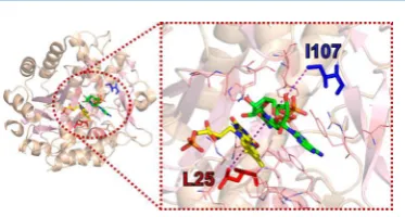
QMT contributes to this catalytic step,43 and several experimental studies have suggested an involvement of fast (picosecond to nanosecond) dynamics in the H-transfer reaction.23,50,51Here, we interrogated the H-transfer mechanism in PETNR by designing four variants of two second sphere residues (L25 and I107) positioned along the FMN-NAD(P)H N5−C4 axis (Figure 1), as inferred from the structure of PETNR in complex with the unreactive NADH analogue 1,4,5,6-tetrahydro-NAD.43L25 is located below the FMN, with
the side chain in van der Waals contact with the isoalloxazine ring of the FMN cofactor, while the side chain of I107 is positioned above the nicotinamide ring of the coenzyme. These residues are“noncatalytic”and are assumed not to contribute to major active site electrostatic effects. We have altered these
residues by making conservative (L25I and I107L) and side chain length shortening (L25A and I107A) variants of PETNR, with the objective of understanding how second sphere residues influence catalysis, H-tunneling and any vibrational modes
linked to H-transfer. We used X-ray crystallography to assess the impact of mutagenesis on overall structural properties and stopped-flow spectroscopy to analyze the kinetics of hydride
transfer through a combination of concentration and temper-ature dependence studies with both coenzymes (NADPH and NADH). Building on the recent NMR work in which a full sequential backbone assignment of PETNR holoenzyme was undertaken,52 we also used the coenzyme analogues 1,4,5,6-tetrahydro-NAD(P) (NAD(P)H4) to investigate coenzyme
binding modes in the ground-state PETNR−NADPH4 and PETNR−NADH4 complexes. Our overall aim was to under-stand how localized dynamics can impact H-transfer in each of these complexes and to investigate how perturbation of second sphere residues through mutagenesis impacts QMT and active site dynamics associated with this reaction with both NADPH and NADH.
■
RESULTS AND DISCUSSIONFirst, to ensure that mutagenesis of PETNR had not influenced
the coordination of the noncovalently bound FMN cofactor, we recorded UV−vis absorption spectra of the PETNR variants. All variants displayed the same spectroscopic features as wild-type PETNR (WT PETNR), with full FMN occupancy in the active site, and maintaining the characteristic PETNR-bound FMN spectral signature, with maxima at 380 and 465 nm (Figure S1 and Table S1). To assess any impact of mutagenesis on the general structure of the enzyme, we determined X-ray crystallographic structures for all variant forms isolated (Figures S2−S3 and Table S2). When compared to WT PETNR (PDB: 3P62), the RMSD values based on Cαpositions corresponding to each variant were in the range 0.3−0.4 Å, indicating a high degree of similarity. No major reorientations occurred in the loops bearing the side chain mutations, and superpositioning of all the structures indicates the tertiary TIM-barrel fold and external secondary features were largely unaffected by
[image:3.625.353.540.59.159.2]muta-genesis. Despite the overall structures of each of the four variants remaining largely unperturbed by mutagenesis, some subtle shifts in side chains are observed. One key shift in the I107A variant has led to a reorientation of the side chain of Q241 (highlighted inFigure S2). Removal of the I107 side chain bulk allows the side chain of Q241 to rotate toward the vacated space,
Figure 1.Crystal structure of the PETNR−NADH4complex (PDB:
3KFT),43showing the location of residues L25 (red) and I107 (blue), which have been targeted for mutagenesis. Residues located 5 Å away from the NADH4C4-H are represented as wireframes in the right panel
leading to a slight increase in the coenzyme binding pocket in this region. Altogether, the spectroscopic and structural evidence supports the hypothesis that the targeted residues are not directly involved in FMN binding and that the mutations do not affect the overall architecture of the enzyme. Secondary
impacts of these mutations resulting in subtle alterations within the active site are investigated below.
One of the benefits in using PETNR to study enzymatic
hydride transfer mechanisms is that individual catalytic steps can be accessed by stopped-flow measurements that track changes in
FMN absorbance during the reductive half-reaction of the enzyme catalytic cycle. The loss of FMN absorbance during reduction of PETNR by NAD(P)H reports directly on the hydride transfer step, which is essentially irreversible. The observed limiting rate constant (kred) at saturating coenzyme
concentration is therefore the rate of hydride transfer from which the intrinsic KIE values can also be obtained when using deuterated coenzyme. Note that previously we have not observed evidence for other processes affecting the kinetics of
H-transfer in OYEs, e.g., isomerization of the enzyme−substrate complex, and we propose that the measured KIE is therefore an intrinsic value, but we accept that the absence of evidence cannot be taken as proof.53To further support this proposition, we have measured the effect of pH on the rate of FMN reduction
with NADPH and (R)-[4-2H]-NADPH. Neither reaction is
significantly pH-dependent (Figure S4 and Table S3),
suggesting kinetic complexity is minimal and observed rate constants report directly on hydride transfer kinetics.
For the PETNR variant enzymes, the coenzyme concen-tration dependencies of hydride transfer rates were recorded (Figure S5). H-transfer was monitored by following the quenching of PETNR-bound FMN absorbance at 465 nm when mixing the enzyme with NAD(P)H (for representative kinetic traces, see Figure 2), as described previously.50,51 Limiting rate constants (kred) and apparent saturation constants
(KS) at 25°C for PETNR-catalyzed hydride transfer are shown
in Table 1. Although a plethora of data is available for WT PETNR from previous investigations, we re-examined all the kinetics of the WT enzyme for the purpose of this study to eliminate any possible disparities arising from slightly different
conditions, coenzyme purity, or experimental setup. We were able to confirm that there is no difference between thekredand
KSvalues obtained in this study and those previously published for WT PETNR.48
The kinetic traces measured at 25°C were adequatelyfit using
a single exponential function for WT PETNR and for some variants. However, the behavior of a number of variants was found to be more complex (vide infra for a more detailed analysis), with the majority of the reaction transientsfitting to a
double-exponential function, comprising a fast rate (kobs1) and a
slow rate (kobs2< 5 s−1). The values presented inTable 1reflect
only the fast rate of the reaction, as extracted from thefits of the
concentration dependence studies (Figure S5). By determining the saturation constant (KS) for each variant, we noticed that
there is no major difference in the apparent affinity for NADPH,
with the I107L variant having the same affinity (107±5μM) as WT PETNR (103 ± 4 μM), while the other variants have slightly higher values forKS, with the most prominent change for
L25A PETNR, which exhibits a 2-fold increase (KS= 199±6
μM).
[image:4.625.64.564.58.243.2]Analyzing the limiting rate of reduction, we observed correlations between the size of the side chain and the H-transfer rates: while the conservative (L25I and I107L) mutations have only a minor effect on the rate of FMN Figure 2.Stopped-flow kinetic traces showing FMN reduction for the PETNR variants with NADPH (top panel) and NADH (bottom panel) at selected representative temperatures (5, 25, and 40°C). All absorbance values were normalized for a better comparison. Conditions: 50 mM potassium
phosphate buffer (pH 7.0), 20μM enzyme, 10 mM NADPH or 25 mM NADH (final concentrations).
Table 1. Observed Rates of FMN Reduction at 25°C for PETNR Variants with NADPH and NADHa
kred(s−1) KS(μM) kred/KS(s−1μM)
NADPH
WT 33.43±0.22 103±4 (3.25±0.13)×10−1
L25A 55.75±0.35 199±6 (2.80±0.09)×10−1
L25I 29.49±0.49 130±8 (2.27±0.14)×10−1
I107A 21.22±0.25 130±9 (1.63±0.11)×10−1
I107L 28.37±0.20 107±5 (2.65±0.12)×10−1
NADH
WT 2.03±0.01 1067±13 (1.90±0.03)×10−3
L25A 3.62±0.04 2294±91 (1.58±0.06)×10−3
L25I 1.74±0.04 306±40 (5.69±0.94)×10−3
I107A 2.05±0.04 329±37 (6.23±0.71)×10−3
I107L 1.94±0.02 889±36 (2.18±0.09)×10−3
aCalculated from fitting data presented in Figure S5 to kobs1 =
[image:4.625.324.564.321.466.2]reduction, the alanine substitutions impose a markedly modified
reaction rate. The rate of H-transfer is 60% faster for the L25A variant than for WT PETNR, while a 40% reduction in rate is observed for the I107A variant (Table 1). This suggests that an increase in the mobility of the FMN binding site, caused by shortening the L25 side chain, is enhancing catalytic rates, while a larger void above the NADPH substrate, induced by truncation of I107 side chain, is decreasing catalytic rates. While the L25A and L25I variants exhibit a very similar change in rate for the reaction with NADH (when compared to NADPH), the mutations introduced at the I107 site do not affect the limiting
rate constant and, moreover, afford better binding of NADH.
When comparingkred/KS(which reports, in this case, on the
efficiency of each variant to perform the reductive half-reaction),
we conclude that the increase in rate for L25A variants comes with a cost of lower binding affinity, leading to a slightly less
efficient enzyme. We have noticed similar compensatory
behavior in the reaction of other OYEs with nicotinamide coenzyme biomimetics, where elevated catalytic rates are associated with inverse changes in binding affinity.54 The
I107A variant is more efficient in the reaction with NADH and is
noticeably less efficient for performing H-transfer from
NADPH. Further, we performed temperature dependence studies of the rate of FMN reduction with both NADPH and NADH coenzymes, along with their corresponding deuterated forms, in the range of 5−40°C. The unusual behavior of the variants is first noticed through the shape of the transients
observed, with all variants manifesting multiple kinetic phases at elevated temperatures (Figure 2). In general, the kinetic traces could be fitted to a single exponential function for low
temperatures (<20 °C), while at higher temperatures the FMN reduction takes place with multiple resolvable kinetic phases.
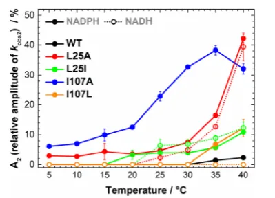
It was observed that the amplitude of the slow kinetic components increases with increasing temperature (Figure 3), with the effect being more pronounced for the L25A and I107A
variants in the case of PETNR reduction by NADPH (∼40% of the total absorbance change attributed tokobs2at 40°C). The
reduction with deuterated substrates follows closely that
observed for NADPH (Figure S6 and Tables S4−S13). This suggests that the reaction chemistry (H vs D transfer) is not kinetically controlled, and the differences observed are solely
attributed to perturbations in the enzyme−coenzyme complex arising from mutagenesis of PETNR. FMN reduction follows the same trend with NADH for L25A and L25I variants, with multiple reactive configurations (MRCs) at elevated
temper-ature and similar changes of amplitude contributions for each kinetic component. However, the mutagenesis of the I107 site does not induce the formation of MRCs in reactions with NADH (the kinetic tracesfit to a single exponential function
across the studied temperature range,Figure 3andFigure S7), a contrasting behavior when compared to reduction by NADPH. This indicates that truncation of the I107 site has no detrimental effect on H-transfer from NADH coenzyme, while the NADPH
reaction is noticeably affected. This suggests truncation of the
I107 side chain induces propagated effects (which extend
beyond the tilting of the Q241 side chain observed from X-ray data) on the structure (and dynamics) of the enzyme.
We have previously detected MRCs in the N189A variant of morphinone reductase (MR), another enzyme belonging to the OYE family, in which four detectable MRCs could be kinetically resolved.36 Likewise, the data for the reduction of PETNR variants suggests that NADPH and NADH are able to bind in multiple conformations in the active site of PETNR variants, which leads to different competent states of the enzyme− coenzyme complex that are able to perform H-transfer, observed as multiple kinetic phases bearing KIEs (Figures S8 and S9). While the kred value is highest for L25A, this fast reactive conformation is very slowly interconverting (<1 s−1) with
another slow reactive conformation. In our previous work involving MR,36we were able to corroborate that the resolved
kinetic phases represent different configurations of the enzyme− coenzyme complex using computational methods. Herein, we propose that our observations with PETNR are consistent with those we have reported previously with MR. However, we cannot exclude other explanations at this stage, e.g., a mechanism involving multiple free enzyme species that have different reactivities. For example, an isotopically insensitive
isomerization from an inactive configuration into an active
configuration would be expected to diminish the observed KIE.
Regardless of the origin of multiple kinetic phases observed in the variants presented here, these data demonstrate that second sphere mutations in the PETNR active site have differing effects
on the reaction with NADH and NADPH.
Given the KIE values at 25°C are only slightly affected by
mutagenesis, with small differences between the studied variants
(Figure S8), we consider that a better way to interpret the data (as we have recently described elsewhere51) is by comparing the activation parameters (presented inTable 2). We analyzed the thermodynamic parameters of the H-transfer reaction byfitting
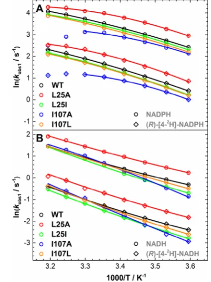
the Arrhenius plots ofkobs1(Figure 4) to the nonlinear
Eyring-based function
i k jjjj y{zzzz
= − Δ + Δ −
+
Δ + Δ
⧧ ⧧
⧧ ⧧
( )
k k T
h
H C T T
RT
S C
R
ln ln ( )
ln T
T
T
T
obs B
p 0
p
o
0
0
(1)
whereΔHT0⧧andΔST0⧧are the apparent activation enthalpy
[image:5.625.85.285.514.658.2]and entropy, respectively, at a reference temperature (T0, 298 K in this study),ΔCp⧧is the difference in heat capacity between Figure 3.Manifestation of multiple reactive configurations as a function
the reactant and transition states, and kB, h, and R are the
Boltzmann, Planck, and ideal gas constants, respectively.55,56 This model has recently been used for interpreting curvatures observed in Eyring plots for a number of enzymes,57,58 and recent computational work suggests changes inΔCp⧧are related
to dynamical changes during catalysis and these changes are attributed from contributions of not only active site regions but also distal domains of the enzyme (dynamical changes that are dispersed throughout the entire structure).59
For the reactions with NADPH and (R)-[4-2H]-NADPH, the
apparent activation enthalpy values for WT PETNR are identical with those previously reported, with a coenzyme isotope effect
of 6.4±0.4 kJ mol−1, which we previously inferred to be an
evidence of a“soft”promoting motion (a promoting vibration with a relatively small force constant) contributing to catalysis.43 The kinetic isotope effect on the FMN reduction with NADH
and (R)-[4-2H]-NADH in WT PETNR manifests less
temper-ature independence (ΔΔHT0⧧ = 4.9 ± 0.2 kJ mol−1) when
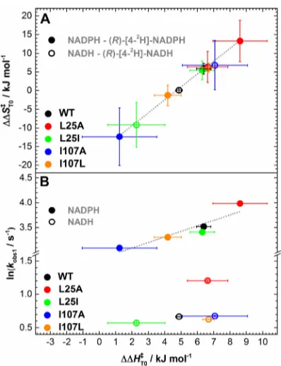
compared with the KIE on the NADPH reaction (note that early work indicated a measurable temperature-independent KIE, which we now consider to be underestimated43). Overall, when comparing the apparent activation enthalpies and entropies for the PETNR variants, we observed a strong compensatory behavior (Figure S10). This linear enthalpy−entropy compen-sation was recently observed in a study involving “heavy”
PETNR isotopologues.51 This compensation effect is closely
observed forΔΔHT0⧧andΔΔS
T0⧧: changes inΔΔHT0⧧values
are reflected by similar changes in ΔΔST0⧧ (Figure 5A).
Moreover, for the NADPH reaction, there is a strong correlation between the rate of the reaction and the coenzyme KIEs (differences in the apparent activation enthalpy, (R)-[4-2
H]-NADPH−NADPH), but there is no correlation for the NADH reaction (Figure 5B).
The more prominent changes inΔCp⧧are observed for the
variants with side chain truncations (L25A and I107A), which manifest a more negativeΔCp⧧when compared to WT PETNR.
A recent study of ketosteroid isomerase andα-glucosidase MalL showed that the decrease inΔCp⧧ can be explained through
significant dynamical contributions of regions remote from the
active site.59 In the case of PETNR, the effect of side chain
truncation on catalytic rates, enthalpy values, coupled with the nonequivalence in reactivity toward the two coenzyme and changes in ΔCp⧧, could inform on vibrational coupling
[image:6.625.62.567.85.295.2](previously observed in “heavy enzyme” studies19,50) that is
Table 2. Extracted Parameters from Kinetic and Thermodynamic Studiesa
ΔHT0⧧(kJ mol−1) ΔST0⧧(J mol−1K−1) ΔCp⧧(kJ mol−1K−1) ΔHT0⧧(kJ mol−1) ΔST0⧧(J mol−1K−1) ΔCp⧧(kJ mol−1K−1)
NADPH NADH
WT 30.4±0.3 −113.9±0.9 −0.50±0.05 35.4±0.1 −120.5±0.5 0.39±0.03
L25A 23.3±1.2 −133.7±4.1 −0.92±0.12 33.6±0.5 −122.1±1.7 0.28±0.05
L25I 30.4±0.6 −114.9±2.0 −0.43±0.07 42.7±1.7 −97.7±6.0 0.32±0.18
I107A 17.8±2.3 −160.2±7.7 −1.08±0.23 46.4±1.1 −83.8±3.6 −0.47±0.22
I107L 31.5±0.7 −111.6±2.4 −0.44±0.08 38.7±0.2 −110.0±0.8 0.23±0.04
(R)-[4-2H]-NADPH (R)-[4-2H]-NADH
WT 36.8±0.3 −108.0±0.1 −0.32±0.04 40.3±0.1 −120.4±0.5 0.46±0.02
L25A 31.9±1.1 −120.5±3.7 −0.78±0.15 40.3±1.1 −115.7±3.8 0.42±0.14
L25I 36.7±0.5 −109.5±1.6 −0.53±0.09 44.9±0.2 −106.8±0.8 0.30±0.07
I107A 19.0±0.2 −172.6±0.7 −1.43±0.02 53.5±1.7 −77.0±5.6 0.17±0.22
I107L 35.7±0.4 −112.9±1.4 −0.65±0.07 45.4±0.3 −103.9±0.9 0.43±0.07
Isotope Effect: (R)-[4-2H]-NADPH−NADPH Isotope Effect: (R)-[4-2H]-NADH−NADH
WT 6.4±0.4 5.8±1.5 0.18±0.06 4.9±0.2 0.1±0.7 0.07±0.04
L25A 8.6±1.27 13.3±5.6 0.14±0.19 6.6±1.2 6.4±4.2 0.13±0.15
L25I 6.3±0.7 5.4±2.5 −0.11±0.11 2.3±1.8 −9.2±6.0 −0.03±0.19
I107A 1.2±2.3 −12.3±7.7 −0.35±0.23 7.1±2.0 6.8±6.7 0.64±0.31
I107L 4.2±0.8 −1.3±2.8 −0.22±0.11 6.7±0.4 6.1±1.2 0.19±0.08
aApparent activation parameters were obtained byfitting the data inFigure 4toeq 1withT0= 298 K. The isotope effects are the differences in
activation parameters (e.g.,ΔHT0⧧,D−ΔHT0⧧,H).
Figure 4.Arrhenius plots of the observed rate of hydride transfer from (A) NADPH and (R)-[4-2H]-NADPH and (B) NADH and (R
)-[4-2H]-NADH to FMN in studied PETNR variants; plots ofkobs1, data
fromTables S4−S13. SeeTable 2for extracted parameters. Note: The
decrease inkobs1values for I107A PETNR reaction with NADPH and (R)-[4-2H]-NADPH at 35−40°C suggests a different mechanism at
[image:6.625.70.283.331.615.2]extended beyond the mutagenesis site(s). While it is assumed thatΔCp⧧values are negative for enzyme systems in which the
chemical reaction is rate limiting (as the heat capacity for enzyme−substrate complex is larger than for the enzyme− transition state complex),59 most of the data for PETNR reduction by NADH show positive values forΔCp⧧.
The kinetic and thermodynamic analysis of the reduction of FMN in the PETNR variants reveals coenzyme-specific
perturbations to the H-transfer chemistry and thermal equilibrium of reactant states. The current knowledge we have at hand to rationalize these findings comes mainly from
crystallographic studies (presented herein) and previously performed molecular dynamics (MD) simulations. The MD data suggest a similar binding mode for both NADPH and NADH coenzymes in the active site of PETNR,43which makes it difficult to rationalize the differences in H-transfer chemistry and
thermal conformation equilibrium presented above. Although the crystal structure of the PETNR−NADH4complex (PDB: 3KFT43) provides valuable insight into the structural proximity of reacting groups in the active site,π−πstacking between the symmetry-related adenosine moieties of NADH4 in the two
PETNR−NADH4monomers present in the asymmetric unit of the crystal lattice results in the pyrophosphate and adenosine groups of NADH4being poorly coordinated by PETNR. The
absence of such interactions limits predictions and analyses that can be made regarding the conformational preferences of NADPH4binding to PETNR. Despite numerous attempts, we
have been unable to crystallize the PETNR−NADPH4complex, which limits further structural and computational investigations. Consequently, we pursued NMR studies of WT PETNR and recently reported the 1H, 15N, and 13C sequential backbone
resonance assignments of the holoenzyme.52Building on this
recent work, we have now performed near-complete backbone resonance assignments of the ground-state PETNR−NADH4
and PETNR−NADPH4complexes at saturating concentrations of either NADH4or NADPH4, which we further used to inspect
differences in the binding modes between the coenzymes.
Given the large size of PETNR (40 kDa), TROSY-based 3D heteronuclear experiments were acquired and analyzed, which enabled a highly successful degree of assignment (97%) of the
1H−15N TROSY spectrum for each complex. Compared to the
extent of assignment achieved for PETNR (333 residues), in the case of the PETNR−NADH4 complex, 334 residues were successfully assigned, as H184 could be identified as a sharp and
intense peak after ligand addition. Further assignment of one more residue of the flexible loop (D274) was also achieved;
however, residue R164 could not be assigned, most probably due to peak overlap. The PETNR−NADPH4 complex was assigned to the greatest extent (335 residues), with all residues identified as in the PETNR−NADH4complex, as well as the additional assignment of residue R164.
In order to assess the impact of the NAD(P)H4 coenzyme
analogue binding on the PETNR structure, we comparatively analyzed the 1H−15N TROSY spectrum of PETNR with 1H−15N TROSY spectra of the PETNR−NADH
4 and
PETNR−NADPH4complexes. Overall, the changes in chemical shift for1H
Nand15N are limited to similar residue segments for
all complex comparisons (Figure S11). The differences in
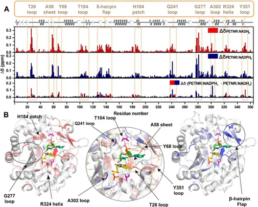
combined chemical shift between each complex and PETNR, and a visual mapping of these changes onto the crystal structure of the PETNR−NADH4complex (PDB: 3KFT43) is illustrated inFigure 6. It can be observed that, while the majority of the peaks are essentially not sensitive to coenzyme analogue binding, localized areas of PETNR are noticeably affected by
the addition of either NADPH4or NADH4(seeFigure S12for a
structural overview of notably affected regions). Most of the
changes that occur in PETNR upon binding of the coenzyme analogues are localized approximately 5 Å away from either NADH4or the FMN cofactor, with similarly affected sites for
both the NADPH4and NADH4analogues. The regions that are
affected significantly by binding of NAD(P)H
4are depicted in
Figure 6B. These encompass several loop regions (with maximum chemical shift differences recorded at residues T26,
Y68, T104, Q241, G277, A302, Y351), a histidine“patch”that is known from crystallographic data to coordinate the NADH4
coenzyme analogue60(denoted the H184 patch) and anα-helix
found in close contact with the FMN cofactor (denoted the R324 helix). Coenzyme analogue binding is also observed to cause significant chemical shift changes in theβ-hairpinflap (a
region encompassing N126-T147), suggesting a major role of this structural motif in binding the NAD(P)H coenzyme in the active site. The significant differences in chemical shift between
PETNR and the PETNR−NAD(P)H4complexes observed in theβ-sheet containing A58 and the loop involving Y351 (which are known to coordinate the FMN cofactor60), along with differences located at the top of the β-barrel that is in close
proximity to FMN, suggest that NAD(P)H4 binding induces
FMN repositioning within the active site for efficient H-transfer.
This is supported by the formation of a charge-transfer complex between NAD(P)H4 and FMN, resulting in changes to the
electrostatic distribution within the system, which contributes to the observed differences in chemical shift. For T26, the large
difference in chemical shift can be explained by the formation of
a hydrogen bond between the side chain hydroxyl group of T26 and the NH2 group of the carboxamide function of the
nicotinamide ring in NAD(P)H4and by perturbations in the
[image:7.625.81.280.64.323.2]hydrogen bond between the backbone amide proton of T26 and
Figure 5.Relationship between the differences (coenzyme KIE) in apparent activation enthalpy (ΔΔHT0⧧) and entropy (A) and
correlations betweenΔΔHT0⧧and observed rate constant (B) during
the N5 atom of FMN. For the resonances assigned in the G277 loop, the NMR data suggest that coenzyme analogue binding induces a dramatic reorganization of the loop. This effect is
propagated into theα-helix from which the G277 loop emerges, which is also moderately affected by NAD(P)H4binding, even
though it is positioned far away from the active site.
Overall, similar residue segments show differences in
backbone amide 15N chemical shifts between the PETNR−
-NADH4and PETNR−NADPH4complexes (Figure 7). Analysis
of amide15N chemical shift changes (Δδ
N) can provide valuable
information on the location of conformational differences
between the complexes, as they report mainly on changes in backbone torsion angles. The only structural difference between
the coenzyme analogues is the presence of a 2′-phosphate group on the ribose ring of the adenosine moiety in NADPH4.
[image:8.625.61.566.68.473.2]Therefore,ΔδNvalues reported between these analogues reflect Figure 6.Observed changes in chemical shift for PETNR upon binding the NADH4or the NADPH4coenzyme analogues. (A) Histograms of the
residue-specific chemical shift changes for the backbone amide groups in PETNR upon binding NADH4(red) or NADPH4(blue). The absolute chemical shift changes were calculated using the following equation:ΔδX= [(δHNPETNR−δHNX)2+ (C(δNPETNR−δNX))2]1/2, whereδHNis the
backbone amide proton chemical shift,δNis the backbone amide nitrogen chemical shift,Cvalue is 0.12 (for rescaling ofδNvalues), andXis either the
PETNR−NADH4complex or the PETNR−NADPH4complex. The lower histogram shows the residue-specific chemical shift differences between the
PETNR−NADH4complex and the PETNR−NADPH4complex, with red and blue bars indicating larger changes in PETNR upon binding NADH4or
NADPH4, respectively. The absolute chemical shift differences between the PETNR−NAD(P)H4 complexes were calculated as Δδ =
[(ΔδPETNR−NADPH4−ΔδPETNR−NADH4)
2]1/2. The secondary structure of PETNR is shown at the top of thefigure with gray helices denoting
α-helices and white arrows indicatingβ-strands. Regions with significantΔδvalues are highlighted with segment labels. (B) Distributions ofΔδvalues plotted onto the structure of the PETNR−NADH4complex (PDB: 3KFT). Residues with significantΔδvalues are colored red (ΔδPETNR−NADH4, left
panel) and blue (ΔδPETNR−NADPH4, right panel). The FMN cofactor is shown as yellow sticks, the NADH4coenzyme analogue is shown as green sticks,
and the mutation sites (L25 and I107) are depicted as purple sticks. The central panel encompasses a zoomed-in view of the active site and highlights theΔδvalues between the two complexes (red, largerΔδupon PETNR−NADH4complex formation; blue, largerΔδupon PETNR−NAPDH4
how PETNR specifically modifies its conformational and
dynamic preferences to accommodate NADPH4 binding in
the active site.
The residue with the largestΔδNvalue involves T131, present in a cluster of contiguous residues (T129−R134 and A140− T147 of theβ-hairpinflap), which have significant15N chemical
shift differences (Figure 7andFigure S11). Although the R130
side chain and the putative location of the 2′-phosphate group
are not within the hydrogen-bonding distance in the crystal structure of the PETNR−NADH4complex, it is likely that bond rotation would allow productive coordination of these groups in solution. Structural perturbations within theβ-hairpinflap are
propagated to Y68 and also to Q241 via a network of side chain hydrogen bonds involving Y186, which are positioned directly over the face of the nicotinamide ring of NADH4 in close
proximity to I107. Conformational differences within the β
-hairpinflap are also propagated through side chain hydrogen
bonding with R134 to the L25−I31 and E348−G352 regions. For the D274−G277 loop with missing structural density in the crystal, chemical shift differences in the G278−E283 region indicate that the loop likely coordinates the coenzyme. Remarkably, chemical shift perturbations are also noted for G301−A302 and F322−G323, which are residues that coordinate the phosphate group present in FMN, indicating that conformational and dynamic preferences for NADH4and
NADPH4coordination by PETNR are propagated through the
charge-transfer complex formed with FMN.
Based on the differences in chemical shift observed between
PETNR and the PETNR−NAD(P)H4complexes, we can now rationalize, in part, the kinetic behavior of the investigated variants. Residue L25 presents large differences in chemical shift
for both PETNR−NADH4and PETNR−NADPH4complexes, and these observations are most readily explained by its close proximity to T26, which is a residue directly involved in FMN cofactor binding.60 In the crystal structure, residue I107 is
positioned∼8 Å away from the face of the NADH4nicotinamide ring and is sandwiched between Y68, Y186, and Q241, which make direct contact with the coenzyme analogue and a segment of the β-hairpin flap. I107 is slightly affected by NADPH4
binding, but not by NADH4 binding, suggesting that the
presence of phosphate in the coenzyme analogue contributes to a more pronounced reorientation of the loop containing I107. Overall, the structural features in the vicinity of the FMN cofactor (and residue L25) are highly affected by both
coenzymes, with higher chemical shift changes occurring upon NADH4addition, while the regions affected more by NADPH4
binding are situated in the vicinity of residue I107 (Figure 6B). The fact that residue I107 is not affected by NADH
4binding,
and H-transfer kinetics are also not majorly affected for the
reduction with NADH (i.e., FMN reduction occurs in a single phase), as in contrast with NADPH4, informs clearly about
differences in the binding modes of the two coenzymes, which
have consequences on the H-transfer mechanism. Along with the observed reorientation of Q241 side chain in I107A variant, the NMR and thermodynamic data suggests extended vibra-tional coupling is present during H-transfer from NADPH to PETNR.
To further the analysis of the binding modes of the two coenzymes, we performed an analysis of the1H line width of
peaks in the 1H−15N TROSY spectra (Figure S13), which
enables detection of residues that have a significant exchange
contribution to the transverse relaxation rate (R2). Broadening of the line width suggests conformational exchange between two or more species on the microsecond to millisecond NMR time scale.61It can be observed that PETNR exhibits a high degree of
conformational exchange on this time scale, with most of the regions affected being located around the active site (Figure
S14). A reduction in the exchange contribution toR2can be
observed for some residues following addition of NADH4, such
as those situated in the G277 loop. The minimization of this dynamic behavior is even more pronounced in the case of the PETNR−NADPH4 complex, for which most of these slow conformational exchange processes are removed. The data indicate that the NADPH4coenzyme mimic is interacting more
tightly with PETNR than its nonphosphorylated counterpart, which explains the higher affinity of PETNR for NADPH than
for NADH (Table 1). The mutations chosen for kinetic studies are located on the edge of these regions that exhibit microsecond to millisecond conformational exchange in PETNR. Residue I107 is not a dynamic center, but it is located in a region with moderate dynamic behavior in PETNR, while line widths for residues in the vicinity of L25 are considerably broadened. Both regions around the targeted residues show a restriction in conformational exchange on addition of coenzyme, suggesting an important role of binding in controlling enzyme motions on the microsecond to millisecond time scale.
This study was undertaken to investigate how second sphere
“noncatalytic”residues influence vibrational modes linked to
H-transfer and H-tunneling in PETNR. The use of a combination of experimental tools (e.g., stopped-flow rapid kinetics, isotope/
temperature dependence studies of H-transfer and NMR spectroscopy) allowed us to underpin a complex, coenzyme-specific kinetic and thermodynamic equilibrium during
H-transfer in the investigated variants. The side chain modifi
[image:9.625.67.298.76.224.2]ca-tions (L25I, L25A, I107A, and I107L PETNR) were designed on the basis of their location within the active site (situated along the FMN-NAD(P)H N5−C4 axis in the PETNR−NAD(P)H complexes). The hypothesis that the targeted side chains are not
Figure 7.Structural mapping of absolute chemical shift differences for b a c k b o n e a m i d e 1 5N a t o m s c a l c u l a t e d a s
ΔδN =
[(δNPETNR−NADPH4 − δNPETNR−NADH4)2]1/2 plotted onto
the structure of the PETNR−NADH4complex (PDB: 3KFT) (Figure
S11). Residues with significantΔδNvalues are colored red, with a stronger intensity of color representing a higherΔδNvalue, with a cutoff
of 0.1 ppm applied. The side chains are shown as wireframes in the putty representation of the protein, the FMN cofactor is shown as yellow sticks, the NADH4coenzyme analogue is shown as green sticks,
involved in major electrostatic interactions within the active site was confirmed through stationary UV−vis spectroscopy and crystallographic data. The side chain modifications are subtle,
but they prompt changes to the conformational landscape of the enzyme that can be observed during the reductive half-reaction using stopped-flow techniques. The rate of H-transfer at room
temperature is broadly maintained through enthalpy−entropy compensation. However, temperature dependence studies of H-transfer revealed a coenzyme-specific and complex
thermody-namic equilibrium between different reactive configurations in
PETNR−coenzyme complexes. We find that mutagenesis of
these second sphere“noncatalytic”residues (L25 and I107) has differing effects on the reactivity (activation entropy and
enthalpy) of PETNR with NADPH and NADH coenzymes. We attribute these differences in coenzyme reactivity to subtle,
dynamic structural changes in the PETNR active site. This interpretation is aided by NMR chemical shift data measured for PETNR−NADH4and PETNR−NADPH4, which show struc-tural differences between the two complexes.
■
CONCLUDING REMARKSEnzymes have evolved to catalyze chemical reactions with extraordinary selectivity and specificity, and it is often assumed
that predominantly one configuration of the enzyme−substrate
complex will be the catalytically active one. This hypothesis has a strong physical basis for the reaction of WT enzymes with their natural substrates. However, when dealing with enzyme variants, the perturbation of the finely tuned enzyme structure by
mutagenesis can change the conformational flexibility and
geometries in the enzyme to the extent that multiple reactive conformations are populated. To date, kinetic studies of OYEs have allowed us to demonstrate that variants of two enzymes in this family (N189A variant of MR36 and this study) exhibit shifted distributions of reactive conformational states when compared to the corresponding WT enzymes. While the N189A variant of MR could be considered a disruptive mutation (N189 is intimately involved in coenzyme binding through H-bonding), it is not the case for the variants in the current study. Herein, the synergistic use of complementary spectro-scopic techniques has allowed us to unravel the nonequivalence of second sphere “noncatalytic” residues in PETNR. The complex kinetic behavior observed upon truncation of L25 and I107 side chains indicates long-range cooperativity throughout the active site. We suggest that formation of MRCs could be a common feature when investigating variant enzymes, and other authors have pointed toward the existence of parallel reaction pathways during catalysis.62−65Moreover, kinetic complexities
on H-transfer caused by (distal) mutagenesis have been observed in dihydrofolate reductase catalysis,31and these effects
have been correlated with protein dynamics contribu-tions.26,33,66−68
While we are aware of the experimental challenges that would allow us to visualize MRCs in a wider range of H-transfer enzymes, we propose that synergistic experimental approaches should aid interpretation of kinetic data. This study is an illustration of how the use of integrated structural and kinetic experimental tools can uncover details often masked in more traditional studies of H-transfer (e.g., those reliant only on the use of steady-state turnover studies with isotopically labeled substrates). These approaches are needed to identify these hidden complexities even for relatively simple reactions such as H-transfer. Similar approaches are therefore likely to be informative with other enzymes to understand the
relative importance of small-scale dynamics in controlling enzymatic rates of reaction.
■
ASSOCIATED CONTENT*
S Supporting InformationThe Supporting Information is available free of charge on the ACS Publications websiteat DOI:10.1021/acscatal.8b02810.
Experimental details including overexpression of PETNR variants and 2H,13C,15N-labeled PETNR, synthesis of
deuterated and reduced coenzymes, conditions used (and data analysis details) for stopped-flow experiments,
crystallography and NMR spectroscopy; additional data including static UV−vis spectroscopy, crystallography, tabulated rate constants, and additionalfigures for kinetic
and NMR data (Figures S1−S14, Tables S1−S13) (PDF)
■
AUTHOR INFORMATIONCorresponding Author
*E-mail:nigel.scrutton@manchester.ac.uk.
ORCID
Andreea I. Iorgu:0000-0002-1363-3697 Jonathan P. Waltho:0000-0002-7402-5492 Sam Hay:0000-0003-3274-0938
Nigel S. Scrutton:0000-0002-4182-3500
Notes
The authors declare no competingfinancial interest.
■
ACKNOWLEDGMENTSA.I.I. acknowledges the funding received as an Early Stage Researcher through the Marie Curie Initial Training Network MAGIC (the EU’s Seventh Framework Programme, Grant Agreement No. 606831). N.S.S. was an Engineering and Physical Sciences Research Council (EPSRC) Established Career Fellow (EP/J020192/1). The work was supported, in part, by the Biotechnology and Biological Sciences Research Council (BBSRC; BB/M007065/1 BB/H021523/1 and BB/ M021637/1). We thank Dr. Alexander Geddes for site-directed mutagenesis and preliminary kinetic characterization of the variant enzymes, Dr. Andrew Guerriero for NMR groundwork, and the Diamond Light Source for access to beamlines i24, i04-1, i04, i03 (proposal nos. 8997-2, 8997-8, 12788-26, 12788-35).
■
REFERENCES(1) Lad, C.; Williams, N. H.; Wolfenden, R. The Rate of Hydrolysis of Phosphomonoester Dianions and the Exceptional Catalytic Proficien-cies of Protein and Inositol Phosphatases.Proc. Natl. Acad. Sci. U. S. A.
2003,100, 5607−5610.
(2) Radzicka, A.; Wolfenden, R. A Proficient Enzyme.Science1995,
267, 90−93.
(3) Edwards, D. R.; Lohman, D. C.; Wolfenden, R. Catalytic Proficiency: The Extreme Case of S-O Cleaving Sulfatases. J. Am. Chem. Soc.2012,134, 525−531.
(4) Warshel, A.; Sharma, P. K.; Kato, M.; Xiang, Y.; Liu, H.; Olsson, M. H. M. Electrostatic Basis for Enzyme Catalysis.Chem. Rev.2006,106, 3210−3235.
(5) Cleland, W. W.; Frey, P. A.; Gerlt, J. A. The Low Barrier Hydrogen Bond in Enzymatic Catalysis.J. Biol. Chem.1998,273, 25529−25532. (6) Nagel, Z. D.; Klinman, J. P. Tunneling and Dynamics in Enzymatic Hydride Transfer.Chem. Rev.2006,106, 3095−3118.
(8) Schwartz, S. D.; Schramm, V. L. Enzymatic Transition States and Dynamic Motion in Barrier Crossing.Nat. Chem. Biol.2009,5, 551− 558.
(9) Hay, S.; Scrutton, N. S. Good Vibrations in Enzyme-Catalysed Reactions.Nat. Chem.2012,4, 161−168.
(10) Eisenmesser, E. Z.; Bosco, D. A.; Akke, M.; Kern, D. Enzyme Dynamics during Catalysis.Science2002,295, 1520−1523.
(11) Agarwal, P. K. Enzymes: An Integrated View of Structure, Dynamics and Function.Microb. Cell Fact.2006,5, 2.
(12) Henzler-Wildman, K.; Kern, D. Dynamic Personalities of Proteins.Nature2007,450, 964−972.
(13) Klinman, J. P.; Kohen, A. Evolutionary Aspects of Enzyme Dynamics.J. Biol. Chem.2014,289, 30205−30212.
(14) Zoi, I.; Suarez, J.; Antoniou, D.; Cameron, S. A.; Schramm, V. L.; Schwartz, S. D. Modulating Enzyme Catalysis through Mutations Designed to Alter Rapid Protein Dynamics.J. Am. Chem. Soc.2016,138, 3403−3409.
(15) Agarwal, P. K.; Billeter, S. R.; Rajagopalan, P. T. R.; Benkovic, S. J.; Hammes-Schiffer, S. Network of Coupled Promoting Motions in Enzyme Catalysis.Proc. Natl. Acad. Sci. U. S. A.2002,99, 2794−2799. (16) Núñez, S.; Antoniou, D.; Schramm, V. L.; Schwartz, S. D. Promoting Vibrations in Human Purine Nucleoside Phosphorylase. A Molecular Dynamics and Hybrid Quantum Mechanical/Molecular Mechanical Study.J. Am. Chem. Soc.2004,126, 15720−15729.
(17) Hatcher, E.; Soudackov, A. V.; Hammes-Schiffer, S. Proton-Coupled Electron Transfer in Soybean Lipoxygenase: Dynamical Behavior and Temperature Dependence of Kinetic Isotope Effects.J. Am. Chem. Soc.2007,129, 187−196.
(18) Johannissen, L. O.; Hay, S.; Scrutton, N. S.; Sutcliffe, M. J. Proton Tunneling in Aromatic Amine Dehydrogenase Is Driven by a Short-Range Sub-Picosecond Promoting Vibration: Consistency of Simu-lation and Theory with Experiment.J. Phys. Chem. B2007,111, 2631− 2638.
(19) Delgado, M.; Görlich, S.; Longbotham, J. E.; Scrutton, N. S.; Hay, S.; Moliner, V.; Tuñón, I. Convergence of Theory and Experiment on the Role of Preorganization, Quantum Tunneling, and Enzyme Motions into Flavoenzyme-Catalyzed Hydride Transfer. ACS Catal.
2017,7, 3190−3198.
(20) Wang, Z.; Antoniou, D.; Schwartz, S. D.; Schramm, V. L. Hydride Transfer in DHFR by Transition Path Sampling, Kinetic Isotope Effects, and Heavy Enzyme Studies.Biochemistry2016,55, 157−166.
(21) Harijan, R. K.; Zoi, I.; Antoniou, D.; Schwartz, S. D.; Schramm, V. L. Catalytic-Site Design for Inverse Heavy-Enzyme Isotope Effects in Human Purine Nucleoside Phosphorylase.Proc. Natl. Acad. Sci. U. S. A.
2017,114, 6456−6461.
(22) Schramm, V. L.; Schwartz, S. D. Promoting Vibrations and the Function of Enzymes. Emerging Theoretical and Experimental Convergence.Biochemistry2018,57, 3299−3308.
(23) Geddes, A.; Paul, C. E.; Hay, S.; Hollmann, F.; Scrutton, N. S. Donor-Acceptor Distance Sampling Enhances the Performance Of
“Better than Nature” Nicotinamide Coenzyme Biomimetics. J. Am. Chem. Soc.2016,138, 11089−11092.
(24) Salna, B.; Benabbas, A.; Russo, D.; Champion, P. M. Tunneling Kinetics and Nonadiabatic Proton-Coupled Electron Transfer in Proteins: The Effect of Electric Fields and Anharmonic Donor-Acceptor Interactions.J. Phys. Chem. B2017,121, 6869−6881.
(25) Ruiz-Pernía, J. J.; Behiry, E.; Luk, L. Y. P.; Loveridge, E. J.; Tuñón, I.; Moliner, V.; Allemann, R. K. Minimization of Dynamic Effects in the Evolution of Dihydrofolate Reductase.Chem. Sci.2016,7, 3248−3255. (26) Wong, K. F.; Selzer, T.; Benkovic, S. J.; Hammes-Schiffer, S. Impact of Distal Mutations on the Network of Coupled Motions Correlated to Hydride Transfer in Dihydrofolate Reductase.Proc. Natl. Acad. Sci. U. S. A.2005,102, 6807−6812.
(27) Klinman, J. P.; Kohen, A. Hydrogen Tunneling Links Protein Dynamics to Enzyme Catalysis.Annu. Rev. Biochem.2013,82, 471− 496.
(28) Kamerlin, S. C. L.; Warshel, A. At the Dawn of the 21st Century: Is Dynamics the Missing Link for Understanding Enzyme Catalysis?
Proteins: Struct., Funct., Genet.2010,78, 1339−1375.
(29) Warshel, A.; Bora, R. P. Perspective: Defining and Quantifying the Role of Dynamics in Enzyme Catalysis.J. Chem. Phys.2016,144, 180901.
(30) Pisliakov, A. V.; Cao, J.; Kamerlin, S. C. L.; Warshel, A. Enzyme Millisecond Conformational Dynamics Do Not Catalyze the Chemical Step.Proc. Natl. Acad. Sci. U. S. A.2009,106, 17359−17364.
(31) Stojkovic, V.; Perissinotti, L. L.; Willmer, D.; Benkovic, S. J.;́
Kohen, A. Effects of the Donor-Acceptor Distance and Dynamics on Hydride Tunneling in the Dihydrofolate Reductase Catalyzed Reaction.J. Am. Chem. Soc.2012,134, 1738−1745.
(32) Bhabha, G.; Lee, J.; Ekiert, D. C.; Gam, J.; Wilson, I. A.; Dyson, H. J.; Benkovic, S. J.; Wright, P. E. A Dynamic Knockout Reveals that Conformational Fluctuations Influence The Chemical Step of Enzyme Catalysis.Science2011,332, 234−238.
(33) Wang, L.; Goodey, N. M.; Benkovic, S. J.; Kohen, A. Coordinated Effects of Distal Mutations on Environmentally Coupled Tunneling in Dihydrofolate Reductase. Proc. Natl. Acad. Sci. U. S. A.2006, 103, 15753−15758.
(34) Knapp, M. J.; Rickert, K.; Klinman, J. P. Temperature-Dependent Isotope Effects in Soybean Lipoxygenase-1: Correlating Hydrogen Tunneling with Protein Dynamics.J. Am. Chem. Soc.2002,124, 3865− 3874.
(35) Wang, Z.; Abeysinghe, T.; Finer-Moore, J. S.; Stroud, R. M.; Kohen, A. A Remote Mutation Affects the Hydride Transfer by Disrupting Concerted Protein Motions in Thymidylate Synthase.J. Am. Chem. Soc.2012,134, 17722−17730.
(36) Pudney, C. R.; Hay, S.; Pang, J.; Costello, C.; Leys, D.; Sutcliffe, M. J.; Scrutton, N. S. Mutagenesis of Morphinone Reductase Induces Multiple Reactive Configurations and Identifies Potential Ambiguity in Kinetic Analysis of Enzyme Tunneling Mechanisms.J. Am. Chem. Soc.
2007,129, 13949−13956.
(37) Hay, S.; Sutcliffe, M. J.; Scrutton, N. S. Promoting Motions in Enzyme Catalysis Probed by Pressure Studies of Kinetic Isotope Effects.Proc. Natl. Acad. Sci. U. S. A.2007,104, 507−512.
(38) Hay, S.; Johannissen, L. O.; Hothi, P.; Sutcliffe, M. J.; Scrutton, N. S. Pressure Effects on Enzyme-Catalyzed Quantum Tunneling Events Arise from Protein-Specific Structural and Dynamic Changes.J. Am. Chem. Soc.2012,134, 9749−9754.
(39) Silva, R. G.; Murkin, A. S.; Schramm, V. L. Femtosecond Dynamics Coupled to Chemical Barrier Crossing in a Born-Oppenheimer Enzyme. Proc. Natl. Acad. Sci. U. S. A. 2011, 108, 18661−18665.
(40) Suarez, J.; Schramm, V. L. Isotope-Specific and Amino Acid-Specific Heavy Atom Substitutions Alter Barrier Crossing in Human Purine Nucleoside Phosphorylase.Proc. Natl. Acad. Sci. U. S. A.2015,
112, 11247−11251.
(41) Wang, Z.; Singh, P.; Czekster, C. M.; Kohen, A.; Schramm, V. L. Protein Mass-Modulated Effects in the Catalytic Mechanism of Dihydrofolate Reductase: Beyond Promoting Vibrations. J. Am. Chem. Soc.2014,136, 8333−8341.
(42) Francis, K.; Sapienza, P. J.; Lee, A. L.; Kohen, A. The Effect of Protein Mass Modulation on Human Dihydrofolate Reductase.
Biochemistry2016,55, 1100−1106.
(43) Pudney, C. R.; Hay, S.; Levy, C.; Pang, J.; Sutcliffe, M. J.; Leys, D.; Scrutton, N. S. Evidence to Support the Hypothesis That Promoting Vibrations Enhance the Rate of an Enzyme Catalyzed H-Tunneling Reaction.J. Am. Chem. Soc.2009,131, 17072−17073.
(44) Harijan, R. K.; Zoi, I.; Antoniou, D.; Schwartz, S. D.; Schramm, V. L. Inverse Enzyme Isotope Effects in Human Purine Nucleoside Phosphorylase with Heavy Asparagine Labels.Proc. Natl. Acad. Sci. U. S. A.2018,115, E6209−E6216.
(45) Vaughn, M. B.; Zhang, J.; Spiro, T. G.; Dyer, R. B.; Klinman, J. P. Activity-Related Microsecond Dynamics Revealed by Temperature-Jump Förster Resonance Energy Transfer Measurements on Thermophilic Alcohol Dehydrogenase.J. Am. Chem. Soc.2018,140, 900−903.
(47) Williams, R. E.; Bruce, N. C.“New Uses for an Old Enzyme”− the Old Yellow Enzyme Family of Flavoenzymes.Microbiology2002,
148, 1607−1614.
(48) Pudney, C. R.; Hay, S.; Scrutton, N. S. Bipartite Recognition and Conformational Sampling Mechanisms for Hydride Transfer from Nicotinamide Coenzyme to FMN in Pentaerythritol Tetranitrate Reductase.FEBS J.2009,276, 4780−4789.
(49) Basran, J.; Harris, R. J.; Sutcliffe, M. J.; Scrutton, N. S. H-Tunneling in the Multiple H-Transfers of the Catalytic Cycle of Morphinone Reductase and in the Reductive Half-Reaction of the Homologous Pentaerythritol Tetranitrate Reductase. J. Biol. Chem.
2003,278, 43973−43982.
(50) Pudney, C. R.; Guerriero, A.; Baxter, N. J.; Johannissen, L. O.; Waltho, J. P.; Hay, S.; Scrutton, N. S. Fast Protein Motions Are Coupled to Enzyme H-Transfer Reactions.J. Am. Chem. Soc.2013,135, 2512− 2517.
(51) Longbotham, J. E.; Hardman, S. J. O.; Görlich, S.; Scrutton, N. S.; Hay, S. Untangling Heavy Protein and Cofactor Isotope Effects on Enzyme-Catalyzed Hydride Transfer.J. Am. Chem. Soc.2016,138(41), 13693−13699.
(52) Iorgu, A. I.; Baxter, N. J.; Cliff, M. J.; Waltho, J. P.; Hay, S.; Scrutton, N. S.1H,15N and13C Backbone Resonance Assignments of
Pentaerythritol Tetranitrate Reductase fromEnterobacter CloacaePB2.
Biomol. NMR Assignments2018,12, 79−83.
(53) Hay, S.; Pudney, C. R.; Scrutton, N. S. Structural and Mechanistic Aspects of Flavoproteins: Probes of Hydrogen Tunnelling.
FEBS J.2009,276, 3930−3941.
(54) Knaus, T.; Paul, C. E.; Levy, C. W.; de Vries, S.; Mutti, F. G.; Hollmann, F.; Scrutton, N. S. Better than Nature: Nicotinamide Biomimetics That Outperform Natural Coenzymes.J. Am. Chem. Soc.
2016,138, 1033−1039.
(55) Arcus, V. L.; Pudney, C. R. Change in Heat Capacity Accurately Predicts Vibrational Coupling in Enzyme Catalyzed Reactions.FEBS Lett.2015,589, 2200−2206.
(56) Arcus, V. L.; Prentice, E. J.; Hobbs, J. K.; Mulholland, A. J.; Van der Kamp, M. W.; Pudney, C. R.; Parker, E. J.; Schipper, L. A. On the Temperature Dependence of Enzyme-Catalyzed Rates.Biochemistry
2016,55, 1681−1688.
(57) Nguyen, V.; Wilson, C.; Hoemberger, M.; Stiller, J. B.; Agafonov, R. V.; Kutter, S.; English, J.; Theobald, D. L.; Kern, D. Evolutionary Drivers of Thermoadaptation in Enzyme Catalysis.Science2017,355, 289−294.
(58) Firestone, R. S.; Cameron, S. A.; Karp, J. M.; Arcus, V. L.; Schramm, V. L. Heat Capacity Changes for Transition-State Analogue Binding and Catalysis with Human 5′-Methylthioadenosine Phosphor-ylase.ACS Chem. Biol.2017,12, 464−473.
(59) van der Kamp, M. W.; Prentice, E. J.; Kraakman, K. L.; Connolly, M.; Mulholland, A. J.; Arcus, V. L. Dynamical Origins of Heat Capacity Changes in Enzyme-Catalysed Reactions.Nat. Commun.2018,9, 1177. (60) Barna, T. M.; Khan, H.; Bruce, N. C.; Barsukov, I.; Scrutton, N. S.; Moody, P. C. E. Crystal Structure of Pentaerythritol Tetranitrate Reductase: “Flipped”Binding Geometries for Steroid Substrates in Different Redox States of the Enzyme.J. Mol. Biol.2001,310, 433−447. (61) Williamson, M. P. Using Chemical Shift Perturbation to Characterise Ligand Binding. Prog. Nucl. Magn. Reson. Spectrosc.
2013,73, 1−16.
(62) Nagel, Z. D.; Dong, M.; Bahnson, B. J.; Klinman, J. P. Impaired Protein Conformational Landscapes as Revealed in Anomalous Arrhenius Prefactors. Proc. Natl. Acad. Sci. U. S. A. 2011, 108, 10520−10525.
(63) Roston, D.; Cheatum, C. M.; Kohen, A. Hydrogen Donor-Acceptor Fluctuations from Kinetic Isotope Effects: A Phenomeno-logical Model.Biochemistry2012,51, 6860−6870.
(64) Reddish, M. J.; Peng, H. L.; Deng, H.; Panwar, K. S.; Callender, R.; Dyer, R. B. Direct Evidence of Catalytic Heterogeneity in Lactate Dehydrogenase by Temperature Jump Infrared Spectroscopy.J. Phys. Chem. B2014,118, 10854−10862.
(65) Świderek, K.; Kohen, A.; Moliner, V. The Influence of Active Site Conformations on the Hydride Transfer Step of the Thymidylate
Synthase Reaction Mechanism. Phys. Chem. Chem. Phys. 2015, 17, 30793−30804.
(66) Cameron, C. E.; Benkovic, S. J. Evidence for a Functional Role of the Dynamics of Glycine-121 of Escherichia Coli Dihydrofolate Reductase Obtained from Kinetic Analysis of a Site-Directed Mutant.
Biochemistry1997,36, 15792−15800.
(67) Rajagopalan, P. T. R.; Lutz, S.; Benkovic, S. J. Coupling Interactions of Distal Residues Enhance Dihydrofolate Reductase Catalysis: Mutational Effects on Hydride Transfer Rates.Biochemistry
2002,41, 12618−12628.
(68) Boehr, D. D.; Schnell, J. R.; McElheny, D.; Bae, S. H.; Duggan, B. M.; Benkovic, S. J.; Dyson, H. J.; Wright, P. E. A Distal Mutation Perturbs Dynamic Amino Acid Networks in Dihydrofolate Reductase.