High Rates of Actin Filament Turnover in Budding
Yeast and Roles for Actin in Establishment and
Maintenance of Cell Polarity Revealed Using
the Actin Inhibitor Latrunculin-A
Kathryn R. Ayscough,* Joel Stryker,* Navin Pokala,* Miranda Sanders,
‡Phil Crews,
‡and David G. Drubin*
*Department of Molecular and Cell Biology, University of California, Berkeley, California 94720-3202; and ‡Department of Chemistry and Biochemistry, University of California, Santa Cruz, California 95064
Abstract.
We report that the actin assembly inhibitor latrunculin-A (LAT-A) causes complete disruption of the yeast actin cytoskeleton within 2–5 min, suggesting that although yeast are nonmotile, their actin filaments undergo rapid cycles of assembly and disassembly in vivo. Differences in the LAT-A sensitivities of strains carrying mutations in components of the actin cytoskel-eton suggest that tropomyosin, fimbrin, capping pro-tein, Sla2p, and Srv2p act to increase actin cytoskeleton stability, while End3p and Sla1p act to decrease stabil-ity. Identification of three LAT-A resistant actin mu-tants demonstrated that in vivo effects of LAT-A are due specifically to impairment of actin function and im-plicated a region on the three-dimensional actin struc-ture as the LAT-A binding site.LAT-A was used to determine which of 19 different proteins implicated in cell polarity development re-quire actin to achieve polarized localization. Results
show that at least two molecular pathways, one actin-dependent and the other actin-inactin-dependent, underlie polarity development. The actin-dependent pathway localizes secretory vesicles and a putative vesicle dock-ing complex to sites of cell surface growth, providdock-ing an explanation for the dependence of polarized cell sur-face growth on actin function. Unexpectedly, several proteins that function with actin during cell polarity de-velopment, including an unconventional myosin (Myo2p), calmodulin, and an actin-interacting protein (Bud6/Aip3p), achieved polarized localization by an ac-tin-independent pathway, revealing interdependence among cell polarity pathways. Finally, transient actin depolymerization caused many cells to abandon one bud site or mating projection and to initiate growth at a second site. Thus, actin filaments are also required for maintenance of an axis of cell polarity.
I
n the budding yeast, Saccharomyces cerevisiae, there aretwo types of actin filament-based structures, cytoplas-mic cables, and cortical patches (Kilmartin and Adams, 1984). Throughout the yeast cell cycle, precisely choreo-graphed changes in the organization of the actin cytoskele-ton underlie spatial control of cell surface growth and thereby determine cell morphology (Kilmartin and Adams, 1984; Ad-ams and Pringle, 1984; Novick and Botstein, 1995; Lew and Reed, 1993). Important questions concern the mechanism by which actin cytoskeleton organization is controlled and how actin-based structures facilitate polarized cell surface growth. Recently, expression of green fluorescent protein–labeled
actin cytoskeleton proteins in yeast has shown that actin cortical patches are in constant motion within the plane of the cell cortex (Waddle et al., 1996; Doyle and Botstein, 1996). This patch motility might facilitate programmed changes in actin organization. An alternative possibility is that disassembly of actin patches in one region of the cell cortex and assembly of new patches in different regions underlie changes in actin organization. Also, the mecha-nism by which actin cortical patches move has not been determined. Movement might be driven by myosin motors or by an actin assembly-coupled process akin to the move-ment of Listeria bacteria and cell surface comets (Tilney and Portnoy, 1989; Tilney et al., 1990; Theriot and Mitchi-son, 1992; Forscher et al., 1992). A possible problem with models proposing actin assembly dynamics in cortical patches is the suggestion that the pool of free actin mono-mers in yeast is too low to be compatible with dynamic ac-tin assembly and disassembly (Karpova et al., 1995). Please address all correspondence to D.G. Drubin, Department of
Molec-ular and Cell Biology, 401 H.A. Barker Hall, University of California, Berkeley, CA 94720-3202. Tel.: (510) 642-3692. Fax: (510) 642-6420. E-Mail: drubin@mendel.berkeley.edu
Nevertheless, there are indications that dynamic assem-bly and disassemassem-bly of actin filaments is a characteristic of actin in all eukaryotes. First, actin from all organisms has an intrinsic ATPase activity, indicating that all actins have the capacity to assemble and disassemble dynamically. Second, all eukaryotic cells, including yeast, are endowed with a full complement of proteins including cofilin (Moon et al., 1993), profilin (Haarer et al., 1990), and Arp2 (Moreau et al., 1996), which are implicated in the dynamic turnover of ac-tin filaments. Third, the yeast cortical acac-tin cytoskeleton appears to have the capacity to continuously nucleate ac-tin filament assembly (Li et al., 1995). Presumably, this as-sembly would be balanced by continuous disasas-sembly. Clearly, knowing whether actin filaments undergo rapid cycles of assembly and disassembly in yeast will greatly help to resolve the issues discussed here and will provide insights into regulation of actin-mediated morphogenetic processes in nonmotile cells.
Here, we characterize the effects on yeast of a drug, la-trunculin-A (LAT-A)1, which had previously been shown to disrupt the actin cytoskeleton in vertebrate cells (Spec-tor et al., 1989). Our results lead us to conclude that the yeast actin cytoskeleton undergoes rapid cycles of assem-bly and disassmassem-bly in vivo and provide novel insights into the contributions of a variety of proteins to modulation of cytoskeleton integrity.
We also used LAT-A to investigate the role of actin in the establishment and maintenance of cell polarity. Based on a multitude of studies, it has been hypothesized that functional hierarchies govern the generation of cell polar-ity in eukaryotic cells as diverse as budding yeast and mam-malian epithelia (reviewed by Drubin and Nelson, 1996). That is, certain proteins must function at the right place and time before other proteins involved in polarity estab-lishment function properly. Numerous proteins have been identified in yeast which accumulate at a specific area of the cell cortex before bud emergence. This area has been termed the presumptive bud site. Several of the proteins localizing to this site have been shown to be important for the formation of the bud or for subsequent cytokinesis of the bud from the mother cell, while the specific roles for many other proteins located at the presumptive bud site are not known. However, the interdependencies between the many polarized proteins for localization and subse-quent function have not been intensively investigated.
While actin is essential for polarized cell growth in yeast (Novick and Botstein, 1985), other proteins are postulated to act upstream of actin in the hierarchy of cell polarity es-tablishment. Three polarity establishment proteins are Cdc24p, Cdc42p, and Bem1p. At the nonpermissive tem-perature, temperature-sensitive cdc24, cdc42, and bem1 mutants accumulate as large, round, unbudded cells (Sloat et al., 1981; Adams et al., 1990; Bender and Pringle, 1991; Chant et al., 1991). In cdc24 and cdc42 mutant cells, nei-ther the neck filament–associated septin proteins nor pro-teins of the actin cytoskeleton are polarized (Adams and Pringle, 1984; Pringle et al., 1995), which is in contrast to the wild-type situation in which both of these cytoskeletal
elements localize to the bud site before bud formation (Kil-martin and Adams, 1984; Ford and Pringle, 1991; Kim et al., 1991). These observations suggest that both septins and pro-teins associated with the actin cytoskeleton require Cdc24p and Cdc42p for localization at the bud site. Thus, the cy-toskeletal proteins would appear to function downstream from the polarity establishment proteins. However, the ability of polarity establishment proteins such as Bem1p and Cdc42p to achieve their normal polarized organiza-tion, which is at the presumptive bud site (Ziman et al., 1993; Pringle et al., 1995), in the absence of the cytoskele-tal structures, has not been tested. Therefore, localization of cytoskeleton proteins and Cdc42p and Bem1p may be interdependent processes. Furthermore, the proteins func-tioning downstream of actin in cell polarity development pathways remain to be defined. Our studies reveal that ac-tin-dependent and actin-independent pathways function downstream of Cdc42p and Bem1p to facilitate polarity development, and that the actin-dependent pathway orga-nizes elements of the secretory pathway to facilitate polar-ized cell surface growth.
Materials and Methods
Materials
Unless otherwise stated, chemicals used were obtained from Sigma Chem. Co. (St. Louis, MO) or Fisher Scientific, (Pittsburgh, PA). LAT-A was pu-rified as described below. However, it is now also available from Molecu-lar Probes (Eugene, OR).
Animal Material
The three sponges which yielded compounds used in this study were as follows: Jasplakinolide was isolated from Jaspis johnstoni (Collection No. 93100); Latrunculin-A was isolated from Cacospongia mycofijiensis (Col-lection No. 93102); Swinholide A was isolated from Stelleta clavosa (Col-lection No. 95004). All three sponges were collected by SCUBA at depths of 40–100 feet from vertical reef habitats in Milne Bay, Papua New Guinea.
Actin Inhibitors
For preparation of jasplakinolide (Inman and Crews, 1989) and swin-holide-A (Kitagawa et al., 1990) sponges were preserved by being im-mersed in an EtOH-H20 (1:1) solution. After z24 h, this solution was de-canted and discarded. The damp organisms were separately placed in Nalgene bottles and shipped back to the home laboratory at ambient tem-perature. Next, 100% MeOH was added and the organisms were soaked for at least 24 h after which time the extraction solution was decanted and evaporated at room temperature in vacuo. This procedure was repeated twice. The MeOH extract was then successively partitioned between equal volumes of aqueous MeOH (percent adjusted to produce a biphasic solu-tion) and hexanes (FH fracsolu-tion) followed by CH2Cl2 (FD fraction). This FD fraction afforded semi-pure oil which was then subjected to Sephadex LH20 chromatography using MeOH:CH2Cl2 (70:30) as an eluent to yield the pure compound. Comparison of the 1H and 13C NMR spectra of jas-plakinolide (Inman and Crews, 1989) and swinholide-A (Kitagawa et al., 1990), respectively, matched those reported in the literature. For prepara-tion of latrunculin-A (Quiñoà et al., 1988), the FD fracprepara-tion was obtained from the sponge as described above. This was then fractionated using sil-ica flash chromatography (35 mm 3 50 cm) using 4.7% MeOH:CH2Cl2 as an eluent. The structure of the pure compound was confirmed by compar-ison of the NMR spectra to those reported in the literature (Quinoa et al., 1988). Latrunculin-A, swinholide-A, and jasplakinolide were made up as 10 mM or 20 mM stocks in DMSO.
Mycalolide-B was a gift from Helen Yin (Southwestern Medical School, Dallas, TX) and Hideaki Karaki (Department of Veterinary Pharmacol-ogy, University of Tokyo). It was made up as a 10 mM stock in 50% etha-nol. Latrunculin-B was from LC Laboratories (Woburn, MA) and was made up as a 10-mM stock in DMSO.
Yeast Strains, Growth Conditions, Measuring Cell Growth, and Viability
Yeast strains used in this study are listed in Table I. Unless otherwise stated cells were grown with rotary shaking at 258C in liquid YPD medium (1% yeast extract, 2% bacto peptone, 2% glucose) except for plasmid car-rying strains which were grown on synthetic medium with appropriate se-lection as described by Kaiser et al. (1994). Cell growth was measured us-ing a counter (Coulter Electronics Ltd., Luton, Bedfordshire, UK) and by measuring the turbidity at OD 600 nm of yeast in suspension. The viability of cells following the addition of LAT-A to a cell suspension was assessed by micromanipulating 120 cells for each condition onto a YPD-agar plate using a micromanipulation needle. Viability was scored as the number of cells that were able to form a colony.
LAT-A Treatment of Cells in Culture
For study of the kinetics of F-actin disruption, cells were grown to log phase and LAT-A was added from a 10-mM DMSO stock to a final concentra-tion of 200 mM. For those experiments in which recovery from LAT-A treat-ment was monitored, cells that were treated for a specified time with LAT-A were washed three times before release into fresh media.
For those experiments observing the effect of LAT-A on shmoo forma-tion, an asynchronous culture of wild-type cells (DDY182) was taken and
a-factor added to 2 mg/ml. When .80% cells were observed to have shmoos, LAT-A (or DMSO as a control) was added to 200 mM for 5 min. Cells were then washed and released back into fresh media containing
a-factor.
Halo Assays and Relative Apparent Sensitivity
An overnight culture of yeast was grown in YPD medium. 10-ml cells were added to 2 ml of 23 YPD, and then 2 ml molten 1% agar (cooled to 558C) was added and the cell suspension was poured onto the surface of a YPD plate. Dilutions of the drugs (or dilutions of solvent alone as a control) were made into dH2O and 10 ml of each dilution of the drug was pipetted onto the center of a sterile 6-mm filter disk (Fisher). The disk was then placed on the top agar. The plate was inverted and left at 258C for 24–48 h before halos were clearly visible. The diameters of halos formed were measured for the various concentrations of drugs applied to the disks. Relative apparent sensitivities were calculated as described by Reneke et al. (1988). For mutant strains, comparison was always made to a congenic wild-type strain.
Yeast Actin Purification
Yeast actin was purified using a procedure modified from that described by Kron et al. (1992). Yeast (6 liters) was grown in YPD to stationary phase (OD 600 nm 6–8). Cells were pelleted and resuspended in a mini-mal volume of G buffer (10 mM Tris, pH 7.5, 0.2 mM CaCl2, 2 mM DTT, 0.5 mM ATP) and then frozen in liquid N2 by squirting through a syringe. 100-g cells were lysed with liquid N2 in a Waring blender (McCormack et al., 1996).The yeast lysate was then thawed and 100 ml G buffer 1 protease inhibitors (0.5 mg/ml each of antipain, leupeptin, pepstatin A, chymostatin, and aprotinin and 1 mM PMSF) was added. The lysate was spun in an SLA-1500 rotor (Sorvall/DuPont, Wilmington, DE) at 12,000 rpm (13,400 g), 48C, 30 min. The supernatant was then spun in a 45Ti rotor (Beckman In-struments Inc, Fullerton, CA), 120 min, 48C, 45,000 rpm. The supernatant was filtered through cheesecloth and then loaded onto a pre-equilibrated 20-ml DNase1 column at z1 ml/min. DNase 1 (Boehringer Mannheim, In-dianapolis, IN) was coupled to Affigel-10 (BioRad, Richmond, CA). The column was washed with 25 ml G buffer 1 PIs 1 10% formamide; with 25 ml G buffer 1 PIs 1 10% formamide 1 0.2M ammonium chloride; with 25 ml G buffer 1 PIs. The column was eluted with 25 ml G-buffer 1 PIs 1
50% formamide at 0.5 ml/min. Ammonium sulfate was added to the elu-ate at 375 mg/ml; the mixture was gently inverted until the ammonium sul-fate was dissolved. The ammonium sulsul-fate mix was transferred to a 30-ml Corex tube and spun at 10,000 rpm (10,300 g) in a Sorvall SA-600 rotor, for 20 min at 48C. The pellet was gently resuspended in 0.4 ml G buffer and 0.5 ml of this was desalted using a G-25 column (NAP-10 column, Pharmacia, Piscataway, NJ).
Nucleotide Exchange Assay
[image:3.612.304.544.54.663.2]The fluorescent signal provided by etheno-ATP (e-ATP; Molecular Probes,
Table I. Yeast Strains Used in This Study
Strain Genotype Source
DDY182 MAT a ura3-52 1
DDY1132 MAT a/a ura3-52/ura3-52 1
R757 MAT aura3-52, his4-15, lys9 2 R2563 MAT aura3-52, his4-15, lys9, Derg6 2
DDY186 MAT aura3-52, leu2-3,112, 1
DDY321 MAT ahis3-200, leu2-3,112, ura3-52,
Dabp1::LEU2
1
DDY332 MAT a, ura3-52, his3-D200, Dsla1-D1::URA3 1 DDY544 MAT aura3-52, ade2, leu2-3,112,
Dsla2-D1::URA3
1
DDY771 MAT a, his4 ura3-52, Dsac6::URA3 1 RH144-3D MAT a, ura3-52, leu2-3,112, his4, bar1-1 3 RH266-1D MAT a, ura3-52, leu2-3,112, his4, bar1-1,
end3-1
3
DDY 816 MAT a, his3-200, leu2-3,112, ura3-52 1 DDY 817 MAT a, his3-200, leu2-3,112, ura3-52,
Dsrv2/CAP::HIS3
1
DDY199 MAT ahis3, leu2, ura3, ade2, ade3, can1, sap3 4 DDY200 MAT ahis3, leu2, ura2, ade3, can1, sap3,
Dtpm1::URA3
4
YJC093 MAT arho1, his3-11, leu2-3,112, trp1-1, ura3-1 5 YJC108 MAT arho1, his3-11, leu2-3,112, trp1-1,
ura3-1, cap2-D1::HIS3
5
DDY280 MATaura3-62, leu2-3,112, trp1-1, his4-619, bem1D::LEU2
6
DDY1157 MATa/aura3-52/ura3-52, Dsec8::URA3/
Dsec8::URA3, leu2,3-112::LEU2-Sec8-myc/ leu2-3,112:LEU2-Sec8-myc
7
JZ414 MAT a/a ura3-52/ura3-52, his3D200/ his3D200, leu2-D1/leu2-Dl lys2-801. trp1-D1/trp1-D1 1 pBUD6, 2m URA3
8
M-442 MATa/a ura3-52/ura3-52, his3D200/
his3D200, leu2-D1/leu2-D1 lys2-801/lys2-801, trp1-D1/trp1-D1, gin4D::HIS3/gin4D::HIS3, GIN4-GST::URA3/GIN4-GST::URA3
8
*DDY336 MAT a, act1-133::HIS3, can1-1, cry1 1 *DDY337 MAT a, act1-108::HIS3, can1-1, cry1 1 *DDY338 MAT a, act1-101::HIS3, can1-1, cry1 1 *DDY339 MAT a, act1-102::HIS3, ade2-101, can1-1 1
*DDY340 MAT a, act1-104::HIS3, can1-1 1
*DDY341 MAT a, act1-111::HIS3, ade2-101, cry1 1
*DDY342 MAT a, act1-113::HIS3, can1-1 1
*DDY343 MAT a, act1-115::HIS3, ade2-201, can1-1 1
*DDY344 MAT a, act1-116::HIS3, can1-1 1
*DDY345 MAT a, act1-117::HIS3, ade2-101, cry1 1 *DDY346 MAT a, act1-119::HIS3, ade2-101, cry1 1 *DDY347 MAT a, act1-120::HIS3, can1-1, cry1 1 *DDY348 MAT a, act1-123::HIS3, ade2-101, can1-1, cry1 1
*DDY349 MAT a, act1-124::HIS3 1
*DDY350 MAT a, act1-125::HIS3 1
*DDY351 MAT a, act1-129::HIS3, can1-1 1
*DDY352 MAT a, act1-132::HIS3, ade2-101, can1-1, cry1 1
*DDY353 MAT a, act1-135::HIS3 1
*DDY354 MAT a, ACT1::HIS3, cry1 1
*DDY355 MAT a, act1-112::HIS3, ade2-101, ade4, cry1 1
*DDY356 MAT a, act1-105::HIS3, can1-1 1
*DDY357 MAT a, act1-136::HIS3, 1
*DDY654 MAT a, act1-121::HIS3, can1-1 1
972 h- wild-type Schizosaccharomyces pombe 9
*These strains are a congenic collection of act1 alleles made as described by Wert-man et al. (1992). Only alleles that vary between strains are listed above. All strains are his3-D200, ura3-52, leu2-3,112, and tub2-201.
(Uni-Eugene, OR) bound to actin was used to measure the rate of exchange of actin nucleotide (Waechter and Engel, 1975) as described by Goldschmidt-Clermont et al. (1991) with the modifications noted (Goldschmidt-Goldschmidt-Clermont et al., 1992). Excitation was at 360 nm and emission at 410 nm. Various concentrations of LAT-A were pre-incubated for 20 min with the reaction buffer (2 mM Tris-HCl, pH 7.5, 1 mM ATP, 200 mM e-ATP, 0.5 mM CaCl2, 1 mM EGTA, 1 mM MgCl2, 50 mM KCl, and 2 mM DTT). Yeast actin was added at 3 mM to initiate the reaction.
Actin Polymerization
Actin (3 mM) was assembled by the addition of one tenth volume of 103
polymerization-inducing solution (20 mM MgCl2, 5 mM ATP, 1 M KCl, pH 7.0) in a final volume of 100 ml. LAT-A (3 mM), or an equal volume of solvent, was added to the actin before addition of polymerization salts. Polymerization was allowed to proceed for 2 h at room temperature. Fila-ments were then pelleted at 90 K rpm in a TLA table top centrifuge using a TLA100 rotor (Beckman Instruments Inc.). Supernatants and pellets were visualized on Coomassie-stained SDS-PAGE gels. Analysis of band intensity was performed using a IS-1000 digital imaging system (Alpha In-notech Corp., San Leandro, CA).
Growth of Cells from G0 in the Presence or Absence of LAT-A
200 ml of an overnight yeast culture was spread onto a YPD-agar plate and the cells were allowed to form a lawn (1–2 d at 308C). Cells were scraped from the plate into YPD liquid medium, pelleted, and then resuspended in 20 ml 1 M Sorbitol, 50% YPD. Cells were spun for 1 min, at 500 g. This step preferentially pellets budded cells. The supernatant was retained. The percentage of unbudded cells was checked under the microscope. Cells were repeatedly centrifuged as above until there was an essentially uniform unbudded population of cells. These cells were then pelleted, re-suspended in YPD at 0.25–0.5 3 107 cells/ml, and allowed to grow out of stationary phase (G0) at 258C over the next few h in the presence or ab-sence of 100 mM LAT-A (added to the media from a 20-mM stock in DMSO). An equal volume of DMSO alone was added to the control pop-ulation of cells.
Purification of Bem1 Antibodies
Antisera raised against a Bem1-TrpE fusion protein and the Bem1-TrpE plasmid construct were generous gifts from Erfei Bi and John Pringle (University of North Carolina, Chapel Hill, NC). Antibodies against Bem1p were affinity purified using a micro-affinity purification technique as de-scribed by Pringle et al. (1991). The insoluble Bem1-TrpE fraction ob-tained in the induction of the fusion protein in E. coli was blotted onto ni-trocellulose and used for the affinity purification. Overexpression of the Bem1-TrpE fusion was as described by Koerner et al. (1991). To further purify the antibodies following micro-affinity purification, the antibodies were pre-absorbed against bem1 null cells.
Fluorescence Procedures
Rhodamine-phalloidin (Rd-phalloidin) staining of actin in yeast was per-formed as described by Pringle et al. (1989). Rd-phalloidin staining of ac-tin filaments in vitro was performed according to Drubin et al. (1993). For analyzing protein localization in cells emerging from stationary phase, 1-ml samples of yeast cells were removed at 1-h intervals following release from G0. Cells were processed for immunofluorescence essentially as described by Pringle et al. (1991) and Ayscough and Drubin (1997). The cold metha-nol/acetone step (Pringle et al., 1991) was required to observe actin, Aip3p/ Bud6p, Abp1p, Arp2p, Cdc10p, Cdc11p, cofilin, calmodulin, Gin4p, Myo2p, Sla2p, and Spa2. To observe Sla1p, cells were treated as in Pringle (1991) except that they were fixed in formaldehyde for 10 min rather than 60 min. To visualize Cdc42p, Bem1p, Sec4p, Sec8p, and Smy1p, we followed the protocol described by Ziman et al. (1993) in which the methanol/ace-tone step was replaced with an incubation of the cells in 0.5% SDS for 5 min. Table II lists the primary antibodies used in this study and the dilu-tions used. Secondary antibodies used were fluorescein-isothiocyanate (FITC) conjugated goat anti–mouse and FITC goat anti–rabbit (Cappell/ Organon Technika, Malvern, PA) at a dilution of 1:1,000 and CY3-conju-gated sheep anti–rabbit (Sigma Chem. Co.) at a dilution of 1:200. Cells were viewed with a Zeiss Axioskop fluorescence microscope with a 100 W mercury lamp and a Zeiss 100X Plan-NeoFluar oil immersion objective.
Images were recorded using T-MAX 400 film (Kodak). In addition, im-ages were captured electronically using a 200-E CCD camera (Sony Elec-tronics Inc. San José, CA) and displayed on a Micron 133 computer (Mi-cron Electronics Inc., Nampa, ID) using Northern Exposure software (Phase 3 Imaging Systems, Milford, MA). All images show cells after 4 h release from the G0 arrest.
Counting Cells and Analyzing Polarized Cell Staining
The staining patterns for each antibody, in the presence or absence of LAT-A, were examined at each time point after release from G0. In each case, the time course was repeated three times and the number of cells counted was at least 200 for each time point. Polarized staining was indi-cated by an intense patch or ring at one end of the ellipsoidal diploid cell.
Results
The Action of Actin Disrupting Drugs in Yeast
[image:4.612.322.562.54.274.2]We tested the effect of six actin disrupting drugs on the growth of wild-type S. cerevisiae cells by pipetting differ-ent concdiffer-entrations of the drugs onto filter disks which were then placed onto nascent lawns of wild-type yeast cells. Cytochalasin B (see Cooper, 1987, and references therein), jasplakinolide (Bubb et al., 1994; Senderowicz et al., 1995), and swinholide-A (Bubb et al., 1995) did not cause zones of inhibited growth at any concentration tested (1 mM–1 mM; Table II). However, cells in which the ERG6 gene has been deleted (Derg6) to increase cell permeability (Gaber et al., 1989), were sensitive to swinholide-A (Table III). In addition, when the Derg6 cells were exposed to 500 mM swinholide-A in suspension, all filamentous-actin (F-actin) structures detectable by rhodamine-phalloidin staining were rapidly lost (data not shown).
Table II. Primary Antibodies Used in This Study
Antigen Raised in Dilution used Source
actin Rabbit 1/40 1
Cdc42p Rabbit 1/300 2
Bem1p Rabbit 1/10 3
Cdc10p Rabbit 1/10 3
Cdc11p Rabbit 1/10 3
Abp1p Rabbit 1/200 1
cofilin Rabbit 1/150 1
Sla1p Rabbit 1/7.5 1
Sla2p Rabbit 1/50 1
Bud6p/Aip3p Rabbit 1/2 3
Arp2p Rabbit 1/30 4
Sec4p Mouse 1/10 5
Myc 9E10 Mouse 1/50 6
(for Sec8p)
Smy1p Rabbit 1/10 7
Myo2p Rabbit 1/20 7
calmodulin Rabbit 1/50 8
Bni4p Rabbit 1/100 3
GST (for Gin4p) Rabbit 1/20 1
Spa2p Rabbit 1/400 9
LAT-A, LAT-B, and mycalolide-B all caused zones of inhibited growth in lawns of wild-type cells (Table III) and all disrupted the actin cytoskeleton (not shown). We used LAT-A for subsequent studies because its reported mech-anism of actin assembly inhibition, via actin monomer binding and sequestration (Coué et al., 1987), best suited the objectives of our studies (see below), and because yeast were more sensitive to LAT-A than LAT-B. Diploid cells are slightly more sensitive (1.7 fold) than congenic haploid cells to LAT-A (data not shown), and both hap-loid and diphap-loid cells show ,15% difference in sensitivity to LAT-A between 148C and 378C. Finally, LAT-A, LAT-B, and mycalolide B all caused inhibition of growth of the fis-sion yeast Schizosaccharomyces pombe (Table III).
The Effect of LAT-A on the Actin Cytoskeleton in S. cerevisiae
To determine the effect of LAT-A on actin itself rather than its effect on the growth of cells on plates, we added the drug to cells in suspension at the lowest concentration at which a halo was formed. Diploid cells were used for this characterization because they are larger, facilitating visu-alization of actin structures. After 15-min or 2-h incuba-tions of log phase cells in 200 mM LAT-A (or an equivalent volume of DMSO, our solvent for LAT-A, as a control) at 258C, samples were fixed for Rd-phalloidin staining of fila-mentous actin structures. No actin structures were seen in cells at either time point (see Fig. 1 for t 5 2 h; t 5 15 min, data not shown) indicating that LAT-A had caused a com-plete disruption of the actin cytoskeleton. We also stained these cells with antibodies to tubulin and observed normal mitotic spindles and cytoplasmic microtubules in LAT-A– treated cells indicating that the tubulin cytoskeleton was not grossly affected by the drug (data not shown).
Kinetics of the LAT-A Effect Suggest a High Rate of Actin Filament Turnover
To determine the rate of the disruption of the actin cytoskel-eton, LAT-A was added to cells and samples were fixed at time points from 1–15 min after addition. Cells were then stained using Rd-phalloidin to observe F-actin structures. The percentage of cells containing actin cables or cortical actin patches was determined (Fig. 1 c). Whether the
corti-cal structures were polarized within the cell was also noted. The actin cables disappeared rapidly with only 5% of cells having any observable cables after 1 min. No ca-bles were seen after 2 min. Cortical patches were observed in only 5% of cells after 5 min and in ,1% of cells after 10 min. Interestingly, many of the patches appeared to be-come depolarized before they were fully disassembled. The effect of LAT-A was also shown to be fully reversible with cells requiring z60 min to regain their normal polar-ized actin cytoskeleton (Fig. 1 d).
The rapidity with which actin filaments disassembled in yeast treated with LAT-A suggests that actin filaments turn-over very rapidly in living yeast cells. However, this con-clusion is based on the assumption that LAT-A inhibits as-sembly by binding to actin monomers and not by actively causing disassembly through a mechanism such as filament severing. Two lines of evidence reported by Coué et al. (1987) suggested that LAT-A binds to actin monomers to form an assembly-incompetent complex. The first was observation of a kinetic lag in polymerization which in-creased with increasing LAT-A concentration. The second was that at steady state, levels of F-actin assembled in the presence of varying concentrations of LAT-A were consis-tent with formation of a 1:1 LAT-A:actin complex incapa-ble of assembly.
We repeated the analysis of Coué et al. (1987) with yeast actin and obtained similar results (not shown). In addition, we incubated F-actin filaments below the critical concen-tration for polymerization in the presence of LAT-A (or DMSO) and monitored F-actin levels by light scattering. If LAT-A were capable of actively promoting disassembly via a mechanism such as severing, we would have expected the level of F-actin to fall dramatically when incubated with the drug under our experimental protocol (see Bubb et al., 1995), but we did not observe this. Rather, we detected a gradual decline in F-actin levels, at a rate only slightly greater than that of the control (data not shown). These data confirmed that LAT-A inhibits assembly by forming an assembly-incompetent complex with monomeric yeast actin, and that it does not actively promote disassembly.
Specificity of the LAT-A Effect
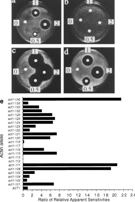
As LAT-A provided an opportunity to determine for the first time the consequences of a total lack of filamentous actin in yeast, it was necessary to first demonstrate the speci-ficity of the drug in living yeast cells. To demonstrate LAT-A specificity, we took advantage of a congenic collection of actin mutants which was generated by a charged-to-ala-nine mutagenesis scan of ACT1, the single, yeast conven-tional actin gene (Wertman et al., 1992). Each allele in the collection expresses a mutant form of actin as the sole source of actin in the cell. The collection, comprising 23 vi-able strains, was previously used to map the binding sites on the actin surface for phalloidin (Drubin et al., 1993), for yeast fimbrin (Holtzman et al., 1994), and for several other actin-associated proteins (Amberg et al., 1995). The LAT-A sensitivities of all of the strains in the collection were com-pared using the halo assay. Fig. 2, a–d shows the halo as-says of several strains in the collection to illustrate the variation in sensitivity among the alleles. In addition, the LAT-A sensitivity of each allele was compared to the sen-Table III. Testing Actin Inhibitors for Effects on Yeast Cells
In Vivo
Halo diameter
Drug Wild-type (ERG6) Derg6 S. pombe mm
Latrunculin-A 13 24 28
Latrunculin-B 10 22 23
Mycalolide-B 11 19 13
Swinholide-A 2 10 2
Jasplakinolide 2 2 2
Cytochalasin-B 2 2 2
Halo assays were used to determine whether the indicated drugs were able to inhibit growth in either wild-type S. cerevisiae cells or in S. cerevisiae cells in which the
ERG6 gene was deleted to increase cell permeability. Results for wild-type S. pombe
[image:5.612.49.290.65.169.2]sitivity of cells containing wild-type actin. This comparison is shown graphically in Fig. 2 e.
The graph clearly shows that there is a large variation in the response of the different alleles to LAT-A. The first observation is that the LAT-A sensitivity does not corre-late with salt or heat sensitivity (phenotypes described by Wertman et al., 1992). It is therefore not simply that the sickest strains are the most sensitive to LAT-A. Indeed, one of the sickest strains in the collection, act1-112, which can only grow between 208C and 308C, and has very re-stricted growth on salt-containing media, is actually resis-tant to LAT-A. In all, there were four strains in the collec-tion of mutants which showed some resistance to LAT-A. act1-119 was partially resistant, while act1-112, act1-113, and act1-117 showed complete resistance at all levels of LAT-A tested. The three mutants that were completely resistant to LAT-A showed variation in their other gross phenotypic properties. As already mentioned, cells carry-ing the act1-112 mutation are very sick with a very re-stricted range of temperatures for growth. Another strain, expressing mutation act1-113, shows slight temperature sensitivity and also exhibits salt sensitivity at low tempera-tures. The third strain, expressing act1-117, has been called a pseudo-wild-type strain as it can grow under the same conditions of temperature and salt concentration as wild-type cells (Wertman et al., 1992).
The existence of actin alleles showing resistance to LAT-A
in an otherwise congenic background was very strong evi-dence that the LAT-A was acting in a very specific manner on actin in yeast cells. However, a final caveat was that these three actin mutations might impair general uptake mechanisms for drugs such as LAT-A. Halo assays per-formed with two other drugs, mycalolide-B and swinholide-A, indicated that this is not likely to be the case. With my-calolide-B, act1-113 and act1-117 cells showed a similar sensitivity to wild-type cells, and act1-112 cells were in fact more sensitive (data not shown). Wild-type cells are resis-tant to swinholide-A at all levels tested, however, all three of the mutants resistant to LAT-A were sensitive to this drug (data not shown) demonstrating that their lack of sensitivity to LAT-A is unlikely to be due to impaired up-take mechanisms.
[image:6.612.65.440.39.394.2]Thus, the halo assay results were highly indicative of a specific interaction of LAT-A with actin in yeast cells. To investigate the possibility that resistance was due to dis-ruption of the normal LAT-A binding site on the actin molecule, we looked at the position of the three resistant alleles within the actin crystal structure. As shown in Fig. 3, the alleles define a small patch on actin adjacent to the nucleotide binding cleft. Residues which have been mu-tated in the three alleles (act1-112 5 K213A, E214A, K215A; act1-113 5 R210A, D211A; act1-117 5 R183A, D184A) are thought to either directly contact through salt bridges or hydrogen bonds, the nucleotide itself, or, to
Figure 1. The effect of LAT-A
on actin structures in yeast cells. Rd-phalloidin was used to visualize the effects of LAT-A addition to yeast cells. (a) Cells incubated in the absence of LAT-A. (b) Cells incubated for 2 h in the presence of 200 mM LAT-A. The kinetics of the effect of LAT-A on actin structures in yeast cells. (c) Cells were fixed at various time points after the addition of LAT-A. The cells were then pro-cessed for visualization of ac-tin using Rd-phalloidin. (d) After 15 min of treatment with LAT-A, the cells were washed twice with fresh dium, resuspended in me-dium, and allowed to resume growth. Cells were fixed at various time points after washing and processed as above. (m) actin cables, (s) depolarized cortical actin patches, and (d) polarized actin patches. In both c and
d, in each of three repeated
form salt bridges to position other residues which them-selves bind the nucleotide (Kabsch et al., 1990). We there-fore determined the effect of LAT-A on nucleotide ex-change.
We tested the nucleotide exchange properties of wild-type yeast actin in the presence of increasing concentra-tions of LAT-A by monitoring the increase in fluorescence observed when e-ATP binds to actin. As shown in Fig. 4, the addition of LAT-A inhibits nucleotide exchange in ac-tin in a dose-dependent manner.
As a final test of LAT-A specificity, we purified actin from two of the LAT-A resistant strains, act1-113 and act1-117. We did not attempt to isolate actin from act1-112 cells because they are very sick and we therefore thought the likelihood that they would yield an actin sufficiently stable to allow in vitro analysis was low. Indeed, actin from an act1-113 mutant polymerized very poorly in vitro (,10% could be pelleted under conditions that nearly quantita-tively pellet polymerized wild-type actin), even though 113 strains are considerably more healthy than
act1-112 strains. Since it would have been difficult to analyze data from assembly of the act1-113 actin, we proceeded with only act1-117 actin. G-actin from ACT1 or act1-117 strains was allowed to polymerize in the absence or pres-ence of LAT-A. F-actin was pelleted, and the resulting su-pernatants and pellets were analyzed by SDS-PAGE (Fig. 5 a). The actin protein bands on this gel were analyzed by densitometry (Fig. 5 b). The results from these experi-ments show that incubation of LAT-A with an equimolar concentration of wild-type actin, but not actin from act1-117 cells, resulted in inhibition of filament assembly. The act1-117 mutation itself appears to affect the ability of ac-tin to form filaments. Only z40% of act1-117 actin was pelleted using the conditions which resulted in .90% pel-leting for wild-type actin. However, the addition of LAT-A to this actin did not significantly affect its ability to poly-merize. To verify that pelleting of the mutant actin was due to filament formation, rather than aggregation of actin monomers, we observed the mutant actin filaments, la-beled with Rh-phalloidin, directly by fluorescence micro-scopy (Fig. 5 c) and found that both wild-type and act1-117 actin were able to form filaments, but only act1-117 actin was able to form filaments in the presence of LAT-A.
Effects of LAT-A on Growth, Viability, Morphogenesis and Endocytosis
Having determined that the effects of LAT-A on actin in yeast cells are rapid, reversible, and specific, we next de-termined how total disruption of actin filaments would af-fect cellular processes. The growth of cells in rich medium was monitored by cell counting following the addition of LAT-A (Fig. 6 a). After addition of the drug there was almost no increase in cell number. However, when cell growth was assessed by measuring turbidity (OD 600 nm), there was an increase after LAT-A addition to about 2.5 times the original OD reading (Fig. 6 b). This indicated a growth in cell volume that was not matched by increasing cell number. The mutant alleles act1-113 and act1-117 were also assessed for growth in the presence of LAT-A. Nei-ther strain showed detectable alteration of its growth rate in the presence of the drug (Fig. 6, c and d). In addition, both act1-113 and act1-117 mutants were stained for F-actin in the presence of LAT-A. Cortical actin patches were clearly present in these cells throughout the 1-h time course (data not shown).
[image:7.612.52.288.46.397.2]The viability of cells following treatment with 100 mM LAT-A was assessed by counting the number of treated cells that could form colonies following a timed incubation (Fig. 6 e). Cells were treated with LAT-A in suspension, struck onto a plate, and 120 cells were dissected out from this population using a micromanipulator. The ability of each manipulated cell to form a colony was assessed. The viability of cells following addition of LAT-A did not drop significantly over the 4-h time course, with 93% of cells treated for 4 h with LAT-A able to form apparently nor-mal colonies when plated. In the control population of cells which were not treated with LAT-A, all of the colo-nies that formed were roughly equal in size. At each time point after LAT-A addition, 2–5% of colonies were much smaller than in the control case, being just visible by eye (data not shown). The reason for the small colonies is not
Figure 2. Sensitivity of a set of congenic actin mutants to LAT-A.
Halo assays were used to assess the sensitivity of 23 actin mutants to LAT-A. (a–d) Representative examples of LAT-A halo as-says. Concentrations measured 0.5 mM, 1 mM, and 2 mM. (a)
ACT1 (wild-type actin), (b) act1-117, (c) act1-129, (d) act1-116.
clear. It is possible that they are petites with the temporary loss in F-actin somehow affecting mitochondrial genome inheritance.
Having established that cell viability remained high for extended periods in LAT-A, we could now use the drug to analyze effects on cell physiology. Thus, we first looked for effects on cell morphogenesis and observed that during incubation in LAT-A cells increase considerably in size but they did not arrest with uniform morphology. When assessed over a 4-h time course, the percentage of unbud-ded cells was observed to increase from 38% unbudunbud-ded in the absence of LAT-A (39% small budded and 23% large-budded) to 77% unbudded in the presence of LAT-A (12% small budded, 11% large budded) demonstrating defects
in bud formation. In addition, many arrested cells con-tained buds which were still attached to the mother cell. These buds could be separated from the mother cells by enzymatic cell wall digestion indicating that actin is not re-quired for cytokinesis, but is necessary for the complete degradation of septal material between mother and daugh-ter cells.
[image:8.612.64.567.42.483.2]Endocytosis in yeast has been shown to be dependent on the presence of an intact actin cytoskeleton (Kübler and Riezman, 1993). We assessed endocytosis by monitor-ing the uptake of the fluid phase marker lucifer yellow (LY; Dulic et al., 1991). In the absence of LAT-A, there is specific uptake of LY into vacuoles. However, in the pres-ence of LAT-A, while the vacuoles were still visible by
Figure 3. Mapping the three LAT-A resistant alleles on the actin molecular structure. Backbone of the actin monomer from the
coordi-nates of rabbit muscle actin as determined by Kabsch et al. (1990). Subdomains are marked I to IV. The side chains of the residues mu-tated to alanine in the LAT-A resistant mutants are shown and are color coded by their allele designation (Wertman et al., 1992).
act1-112 (yellow), act1-113 (red), act1-117 (green). The adenine nucleotide (cyan) is shown in the prominent cleft as a ball and stick model,
Nomarski optics, there was no uptake of LY, indicating in-hibition of endocytosis (data not shown).
The Sensitivity to LAT-A of Strains with a Disrupted Actin Cytoskeleton
To gain new insights into the role of actin cytoskeleton proteins in modulating cytoskeletal integrity and function,
we investigated the effect on LAT-A sensitivity of muta-tions in genes encoding various proteins that are impor-tant for actin cytoskeleton function. We performed halo assays on each mutant strain, and also on the congenic par-ent strain. This control was necessary because we noticed some variation in LAT-A sensitivity between wild-type strains of different strain backgrounds. There was a range of sensitivities of the mutant cells to LAT-A when as-sessed by halo assays. Two mutants showed a slight resis-tance to LAT-A. These carried a deletion of the SLA1 gene, and a mutation in the END3 gene (end3-1), with ra-tios of apparent sensitivities compared to controls of 0.5 and 0.3, respectively (see Materials and Methods). An in-creased sensitivity to LAT-A was shown by several mu-tants including Dsrv2, Dcap2, Dtpm1, Dsac6p, and Dsla2 with ratios of apparent sensitivities of 2.9, 4.0, 2.8, 1.8, and 2.6, respectively. Strains carrying the Dabp1 mutation showed a barely detectable increase in resistance. These results highlight differences among cytoskeletal proteins in their contributions to cytoskeleton stability.
The Role of Actin in the Development of Polarity at the Presumptive Bud Site
[image:9.612.54.288.42.239.2]We used sensitivity to LAT-A to determine which of 19 proteins that normally localize to the presumptive bud site depend on an intact actin cytoskeleton for localization. We used diploid cells because they are more sensitive than haploids to LAT-A, their larger size facilitates observa-tions made by immunofluorescence microscopy and they have an ellipsoidal shape allowing the ends of a cell to be distinguished. Being able to identify the ends of a cell is important so one can determine whether a bud site has formed at the correct position (see Drubin and Nelson,
Figure 4. The effect of LAT-A on actin nucleotide exchange.
[image:9.612.52.214.433.721.2]Pu-rified wild-type yeast actin was added to buffer containing a fluo-rescent nucleotide analogue (e-ATP) and increasing concentra-tions of LAT-A. The kinetics of nucleotide exchange were monitored using e-ATP fluorescence as described in the Materials and Methods section.
Figure 5. The effect of LAT-A
on actin polymerization. Pu-rified actin from ACT1 or
act1-117 cells was incubated
1996). Cells were released from G0, as described in Materi-als and Methods, in the presence or absence of LAT-A. At each time point, immunofluorescence was used to localize the proteins of interest.
Actin Is Required for Bud Formation in Cells Exiting G0
To establish a baseline for comparisons, we determined the rate at which polarization of actin was achieved in cells exiting G0. Hourly time points were taken from G0 cells released into fresh media in the presence or absence of LAT-A. Fig. 7 a shows the percentage of cells with polar-ized actin staining obtained by this procedure. Polarization of actin staining in the absence of LAT-A was clear after 2 h and increased to a peak of 80% cells with polarized stain-ing after 4 h. In longer time courses, the number of cells with polarized staining did not get higher than 80%, pre-sumably because the level of synchrony in the cells was not sufficiently high (i.e., the remaining cells are at early G1 or late G2/mitosis, stages in which the cortical actin cytoskele-ton is depolarized). Fig. 7 c shows the anti-actin immuno-fluorescence staining pattern for cells after 4 h of growth following the G0 release in the absence of LAT-A. During this 4-h period, cell morphology was also monitored.
Al-though growth in culture after release was not completely synchronous, there was a significant enrichment for small-budded cells at 2–3 h after release, and for medium- to large-budded cells at 4 h after the release (Fig. 7 b).
In the presence of LAT-A there was no observable po-larization of the actin cytoskeleton at any time (Fig. 7, a and d). At early time points (0–1 h), no actin structures were seen. However, after this stage an increasing number of cells contained actin bars (Fig. 7 d). Similar bars have been reported previously in mutant strains that have actin de-fects (Johnston et al., 1991; Holtzman et al., 1993; Welch and Drubin, 1993). The actin bars do not stain with the F-actin staining dye rhodamine-phalloidin and so have been proposed to be aggregates of monomeric actin. At the later time points, smaller dots of actin staining can also be seen which appear to be similar in size to cortical patches. However, these aggregates do not label with Rd-phalloi-din, nor, in most cases, do they appear to be at the cortex, indicating that these structures are most likely aggregates of monomeric actin. In addition, in a small percentage of cells (,5%), actin was seen to stain within the nucleus.
[image:10.612.66.359.40.411.2]Because no filamentous actin could be detected in LAT-A–treated cells, any proteins that were able to achieve a polarized staining pattern in the presence of LAT-A could be concluded to be doing so in the absence of F-actin.
Figure 6. The effect of LAT-A on growth and
Polarity Establishment Proteins, Cdc42p and Bem1p
To determine whether the actin cytoskeleton has any role in the early stage of polarity establishment involving local-ization of Cdc42p and Bem1p at the cell cortex, we ana-lyzed the localization of Cdc42p and Bem1p upon exit from G0 in the presence or absence of LAT-A. As de-picted in Fig. 8, a and d, in the absence of LAT-A, Cdc42p and Bem1p became polarized with kinetics similar to ac-tin. As reported in the literature (Ziman et al., 1993; Prin-gle et al., 1995), both proteins showed highly localized staining at the presumptive bud site and at the tip of small buds (Fig. 8, b and e). In cells with larger buds, the staining was more diffuse but still polarized. Staining was also seen at the mother-bud neck before cytokinesis.
In the absence or presence of LAT-A, Cdc42p and Bem1p achieved polarized cellular localization and did so with similar kinetics (Fig. 8). In the LAT-A–treated cells, staining was in the form of a bright spot at one end of the ellipsoidal diploid cell (Fig. 8, c and f) which was the ap-propriate position for the formation of the bud site. In the case of Cdc42p localization in the presence of LAT-A, in addition to the bright spot of staining at the end of the cell, a low level of diffuse cortical staining could also be seen. To further address whether the polarized localization of Bem1p and Cdc42p reflected elements of a natural path-way for presumptive bud site assembly, we performed double labeling with antibodies to another presumptive bud-site protein, Cdc11p (see following section for
de-scription of Cdc11p localization in LAT-A). The patterns of staining of the proteins are different, a small patch of staining is observed for Cdc42p or Bem1p, while a ring is observed for Cdc11p staining, so the patterns of staining could be distinguished. In the presence of LAT-A, staining could be observed to be coincident (a Cdc11p ring encir-cled the Cdc42p or Bem1p patch) in 99% of the cells (400 cells counted) suggesting further that Cdc42p, Bem1p, and Cdc11p had localized to what was a natural bud site except for the absence of actin and associated proteins.
Actin-binding Proteins and Proteins Implicated Genetically in Actin Function
[image:11.612.52.291.40.263.2]The polarized localization of the actin-binding proteins Abp1p and cofilin in cells released from G0 was found to depend on the presence of F-actin structures. The percent-age of cells with polarized staining in the presence and ab-sence of LAT-A was quantified for each protein and these data are illustrated graphically in Fig. 13. In contrast, both Sla1p and Sla2p which also colocalize with cortical actin structures (Holtzman et al., 1993; Ayscough, K., and S.
Figure 7. Immunolocalization of actin in cells exiting G0 at 258C
in the absence or presence of LAT-A. (a) Quantification of im-munofluorescence to assess the percentage of cells incubated in the presence (d) or absence (s) of LAT-A that exhibited polarized actin staining. (b) Morphology of cells exiting G0 in the absence of LAT-A. Cells were classified as unbudded (s), small-budded (d), or medium- to large-budded (h). (c) Immunolocalization of actin in cells grown for 4 h in the absence of LAT-A. (d) Immu-nolocalization of actin in cells grown for 4 h in the presence of LAT-A. Note the presence in some cells of actin bars and actin in the nucleus (as confirmed by DAPI staining, not shown). Bar,
5 mm. Figure 8. Immunolocalization of polarity establishment proteinsin cells exiting stationary phase. Quantification of
[image:11.612.309.549.47.368.2]Yang, unpublished observations), were able to localize to the cell cortex and were in patch-like structures in the ab-sence of F-actin (data not shown). In a small population of cells, these proteins appeared to have a polarized distribu-tion.
In the absence of LAT-A, Aip3p/Bud6p, a protein which interacts with yeast actin in a two-hybrid screen and which localizes to cortical sites (Amberg et al., 1996), achieved its normal polarized staining pattern with staining being ob-served at the presumptive bud site, at the bud tip, and, in large budded cells, at the mother-bud neck (see above; Fig. 13). In the presence of LAT-A, the number of cells showing localized staining was reduced. However, it was clear that in a significant number of cells, Aip3p/Bud6p was still able to localize to the cell cortex, and in many cases it showed a polarized localization. 22% of cells showed po-larized staining at the ends of the cell (Fig. 13) whereas in other cases, the staining was confined to discrete patches which were not at the end of the ellipsoidal cells (3.5%), or there were two patches at the cortex with at least one patch not being at the end of the cell (9.5%).
Kelleher (1995) proposed that an Arp2/Arp3 hetero-dimer might serve as a nucleus for actin polymerization. It was therefore of interest to determine whether Arp2 in yeast could function independently of actin and achieve its normal polar localization in the absence of conventional actin. In the absence of LAT-A, Arp2p has a polarized cortical patch-like staining pattern (Moreau et al., 1996) with kinetics of localization similar to actin. However, in the presence of LAT-A, Arp2p did not polarize, though it did show a different distribution from actin (see Fig. 13 for summary of data). In the presence of LAT-A, actin, but not Arp2p, localizes to bar structures and, in some cases, to the nucleus. In contrast to actin, a significant amount of Arp2 staining appeared to be associated with the cell cor-tex where its staining was diffuse. This was particularly ap-parent at early time points (1–2 h) after release of cells from G0.
Proteins Involved in Secretory Processes
We localized several proteins (Sec4p, Sec8p, Myo2p, cal-modulin, and Smy1p) implicated in secretion in the ab-sence and preab-sence of LAT-A. Sec4p is a member of the Rab-GTPase family of proteins and acts at a late stage of the secretory pathway between the Golgi apparatus and the plasma membrane (Salminen and Novick, 1987). Sec4p localizes to the presumptive bud site and the tip of small-budded cells (Novick and Brennwald, 1993). Sec8p is a com-ponent of a protein complex that localizes to the presump-tive bud site and the bud tip, and may be involved in the docking of vesicles at the plasma membrane (TerBush and Novick, 1995; TerBush, D., personal communication). Myo2p is an unconventional myosin with calmodulin as an associ-ated subunit (Johnston et al., 1991; Brockerhoff et al., 1994). Mutations in Myo2p and calmodulin can cause vesicle ac-cumulation, delocalized cell wall synthesis, chitin deposi-tion and often an arrest as unbudded cells (Johnston et al., 1991; Brockerhoff and Davis, 1992; Brockerhoff et al., 1994; Lillie and Brown, 1994; Govindan, et al., 1995). Smy1p is a kinesin-like protein which was identified as a dosage sup-pressor of a mutation in MYO2. (Lillie and Brown, 1994).
We found that actin is required for the localization of Sec4p, Sec8p, and Smy1p to the presumptive bud site. In contrast, both Myo2p and calmodulin were able to localize to this region in the absence of actin, albeit to a lesser ex-tent than in control cells. Examples of kinetic and photo-graphic data are shown for Sec4p and calmodulin (Figs. 9 and 10). In cells growing out from G0, Sec4p became highly polarized at the presumptive bud site, remaining at the bud tip in small-budded cells and then becoming slightly more diffuse as the bud grows. It was not clearly visible in most medium- to large-budded cells but was observed at the mother-bud neck before cytokinesis (Fig. 9, a and b). However, when LAT-A was added to the growth medium during the exit from stationary phase, Sec4p did not ex-hibit polarized localization (Fig. 9, a and c).
In the absence of LAT-A, calmodulin and Myo2p local-ized to the presumptive bud site, the tip of small-budded cells and the septal region. Staining in medium- to large-budded cells was fainter (Fig. 10, a and b; only calmodulin data is shown since Myo2p staining was essentially identi-cal). In the presence of LAT-A, there was a high level of cytoplasmic staining with most of the cells showing no po-larization of staining. However, in z30% of cells, calmod-ulin appeared in a discrete patch located at the end of the ellipsoidal cell, a position that is appropriate for the bud site (Fig. 10 c). When cells were followed for longer than 4 h, the number of cells with this staining pattern increased slightly but did not rise above 35% of cells observed.
Proteins Required for Cytokinesis
Several proteins become localized to the presumptive bud site but are most important for processes that occur subse-quent to bud formation. The yeast septins, required for cy-tokinesis and proposed to be subunits of the filaments ob-served at the mother-bud neck by electron microscopy, are such proteins (Hartwell, 1971; Haarer and Pringle, 1987; Ford and Pringle, 1991; Kim et al., 1991).
We examined the localization of two septin proteins, Cdc10p and Cdc11p. Because the results were
indistinguish-Figure 9. Immunolocalization of Sec4p in cells exiting stationary
[image:12.612.323.564.41.205.2]able for these two proteins, we have only presented the data for Cdc11p. In both the absence and presence of LAT-A, Cdc11p became polarized and the kinetics of localization were similar (Fig. 11). The staining pattern was a bright ring structure at the presumptive bud site. In the absence of LAT-A, cells with larger buds were seen to contain a double ring of staining at the bud neck (Fig. 11 b). In di-viding cells, one ring appeared to remain with the mother and the other with the daughter cell. In the presence of LAT-A, only single rings were seen, indicating that ring duplication is dependent on bud formation (Fig. 11 c).
Other Proteins of the Pre-Bud Site
Several other proteins have been localized to the pre-sumptive bud site, but their cellular activities are not cur-rently understood well enough to permit them to be classi-fied functionally.
Gin4p and Bni4p localize to a ring structure at the pre-sumptive bud site which persists at the mother-bud neck throughout bud growth (Longtine, M., D. DeMarini, J. Prin-gle, personal communication). In the presence of LAT-A, both Gin4p and Bni4p achieved a polarized localization (Fig. 13).
Spa2p localizes to the presumptive bud site and to the tip of mating projections (Snyder, 1989; Snyder et al., 1991). Cells deleted for SPA2 exhibit no defects in the ability to bud but show a defect in bud site selection (Snyder, 1989). In the absence of LAT-A, Spa2p became polarized with similar kinetics to actin (Fig. 12, a and b). In the presence of LAT-A, Spa2p was still able to polarize. However, the kinetics of polarization were delayed (Fig. 12, a and c). In three separate experiments, polarized localization of Spa2p was always 1–2 h slower than in the absence of LAT-A.
A Role for Actin in the Maintenance of Cell Polarity
[image:13.612.52.291.41.209.2]To determine whether actin plays a role in the mainte-nance of the axis of cell polarity, we released both haploid and diploid cells from stationary phase to a stage where more than 50% of cells had small buds. We then treated these cells with LAT-A for 5 min to completely disrupt the actin cytoskeleton, washed the cells, and allowed them to resume growth. After 2 h, we analyzed the morphology of the cells (Fig. 14). For both haploid and diploid cells, the disruption of the actin cytoskeleton caused a significant percentage of cells to become two-budded. Interestingly, in nearly all cases, this second bud was correctly placed with respect to the expected position of bud site selection. That is, haploid buds were adjacent to one another, and diploid buds were either at both poles or at the same pole
Figure 10. Immunolocalization of calmodulin in cells exiting
sta-tionary phase in the absence or presence of LAT-A. (a) Quantifi-cation of immunofluorescence to assess the percentage of cells that exhibited polarized calmodulin staining when incubated in the presence (d) or absence (s) of LAT-A. Immunolocalization of calmodulin in cells grown in the absence (b) or presence (c) of LAT-A. Bar, 5 mm.
Figure 11. Immunolocalization of Cdc11p in cells exiting
[image:13.612.309.549.44.199.2]station-ary phase in the absence or presence of LAT-A. (c) Quantifica-tion of immunofluorescence to assess the percentage of cells that exhibited polarized Cdc11p staining when incubated in the pres-ence (d) or absence (s) of LAT-A. Immunolocalization of Cdc11p in cells grown in the absence (b) or presence (c) of LAT-A. Note the lack of double ring structures in the cells incubated with LAT-A. Bar, 5 mm.
Figure 12. Immunolocalization of Spa2p in cells exiting
[image:13.612.51.291.465.633.2]of the cell (for a discussion of budding patterns, see Dru-bin and Nelson, 1996).
In a similar set of experiments, haploid MATa cells were exposed to a-factor until more than 90% of the population had formed a mating projection. The population was then exposed to LAT-A for 5 min to completely disrupt the ac-tin cytoskeleton, washed, and released into fresh media.
a-Factor was present at all stages. In a control population to which DMSO was added at the intermediate stage, two shmoos were observed in 19% cells. By contrast, 53% of cells exposed to LAT-A contained two shmoos.
Discussion
LAT-A Action is Rapid and Reversible Indicating Actin Filaments Undergo Rapid Cycles of Assembly and Disassembly in S. cerevisiae
The rapidity of the disappearance of actin structures upon treatment of yeast with LAT-A suggests two mechanistic possibilities. LAT-A might (1) actively destabilize actin fil-aments, or, (2) it might bind to monomers, preventing the assembly step in a rapid cycle of assembly and disassem-bly. Previously published data are most consistent with the latter possibility, a sequestering mechanism via the forma-tion of a 1:1 complex with actin monomer (Coué et al., 1987). We obtained similar data with yeast actin, verifying the conclusions of the earlier studies and demonstrating that they apply to yeast actin. We also observed that LAT-A did not induce rapid F-actin disassembly in vitro, arguing further against a filament-destabilizing or severing mode of action. Thus, the data we generated, and the previously reported data, indicate that the most likely mode of action for LAT-A is monomer sequestration. Such a sequestra-tion mechanism for LAT-A acsequestra-tion, in combinasequestra-tion with the rapidity of its action in vivo, would indicate that the ac-tin cytoskeleton in yeast is highly dynamic. The
observa-tions that an inherent capacity for dynamic turnover is a universal property of actin, and that a nonmotile organism has a dynamic actin cytoskeleton, lead us to suggest that rapid assembly and disassembly is a fundamental property of eukaryotic actin cytoskeletons.
The LAT-A Sensitivity of Actin-associated Protein Mutants
Several actin cytoskeleton-associated protein mutants showed significantly increased sensitivity to LAT-A com-pared with wild-type cells. Where previously derived in-formation about the contributions of individual proteins to cytoskeletal integrity was available, LAT-A sensitivities confirmed previously reached conclusions. For example, mutations in capping protein (encoded by the CAP1 and CAP2 genes) and yeast fimbrin (Sac6p) lead to decreased F-actin and increased G-actin in cells (Karpova et al., 1995). With an already depleted polymer pool, it is expected that sensitivity to LAT-A would be heightened in these mu-tants. Indeed, these mutants showed elevated sensitivity to the drug. Tropomyosin is also postulated to stabilize actin cables in yeast (Liu and Bretscher, 1992; Drees et al., 1995). tpm1 mutants showed elevated sensitivity to LAT-A, providing further evidence that tropomyosin stabilizes ac-tin structures in vivo. While the above examples validated this approach to analysis of cytoskeleton protein function, the opportunity to obtain novel insights into cytoskeleton protein function comes from the application of this ap-proach to the remaining proteins.
[image:14.612.322.561.42.232.2]Srv2p has an actin monomer binding activity (Freeman et al., 1995) so one might predict that deletion of SRV2
Figure 13. Polarization of presumptive bud site proteins in the
[image:14.612.67.302.44.232.2]absence ( ) or presence (j) of LAT-A. A summary of data col-lected for all the proteins analyzed in this study. The cell counts shown here are those at the 4-h time point after release of cells from the stationary phase.
Figure 14. The formation of two-budded cells after incubation of
would lead to an increased resistance to LAT-A. How-ever, the deletion of SRV2 causes increased LAT-A sensi-tivity. This result could be explained if Srv2p plays a role in facilitating filament assembly, perhaps by delivering ac-tin monomers to the ends of acac-tin filaments.
Two of the mutants assayed, Dsla1 and end3-1, had a slightly increased resistance to LAT-A compared with wild-type cells. These mutants have similar actin phenowild-types to one another, with the cortical patches being larger, though fewer, than in the wild-type situation (Holtzman et al., 1993; Benedetti et al., 1994). In addition, actin cables ap-pear to be similarly prominent or even more pronounced compared to wild-type. These actin phenotypes might sug-gest that the mutant cells have increased levels of F-actin. In light of the elevated resistance of the Dsla1 and end3-1 mutant strains to LAT-A, we propose that Sla1p and End3p might promote destabilization of the yeast actin cy-toskeleton.
Actin Mutants Resistant to LAT-A Demonstrate Specificity and Suggest a Binding Site
The demonstration that mutation of actin was sufficient to
make a strain resistant to LAT-A provided powerful evi-dence for a highly specific interaction between LAT-A and actin. Furthermore, the amino acid substitutions in the three resistant alleles mapped to a distinct region on the actin molecule, implicating this region as the binding site for LAT-A. This region was adjacent to the nucleotide bind-ing cleft. In vitro assays tested the prediction that LAT-A might impair nucleotide exchange and demonstrated that this was the case. In addition, actin purified from a LAT-A resistant mutant was resistant to the action of LAT-A in vitro, adding further support to the proposal that the muta-tions identified the LAT-A binding site on actin.
A Functional Hierarchy of Proteins Is Involved in the Establishment of Cell Polarity
[image:15.612.52.429.36.459.2]Fig. 15 shows a hierarchy of protein functions required for cell polarity development. Previous studies suggested that Cdc42p and Bem1p function early in the pathways leading to polarity establishment. Temperature-conditional cdc42 and bem1 mutations caused the actin cytoskeleton and septins to be delocalized at the nonpermissive tempera-ture, indicating that cytoskeleton localization depends on
Figure 15. Model depicting
the functions of the polarity establishment proteins (Ad-ams et al., 1990; Bender and Pringle, 1991; Chant et al., 1991; Pringle et al., 1995). We showed here that neither Cdc42p nor Bem1p requires actin for localization, an ob-servation consistent with the conclusion that Cdc42p be-comes localized independent of actin and then organizes the actin cytoskeleton.
Proteins That Require Actin to Achieve Their Normal Polarized Localization
As may have been predicted, proteins that bind directly to actin, Abp1p and cofilin, require actin to achieve their normal localization at the presumptive bud site. However, Sla1p, Sla2p, and Arp2p, which in control cells colocalize with actin cortical patches, show a level of actin-indepen-dent association with the cell cortex, but not polarized lo-calization. This indicates that these proteins localize to the cortex in an actin-independent manner but require actin to polarize to the presumptive bud site.
Actin was shown to be required for the localization of Sec4p, Sec8p, and Smy1p (discussed further below) to the presumptive bud site. In contrast, Myo2p and calmodulin were able to localize to this region in the absence of actin, albeit to a lesser extent than in control cells. This observa-tion indicates that Myo2p/calmodulin complexes are able to interact with proteins other than actin at the presump-tive bud site. Interestingly, previous studies on the local-ization of these proteins in actin mutants indicated that both Myo2p (Lillie and Brown, 1994) and calmodulin (Brockerhoff and Davis, 1992) showed actin-dependent localization. Both of these studies were performed in actin mutants in which asynchronous cultures of log phase cells were shifted to the nonpermissive temperature. Brocker-hoff and Davis (1992) reported that, at the nonpermissive temperature, no budded cells show polarized calmodulin whereas in 5% of the unbudded cells, polarized staining is seen. In our studies we have assessed the ability of cells to polarize proteins during entry into the cell cycle and so, have focused on the unbudded population of cells. An ex-planation of the results which could encompass the previ-ous findings (Brockerhoff and Davis, 1992; Lillie and Brown, 1994) would be that localization of calmodulin and Myo2p to the presumptive bud site can occur in the ab-sence of actin. However, stabilization of this association throughout the cell cycle, and/or at elevated temperatures, requires the presence of an intact actin cytoskeleton.
A Role for Actin in Polarized Secretion
The formation of new buds involves localized secretion (Field and Schekman, 1980) and is an actin-dependent process (Novick and Botstein, 1985). Several proteins pre-viously shown to be involved in vesicle trafficking and se-cretion were demonstrated here to require actin to be-come localized to the presumptive bud site. This observation strongly suggests that actin facilitates bud growth by ori-enting spatially elements of the secretory pathway. Our data establish that actin functions at at least two levels to mediate polarized exocytosis. We demonstrated for the first time that actin is necessary to localize secretory vesi-cles, marked by Sec4p, to the bud site, a role which had been postulated previously (Adams and Pringle, 1984;
Kil-martin and Adams, 1984; Johnston et al., 1991). In addi-tion, we have established that actin is involved in localiza-tion of the putative vesicle docking complex, marked by Sec8p, to the presumptive bud site.
Proteins That Do Not Require Actin for Localization to the Presumptive Bud Site
The septins are a group of related proteins that localize to the presumptive bud site (Haarer and Pringle, 1987; Ford and Pringle, 1991; Kim et al., 1991; Longtine et al., 1996). Septin mutants are able to polarize their actin cytoskele-tons, indicating independence of actin and septin cytoskel-etal components for localization (Adams and Pringle, 1984). Earlier studies by Ford and Pringle (1991) investi-gated localization of septins and actin at the presumptive bud site. Their results suggested that actin may arrive at this site slightly before Cdc11p. However, a few cells in the population were observed with only septins or only actin localized at the presumptive bud site, suggesting that local-ization of actin and septins at the site might be indepen-dent processes. Here, using LAT-A to disrupt F-actin structures, we demonstrated unequivocally that septin lo-calization is independent of actin lolo-calization. Thus, while both actin and the septins are delocalized in the absence of Cdc42p, they are not dependent on one another for their own localization. This allows us to demonstrate a bifurca-tion point in the hierarchy of polarity establishment func-tions with actin-dependent, and actin-independent funcfunc-tions defining the division (Fig. 15).
The proteins Bni4p and Gin4p have immunofluores-cence staining patterns at the presumptive bud site similar to those of the septins, and, like the septins, neither Bni4p nor Gin4p required the presence of an intact actin cyto-skeleton for localization. Their actin-independent localiza-tion suggests that the primary role of these proteins is not in bud formation processes but is most likely in subse-quent processes, possibly associated with cytokinesis.
Finally, we showed that localization of Spa2p at the pre-sumptive bud site was not dependent on an intact actin cy-toskeleton, but there was a kinetic delay in its polarized lo-calization in cells incubated with LAT-A. It is possible that Spa2p, or a protein that binds Spa2p, normally uses actin-based structures to localize at the presumptive bud site. However, when at the bud site, Spa2p makes interactions with other proteins not associated with actin, and in LAT-A–treated cells these interactions are responsible for Spa2p localization. Alternatively, the kinetic delay might reflect perturbation by LAT-A of a mechanism to coordi-nate temporally actin and Spa2p localization.
A Role for Actin in Maintaining an Axis of Cell Polarity